Journal club
-
Upload
kannangunasekaran -
Category
Documents
-
view
18 -
download
1
Transcript of Journal club

Dr Vijayakumar A.R,Senior resident.

INTRODUCTIONEVAR is the preferred treatment for infra-
renal abdominal aortic aneurysms when appropriate patient selection is practiced.
Reduced short term mortality, hospital stay and ICU care compared to that of open surgical repair.
Repeat intervention owing to endograft related complications is necessary in approximately 15% of patients and hence requires long-term postoperative surveillance.
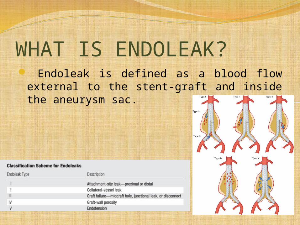
WHAT IS ENDOLEAK? Endoleak is defined as a blood flow external
to the stent-graft and inside the aneurysm sac.

Static biphasic CT angiography is the current standard method for pre- and postoperative imaging evaluation of abdominal aortic aneurysms.
The recent introduction of time-resolved dynamic CT angiography with repetitive bidirectional table movement allows the assessment of temporal enhancement patterns in the endograft, aneurysm sac, and adjacent aortic branches with a high temporal resolution.

AIM
To determine the time course of enhancement patterns in the aorta and endoleaks at dynamic CT angiography as well as their effect on the endoleak detection rate in patients who have undergone abdominal aortic EVAR.

MATERIALS AND METHODSRetrospective study.All of the consecutive patients who were scheduled
for periodic CT follow-up of the abdominal aorta after EVAR between February 2010 and November underwent dynamic CT angiography.
Dynamic CT angiograms with complete coverage of the entire endograft, followed by a static venous phase and adequate arterial contrast with a minimal attenuation of 150 HU in the abdominal aorta during at least one phase of dynamic CT angiography.
Exclusion criteria were incomplete image data or low arterial contrast (attenuation <150 HU).
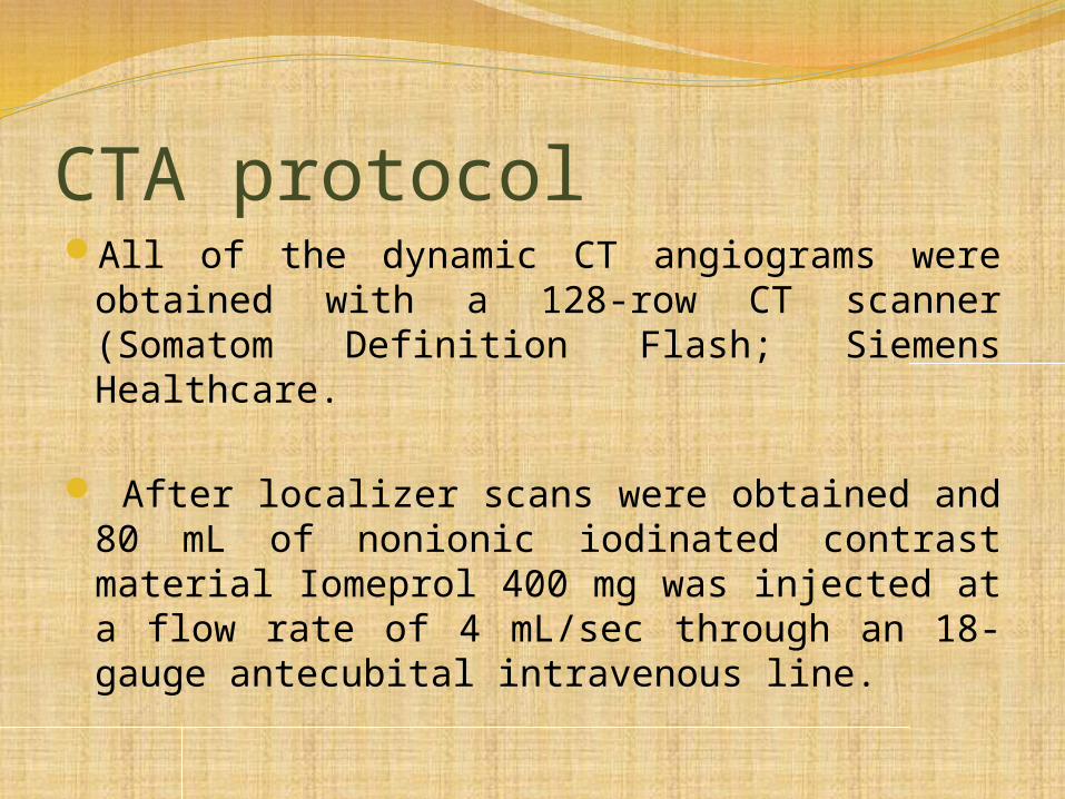
CTA protocolAll of the dynamic CT angiograms were
obtained with a 128-row CT scanner (Somatom Definition Flash; Siemens Healthcare.
After localizer scans were obtained and 80 mL of nonionic iodinated contrast material Iomeprol 400 mg was injected at a flow rate of 4 mL/sec through an 18-gauge antecubital intravenous line.

10 unidirectional scan phases of 2.5 seconds with a temporal resolution of 5 seconds and a field of view of 283 mm on the long axis. The post threshold delays were 2, 7, 12, 17, 22, 27, 32, 37, 42, and 47 seconds. A delay of 7 seconds after the threshold represented the arterial phase of a biphasic study.
Static venous phase scanning that covered the entire abdominal aorta with a delay of 100 seconds after reaching threshold.

IMAGE ANALYSISImage analysis was performed by using a
commercially available diagnostic workstation (Easyvision R11.4.1; Philips Medical Systems.
Analysis of all of the acquisition phases was carried out in random order without any image annotations in consensus by two experienced radiologists with 10 and 3 years of experience who were blinded to the clinical data and to the results of the CT examination.

Enhancement was calculated by subtracting the mean attenuation measured in a region of interest from the attenuation of the distal inferior vena cava in the first available scan phase to exclude the attenuation of unenhanced blood.
The degree of contrast material enhancement at each time point was determined in the aortic lumen 1 cm cranial to the stent-graft and in all of the detectable endoleaks larger than 2 mm in the same position throughout all of the scan phases.
The size of the endoleak region of interest was determined by using the scan phase with the greatest endoleak extent in a transverse section, and the size of this region of interest was kept constant throughout all of the scan phases. The distribution of the contrast material from the endoleaks within the aneurysm sac was not assessed.

the overall image quality of each scan series was rated on a five-point ordinal scale, taking into account image noise and beam-hardening artifacts, as follows:
0 = very poor, 1 =poor, 2 = reduced, 3 = good, and 4 = very good.
The ratings of all images obtained in the dynamic part of dynamic CT angiography were combined, whereas the static scan series of dynamic CT angiography was rated separately.
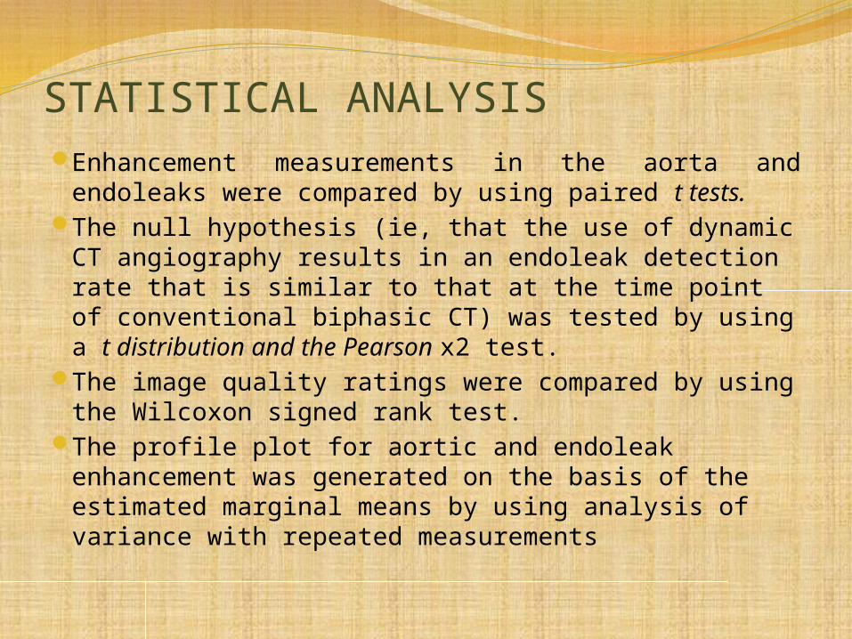
STATISTICAL ANALYSISEnhancement measurements in the aorta and
endoleaks were compared by using paired t tests.The null hypothesis (ie, that the use of dynamic CT
angiography results in an endoleak detection rate that is similar to that at the time point of conventional biphasic CT) was tested by using a t distribution and the Pearson x2 test.
The image quality ratings were compared by using the Wilcoxon signed rank test.
The profile plot for aortic and endoleak enhancement was generated on the basis of the estimated marginal means by using analysis of variance with repeated measurements

RESULTSTotal: 71 patients. Men 66. mean age 72.2.
women 5. mean age 74.2.Mean BMI: 28.2.Ten types of endografts were used.

ENDOLEAK DETECTION RATE
TOTAL: 44.Maximum in phase 6 at 27 sec. Least in
phase 1 at 2 sec.
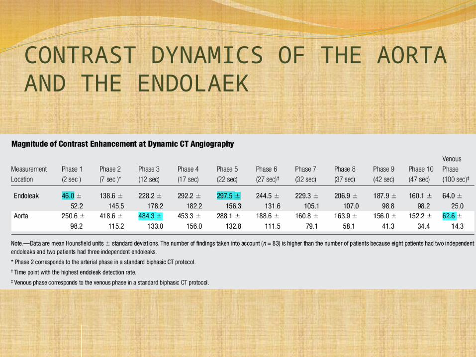
CONTRAST DYNAMICS OF THE AORTA AND THE ENDOLAEK

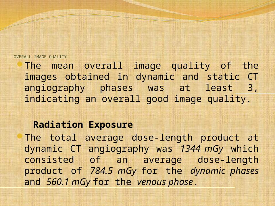
OVERALL IMAGE QUALITY
The mean overall image quality of the images obtained in dynamic and static CT angiography phases was at least 3, indicating an overall good image quality.
Radiation ExposureThe total average dose-length product at
dynamic CT angiography was 1344 mGy which consisted of an average dose-length product of 784.5 mGy for the dynamic phases and 560.1 mGy for the venous phase.

DISCUSSIONImaging after EVAR is essential.CT is considered to be the method of choice for these
follow-up examinations.Contrast-enhanced US cannot replace CT
angiography in the post-EVAR follow-up with regard to graft anchoring integrity, aneurysm morphology, or visceral vessel patency.
At most centres, CT is usually performed at least during the contrast-enhanced arterial and venous phases.
Dynamic CT angiography with a large z-axis field of view, such as is needed for coverage of the abdominal aorta, is currently only available with two 128-sec-tion scanners (Somatom Definition AS+ and Somatom Definition Flash, Siemens Healthcare)

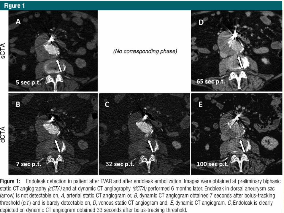
The maximum endoleak enhancement was reached at 22 seconds after the bolus-tracking threshold, during the phase when the bolus of contrast material had already passed the aorta.
The highest endoleak detection rate was achieved later, at 27 seconds after the bolus-tracking threshold, during the phase when the mean peak enhancement of the aorta and the endoleak had already passed and maximum contrast could be achieved between the remaining endoleak enhancement and the rapidly de-enhancing aorta.
For the commonly used biphasic static CT angiography, we conclude that the arterial phase is acquired too early and the venous phase too late to identify the maximum number of endoleaks

Scan phases 3 and 6, at 12 and 27 seconds after the bolus-tracking threshold, respectively, are the most useful scan phases in patients have undergone EVAR.
The first shows the highest aortic enhancement and should be used to evaluate the aorta and its branches, detect early endoleaks, and assess the endograft anchoring and position.
The second of the two phases is used to detect endoleaks.
A late venous phase (with a delay of 300 seconds) can help detect low-flow endoleaks.

The percentage of endoleaks after EVAR is generally reported to be 20%–30%.
The endoleak rate of approximately 45% found with dynamic CT angiography in this series.
Most of the detected endoleaks were type II leaks and the current knowledge regarding their management differs widely.
This prevalence might explain some of the type V endoleaks observed in bi-phasic CT angiography, where sac enlargement is detected but no contrast material can be detected outside the graft.

RADIATION DOSE Dynamic CT angiography had a 10% higher
radiation dose and the venous phase had a 20% lower radiation dose.
Exclusion of the dynamic CT angiography scan phases with low diagnostic value (phases 1, 2, 9, and 10 at 2, 7, 42, and 47 seconds after the bolus-tracking threshold, respectively), reduction of the tube voltage to 80 Kv would reduce the dose by more than 20% compared with that in our current dynamic CT angiography protocol.
Restricting the venous phase acquisition to the region of interest would result in an overall dose reduction of approximately 65%.

With use of this reduced dose protocol, the dynamic information of the enhancement patterns would not be lost because the protocol would still include the phases with the highest endoleak enhancement and highest endoleak detection rates.
If the scanner used does not allow a dynamic examination and the post-EVAR follow-up is performed as biphasic static CT angiography, an additional scan phase over the period of 22–32 seconds after the bolus-tracking threshold should be added.
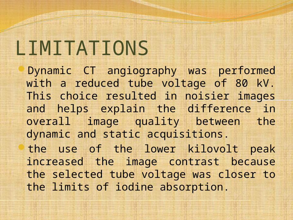
LIMITATIONSDynamic CT angiography was performed with
a reduced tube voltage of 80 kV. This choice resulted in noisier images and helps explain the difference in overall image quality between the dynamic and static acquisitions.
the use of the lower kilovolt peak increased the image contrast because the selected tube voltage was closer to the limits of iodine absorption.

CONCLUSIONDynamic CT angiography during post-EVAR
follow-up revealed that the peak enhancement of endoleaks is significantly different than that of the aorta and is not adequately evaluated with conventional biphasic CT angiography protocols.
Use of dynamic CT angiography is associated with an increased endoleak detection rate compared with the detection rates at the time points of conventional biphasic CT.

Thank you