Journal #1: How is the integumentary system (skin) like an onion? Vocabulary 1. Integument 2....
17
Journal #1: How is the integumentary system (skin) like an onion? Vocabulary 1. Integument 2. Epidermis 3. Dermis 4. Subcutaneous layer 5. Basal Cells 6. Keratin 7. Carotene 8. Melanin Objective: List the components of the integumentary system and their relationship to each other Specify the functions of the integumentary system
-
Upload
garry-collins -
Category
Documents
-
view
220 -
download
0
Transcript of Journal #1: How is the integumentary system (skin) like an onion? Vocabulary 1. Integument 2....
Journal #1: How is the integumentary system (skin) like an onion?
Vocabulary1. Integument2. Epidermis3. Dermis4. Subcutaneous layer5. Basal Cells6. Keratin7. Carotene8. Melanin
Objective: List the components of
the integumentary system and their relationship to each other
Specify the functions of the integumentary system

Ch.5 Integumentary System
Part 1: Layers
Pages 153-164
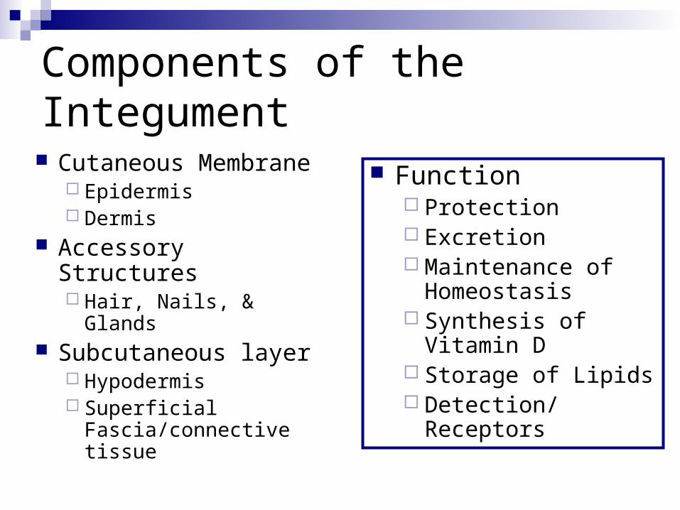
Components of the Integument
Cutaneous Membrane Epidermis Dermis
Accessory Structures Hair, Nails, & Glands
Subcutaneous layer Hypodermis Superficial
Fascia/connective tissue
Function Protection Excretion Maintenance of
Homeostasis Synthesis of Vitamin D Storage of Lipids Detection/ Receptors
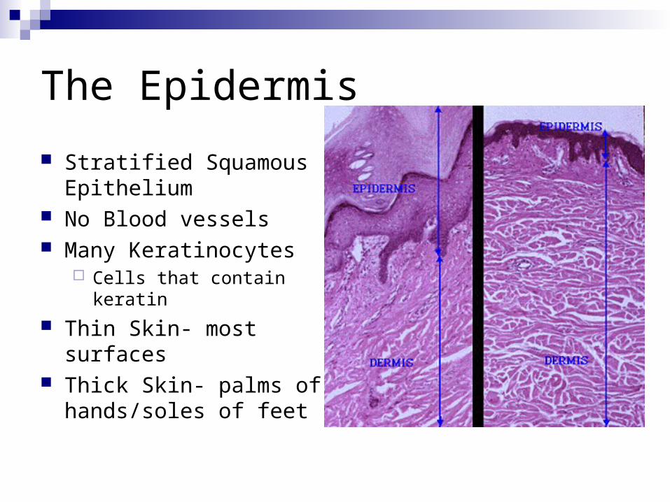
The Epidermis
Stratified Squamous Epithelium
No Blood vessels Many Keratinocytes
Cells that contain keratin
Thin Skin- most surfaces Thick Skin- palms of
hands/soles of feet
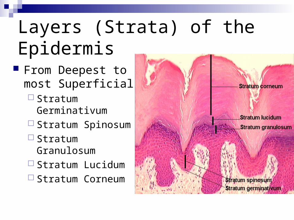
Layers (Strata) of the Epidermis
From Deepest to most Superficial Stratum Germinativum Stratum Spinosum Stratum Granulosum Stratum Lucidum Stratum Corneum
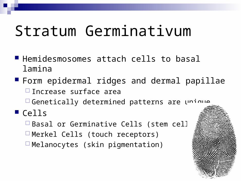
Stratum Germinativum
Hemidesmosomes attach cells to basal lamina Form epidermal ridges and dermal papillae
Increase surface area Genetically determined patterns are unique
Cells Basal or Germinative Cells (stem cells) Merkel Cells (touch receptors) Melanocytes (skin pigmentation)
Stratum spinosum
“Spiny Layer” of 8-10 rows of keratinocytes
Results from one of the daughter cells from stem cell division being pushed up from the stratum germinativum
Langerhans cellsStimulate immune response to
microorganisms and cancer
Stratum Granulosum
“Grainy Layer” of 3-5 layers of keratinocytes
Lots of Keratin (tough protein) & Keratohyaline (promotes dehydration)
Thin flat cells with decreased permeability
Stratum Lucidum
In thick skin of palms and soles only Clear layer that covers the stratum
granulosum Flat, dense, and filled with keratin
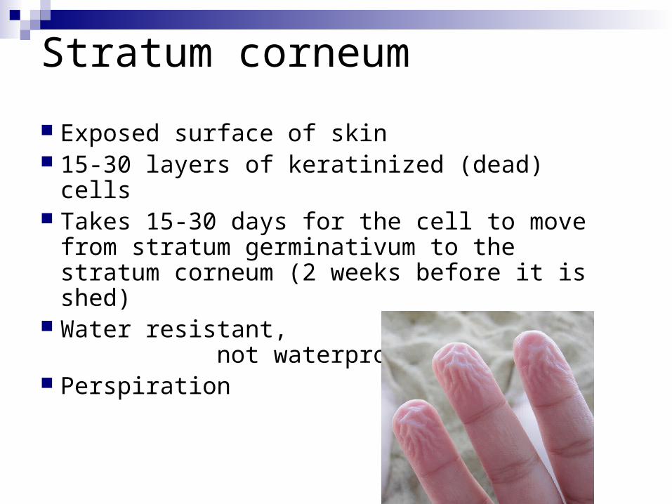
Stratum corneum
Exposed surface of skin 15-30 layers of keratinized (dead) cells Takes 15-30 days for the cell to move
from stratum germinativum to the stratum corneum (2 weeks before it is shed)
Water resistant, not waterproof
Perspiration
Journal #2: Give the layers of the epidermis from the most superficial to the deepest.
Vocabulary9. UV Radiation10. Cyanosis11. Vitamin D12. Epidermal Growth
Factor13. Papillary Layer14. Reticular Layer15. Hypodermis
Objective: List the components of
the integumentary system and their relationship to each other
Specify the functions of the integumentary system
Skin Color Pigmentation
Carotene Orange yellow pigment in epidermal cells Can convert to Vitamin A (needed in the growth of epidermal
cells) Melanin
Brown, yellow, or black pigment Melanocytes produce it in the stratum germinativum and
store in vesicles called melanosomes Dark skinned people have larger melanosomes Synthesis increases with UV exposure
Dermal Circulation Red tones produced by hemoglobin in RBC’s Cyanosis- blue tone to skin
Other Roles of the Epidermis
Steroid Production UV Radiation in epidermal
cells in the stratum spinosum and germinativum convert a steroid into cholecalciferol or Vitamin D
Helps in bone development and maintenance (Rickets-abnormal bone development due to lack of Vitamin D
Epidermal Growth Factor Made by salivary glands and
the glands of the duodenum Functions
Promotes cell division in stratum germinativum and spinosum
Speeds up production of keratin in keratinocytes
Stimulates skin repair and development
Stimulates activity and secretion in epithelial glands
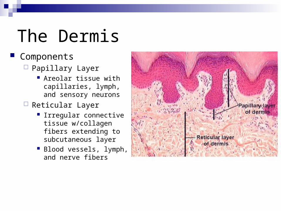
The Dermis Components
Papillary Layer Areolar tissue with
capillaries, lymph, and sensory neurons
Reticular Layer Irregular connective tissue
w/collagen fibers extending to subcutaneous layer
Blood vessels, lymph, and nerve fibers
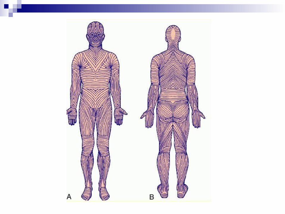
Characteristics of the Dermis Strength and Elasticity
Collagen & elastic fibers Water content
Lines of Cleavage (pattern fibers make) Cut parallel will heal with little scarring Cut at a right angle will have greater scarring
Blood Supply Arteries to subcutaneous and border reticular layer called
cutaneous plexus Innervation
Sensory reception Light touch: tactile (Meisners’s) corpuscles Deep pressure & vibration: lamellated (Pacinian) corpuscles
Subcutaneous Layer
HypodermisStabilizes skin in relation to muscles and
other organsAreolar and adipose (baby fat) tissueVenous circulation contains a great amount of
blood Subcutaneous injection effective