Joints
40
SKELETAL JOINTS Dr. Deepak K Gupta
-
Upload
deepak-kumar-gupta -
Category
Health & Medicine
-
view
37 -
download
7
Transcript of Joints
SKELETAL JOINTSDr. Deepak K Gupta
Joints (Articulations)
Weakest parts of the skeleton
Articulation – site where two or more bones meet
Functions of joints
Give the skeleton mobility
Hold the skeleton together
Classification of Joints: Structural
Structural classification focuses on the material binding bones
together and whether or not a joint cavity is present
The three structural classifications are:
Fibrous
Sutures
Gomphoses
Syndesmoses
Cartilaginous
Synchondroses
symphyses
Synovial
Classification of Joints: Functional
Functional classification is based on the amount of
movement allowed by the joint
The three functional classes of joints are:
Synarthroses – immovable
Amphiarthroses – slightly movable
Diarthroses – freely movable
Fibrous Structural Joints
The bones are joined by fibrous tissues
There is no joint cavity
Most are immovable
There are three types – sutures, syndesmoses, and
gomphoses
Fibrous Structural Joints: Sutures
Occur between the bones of the skull
Comprised of interlocking junctions completely
filled with connective tissue fibers
Bind bones tightly together, but allow for growth
during youth
In middle age, skull bones fuse and are called
synostoses
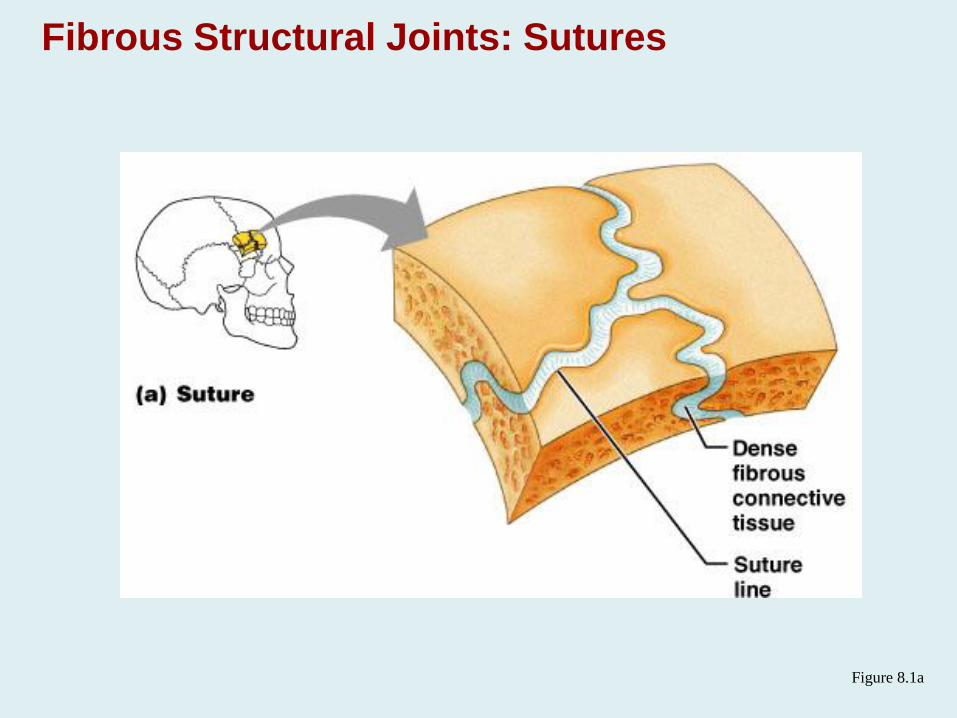
Fibrous Structural Joints: Sutures
Figure 8.1a
Fibrous Structural Joints: Syndesmoses
Bones are connected by a fibrous tissue ligament
Movement varies from immovable to slightly
variable
Examples include the connection between the tibia
and fibula, and the radius and ulna
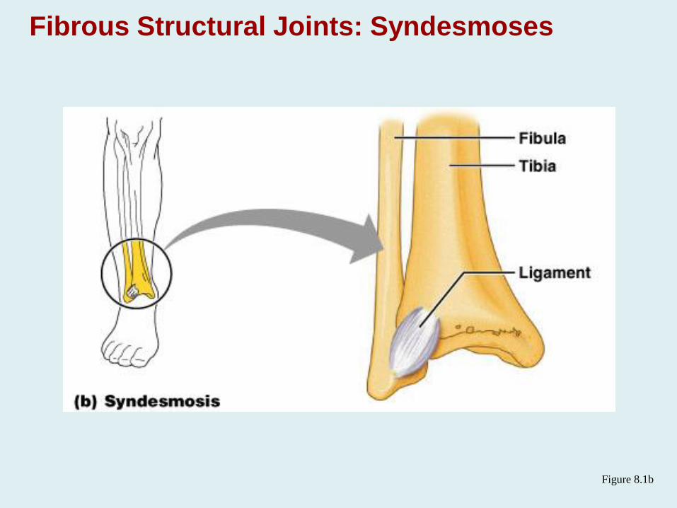
Fibrous Structural Joints: Syndesmoses
Figure 8.1b
Fibrous Structural Joints: Gomphoses
The peg-in-socket fibrous joint between a tooth and
its alveolar socket
The fibrous connection is the periodontal ligament
Cartilaginous Joints
Articulating bones are united by cartilage
Lack a joint cavity
Two types – synchondroses and symphyses
Cartilaginous Joints: Synchondroses
A bar or plate of hyaline cartilage unites the bones
All synchondroses are synarthrotic
Examples include:
Epiphyseal plates of children
Joint between the costal cartilage of the first rib
and the sternum
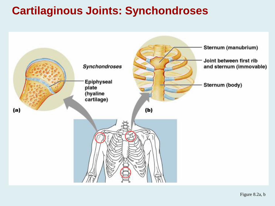
Cartilaginous Joints: Synchondroses
Figure 8.2a, b
Cartilaginous Joints: Symphyses
Hyaline cartilage covers the articulating surface of
the bone and is fused to an intervening pad of
fibrocartilage
Amphiarthrotic joints designed for strength and
flexibility
Examples include intervertebral joints and the
pubic symphysis of the pelvis
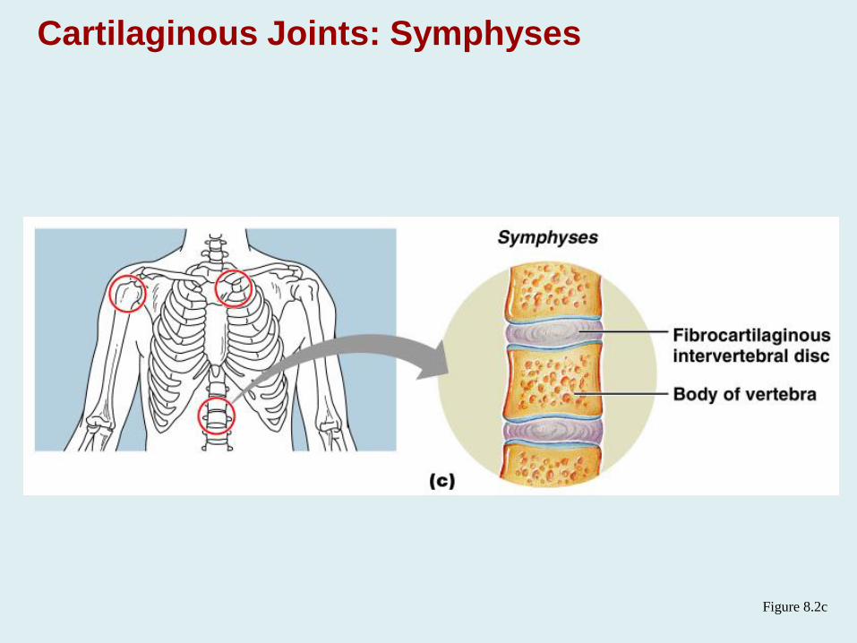
Cartilaginous Joints: Symphyses
Figure 8.2c
Synovial Joints
Those joints in which the articulating bones are
separated by a fluid-containing joint cavity
All are freely movable diarthroses
Examples – all limb joints, and most joints of the
body
Synovial Joints: General Structure
Synovial joints all have the following
Articular cartilage
Joint (synovial) cavity
Articular capsule
Synovial fluid
Reinforcing ligaments
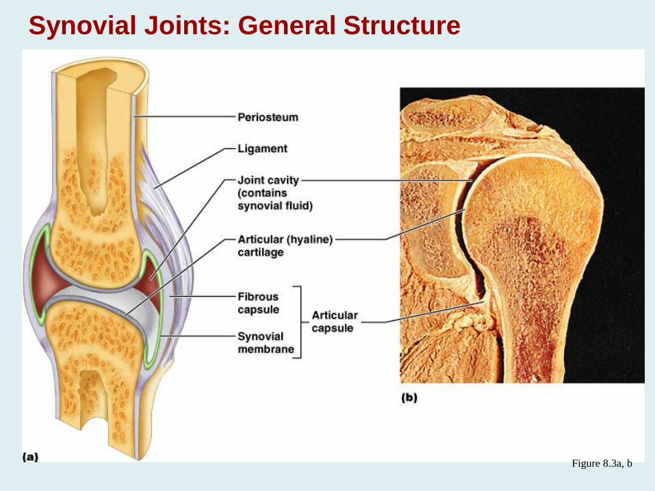
Synovial Joints: General Structure
Figure 8.3a, b
Synovial Joints: Stability
Stability is determined by:
Articular surfaces – shape determines what movements are possible
Ligaments – unite bones and prevent excessive or undesirable motion
Muscle tone is accomplished by:
Muscle tendons across joints acting as stabilizing factors
Tendons that are kept tight at all times by muscle tone
Synovial Joints: Movement
The two muscle attachments across a joint are:
Origin – attachment to the immovable bone
Insertion – attachment to the movable bone
Described as movement along transverse, frontal, or
sagittal planes
Synovial Joints: Range of Motion
Nonaxial – slipping movements only
Uniaxial – movement in one plane
Biaxial – movement in two planes
Multiaxial – movement in or around all three planes
Types of Synovial Joints
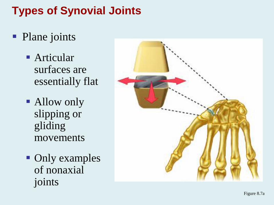
Plane joints
Articularsurfaces are essentially flat
Allow only slipping or gliding movements
Only examples of nonaxialjoints
Figure 8.7a
Types of Synovial Joints
Hinge joints
Cylindrical projections of one bone fits into a
trough-shaped surface on another
Motion is along a single plane
Uniaxial joints permit flexion and extension only
Examples: elbow and interphalangeal joints
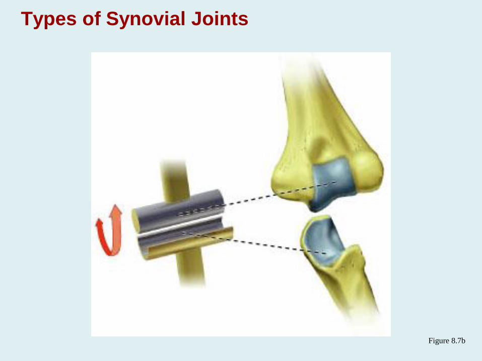
Types of Synovial Joints
Figure 8.7b
Pivot Joints
Rounded end of one bone protrudes into a “sleeve,”
or ring, composed of bone (and possibly ligaments)
of another
Only uniaxial movement allowed
Examples: joint between the axis and the dens, and
the proximal radioulnar joint
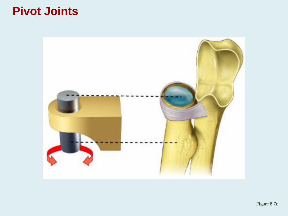
Pivot Joints
Figure 8.7c
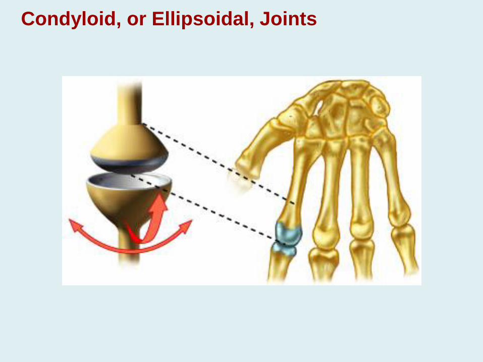
Condyloid, or Ellipsoidal, Joints
Oval articular surface of one bone fits into a
complementary depression in another
Both articular surfaces are oval
Biaxial joints permit all angular motions
Examples: radiocarpal (wrist) joints, and
metacarpophalangeal (knuckle) joints
Condyloid, or Ellipsoidal, Joints
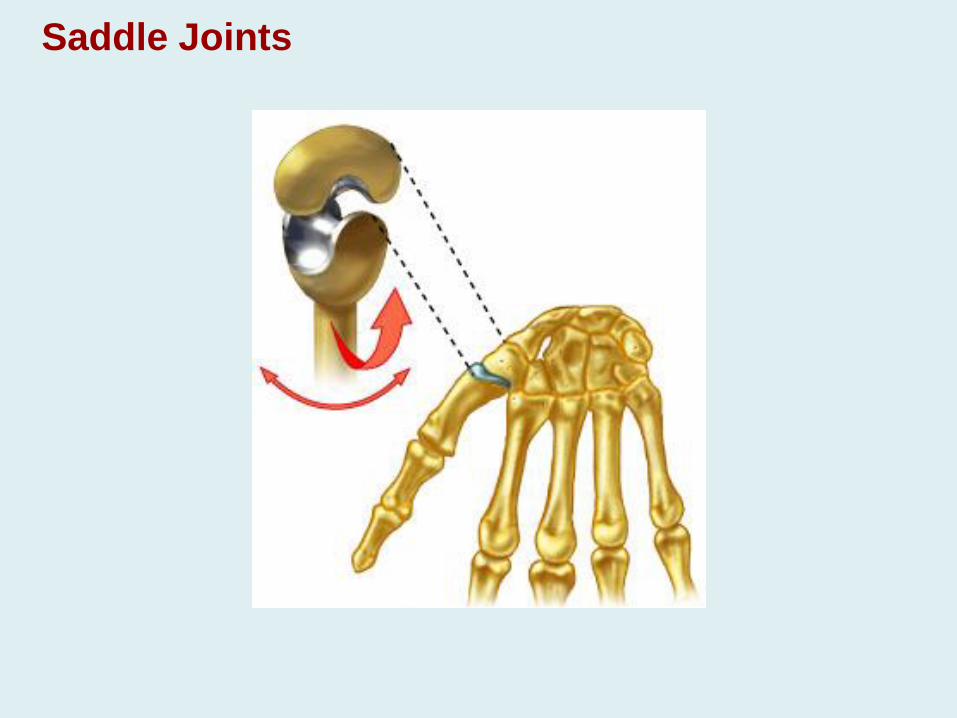
Saddle Joints
Similar to condyloid joints but allow greater
movement
Each articular surface has both a concave and a
convex surface
Example: carpometacarpal joint of the thumb
Saddle Joints
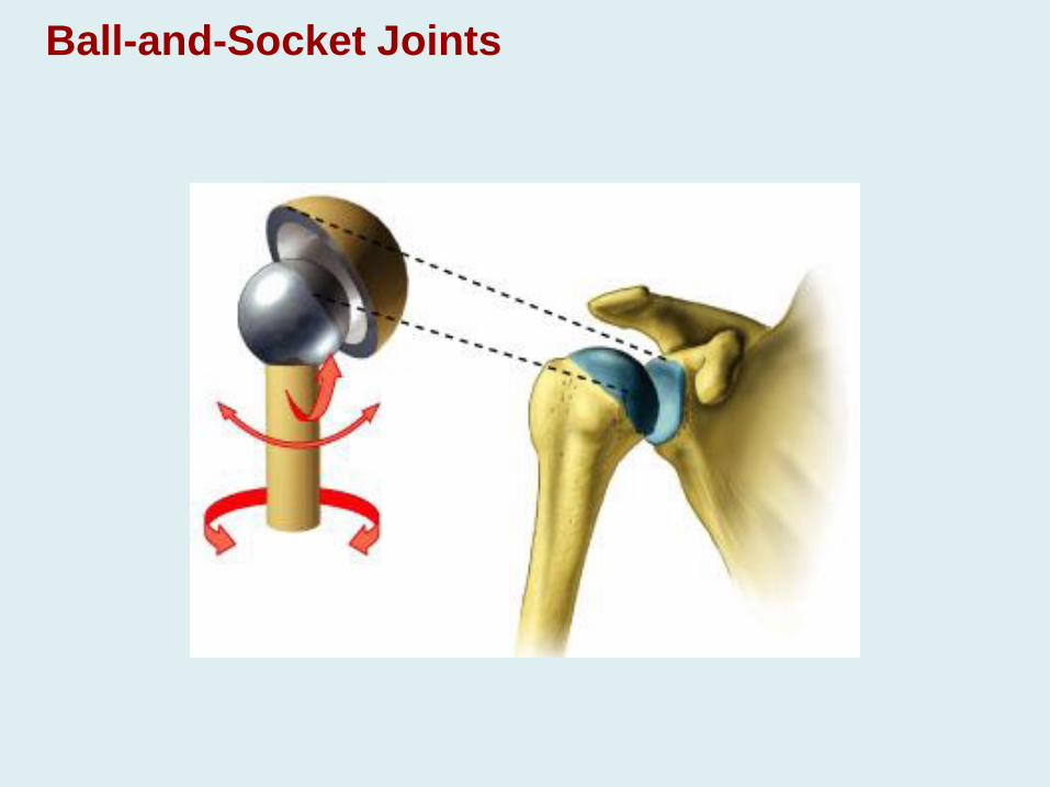
Ball-and-Socket Joints
A spherical or hemispherical head of one bone
articulates with a cuplike socket of another
Multiaxial joints permit the most freely moving
synovial joints
Examples: shoulder and hip joints
Ball-and-Socket Joints
Clinical Aspect
Sprains
The ligaments reinforcing a joint are stretched or
torn
Partially torn ligaments slowly repair themselves
Completely torn ligaments require prompt surgical
repair
Cartilage Injuries
The snap and pop of overstressed cartilage
Common aerobics injury
Repaired with arthroscopic surgery
Dislocations
Occur when bones are forced out of alignment
Usually accompanied by sprains, inflammation, and
joint immobilization
Caused by serious falls and are common sports
injuries
Subluxation – partial dislocation of a joint
Inflammatory and Degenerative Conditions
Bursitis
An inflammation of a bursa, usually caused by a blow or friction
Symptoms are pain and swelling
Treated with anti-inflammatory drugs; excessive fluid may be aspirated
Tendonitis
Inflammation of tendon sheaths typically caused by overuse
Symptoms and treatment are similar to bursitis
Arthritis
More than 100 different types of inflammatory or
degenerative diseases that damage the joints
Most widespread crippling disease in the U.S.
Symptoms – pain, stiffness, and swelling of a joint
Acute forms are caused by bacteria and are treated
with antibiotics
Chronic forms include osteoarthritis, rheumatoid
arthritis, and gouty arthritis
Osteoarthritis (OA)
Most common chronic arthritis; often called “wear-
and-tear” arthritis
Affects women more than men
85% of all Americans develop OA
More prevalent in the aged, and is probably related
to the normal aging process
Rheumatoid Arthritis (RA)
Chronic, inflammatory, autoimmune disease of
unknown cause, with an insidious onset
Usually arises between the ages of 40 to 50, but may
occur at any age
Signs and symptoms include joint tenderness,
anemia, osteoporosis, muscle atrophy, and
cardiovascular problems
The course of RA is marked with exacerbations and
remissions