
Johnson & Johnson Vision Optometry’s Partner...The TECNIS Symfony Extended Range of Vision IOL,...
12
Johnson & Johnson Vision Sondra Black, OD Crystal Clear Vision Ontario, Canada Mark R. Bloomenstein, OD, FAAO Schwartz Laser Eye Center Scottsdale, Arizona Doug Devries, OD Eye Care Associates of Nevada Sparks, Nevada SPONSORED BY Optometry’s Partner in Bringing Healthy Vision to Everyone, Everywhere, Every Day Paul M. Karpecki, OD, FAAO Kentucky Eye Institute Lexington, Kentucky Charissa Lee, OD, FAAO Johnson & Johnson Vision Jacksonville, Florida This report is for and on behalf of Johnson & Johnson Vision. Drs. Karpecki, Black, Devries and Bloomenstein are paid consultants for Johnson & Johnson Vision. Dr. Lee is an employee of Johnson & Johnson Vision.
Transcript of Johnson & Johnson Vision Optometry’s Partner...The TECNIS Symfony Extended Range of Vision IOL,...

1
Johnson & Johnson Vision
Sondra Black, ODCrystal Clear VisionOntario, Canada
Mark R. Bloomenstein, OD, FAAOSchwartz Laser Eye CenterScottsdale, Arizona
Doug Devries, ODEye Care Associates of NevadaSparks, Nevada
SPONSORED BY
Optometry’s Partner
in Bringing Healthy Vision to
Everyone, Everywhere, Every Day
Paul M. Karpecki, OD, FAAOKentucky Eye InstituteLexington, Kentucky
Charissa Lee, OD, FAAOJohnson & Johnson Vision Jacksonville, Florida
This report is for and on behalf of Johnson & Johnson Vision. Drs. Karpecki, Black, Devries and Bloomenstein are paid consultants for Johnson & Johnson Vision. Dr. Lee is an employee of Johnson & Johnson Vision.
1217_JJ_CataractCLs-V3.indd 11217_JJ_CataractCLs-V3.indd 1 12/14/17 3:03 PM12/14/17 3:03 PM

2
Dear Colleague,In 2017, Johnson & Johnson completed the acquisition of Abbott Medical Optics (AMO),
adding ophthalmic products in three areas of patient care: cataract surgery, laser refractive surgery (LASIK), and consumer eye health. These products joined with the world-leading ACUVUE® Brand Contact Lens business, and formed our combined organization known as Johnson & Johnson Vision.
Johnson & Johnson Vision recently completed the acquisition of TearScience, Inc.—a medical device manufacturer dedicated to evaluating and treating meibomian gland dysfunction, the leading cause of dry eye disease—adding innovative solutions to our dry eye portfolio.
Johnson & Johnson Vision brings together insights, technology, and people to encourage professionals and patients to proactively preserve and enhance vision for a lifetime of overall well-being. We deliver meaningful innovation to improve patients’ sense of sight and enhance professionals’ abilities to deliver better outcomes. In the pages that follow you’ll learn more about the steps we’re taking to advance eye health for the good of your patients.
Sincerely,
Millicent L. Knight, OD, FAAO, FAARM Vice President, Professional Education & DevelopmentNorth America, Vision Care
TABLE OF CONTENTSContemporary Strategies for Mitigating the Effects of Presbyopia
By Paul M. Karpecki, OD, FAAO
New Possibilities for Astigmatism Management
By Sondra Black, OD, and Charissa Lee, OD, FAAO
IOL Technology: The Relevance of Optical Design
By Sondra Black, OD
Dry Eye and Meibomian Gland Dysfunction
By Doug Devries, OD
Perioperative Management of Cataract Surgery Patients
By Doug Devries, OD, and Paul M. Karpecki, OD, FAAO
The Evolution of Refractive Surgery
By Marc R. Bloomenstein, OD, FAAO
3
4
6
8
7
9
1217_JJ_CataractCLs-V3.indd 21217_JJ_CataractCLs-V3.indd 2 12/14/17 3:03 PM12/14/17 3:03 PM
3
Contemporary Strategies for Mitigating the Effects of Presbyopia
By Paul M. Karpecki, OD, FAAO
Optometrists perform
an estimated 88
million comprehensive
eye exams annually
of the 104 million
performed by all eye
care professionals.2
“
”
Every patient over the age of 50 is impacted by presbyopia,1 yet only 6.5% of patients receive a presbyopia-correcting IOL during cataract surgery. Without a doubt, cataract patients can benefit tremendously by the guidance of a clinically astute and caring OD. Optometrists
perform an estimated 88 million comprehensive eye exams annually of the 104 million performed by all eye care professionals.2 That’s 85 percent of all comprehensive eye exams.
The combination of excellent care and an expanded menu of IOL options opens up a whole new world of possibilities to patients.
A Different IOL Type
Until recently, there were three primary classes of IOLs—monofocals, accommodating lenses, and multifocals. Each of these are also available in toric designs. However, last year an entirely different class of lens was approved by the FDA. The TECNIS Symfony® IOL is the first Extended Depth of Focus (EDOF) Presbyopia-Correcting IOL for patients with and without astigmatism. This lens is worth learning more about.
Make no mistake, the TECNIS Symfony® IOL is not a multifocal IOL. A multifocal IOL works by taking different images at different distances and separating them. Conversely, the TECNIS Symfony® IOL elongates the focus—hence the term “extended depth of focus.” The unique shape of the echelettes on the TECNIS Symfony® IOL is designed to create an elongated area of focus. This results in a high-quality continuous range of vision for patients, and helps provide intermediate and near vision with low incidence of glare and halo and without compromising distance vision.3 The TECNIS Symfony® IOL also corrects for chromatic aberration, which improves image contrast, allowing you to further increase visual quality.
Be Part of the Legacy
Consider life expectancy when considering an IOL; will your patient be missing out on many years, or decades, of quality vision? We now have the technology to strive for outstanding versus satisfactory. With this comes an opportunity to give patients better vision.
With this in mind, work with a surgeon who uses technology that can help deliver excellent out-comes for your patients. IOLs leave a lasting legacy and we can be part of it.
1. 2016 Market Scope
2. http://reviewob.com/wp-content/uploads/2016/11/8-21-13stateofoptometryreport.pdf
3. TECNIS® Symfony DFU
INDICATIONS FOR USE
The TECNIS Symfony Extended Range of Vision IOL, Model ZXR00, is indicated for primary implantation for the visual correction of aphakia, in adult patients with less than 1 diopter of pre-existing corneal astigmatism, in whom a cataractous lens has been removed. The lens mitigates the effects of presbyopia by providing an extended depth of focus. Compared to an aspheric monofocal IOL, the lens provides improved intermediate and near visual acuity, while maintaining comparable distance visual acuity. The Model ZXR00 IOL is intended for capsular bag placement only. The TECNIS Symfony Toric Extended Range of Vision IOLs, Models ZXT150, ZXT225, ZXT300, and ZXT375, are indicated for primary implantation for the visual correction of aphakia and for reduction of residual refractive astigmatism in adult patients with greater than or equal to 1 diopter of preoperative corneal astigmatism, in whom a cataractous lens has been removed. The lens mitigates the effects of presbyopia by providing an extended depth of focus. Compared to an aspheric monofocal IOL, the lens provides improved intermediate and near visual acuity, while maintaining comparable distance visual acuity. The Model Series ZXT IOLs are intended for capsular bag placement only.
See Important Safety Information in back of Supplement.
1217_JJ_CataractCLs-V3.indd 31217_JJ_CataractCLs-V3.indd 3 12/14/17 3:04 PM12/14/17 3:04 PM

4
New Possibilities for Astigmatism Management
When you compare astigmatism correction options that were available a decade ago to what we
have today, it’s truly remarkable. Whether our astigmatic patient is a 30-year old contact lens wearer or a 75-year old cataract patient, we can help guide him or her to solutions that meet or exceed expectations, without the excessive compromise that patients with astigmatism have historically endured.
Overcome Compromise
with Contact Lenses
In a study assessing contact lens wearer priorities, astigmats were six times more likely to experience visual compromise than their spherical counterparts.1 And, just like their spherical counterparts, astigmats struggle with comfort as well. For example, evidence suggests that more than two-thirds of monthly wearers are struggling with comfort issues.2 For some, a daily disposable may be
the answer, but there are certain patients who are always going to be better suited to monthly replacement lenses. In fact, 40% of spherical contact lens wearers are in the monthly cate-gory.3 This may be due to cost, the familiarity of a monthly schedule, or simply a desire to get the most out of their lenses. In any case, it is our responsibility to meet the needs of these monthly wearers—even when they have the added burden of astigmatism.
Johnson & Johnson Vision’s ACUVUE® VITA® Brand Contact Lenses with HydraMax® Technology can help us do this. To begin, the lenses incorporate beneficial lipids from the tear film during wear to help address a change in lens hydration, which may be one of the key factors involved in contact lens discomfort.4 By maximizing and maintaining lens hydra-tion, HydraMax® Technology is able to deliver reliable, exceptional comfort—all month long.
Furthermore, ACUVUE® VITA® for ASTIGMATISM is the first monthly replace-ment lens with BLINK STABILIZED® Design. Inspired by the anatomy of the eyelids and their dynamic interaction with a contact lens during blinks, lenses with this design have active zones or points of stabilization at the top and bottom of two carefully designed contour areas that fit between the eyelids. Their vertical and horizontal symmetry mini-mizes the influence of gravity, helping the lens stay in position (or quickly rotate back into position), even with eye and head movements. BLINK STABILIZED® Design makes it easy to fit astigmats because the symmetrical design helps the lenses settle quickly.
Many factors impact the experi-ence of astigmatic patients who wear
By Sondra Black, OD, and Charissa Lee, OD, FAAO
Research shows
that most contact
lens patients
who experience
discomfort say
they don’t plan to
tell their eye care
providers about
it,5 so we shouldn’t
wait for patients to
complain before
considering another
option.
“
” See Important Safety Information in back of Supplement.
1217_JJ_CataractCLs-V3.indd 41217_JJ_CataractCLs-V3.indd 4 12/14/17 3:04 PM12/14/17 3:04 PM

5
monthly lenses, and we should endeavor to address them. ACUVUE® VITA® for ASTIG-MATISM helps us do that. Finally, research shows that most contact lens patients who experience discomfort say they don’t plan to tell their eye care providers about it,5 so we shouldn’t wait for patients to complain before considering another option.
Addressing Astigmatism
with Cataract Surgery
Toric IOLs provide more stable and pre-dictable refraction than manual incision sur-gery and can reduce dependence on glasses.8,9 These benefits, combined with mitigating the effects of presbyopia, make the TECNIS Symfony® Toric IOL a welcome addition to our vision-correction armementarium.
One-third of patients have >1.0D of astig-matism but only one-fourth of those patients are receiving a toric IOL. Furthermore, every patient over the age of 50 is impacted by pres-byopia,6 yet only 6.5% of patients receive a pres-byopia-correcting IOL. Previously, there have been no FDA-approved IOL options capable of addressing both visual deficits. Now that these options exist, it is our responsibility to educate patients when surgery may be able to more fully meet their needs and match their expectations.
The approval of the TECNIS Symfony® Toric IOL gives patients the choice to address both presbyopia and astigmatism in one surgery. This is the first presbyopia-correcting lens available in the United States for the 33% of the population who have significant levels of astigmatism. While it is true that monovi-sion toric lenses have been around for a long time, for many patients, they weren’t per-ceived as good enough. Offering a premium procedure at a premium price, yet explaining that most presbyopia symptoms will remain is a difficult conversation that many patients find confusing. With the TECNIS Symfony®
Toric IOL, the premium lens conversation is much easier.
The lens uses TECNIS Symfony® IOL extended depth of focus technology, providing a wide range of vision. In addition, it has the TECNIS® Toric IOL astigmatism correction that surgeons are accustomed to using. In-deed, many surgeons who have long implant-ed torics, yet shied away from presbyopia-cor-recting technology have adopted the TECNIS Symfony® Toric IOL. There are many reasons for this. For example, the TECNIS Symfony® IOL is often referred to as a “forgiving lens” insofar as it offers extended depth of focus with tolerance to astigmatism and decentra-tion. This means patients achieve quality of vision benefits across the entire range with low glare and halo. How? The TECNIS Sym-fony® IOL echelettes elongate the focal range rather than splitting the light and creating a second focal point. This helps provide inter-mediate and near vision with low incidence of glare and halo and without compromising distance vision.7 The TECNIS Symfony® IOL also corrects for chromatic aberration, which improves image contrast.
We now have the technology to strive for outstanding versus satisfactory. Don’t let your patient miss out on years of quality vision. It’s time to learn about extended depth of focus IOLs, educate patients about the benefits, and work with surgeons who use technology that can help deliver excellent outcomes.
1. Data on fi le, Johnson & Johnson Vision, 2008.2. Schnider CM, Wales M. Optician, April, 2017;28-30.3. ProVoice, November 2015. 4. Dumbleton K, et al. IOVS 2013;54(11):TFOS20-36. 5. Schnider CM, Wales M. Optician, April, 2017;28-30.6. 2016 Market Scope7. TECNIS® Symfony DFU8. Mingo-Botin D, et al. Comparison of toric intraoc-ular lenses and peripheral corneal relaxing incisions to treat astigmatism during cataract surgery. J Cataract Refract Surg. 2010;36:1700-8.9. Hirnschall N, et al. Correction of moderate corneal astigmatism during cataract surgery: Toric intraocu-lar lens versus peripheral corneal relaxing incisions. J Cataract Refract Surg. 2014;40:354-61.
See Important Safety Information in back of Supplement.
“The approval of the
TECNIS Symfony®
Toric IOL gives
patients the choice
to address both
presbyopia and
astigmatism in one
surgery. This is the
fi rst presbyopia-
correcting lens
available in the
United States for
the 33% of the
population who have
signifi cant levels of
astigmatism.
”
1217_JJ_CataractCLs-V3.indd 51217_JJ_CataractCLs-V3.indd 5 12/14/17 3:04 PM12/14/17 3:04 PM
6
IOL Technology: The Relevance of Optical Design
The TECNIS Symfony® IOL and the TECNIS Symfo-ny® Toric IOL feature a different kind of IOL technology that was categorized differently by the FDA. The TECNIS
Symfony® is not a monofocal or a multifocal IOL. Rather, it incorporates an innovative diffractive design and achromatic technology that make the lens different compared to predeces-sors. A multifocal IOL works by taking different images at dif-ferent distances and separating them. Conversely, the TECNIS Symfony® IOL elongates the focus—hence the term “extended depth of focus.”
It All Begins with the EcheletteThe TECNIS Symfony® IOL features the same single-piece
acrylic material as other lenses on the TECNIS platform. As such, it offers the same remarkable spherical aberration correc-tion that’s typical with this entire family of lenses. It also has a diffractive grating that makes it look a little bit like a multifocal lens; however, there are big differences.
The proprietary diffractive echelette design introduces a nov-el pattern of light diffraction that elongates the focus, resulting in an extended range of vision. In addition, the achromat tech-nology reduces chromatic aberration to boost image quality.
The nine echelettes on a TECNIS Symfony® IOL elongate the focus area rather than splitting the light and creating a second focal point. This helps to enhance near and intermediate vision without compromising distance vision.1
Qualitative VisionThere is so much more to vision than vision charts reveal.
Qualitative vision is just as important as quantitative vision, and the optical design of the TECNIS Symfony® IOL addresses both of these.
To begin, the TECNIS Symfony® IOL design also allows for less chromatic aberration, which delivers contrast sensitivity with no clinically significant difference compared to a mono-focal IOL.1 By correcting the chromatic aberration, potential acuity or quality of vision may increase. And, if needed, the lens demonstrates decentration tolerance, which is important to many surgeons.
Patients implanted with the TECNIS Symfony® IOL achieved mean binocular uncorrected vision of 20/20 for intermediate
distance and between 20/25 and 20/32 for near in clinical trials.1
The TradeoffsWe all know how important distance vision is to our patients,
which is why we want to make sure that these needs are satisfied with the IOL they choose. With the TECNIS Symfony® IOL, monocular and binocular distance visual acuities were compa-rable to those of the monofocal control group in clinical trials.1
In other words, patients had high quality of vision at all distances, and gains in near vision did not come at the expense of distance vision.
Night-vision symptoms are another valid concern when choosing a presbyopia-correcting IOL. The TECNIS Symfony® IOL does not eliminate all concerns of glare and halo, but the spontaneous reports of night vision symptoms were low in clini-cal studies.1 In fact, the vast majority of TECNIS Symfony® pa-tients had no spontaneous reports of halo, glare, or starbursts.
Among those who did report night vision symptoms, most experienced only mild or moderate symptoms, with less than 3% classifying their symptoms as “severe.”1
1. TECNIS® Symfony DFU
2. Data on File._Data on File_Tecnis Symfony Green Light Bundle
Bench Test DOF2014CT0005. Abbott Medical Optics Inc. 2014
See Important Safety Information in back of Supplement.
By Sondra Black, OD
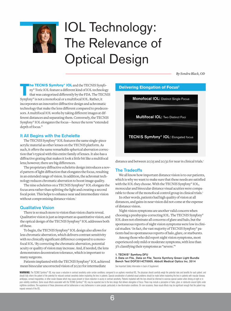
Delivering Elongation of Focus2
Monofocal IOL: Distinct Single Focus
Multifocal IOL: Two Distinct Foci
TECNIS Symfony® IOL: Elongated focus
WARNING: The TECNIS Symfony® IOL may cause a reduction in contrast sensitivity under certain conditions, compared to an aspheric monofocal IOL. The physician should carefully weigh the potential risks and benefi ts for each patient, and should fully inform the patient of the potential for reduced contrast sensitivity before implanting the lens in patients. Special consideration of potential visual problems should be made before implanting the lens in patients with macular disease, amblyopia, corneal irregularities, or other ocular disease which may cause present or future reduction in acuity or contrast sensitivity. Patients implanted with the lens should be informed to exercise special caution when driving at night or in poor visibility conditions. Some visual effects associated with the TECNIS Symfony® IOL may be expected due to the lens design that delivers elongation of focus. These may include a perception of halos, glare, or starbursts around lights under nighttime conditions. The experience of these phenomena will be bothersome or very bothersome in some people, particularly in low-illumination conditions. On rare occasions, these visual effects may be signifi cant enough that the patient may request removal of the IOL.
1217_JJ_CataractCLs-V3.indd 61217_JJ_CataractCLs-V3.indd 6 12/14/17 3:04 PM12/14/17 3:04 PM
7
By Doug Devries, OD
Dry Eye and Meibomian Gland Dysfunction
Dry eye is the largest patient segment within eye care practices, with an estimated 300 million dry eye suffer-ers worldwide and 23 million sufferers over the age of
20 in the United States alone.1 Whether or not the condition is being diagnosed and treated, most primary care optometrists likely see several dry eye patients every day. Indeed, research shows that 38% of patients visiting their eye care professional suffer from dry eye.1
Until recently, diagnosing and treating dry eye has been a tremendous challenge. However, as our knowledge of what causes dry eye improves, so too does our ability to effectively address it.
Evaluation and Imaging Optometrists have traditionally utilized tests that identify
the impact of the sequelae of dry eye on the tear film and oc-ular surface. These tests examine aspects of the disease such as ocular surface damage, tear film stability, osmolarity, and inflammation. All of these are helpful, but they don’t identify the primary cause or causes of dry eye.
In our practice, to get to the root cause, we turn to dynamic meibography in many cases—especially in a dry eye exam. As the International Workshop on MGD revealed, meibo-mian gland function and structure are essential pieces of the dry eye puzzle.2 The workshop authors concluded that the obstruction of meibomian glands is an underestimated condi-tion and is likely the leading cause of dry eye disease.2
Imaging the eyelids with LipiScan™ Dynamic Meibomian Imager or LipiView® II Ocular Surface Interferometer makes visualization of gland structure straightforward. Both devices feature Dynamic Meibomian Imaging™ that offers fast, high-definition gland scans. The LipiView® II also provides real-time measurement and visualization of lipid layer thick-ness and allows you to evaluate the dynamic response of the lipids to blinking.
TreatmentWhether your patient is a contact lens wearer, a cataract
surgery candidate, or is simply uncomfortable, the underlying dry eye disease must be addressed. Make no mistake, dry eye with MGD can be problematic whether or not a patient has symptoms. Furthermore, our treatments shouldn’t be aimed at addressing only symptoms.
A modern approach includes treating the primary cause of
dry eye rather than just treating the symptoms. The LipiFlow® Thermal Pulsation System has allowed us to achieve this in a growing number of cases. The LipiFlow® Activator, a single-use sterile device, comfortably delivers automated therapeutic energies to the meibomian glands simultaneously while pro-tecting the delicate structures of the patient’s eye.
Its contoured design vaults the cornea and protects the eye, allowing a maximum therapeutic temperature of 43 degrees Celsius to reach glands from the inner eyelid, without dam-aging the eyelid or delicate structures of the globe. Vectored Thermal Pulse™ (VTP) technology applies a combination of heat and pressure to the inner eyelid to remove gland obstruc-tions and stagnant gland content. The therapeutic motion also provides proximal-to-distal peristalsis to clear gland contents.
Over the years, we’ve turned to many products and pre-scriptions to treat the symptoms of dry eye, but few address the root cause: MGD. As we learn more about the condition, it becomes clear that we need to do more than provide limited symptomatic relief. Fortunately, today we can.
1. Yu J, Asche CV, Fairchild CJ. The economic burden of dry eye disease
in the United States: A decision tree analysis. Cornea 2011; 30(4):379-87.
2. Nichols KK, Foulks GN, Bron AJ, et al. The international workshop on
meibomian gland dysfunction: executive summary. Invest Ophthalmol
Vis Sci 2011;52(4):1922–9.
See Important Safety Information in back of Supplement.
As the Interna-
tional Workshop
on MGD revealed,
meibomian gland
function and struc-
ture are essential
pieces of the dry
eye puzzle.2
“
”
1217_JJ_CataractCLs-V3.indd 71217_JJ_CataractCLs-V3.indd 7 12/14/17 3:04 PM12/14/17 3:04 PM
8
In order to responsibly care for cataract patients, optom-etrists need to take a more hands-on approach. This begins with an improved understanding of IOL technology and a
greater comfort level when it comes time to educate patients on options that will meet their needs and match their expec-tations. In addition to patient education, we need to be able to confidently care for cataract patients in preoperative and postoperative periods.
Preoperative CarePreoperatively, we need to be very confident that the ocular
surface is healthy before referring the patient to surgery. Failure to do so puts the patient at risk of suboptimal out-comes. For example, MGD decreases refractive accuracy1,2 and stability of visual acuity.2
Always perform a complete ocular surface exam before re-ferring patients for cataract surgery. If the surgeon catches it, the patient may be sent back to our practice for pretreatment, feeling less confident in our skills.
When evaluating the ocular surface, never overlook the meibomian glands. Pay close attention to gland structure and function, and examine the lid margin for telangectasias, notch-ing in the lid, or posterior dragging of the glands. You can also measure the lipid layer with LipiView ocular surface interfer-ometer. The Meibomian Gland Evaluator is also a helpful tool to express the glands and assess the quality and flow of the secretions.
In the event you uncover a problem, there may be benefits to pretreating ocular surface disease.1,2 Most importantly, it may help optimize ocular biometry for optimum IOL power calculations and may minimize postop complications.1,2 It also may eliminate or reduce the chance of ocular discomfort that patients may experience with dry eye.1,2
Day 1
If the patient returns to your care at postop Day 1, review medications and check IOP. Also ensure that the wound is secure and the cornea is clear, noting any edema. The anterior chamber should be well-formed with about 2+ cell, and the IOL should be well-centered in the pupil. At the Day 1 postop visit, check distance vision only. Do not check near vision at this visit. Finally, review instructions with the patient and fax the results of your exam to the surgeon.
Week 1
At the Week 1 visit, review the history, confirm medications and check IOP. The slit-lamp exam should be clear to < grade 2 cell. Look closely for infection or increased signs of inflamma-tion. Check uncorrected vision at distance and near with good lighting. Finally, fax your exam results to the surgeon.
One Month
At the one-month visit, ascertain how the patient is functioning. Check uncorrected vision at distance and near with good lighting, and determine the final refraction. Check IOP again. At the slit lamp, the cornea should be clear. The anterior chamber should be well-formed with no cell, and the IOL should be well-centered in the pupil. Look for any surface disease. Evaluate posterior capsule. A fundus exam should also be performed at the one-month visit. Confirm that there is no CME, and check peripheral retina. Finally, fax your results to the surgeon.
Three Months
The main purpose of the three-month exam is to assess for the presence of posterior capsular opacification and to optimize outcomes by treating any visual fluctuation resulting from ocular surface disease. Again, all of this should be com-municated back to the surgeon.
1. Epitropoulos AT. Visual and refractive outcomes of a toric presby-
opia-correcting intraocular lens. J Ophthalmol. 2016.
2. Liang Q, Dong Z, Wang N. Perioperative ocular surface evaluation
and management in meibomian gland dysfunction patients undergoing
cataract surgery. Zhonghua Yan Ke Za Zhi. 2014 Apr;50(4):244-6.
See Important Safety Information in back of Supplement.
Perioperative Management of Cataract Surgery Patients
By Doug Devries, OD, and Paul M. Karpecki, OD, FAAO
The Meibomian Gland Evaluator is
also a helpful tool to express the
glands and assess the quality and
fl ow of the secretions.
“”
1217_JJ_CataractCLs-V3.indd 81217_JJ_CataractCLs-V3.indd 8 12/14/17 3:05 PM12/14/17 3:05 PM

9
The Evolution of Refractive Surgery
As the refractive surgery landscape evolves, so too must optometry’s narrative when educating patients. Although many optometrists are stretched thin with
waiting rooms full of spectacle and contact lens patients, ensuring that LASIK touch-points also have a presence in your practice is important. Without these, your patients may as-sume that you aren’t involved in refractive surgery. Therefore, if they start to develop an interest, they may not turn to you first for advice.
The good news is you don’t have to spend a ton of time get-ting into specifics about refractive surgery. The first and most important step is educating yourself. It is incumbent upon us as referring optometrists to have in-depth knowledge about the technology that is used by surgeons to whom we direct our patients.
The Path to PrecisionIn the past, LASIK was performed using a standard laser
pattern that was based on a single measurement. This method relied on refraction results and the same type of patient preference methodology that we use to compare corrective lens options. The prescription, as generated from a phoropter, was then entered into the laser for the treatment. From there, a theoretical curve, similar to that of glasses, determined the spot pattern that was applied to the eye. Every patient with the same prescription received the same laser treatment. But, be-cause the eye’s curvature varies and the compensation for the steepness of the curvature is based on averages, some spheri-cal aberration and other aberrations were often induced.
Later, when wavefront-optimized LASIK was introduced, surgeons could use special software to reduce LASIK-induced spherical aberration. Using wavefront modeling and theory, surgeons can administer extra pulses in the periphery of the laser ablation area to manage the induced spherical aberration. However, as with conventional LASIK, the same number of laser pulses are used for every patient with the same pre-scription. In other words, the procedure is customized to be prescription-specific, but it is not specific to the individual aberrations of each eye. Furthermore, “optimized” simply implies that adjustments have been made to treatments to minimize induction of spherical aberration; it does not mean you can correct ones that exist already. In some patients, depending on their unique curvature, it can actually make the
aberrations worse. That is where wavefront guided excels.
Another Step ForwardThere is a difference between wavefront-optimized LASIK
and wavefront-guided LASIK. With wavefront-guided LASIK, the goal is to reduce all higher order aberrations. This is achieved by projecting a small beam of infrared light onto the retina and then analyzing the scattered light that is collected by the lens and cornea by breaking it into a grid of small sam-ples. The wavefront technology captures and measures each ray of light and determines the aberrations of the entire eye based upon the deflection of the light from its ideal path.
This technique can measure hundreds of individualized light rays that go beyond the ability of the phoropter. From there, the treatment software calculates the number and diam-eter of laser pulses to reshape the cornea so that the light from objects will focus perfectly on the fovea.
New FrontiersAdvanced technology doesn’t merely provide excellent
results, it also often makes it available to more patients. With the iDESIGN® System, many more patients may now be eligible for iLASIK—even those who may have previously been told they did not qualify. That’s because iDESIGN® can provide a more detailed map of the eye, making it possible to treat patients with higher levels of astigmatism, a wider range of pupil sizes, and even mixed astigmatism. iDESIGN’s ability to measure patients with higher astigmatism gives high-reso-lution treatment accessibility to a population that can benefit significantly from it.
Patients also love that the iDESIGN® uses the same imaging technology that NASA used in developing the James Webb Space Telescope.
Patients rely on their optometrists to educate and guide them to appropriate procedures. While it is incumbent upon the surgeon to deliver excellent outcomes, it often is up to the optometrist to help educate and advise the patient to such possibilities as well as the safety concerns of LASIK surgeries. LASIK can be performed using many different technologies and techniques. It’s up to us to frame the conversation prop-erly and match the patient with a surgeon who can deliver the result we all hope for.
By Marc R. Bloomenstein, OD, FAAO
See Important Safety Information in back of Supplement.
1217_JJ_CataractCLs-V3.indd 91217_JJ_CataractCLs-V3.indd 9 12/14/17 3:05 PM12/14/17 3:05 PM

iDESIGN® Advanced WaveScan Studio System and STAR S4 IR® Exci-mer Laser System Healthcare Professional Indications and Important Safety Information
CAUTION: U.S. Federal Law restricts this device to sale, distribution, and use by or on the order of a physician or other licensed eye care practitioner. ATTENTION: Reference the Operator’s Manuals for the STAR S4 IR® Excimer Laser System and iDESIGN® Advanced WaveScan Studio (iDESIGN®) System for a complete listing of Indications and Important Safety Information. INDICATIONS: The STAR S4 IR® Excimer Laser and the iDESIGN® System is indicated for wavefront guided LASIK in patients with myopia as measured by the iDESIGN® System up to -11.00 D SE, with up to -5.00 D cylinder; in patients with hyperopia with or without astigmatism as measured by the iDESIGN® System up to +4.00 D SE, with up to +2.00 D cylinder; and in patients with mixed astigmatism as measured by the iDESIGN® System where the magnitude of the cylinder (1.0 D to 5.0 D) is greater than the magnitude of the sphere, and the cylinder and sphere have opposite signs; with agreement between manifest refraction (adjusted for optical infi nity) and the iDESIGN® System refraction of 1) SE: magnitude of the difference is < 0.625 D, and 2) cylinder: magnitude of the difference is ≤ 0.5 D; with patients 18 years of age and older, and with refractive stability (a change of ≤ 1.0 D in sphere or cylinder for a minimum of 12 months prior to surgery). CONTRAINDICATIONS: Laser refractive surgery is contraindicated for: patients with collagen vascular, autoim-mune, or immunodefi ciency diseases, pregnant or nursing women, patients with signs of corneal abnormalities including signs of keratoconus, abnormal corneal topography, epithelial basement membrane disease (EBMD) and degenerations of the structure of the cornea, patients with symptoms of signifi cant dry eyes, patients whose corneal thickness would cause the anticipated treatment to violate the posterior 250 microns (µm) of corneal stroma, and in patients with advanced glaucoma, and uncontrolled diabetes. If the patients have severely dry eyes, LASIK may increase the dryness; this may or may not go away. Severe eye dryness may delay healing of the fl ap or interfere with the surface of the eye after surgery; it may result in poor vision after LASIK. WARNINGS AND PRECAUTIONS: LASIK is not recommended in patients who: have systemic diseases likely to affect wound healing, such as autoimmune connective tissue disease, diabetes or an immunocompromised status, have a history of Herpes simplex or Herpes zoster keratitis, have severe allergies or tendency rub their eyes often, have glaucoma, elevated IOP, ocular hypertension or being followed for possible glaucoma (glaucoma suspect), are taking the medication Isotretinoin (Accutane®), are taking antimetabolites for any medical conditions. The safety and effectiveness of this laser for LASIK correction have NOT been established in patients: with progressive refractive errors, ocular disease, corneal abnormal-ity, previous corneal or intraocular surgery, or trauma in the ablation zone, who are taking the medication Sumatriptan (Imitrex®), or Amiodarone hydrochloride (Cordarone®), with corneal neovascularization within 1.0 mm of the ablation zone, over the long term (more than 1 year after surgery for myopia and more than 2 years for mixed astigmatism), for patients who engage in activities that could endanger or damage the LASIK fl ap, for patients who have a family history of degenerative corneal disease, history of infl ammation of the eye, for patients who have a history of crossed eyes (strabismus) or who have undergone strabis-mus surgery, prior LASIK or Refractive Surgery, with history of any eye diseases or abnormalities such as corneal scars or active disease, and whose BSCVA is worse than 20/20. To reduce the risk of corneal ectasia, the posterior 250 microns (µm) of corneal stroma should not be violated. The treatment of highly myopic eyes necessitates the removal of signifi cant amounts of corneal tissue. The iDESIGN® System calculates the estimated residual bed depth using the pachymetry and intended fl ap thickness entered by the user. Actual fl ap thick-nesses may vary. If the estimated residual stromal bed is ≤ 320 microns, an in-the-bed pachymetric measurement should be performed. ADVERSE EVENTS: Possible adverse events include loss of best spectacle corrected visual acuity (BSCVA), serious Transient Light Sensitivity Syndrome, serious primary open angle glaucoma, miscreated fl ap, melting of the fl ap, severe glare, and severe dry eyes. Complications can include corneal edema, epithelial ingrowth, diffuse lamellar keratitis, foreign body sensation, and pain.
©2017 AMO Manufacturing USA, LLC. iDESIGN, iDESIGN Advanced WaveS-can Studio, and STAR S4 IR are trademarks owned by or licensed to AMO Manufacturing USA, LLC, its subsidiaries, or affi liates. All other trademarks are the intellectual property of their respective owners.
INDICATIONS AND IMPORTANT SAFETY INFORMATION FOR TECNIS SYMFONY AND TECNIS SYMFONY TORIC EXTENDED RANGE OF VISION IOLsRx Only
INDICATIONS FOR USEThe TECNIS Symfony Extended Range of Vision IOL, Model ZXR00, is indicated for primary implantation for the visual correction of aphakia, in adult patients with
less than 1 diopter of pre-existing corneal astigmatism, in whom a cataractous lens has been removed. The lens mitigates the effects of presbyopia by providing an ex-tended depth of focus. Compared to an aspheric monofocal IOL, the lens provides improved intermediate and near visual acuity, while maintaining comparable distance visual acuity. The Model ZXR00 IOL is intended for capsular bag placement only.
The TECNIS Symfony Toric Extended Range of Vision IOLs, Models ZXT150, ZXT225, ZXT300, and ZXT375, are indicated for primary implantation for the visual correction of aphakia and for reduction of residual refractive astigmatism in adult patients with greater than or equal to 1 diopter of preoperative corneal astigmatism, in whom a cataractous lens has been removed. The lens mitigates the effects of presbyopia by providing an extended depth of focus. Compared to an aspheric monofocal IOL, the lens provides improved intermediate and near visual acuity, while maintaining comparable distance visual acuity. The Model Series ZXT IOLs are intended for capsular bag placement only.
WARNINGSPhysicians considering lens implantation under any of the following circumstances should weigh the potential risk/benefi t ratio:1. Patients with any of the following conditions may not be suitable candidates for an intraocular lens because the lens may exacerbate an existing condition, may interfere with diagnosis or treatment of a condition, or may pose an unreasonable risk to the patient’s eyesight:
a) Patients with recurrent severe anterior or posterior segment infl ammation or uveitis of unknown etiology, or any disease producing an infl ammatory reaction in the eye.
b) Patients in whom the intraocular lens may affect the ability to observe, diag-nose or treat posterior segment diseases.
c) Surgical diffi culties at the time of cataract extraction, which may increase the potential for complications (e.g., persistent bleeding, signifi cant iris damage, uncontrolled positive pressure or signifi cant vitreous prolapse or loss).
d) A compromised eye due to previous trauma or developmental defects in which appropriate support of the IOL is not possible.
e) Circumstances that would result in damage to the endothelium during implantation.
f) Suspected microbial infection.g) Patients in whom neither the posterior capsule nor the zonules are intact
enough to provide support for the IOL.h) Children under the age of 2 years are not suitable candidates for intraocular
lenses.i) Congenital bilateral cataracts.j) Previous history of, or a predisposition to, retinal detachment.k) Patients with only one good eye with potentially good vision.l) Medically uncontrollable glaucoma.m) Corneal endothelial dystrophy.n) Proliferative diabetic retinopathy.
2. The Tecnis Symfony IOL should be placed entirely in the capsular bag and should not be placed in the ciliary sulcus.
3. The Tecnis Symfony IOL may cause a reduction in contrast sensitivity under certain conditions, compared to an aspheric monofocal IOL. The physician should carefully weigh the potential risks and benefi ts for each patient, and should fully inform the patient of the potential for reduced contrast sensitivity before implanting the lens in patients. Special consideration of potential visual problems should be made before implanting the lens in patients with macular disease, amblyopia, corneal irregularities, or other ocular disease which may cause present or future reduction in acuity or contrast sensitivity.
4. Because the Tecnis Symfony IOL may cause a reduction in contrast sensitivity compared to a monofocal IOL, patients implanted with the lens should be informed to exercise special caution when driving at night or in poor visibility conditions.
5. Some visual effects associated with the Tecnis Symfony IOL may be expected due to the lens design that delivers elongation of focus. These may include a percep-tion of halos, glare, or starbursts around lights under nighttime conditions. The expe-rience of these phenomena will be bothersome or very bothersome in some people, particularly in low-illumination conditions. On rare occasions, these visual effects may be signifi cant enough that the patient may request removal of the IOL.
6. Patients with a predicted postoperative astigmatism greater than 1.0 diopter may not be suitable candidates for implantation with the Tecnis Symfony and Tecnis Sym-fony Toric IOLs, Models ZXR00, ZXT150, ZXT225, ZXT300, and ZXT375, as they may not obtain the benefi ts of reduced spectacle wear or improved intermediate and near vision seen in patients with lower astigmatism. 7. The effectiveness of Tecnis Symfony Toric IOLs in reducing postoperative residual astigmatism in patients with preoperative corneal astigmatism < 1.0 diopter has not been demonstrated.
8. Rotation of Tecnis Symfony Toric IOLs away from their intended axis can reduce their astigmatic correction. Misalignment greater than 30° may increase postopera-tive refractive cylinder. If necessary, lens repositioning should occur as early as possible prior to lens encapsulation.
9. AMO IOLs are single-use devices only. Do not reuse this IOL.
PRECAUTIONS
1. Prior to surgery, the surgeon must inform prospective patients of the possible risks and benefi ts associated with the use of this device and provide a copy of
1217_JJ_CataractCLs-V3.indd 101217_JJ_CataractCLs-V3.indd 10 12/14/17 3:05 PM12/14/17 3:05 PM

the patient information brochure to the patient.
2. When performing refraction in patients implanted with the Tecnis Symfony IOL, interpret results with caution when using autorefractors or wavefront aberrometers that utilize infrared light, or when performing a duochrome test. Confi rmation of refraction with maximum plus manifest refraction technique is recommended.
3. The ability to perform some eye treatments (e.g. retinal photocoagulation) may be affected by the Tecnis Symfony IOL optical design.
4. Recent contact lens usage may affect the patient’s refraction; therefore, in contact lens wearers, surgeons should establish corneal stability without contact lenses prior to determining IOL power.
5. Do not resterilize the lens. Most sterilizers are not equipped to sterilize the soft acrylic material without producing undesirable side effects.
6. Do not soak or rinse the intraocular lens with any solution other than sterile balanced salt solution or sterile normal saline.
7. Do not store the lens in direct sunlight or at a temperature greater than 113°F (45°C). Do not autoclave the intraocular lens.
8. The surgeon should target emmetropia as this lens is designed for optimum visual performance when emmetropia is achieved.
9. Care should be taken to achieve IOL centration, as lens decentration may result in a patient experiencing visual disturbances under certain lighting conditions.
10. When the insertion system is used improperly, Tecnis Symfony IOLs may not be delivered properly (i.e., haptics may be broken). Please refer to the specifi c instructions for use provided with the insertion instrument or system.
11. The safety and effectiveness of Tecnis Symfony IOLs have not been sub-stantiated in patients with preexisting ocular conditions and intraoperative complications (see below for examples). Careful preoperative evaluation and sound clinical judgment should be used by the surgeon to decide the benefi t/risk ratio before implanting a lens in a patient with one or more of these conditions:
Before Surgery• Pupil abnormalities• Prior corneal refractive or intraocular surgery• Choroidal hemorrhage• Chronic severe uveitis• Concomitant severe eye disease• Extremely shallow anterior chamber• Medically uncontrolled glaucoma• Microphthalmos• Non-age-related cataract• Proliferative diabetic retinopathy (severe)• Severe corneal dystrophy• Severe optic nerve atrophy• Irregular corneal astigmatism• Amblyopia• Macular disease• Pregnancy
During Surgery• Excessive vitreous loss• Non-circular capsulotomy/capsulorhexis• The presence of radial tears known or suspected at the time of surgery• Situations in which the integrity of the circular capsulotomy/capsulorhexis
cannot be confi rmed by direct visualization• Cataract extraction by techniques other than phacoemulsifi cation or
liquefaction• Capsular rupture• Signifi cant anterior chamber hyphema• Uncontrollable positive intraocular pressure• Zonular damage
12. Carefully remove all viscoelastic and do not over-infl ate the capsular bag at the end of the case. Residual viscoelastic and/or overinfl ation of the capsular bag may allow the lens to rotate, causing misalignment of the Tecnis Symfony Toric IOL with the intended axis of placement.
13. The PCA is based on an algorithm that combines published literature (Koch et.al, 2012) and a retrospective analysis of data from a TECNIS Toric multi-center clinical study. The PCA algorithm for the selection of appropriate cylinder power and axis of implantation was not assessed in a prospective clinical study and may yield results different from those in the TECNIS Toric intraocular lens labeling. Please refer to the AMO Toric Calculator user manual for more information.
14. The use of methods other than the Tecnis Toric Calculator to select cylinder power and appropriate axis of implantation were not assessed in the parent Tecnis Toric IOL U.S. IDE study and may not yield similar results. Accurate keratometry and biometry, in addition to the use of the Tecnis Toric Calculator (www.TecnisToric-Calc.com), are recommended to achieve optimal visual outcomes for the Tecnis SymfonyToric IOL.
15. All preoperative surgical parameters are important when choosing a Tecnis Symfony Toric IOL for implantation, including preoperative keratometric cylinder (magnitude and axis), incision location, surgeon’s estimated surgically induced astigmatism (SIA) and biometry. Variability in any of the preoperative measurements can infl uence patient outcomes, and the effectiveness of treating eyes with lower amounts of preoperative corneal astigmatism.
16. All corneal incisions were placed temporally in the parent Tecnis Toric IOL U.S. IDE study. If the surgeon chooses to place the incision at a different location, outcomes may be different from those obtained in the clinical study for the parent Tecnis Toric IOL. Note that the Tecnis Toric Calculator incorporates the surgeon’s estimated SIA and incision location when providing IOL options.
17. Potential adverse effects (e.g., complications) associated with the use of the device include the following:
• Infection (endophthalmitis)• Hypopyon• IOL dislocation• Cystoid macular edema• Corneal edema• Pupillary block • Iritis• Retinal detachment/tear • Raised IOP requiring treatment • Visual symptoms requiring lens removal • Tilt and decentration requiring repositioning • Residual refractive error resulting in secondary intervention.
Secondary surgical interventions include, but are not limited to: • Lens repositioning (due to decentration, rotation, subluxation, etc.) • Lens replacement • Vitreous aspirations or iridectomy for pupillary block • Wound leak repair • Retinal detachment repair • Corneal transplant • Lens replacement due to refractive error • Unacceptable optical/visual symptoms• Severe infl ammation
SERIOUS ADVERSE EVENTS
The most frequently reported serious adverse events that occurred during the clinical trial of the Tecnis Symfony lens were cystoid macular edema (2 eyes, 0.7%) and surgical reintervention (treatment injections for cystoid macular edema and en-dophthalmitis, 2 eyes, 0.7%). One eye was reported with pupillary capture and the eye that had endophthalmitis also had a small hypopyon. No other serious adverse events and no lens-related adverse events occurred during the trial.
INDICATIONS AND IMPORTANT SAFETY INFORMATION for the TECNIS
Toric 1-Piec IOL
Rx Only
INDICATIONS: The TECNIS Toric 1-Piece posterior chamber lensesare indicated for the visual correction of aphakia and pre-existing corneal astigmatism of one di-opter or greater in adult patients with or without presbyopia in whom a cataractous lens has been removed by phacoemulsifi cation and who desire improved uncorrect-ed distance vision, reduction in residual refractive cylinder, and increased spectacleindependence for distance vision. The device is intended to be placed in the capsular bag. IMPORTANT SAFETY INFORMATION: Rotation can reduce astigmatic correction. Misalignment greater than 30° may induce refractive error. Accurate keratometry, biometry and www.TecnisToricCalc.com are recommended to optimize visual outcomes. Weigh the potential risk/benefi t ratio that could increase pre-existing complications or impact patient outcomes. Variability in any preoperative measurements can infl uence outcomes.ATTENTION: Reference the Directions for Use labeling for a complete listing of Indications and Important Safety Information.
ACUVUE® Brand Contact Lenses are indicated for vision correction. As with any contact lens, eye problems, including corneal ulcers, can develop. Some wearers may experience mild irritation, itching or discomfort. Lenses should not be pre-scribed if patients have any eye infection, or experience eye discomfort, excessive tearing, vision changes, redness or other eye problems. Consult the package insert for complete information. Complete information is also available from Johnson & Johnson Vision Care, Inc., by calling 1-800-843-2020 or by visiting www.acuvueprofessional.com
ACUVUE® VITA® Brand Contact Lenses are indicated for vision correction as a daily wear lens with one-month recommended replacement. As with any contact lens, eye problems, including corneal ulcers, can develop. Some wearers may experience mild irritation, itching or discomfort. Lenses should not be prescribed if patients have any eye infection, or experience eye discomfort, excessive tearing, vision changes, redness or other eye problems. Consult the package insert for complete information. Complete information is also available by visiting acuvueprofessional.com or by calling 1-800-843-2020.
1217_JJ_CataractCLs-V3.indd 111217_JJ_CataractCLs-V3.indd 11 12/14/17 3:05 PM12/14/17 3:05 PM

12All trademarks are the intellectual property of their respective owners.© Johnson & Johnson Vision Care, Inc. 2017PP2017CT2031
1217_JJ_CataractCLs-V3.indd 121217_JJ_CataractCLs-V3.indd 12 12/14/17 3:05 PM12/14/17 3:05 PM