)JOEBXJ1VCMJTIJOH$PSQPSBUJPO …downloads.hindawi.com/journals/bmri/2014/378235.pdf · Inflammation...
9
Research Article Neutralizing Effects of Mimosa tenuiflora Extracts against Inflammation Caused by Tityus serrulatus Scorpion Venom Mariana Angélica Oliveira Bitencourt, 1 Maira Conceição Jerônimo de Souza Lima, 1 Manoela Torres-Rêgo, 1 Júlia Morais Fernandes, 2 Arnóbio Antônio da Silva-Júnior, 1 Denise Vilarinho Tambourgi, 3 Silvana Maria Zucolotto, 2 and Matheus de Freitas Fernandes-Pedrosa 1 1 Laboratory of Pharmaceutical Technology and Biotechnology, Department of Pharmacy, Federal University of Rio Grande do Norte, Avenue General Gustavo Cordeiro de Farias, Petr´ opolis, 59012-570 Natal, RN, Brazil 2 Laboratory of Pharmacognosy, Department of Pharmacy, Federal University of Rio Grande do Norte, Natal, RN, Brazil 3 Laboratory of Immunochemistry, Butantan Institute, S˜ ao Paulo, SP, Brazil Correspondence should be addressed to Matheus de Freitas Fernandes-Pedrosa; [email protected] Received 24 February 2014; Revised 5 May 2014; Accepted 8 May 2014; Published 11 June 2014 Academic Editor: Ruxana Sadikot Copyright © 2014 Mariana Ang´ elica Oliveira Bitencourt et al. is is an open access article distributed under the Creative Commons Attribution License, which permits unrestricted use, distribution, and reproduction in any medium, provided the original work is properly cited. Scorpion bite represents a significant and serious public health problem in certain regions of Brazil, as well as in other parts of the world. Inflammatory mediators are thought to be involved in the systemic and local immune response induced by Tityus serrulatus scorpion envenomation. e aim of this study was to evaluate the effect of extracts of Mimosa tenuiflora on model envenomation. In mice, the envenomation model is induced by Tityus serrulatus venom. Previous treatment of mice with fractions from M. tenuiflora was able to suppress the cell migration to the peritoneal cavity. e treatment of mice with M. tenuiflora extracts also decreased the levels of IL-6, IL-12, and IL-1. We concluded that the administration of the extract and fractions resulted in a reduction in cell migration and showed a reduction in the level of proinflammatory cytokines. is study demonstrates, for the first time, the anti-inflammatory effect of aqueous extract from the Mimosa tenuiflora plant on T. serrulatus venom. 1. Introduction Scorpion bite represents a significant and serious public health problem in certain regions of Brazil, as well as in other parts of the world, due to the frequency of their occurrence and to their potential for inducing severe, even fatal, clinical manifestations, especially among children [1]. In Brazil, most fatalities result from bites received from the Tityus serrulatus scorpion. e Brazilian Ministry of Health reports approximately 8000 scorpion bites/year, and the mortality rate among children is 1% [2]. e specific signs of scorpion envenomation are directly related to the venom components, with some patients devel- oping an inflammatory response. Although the production of pro- and anti-inflammatory cytokines in response to tissue injury is essential to repair tissue structure and function, excessive generation of proinflammatory cytokines can aggravate tissue damage [3]. Many different cytokines are released following severe envenomation. Increased inter- leukin IL-6 levels have been observed in plasma from patients with different grades of T. serrulatus envenomation. High levels of IL-6 and IL-1 were also observed in mice exposed to Centruroides noxius and T. serrulatus scorpion venoms [4– 6]. Injection of scorpion venom into experimental animals produces systemic effects, with signs and symptoms similar to those observed in human envenomation, being fever, psy- chomotor agitation, salivation, lachrymation, increased gas- trointestinal tract mobility, cardiac and respiratory arrhyth- mias, and arterial hypertension followed by hypotension, car- diac failure, pulmonary edema, and shock, among others [2, 7, 8]. ese features have been traditionally explained by the effects of neurotransmitters released by the scorpion venom, Hindawi Publishing Corporation BioMed Research International Volume 2014, Article ID 378235, 8 pages http://dx.doi.org/10.1155/2014/378235
-
Upload
nguyentram -
Category
Documents
-
view
215 -
download
0
Transcript of )JOEBXJ1VCMJTIJOH$PSQPSBUJPO …downloads.hindawi.com/journals/bmri/2014/378235.pdf · Inflammation...

Research ArticleNeutralizing Effects of Mimosa tenuiflora Extracts againstInflammation Caused by Tityus serrulatus Scorpion Venom
Mariana Angélica Oliveira Bitencourt,1 Maira Conceição Jerônimo de Souza Lima,1
Manoela Torres-Rêgo,1 Júlia Morais Fernandes,2
Arnóbio Antônio da Silva-Júnior,1 Denise Vilarinho Tambourgi,3
Silvana Maria Zucolotto,2 and Matheus de Freitas Fernandes-Pedrosa1
1 Laboratory of Pharmaceutical Technology and Biotechnology, Department of Pharmacy, Federal University of Rio Grande do Norte,Avenue General Gustavo Cordeiro de Farias, Petropolis, 59012-570 Natal, RN, Brazil
2 Laboratory of Pharmacognosy, Department of Pharmacy, Federal University of Rio Grande do Norte, Natal, RN, Brazil3 Laboratory of Immunochemistry, Butantan Institute, Sao Paulo, SP, Brazil
Correspondence should be addressed to Matheus de Freitas Fernandes-Pedrosa; [email protected]
Received 24 February 2014; Revised 5 May 2014; Accepted 8 May 2014; Published 11 June 2014
Academic Editor: Ruxana Sadikot
Copyright © 2014 Mariana Angelica Oliveira Bitencourt et al. This is an open access article distributed under the CreativeCommons Attribution License, which permits unrestricted use, distribution, and reproduction in any medium, provided theoriginal work is properly cited.
Scorpion bite represents a significant and serious public health problem in certain regions of Brazil, as well as in other parts of theworld. Inflammatory mediators are thought to be involved in the systemic and local immune response induced by Tityus serrulatusscorpion envenomation.The aim of this study was to evaluate the effect of extracts ofMimosa tenuiflora onmodel envenomation. Inmice, the envenomationmodel is induced by Tityus serrulatus venom. Previous treatment of mice with fractions fromM. tenuiflorawas able to suppress the cell migration to the peritoneal cavity. The treatment of mice with M. tenuiflora extracts also decreasedthe levels of IL-6, IL-12, and IL-1𝛽. We concluded that the administration of the extract and fractions resulted in a reduction incell migration and showed a reduction in the level of proinflammatory cytokines. This study demonstrates, for the first time, theanti-inflammatory effect of aqueous extract from theMimosa tenuiflora plant on T. serrulatus venom.
1. Introduction
Scorpion bite represents a significant and serious publichealth problem in certain regions of Brazil, as well as in otherparts of the world, due to the frequency of their occurrenceand to their potential for inducing severe, even fatal, clinicalmanifestations, especially among children [1]. In Brazil,most fatalities result from bites received from the Tityusserrulatus scorpion. The Brazilian Ministry of Health reportsapproximately 8000 scorpion bites/year, and the mortalityrate among children is 1% [2].
The specific signs of scorpion envenomation are directlyrelated to the venom components, with some patients devel-oping an inflammatory response. Although the production ofpro- and anti-inflammatory cytokines in response to tissueinjury is essential to repair tissue structure and function,
excessive generation of proinflammatory cytokines canaggravate tissue damage [3]. Many different cytokines arereleased following severe envenomation. Increased inter-leukin IL-6 levels have been observed in plasma frompatientswith different grades of T. serrulatus envenomation. Highlevels of IL-6 and IL-1 were also observed in mice exposedto Centruroides noxius and T. serrulatus scorpion venoms [4–6]. Injection of scorpion venom into experimental animalsproduces systemic effects, with signs and symptoms similarto those observed in human envenomation, being fever, psy-chomotor agitation, salivation, lachrymation, increased gas-trointestinal tract mobility, cardiac and respiratory arrhyth-mias, and arterial hypertension followed by hypotension, car-diac failure, pulmonary edema, and shock, among others [2,7, 8]. These features have been traditionally explained by theeffects of neurotransmitters released by the scorpion venom,
Hindawi Publishing CorporationBioMed Research InternationalVolume 2014, Article ID 378235, 8 pageshttp://dx.doi.org/10.1155/2014/378235

2 BioMed Research International
but the release of mediators from the systemic inflammatoryresponse syndrome may also play an important role [9].
The recommended treatment for envenomation is theintravenous administration of antivenom according to theseverity of envenoming. The serum therapy is indicated inneutralizing venoms inoculated after incidence with ven-omous animals [10–12]. The serum has some disadvantages,having a series of adverse effects on the victim, such asanaphylaxis and hypersensitivity to heterologous proteinsfrom serum and inefficiency in combating the local effectsof the venom [13]. For these reasons, several studies havebeen conducted in the search for natural compounds that cancomplement the currently available serotherapy.
Mimosa tenuiflora (Willd.) Poiret (Leguminosae) is atree popularly known in Brazil as jurema-preta [14]. Theplant is distributed in areas of tropical deciduous forestsin the Americas, from the southeastern regions of Mexicoto northern Brazil and Venezuela, growing as secondaryopportunistic vegetation. According to Brazilian and Mexi-can ethnopharmacological sources, the bark of this plant—once dried, powdered, and directly applied to the lesion—isan effective remedy for treating skin burns and wounds andpreventing inflammation [15–18].
Basic preclinical studies report that aqueous and alcoholicextracts from driedM. tenuiflora bark are particularly rich intannins, compounds which seem to play an important role inthe healing mechanism; they possess in vitro antimicrobialproperties against a broad group of Gram-positive and-negative microorganisms, yeasts, and dermatophytes [19,20]. Also with treatment for severe skin ulcer, the positiveeffects ofM. tenuiflora extracts were clearly related to the highcontent of polyphenols with a high cicatrization potential[18]. Other studies allowed identification of a group of triter-penoidal saponins from the bark, designated asmimonosidesA–C [21], which, according to in vitro observations, inducedcultured human-cell proliferation, possessed immunomod-ulation capacity, and, therefore, were attributed to at leastpart of the potential cicatrizing properties of the plant’s bark[22, 23].
The aim of this study is to evaluate the neutralizing capac-ity of the extract of Mimosa tenuiflora on the inflammationinduced by Tityus serrulatus scorpion venom.
2. Materials and Methods
2.1. Extraction. The bark of Mimosa tenuiflora wascollected in the rural region of “Queimadas Mountain”(7∘03 52 S/34∘49 51 W), Parelhas, Rio Grande do NorteState, Brazil, in April 2011. The specimen was identified byAllan A. Roque. Voucher specimens of M. tenuiflora (JPB13985) have been deposited in the Herbarium UFRN at theUniversidade Federal do Rio Grande do Norte, Brazil.
2.2. Isolation. The bark ofM. tenuiflora (500 g) was air-driedat room temperature, powdered, and extracted using hotwater (100∘C) by decoction (plant: solvent, 1 : 10, w/v) for10min. Then, the extracts were filtered through Whatmanpaper no.1 and lyophilized.
Thereafter, the aqueous extract was resuspended indistilled water and partitioned with the following sol-vents: dichloromethane (3 × 300mL), ethyl acetate (3 ×300mL), and 𝑛-butanol (3 × 300mL), yielding 750mg ofdichloromethane (CH
2Cl2), 1.0 g of ethyl acetate (EtOAc),
and 2.5 g of 𝑛-butanol (𝑛-BuOH) fractions. The CH2Cl2,
EtOAc, and 𝑛-BuOH fractions were analyzed by TLC usingaluminum sheets of silica gel F254 (Merck). All chro-matograms were developed in a saturated chamber. Thefollowing solvent system was used: ethyl acetate: aceticacid: formic acid: water (8 : 0.5 : 0.5 : 0.5 v/v/v/v). After thechromatogramswere developed, the plates were dried and thespots were visualized sequentially under UV light at 254 and365 nm. The plates were then sprayed with a methanol solu-tion of sulfuric vanillin (4%) and Natural Product Reagent A0.1% (NP-Reagent). Production and purification of extractsendowed with antivenom activity are patented processes(patent number PI033120000116, INPI).
2.3. Animals. Male BALB/c mice (6–8 weeks old) were usedin the experiment. All mice were housed, 5-6 per cage, at aroom temperature of 22± 2∘Cand a 12 h : 12 h light/dark cycle.They had free access to food andwater. Groups of five animalswere used in each test group and control animals receivedsaline only. All in vivo experiments were approved by “EthicsCommittee on Animal Use, CEUA/UFRN,” under protocolnumber 008/2011, which was in accordance with the guide-lines of the BrazilianCommittee forAnimal Experimentation(COBEA).
2.4. Venom. Lyophilized Tityus serrulatus scorpion venomwas kindly supplied by the Butantan Institute, Sao Paulo,SP, Brazil. The venom was prepared in PBS at 1mg/mLconcentration and was stored at −20∘C until used.
2.5. Evaluation of T. serrulatusVenom-Induced Envenomation.In order to evaluate the envenomation induced by T. serrula-tus and establish the challenge dose, groups of six male micewere injected intraperitoneally (i.p.) with 0.1, 0.2, 0.3, 0.4, and0.8mg/kg of T. serrulatus venom dissolved in isotonic saline.At the selected time points (4, 6, and 8 hours), the animalswere anesthetized with ketamine/xylazine (80/10mg/kg i.p.)and sacrificed by cervical dislocation, and peritoneal exudateswere harvested by peritoneal wash with 3mL of cold PBS.Exudates were centrifuged at 250×g for 10min, at 4∘C, andthe total cell numbers were determined in a Neubauer cham-ber following stainingwith Turk’s solution. After selecting theappropriate dose of venom and the time, the neutralizationassay of venoming induced by Tityus serrulatus venom wasperformed by extracts ofM. tenuiflora.
2.6. Assessment of Antivenom Activity. The envenomationwas induced according to the procedure described previ-ously in Pessini, 2006, with slight modifications. Animalswere inoculated intravenously (i.v.) with saline, aqueousextracts from M. tenuiflora (20, 30, or 40mg/kg), CH
2Cl2,
EtOAc, and BuOH fractions (40mg/kg) and, five minuteslater, the animals were injected intraperitoneally with sterile

BioMed Research International 3
Sal 0.1 0.2 0.3 0.4 0.8
0
5
10
15
20
25
Tityus serrulatus venom
∗∗
Perit
onea
l leu
kocy
tes (×10
6/m
L)
∗∗∗
∗∗∗
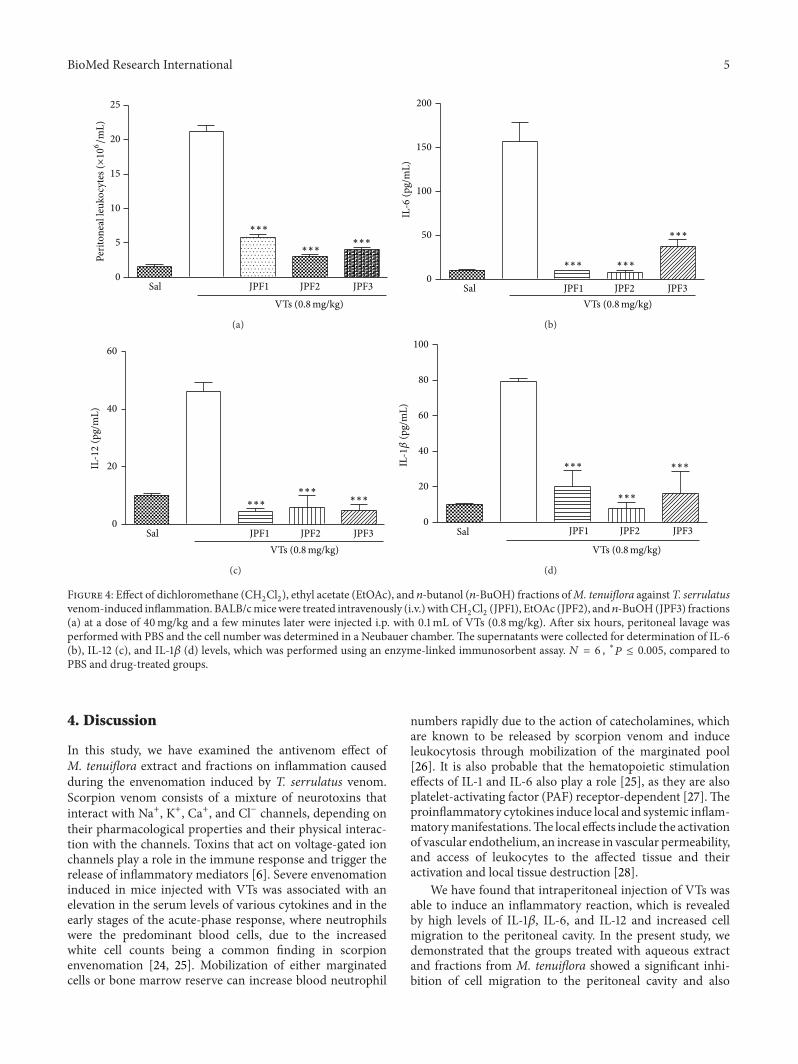
Figure 1: Tityus serrulatus venom-induced inflammation. BALB/cmice were injected i.p. with 0.1mL of VTs (0.1, 0.2, 0.3, 0.4, or0.8mg/kg). After 6 hours, peritoneal lavage was performed withPBS and the cell number was determined in a Neubauer chamber.𝑁 = 6 , ∗∗∗𝑃 ≤ 0.001, compared to PBS and 0.8mg/kg group.
PBS or T. serrulatus venom (VTs) freshly prepared (0.1mLof 0.8mg/kg) in sterile PBS. According to the observedresults, AcOEt fraction showed the best inhibition profileof inflammatory cells; therefore a dose curve response toEtOAc fraction (20, 30, or 40mg/kg) was evaluated. After6 h, the animals were anesthetized with ketamine/xylazine(80/10mg/kg i.p.) and sacrificed by cervical dislocation, andperitoneal exudates were harvested by peritoneal wash with3mL of cold PBS. Exudates were centrifuged at 250×g for10min, at 4∘C, and the total cell numbers were determined ina Neubauer chamber following staining with Turk’s solution.Results were expressed as number of neutrophils/cavity. Thesupernatants were collected for determination of IL-6, IL-12, and IL-1𝛽 (pg/mL) levels, which was performed using anenzyme-linked immunosorbent assay kit from eBioscience(San Diego, CA, USA).
2.7. Statistical Analyses. Data are expressed as the mean ±standard deviation. Statistical analyses were performed byANOVA and Tukey test and the level of significance was setat 𝑃 < 0.0001.
3. Results
The envenomation induced by VTs was assessed using ananimal model of peritonitis. A significant increase in cellmigration to peritoneal cavity was observed upon intraperi-toneal injection of VTs. Among the doses tested, the strongesteffect was obtained with 0.8mg/kg (Figure 1). This dose waschosen as the challenge dose for evaluation of the antivenomactivity ofM. tenuiflora extract (after evaluating the effect ofthree different doses of T. serrulatus-induced envenomation).After selecting the dose, the kinetics (4, 6, and 8 h) of thevenom-induced peritoneal cell migration was evaluated. Theresults presented in Figure 2 show that VTs induced amarkedincrease in the peritoneal cell migration at the three times
Perit
onea
l leu
kocy
tes (×10
6/m
L)
0
5
10
15
20
25
VTs 0.8mg/kg
∗∗∗
∗
8Hs6Hs4Hs
Figure 2: Tityus serrulatus venom-induced inflammation. BALB/cmice were injected i.p. with 0.1mL of VTs (0.8mg/kg). After 4, 6,and 8 hours, peritoneal lavage was performed with PBS and the cellnumber was determined in a Neubauer chamber. 𝑁 = 6 , ∗∗∗𝑃 ≤0.001, compared to PBS and 6-hour group.
Table 1: Anti-inflammatory activity ofMimosa tenuiflora against T.serrulatus venom-induced envenomation.
Groups Dose (mg/kg) Cell migration Inhibition (%)Saline — 21.17 ± 0.8292Aqueous extract 20 6.167 ± 1.600 71∗∗∗
Aqueous extract 30 5.000 ± 1.366 76∗∗∗
Aqueous extract 40 7.200 ± 2.498 66∗∗∗
CH2Cl2 fraction 40 5.750 ± 0.5204 73∗∗∗
n-BuOH fraction 40 4.000 ± 0.2887 81∗∗∗
EtOAc fraction 20 3.500 ± 0.5774 83∗∗∗
EtOAc fraction 30 6.900 ± 0.8124 67∗∗∗
EtOAc fraction 40 3.000 ± 0.2887 86∗∗∗
Values aremean± standard deviation (S.D.),n=6, ∗∗∗𝑃 < 0.0001, comparedto saline group.
analyzed (4, 6, and 8 h). The highest influx of cells in theperitoneal cavitywas observed at 6 h.This timewas, therefore,selected for the neutralization assay, where the antivenomactivity ofM. tenuiflora extract against venoming induced byT. serrulatus was evaluated.
The effect of aqueous extract of M. tenuiflora wasevaluated in a T. serrulatus venom-induced envenomationmodel. As expected, the animals treated intravenously (i.v.)with saline and 5min later intraperitoneally (i.p.) with VTsshowed intense leukocyte migration to the peritoneal cavity(Figure 3(a)). On the other hand, groups treated with threedifferent doses (20, 30, or 40mg/kg) ofM. tenuiflora aqueousextract showed a significant inhibition of cell migration to theperitoneal cavity, as well as a reduction in the levels of IL-6 (Figure 3(b)), IL-12 (Figure 3(c)), and IL-1𝛽 (Figure 3(d)),when compared with the group that received saline i.v. andVTs i.p. Table 1 summarizes the anti-inflammatory activity ofall plant extracts tested regarding the inhibition of peritoneal

4 BioMed Research International
Sal 20 30 40
0
5
10
15
20
25
VTs (0.8mg/kg)Aqueous extract (mg/kg)
∗∗∗∗∗∗
∗∗∗
Perit
onea
l leu
kocy
tes (×10
6/m
L)
(a)
Sal 20 30 400
50
100
150
VTs (0.8mg/kg)
Aqueous extract (mg/kg)
IL-6
(pg/
mL)
200
∗∗∗
∗∗∗∗
(b)
VTs (0.8mg/kg)Aqueous extract (mg/kg)
Sal 20 300
20
40
40
60
IL-12
(pg/
mL)
∗∗∗ ∗∗∗
∗∗∗
(c)
VTs (0.8mg/kg)Aqueous extract (mg/kg)
Sal 20 30 400
20
40
60
80
100
∗∗∗
∗∗∗∗∗∗IL-1𝛽
(pg/
mL)
(d)
Figure 3: Effect of aqueous extracts of M. tenuiflora against T. serrulatus venom-induced inflammation. BALB/c mice were treatedintravenously (i.v.) with PBS or aqueous extract at doses of 20, 30, and 40mg/kg and, a few minutes later, were injected i.p. with 0.1mLof VTs (0.8mg/kg). After six hours, peritoneal lavage was performed with PBS and the cell number was determined in a Neubauer chamber(a).The supernatants were collected for determination of IL-6 (b), IL-12 (c), and IL-1𝛽 (d) levels, whichwas performed using an enzyme-linkedimmunosorbent assay.𝑁 = 6, ∗𝑃 ≤ 0.005, compared to PBS and drug-treated groups.
cell migration, which was similar for all three doses ofaqueous extract analyzed.
Through phytochemical analysis of aqueous extract byTLC, a yellow spot (𝑅𝑓 0.36/UV 365 nm) was observed afterspraying the plate with NP-Reagent, suggesting the presenceof flavonoids. Furthermore, when the plate was revealed withvanillin sulfuric, three red spots (𝑅𝑓𝑠 0.24, 0.34, and 0.43) anda yellow spot (𝑅𝑓 0.36) were observed. According to literaturedata, these results suggest the presence of terpenes and/orsteroids. Following a bioassay-guided fractionation process,the aqueous extract was partitioned with CH
2Cl2, EtOAc,
and 𝑛-BuOH. Each fraction was submitted to a screening inthe same experimental model at the dose of 40mg/kg. Allgroups treated with the fractions showed significant inhibi-tion of cell migration to the peritoneal cavity (Figure 4(a)
and Table 1). The CH2Cl2, EtOAc, and 𝑛-BuOH fractions
showed a reduction in IL-6 (Figure 4(b)), IL-12 (Figure 4(c)),and IL-1𝛽 (Figure 4(d)) levels, respectively, when comparedwith the group administered with saline i.v. and VTs i.p. Adose-response profile (20, 30, and 40mg/kg) was made forthe EtOAc fraction (Figure 5(a)) which displayed a higherantivenom effect than other fractions. The effect on cellmigration to the peritoneal cavity was not dose-dependent,since the three doses tested induced similar inhibition. Onthe other hand, the ability of EtOAc fraction to reduce theproduction of IL-6 (Figure 5(b)), IL-12 (Figure 5(c)), and IL-1𝛽 (Figure 5(d)) was dose-dependent. A study of toxicity wasalso performed “in vitro” using cell T3T, where the aqueousextract showed dose-dependent toxic effect but only at veryhigh doses (data not shown).
BioMed Research International 5
∗∗∗
∗∗∗∗∗∗
Perit
onea
l leu
kocy
tes (×10
6/m
L)
VTs (0.8mg/kg)Sal JPF1 JPF2 JPF3
0
5
10
15
20
25
(a)
∗∗∗ ∗∗∗
∗∗∗
VTs (0.8mg/kg)
IL-6
(pg/
mL)
Sal JPF1 JPF2 JPF30
50
100
150
200
(b)
∗∗∗∗∗∗
∗∗∗
VTs (0.8mg/kg)
IL-12
(pg/
mL)
Sal JPF1 JPF2 JPF30
20
40
60
(c)
∗∗∗
∗∗∗
∗∗∗
VTs (0.8mg/kg)
Sal JPF1 JPF2 JPF30
20
40
60
80
100
IL-1𝛽
(pg/
mL)
(d)
Figure 4: Effect of dichloromethane (CH2
Cl2
), ethyl acetate (EtOAc), and 𝑛-butanol (𝑛-BuOH) fractions ofM. tenuiflora against T. serrulatusvenom-induced inflammation. BALB/cmicewere treated intravenously (i.v.) withCH
2
Cl2
(JPF1), EtOAc (JPF2), and 𝑛-BuOH(JPF3) fractions(a) at a dose of 40mg/kg and a few minutes later were injected i.p. with 0.1mL of VTs (0.8mg/kg). After six hours, peritoneal lavage wasperformed with PBS and the cell number was determined in a Neubauer chamber.The supernatants were collected for determination of IL-6(b), IL-12 (c), and IL-1𝛽 (d) levels, which was performed using an enzyme-linked immunosorbent assay. 𝑁 = 6 , ∗𝑃 ≤ 0.005, compared toPBS and drug-treated groups.
4. Discussion
In this study, we have examined the antivenom effect ofM. tenuiflora extract and fractions on inflammation causedduring the envenomation induced by T. serrulatus venom.Scorpion venom consists of a mixture of neurotoxins thatinteract with Na+, K+, Ca+, and Cl− channels, depending ontheir pharmacological properties and their physical interac-tion with the channels. Toxins that act on voltage-gated ionchannels play a role in the immune response and trigger therelease of inflammatory mediators [6]. Severe envenomationinduced in mice injected with VTs was associated with anelevation in the serum levels of various cytokines and in theearly stages of the acute-phase response, where neutrophilswere the predominant blood cells, due to the increasedwhite cell counts being a common finding in scorpionenvenomation [24, 25]. Mobilization of either marginatedcells or bone marrow reserve can increase blood neutrophil
numbers rapidly due to the action of catecholamines, whichare known to be released by scorpion venom and induceleukocytosis through mobilization of the marginated pool[26]. It is also probable that the hematopoietic stimulationeffects of IL-1 and IL-6 also play a role [25], as they are alsoplatelet-activating factor (PAF) receptor-dependent [27].Theproinflammatory cytokines induce local and systemic inflam-matorymanifestations.The local effects include the activationof vascular endothelium, an increase in vascular permeability,and access of leukocytes to the affected tissue and theiractivation and local tissue destruction [28].
We have found that intraperitoneal injection of VTs wasable to induce an inflammatory reaction, which is revealedby high levels of IL-1𝛽, IL-6, and IL-12 and increased cellmigration to the peritoneal cavity. In the present study, wedemonstrated that the groups treated with aqueous extractand fractions from M. tenuiflora showed a significant inhi-bition of cell migration to the peritoneal cavity and also

6 BioMed Research International
∗∗∗
∗∗∗∗∗∗
Perit
onea
l leu
kocy
tes (×10
6/m
L)
VTs (0.8mg/kg)Ethyl acetate fraction (mg/kg)
Sal 20 30 40
0
5
10
15
20
25
(a)
∗∗∗ ∗∗∗
VTs (0.8mg/kg)
IL-6
(pg/
mL)
Ethyl acetate fraction (mg/kg)
∗∗
Sal 20 30 400
50
100
150
200
(b)
∗∗∗∗∗∗
VTs (0.8mg/kg)
IL-12
(pg/
mL)
Ethyl acetate fraction (mg/kg)
∗∗
Sal 20 30 400
20
40
60
(c)
∗∗∗ ∗∗∗∗∗∗
VTs (0.8mg/kg)Ethyl acetate fraction (mg/kg)
Sal 20 30 400
20
40
60
80
100
IL-1𝛽
(pg/
mL)
(d)
Figure 5: Effect of ethyl acetate fraction of M. tenuiflora against T. serrulatus venom-induced inflammation. BALB/c mice were treatedintravenously (i.v.) with EtOAc fractions (a) at doses of 20, 30, and 40mg/kg and a few minutes later were injected i.p. with 0.1mL of VTs(0.8mg/kg). After six hours, peritoneal lavage was performed with PBS and the cell number was determined in a Neubauer chamber. Thesupernatants were collected for determination of IL-6 (b), IL-12 (c), and IL-1𝛽 (d) levels, which was performed using an enzyme-linkedimmunosorbent assay.𝑁 = 6 , ∗𝑃 ≤ 0.005, compared to PBS and drug-treated groups.
showed a reduction in the levels of IL-6, IL-12, and IL-1𝛽.Theextract-treated groups exhibited antivenomactivity, revealingimpairment of leukocyte migration.
There are, to our knowledge, no previous studies inthe literature presenting antivenom activity of extracts fromM. tenuiflora; however, it is known that there is a signifi-cant presence of saponins in this species [18, 21, 29]. Onehypothesis for this activity is the inhibition of the pro-duction of leukotrienes and prostaglandin E2 by inhibitingthe enzyme phospholipase A2 and COX2, respectively, aswell as blocking the release of all other inflammatorymediators involved in the venoming, as studies being con-ducted in parallel to our laboratory previously demon-strated the anti-inflammatory potential of M. tenuiflora inexperimental models of inflammation using carrageenan.Regarding envenomation-induced inflammation as studiedin the present work, it is known that the scorpion venomscan stimulate the immune-neuroendocrine axis by inducing
the release of catecholamines, corticosteroids, bradykinin[30–33], and eicosanoids mediators, such as prostaglandin(PG)E2, lipoxin A2 (LXA2), and leukotriene (LT)B4 [2,34], which are derived from the enzymatic oxygenationof arachidonic acid (AA). These signal molecules controlkey cellular processes, including cell activation, metabolism,migration, cell proliferation, and death [35, 36].
Other studies in the literature have already demonstratedthat PGE2 is involved in the inflammatory response andin the neutrophil recruitment [37] in mice inoculated withT. serrulatus scorpion venom [38]. As in certain sodiumchannel toxins and potassium (Ts2 and Ts6), T. serrulatusis involved in the release of cytokines and cell migrationby inducing the production and release of PGE2 and LTB4[39]. As already has been stated, the aqueous extract of M.tenuiflora is rich in saponins and tannins [18]. Some studiesshowed the presence of flavonoids and saponins which havebeen previously reported to have anti-inflammatory efforts in

BioMed Research International 7
vivo and in vitro. For example, some saponins and flavonoidswere reported to suppress the release of proinflammatorymediator production by inflammatory agents inmacrophages[40–43]. Some studies have shown that the mechanism ofsaponins and flavonoids in anti-inflammatory activity maybe mediated by inhibiting the activation of nuclear factor-kB,thus resulting in decreased expression of NF-kB-regulatedproteins such as inducible nitric oxide synthase (iNOS) [44–46]. Other reports revealed that tannins have the abilityto bind to proteins [47, 48], suggesting that tannins couldinactivate venom toxins preventing their toxic activity. Asenvenomation byTityus serrulatus triggers an intense inflam-matory response, it is possible that the anti-inflammatorypotential of active components in M. tenuiflora extract, aswell as other possible biological activities, contributes to theantivenom activity of the plant.
5. Conclusion
In conclusion, this is the first study to evaluate the anti-inflammatory activity of extracts of Mimosa tenuiflora usingscorpion venom. We demonstrated that the aqueous extractand fractions from M. tenuiflora exhibited important anti-inflammatory activity in a T. serrulatus venom-inducedenvenomation model. As expected, we have found that theanimals that received intraperitoneal injection of VTs wereable to induce an inflammatory reaction, which is revealedby high levels of IL-1𝛽, IL-6, and IL-12 and increased cellmigration to the peritoneal cavity. On the other hand, groupstreated with fractions showed a significant inhibition of cellmigration to the peritoneal cavity and showed a reduction inlevels of IL-6, IL-12, and IL-1𝛽. Further studies are required todetermine the chemical composition ofM. tenuiflora extractand its possible anti-inflammatory mechanisms of action.
Conflict of Interests
The authors declare that there is no conflict of interestsregarding the publication of this paper.
Acknowledgments
The authors acknowledge all participants for their valuabletime and commitment to the study and thank the CNPq andCAPES for financial support. MFFP andDVT are researchersfrom CNPq. The authors were also grateful to AndrewAlastair Cumming for editing this paper.
References
[1] J. P. Zuliani, T. A. Freitas, I. M. Conceicao, and F. H. Kwas-niewski, “Tityus serrulatus venom increases vascular permeabil-ity in selected airway tissues in a mast cell-independent way,”Experimental and Toxicologic Pathology, vol. 65, no. 3, pp. 229–234, 2013.
[2] E. B. Nascimento Jr., K. A. Costa, C. M. Bertollo et al.,“Pharmacological investigation of the nociceptive response andedema induced by venom of the scorpion Tityus serrulatus,”Toxicon, vol. 45, no. 5, pp. 585–593, 2005.
[3] E. M. S. Fialho, M. C. G. Maciel, A. C. B. Silva et al., “Immunecells recruitment and activation by Tityus serrulatus scorpionvenom,” Toxicon, vol. 58, no. 6-7, pp. 480–485, 2011.
[4] V. L. Petricevich, “Effect of Tityus serrulatus venom on cytokineproduction and the activity of murine macrophages,”Mediatorsof Inflammation, vol. 11, no. 1, pp. 23–31, 2002.
[5] V. L. Petricevich, A. Hernandez Cruz, F. I. V. Coronas, and L. D.Possani, “Toxin gamma from Tityus serrulatus scorpion venomplays an essential role in immunomodulation of macrophages,”Toxicon, vol. 50, no. 5, pp. 666–675, 2007.
[6] V. L. Petricevich, “Scorpion venom and the inflammatoryresponse,” Mediators of Inflammation, vol. 2010, Article ID903295, 16 pages, 2010.
[7] D. T. Bertazzi, A. I. de Assis-Pandochi, A. E. C. S. Azzolini, V.L. Talhaferro, M. Lazzarini, and E. C. Arantes, “Effect of Tityusserrulatus scorpion venom and its major toxin, TsTX-I, on thecomplement system in vivo,” Toxicon, vol. 41, no. 4, pp. 501–508,2003.
[8] A. F. Toro, M. B. Malta, S. L. Soares et al., “Role of IgG(T)and IgGa isotypes obtained from arachnidic antivenom toneutralize toxic activities of Loxosceles gaucho, Phoneutrianigriventer and Tityus serrulatus venoms,” Toxicon, vol. 48, no.6, pp. 649–661, 2006.
[9] P. Cupo, M. M. Azevedo-Marques, S. E. Hering et al., “Animaispeconhentos no Brasil: biologia clinica e terapeutica dos aci-dentes,” Salvier, vol. 2003, pp. 198–208, 2003.
[10] M. N. Krifi, S. Savin, M. Debray, C. Bon, M. El Ayeb, and V.Choumet, “Pharmacokinetic studies of scorpion venom beforeand after antivenom immunotherapy,”Toxicon, vol. 45, no. 2, pp.187–198, 2005.
[11] C. F. S. Amaral and N. A. Rezende, “Treatment of scorpionenvenoming should include both a potent specific antivenomand support of vital functions,” Toxicon, vol. 38, no. 8, pp. 1005–1007, 2000.
[12] L. Freire-Maia, J. A. Campos, and C. F. S. Amaral, “Approachesto the treatment of scorpion envenoming,” Toxicon, vol. 32, no.9, pp. 1009–1014, 1994.
[13] I. Amaro, L. Riano-Umbarila, B. Becerril, and L. D. Possani,“Isolation and characterization of a human antibody fragmentspecific for Ts1 toxin fromTityus serrulatus scorpion,” Immunol-ogy Letters, vol. 139, no. 1-2, pp. 73–79, 2011.
[14] M. R. Oliveira, J. M. E. Rodrigues, O. Chiavone-Filho et al.,“Estudo das condicoes de cultivo da Algaroba e Jurema pretae determinacao do poder calorifico,” Revista de Ciencia eTecnologia, vol. 14, pp. 93–104, 1999.
[15] D. A. C. Bezerra, F. F. G. Rodrigues, J. G. M. Costa et al.,“Abordagem fitoquımica, coposicao bromatologica e atividadeantibacteriana deMimosa tenuiflora (Willd) poired ePiptadeniastipulacea (Benth) ducke,” Acta Scientiarum Biological Sciences,vol. 33, pp. 99–106, 2011.
[16] A. L. Goncalves, A. A. Filho, and H. Menezes, “Estudo Com-parativo da Atividade Antimicrobiana de Extratos de AlgumasArvores Nativas,” Arquivos do Instituto Biologico, vol. 72, pp.353–358, 2005.
[17] G. N.Maia,Caatinga: Arvores e Arbustos e Suas Utilidades, D&ZComputacao, Sao Paulo, Brazil, 2004.
[18] E. Rivera-Arce, M. A. Chavez-Soto, A. Herrera-Arellano et al.,“Therapeutic effectiveness of aMimosa tenuiflora cortex extractin venous leg ulceration treatment,” Journal of Ethnopharmacol-ogy, vol. 109, no. 3, pp. 523–528, 2007.

8 BioMed Research International
[19] X. Lozoya, V. Navarro, J. T. Arnason, and E. Kourany, “Experi-mental evaluation ofMimosa tenuiflora (Willd.) poir. (Tepesco-huite) I. —screening of the antimicrobial properties of barkextracts,”Archivos de InvestigacionMedica, vol. 20, no. 1, pp. 87–93, 1989.
[20] M. Meckes, X. Lozoya, L. Gonzalez, and M. Martinez, “Efectoproducido por la fraccion de alcaloides de Mimosa tenuiflora(Tepescohuite) sobre el reflejo peristaltico del ileon del cobayo,”Archivos de Investigacion Medica, vol. 21, pp. 171–174, 1990.
[21] Y. Jiang, M. Haag-Berrurier, R. Anton et al., “Structure of a newsaponin from the bark ofMimosa tenuiflora,” Journal of NaturalProducts, vol. 54, no. 5, pp. 1247–1253, 1991.
[22] Y. Jiang, B. Weniger, M. Haag-Barrurier, R. Anton, J.-P. Beck,and L. Italiano, “Effects of saponins fromMimosa tenuiflora onlymphoma cells and lymphocytes,” Phytotherapy Research, vol.6, no. 6, pp. 310–313, 1992.
[23] R. Anton, Y. Jiang, B. Weniger, J. P. Beck, and L. Rivier,“Pharmacognosy ofMimosa tenuiflora (Willd.) poiret,” Journalof Ethnopharmacology, vol. 38, no. 2-3, pp. 153–157, 1993.
[24] A.-R. M. A. Meki and Z. M. Mohey El-Dean, “Seruminterleukin-1𝛽, interleukin-6, nitric oxide and 𝛼1-antitrypsin inscorpion envenomed children,”Toxicon, vol. 36, no. 12, pp. 1851–1859, 1998.
[25] A. C. Pessini, A. M. de Souza, L. H. Faccioli, Z. M. O. Gregorio,and E. C. Arantes, “Time course of acute-phase responseinduced by Tityus serrulatus venom and TsTX-I in mice,”International Immunopharmacology, vol. 3, no. 5, pp. 765–774,2003.
[26] C. A.M. deDavila, D. F. Davila, J. H. Donis, G. A. de Bellabarba,V. Villarreal, and J. S. Barboza, “Sympathetic nervous systemactivation, antivenin administration and cardiovascular man-ifestations of scorpion envenomation,” Toxicon, vol. 40, no. 9,pp. 1339–1346, 2002.
[27] C. M. Borges, M. R. Silveira, M. A. C. L. Beker, L. Freire-Maia,and M. M. Teixeira, “Scorpion venom-induced neutrophilia isinhibited by a PAF receptor antagonist in the rat,” Journal ofLeukocyte Biology, vol. 67, no. 4, pp. 515–519, 2000.
[28] V. L. Petricevich and I. Lebrun, “Immunomodulatory effects ofthe Tityus serrulatus venom on murine macrophage functionsin vitro,”Mediators of Inflammation, vol. 2005, no. 1, pp. 39–49,2005.
[29] Y. Jiang, G. Massiot, C. Lavaud et al., “Triterpenoid glycosidesfrom the bark ofMimosa tenuiflora,” Phytochemistry, vol. 30, no.7, pp. 2357–2360, 1991.
[30] M. C. J. S. Lima, M. A. O. Bitencourt, A. A. Furtado et al.,“Ipomoea asarifolia neutralizes inflammation induced by Tityusserrulatus scorpion venom,” Journal of Ethnopharmacology, vol.153, no. 3, pp. 890–895, 2014.
[31] I. H. Chaudry, R. N. Stephan, J. M. Harkema, and R. E. Dean,“Immunological alterations following simple hemorrhage,” inImmune Consequences of Trauma, Schock and Sepsis, F. Fasit, J.Ninnemann, and D. Green, Eds., pp. 363–373, 1989.
[32] S. Sofer, M. Gueron, R. M. White, M. Lifshitz, and R. N. Apte,“Interleukin-6 release following scorpion sting in children,”Toxicon, vol. 34, no. 3, pp. 389–392, 1996.
[33] M.M.Magalhaes, M. E. S. Pereira, C. F. S. Amaral et al., “Serumlevels of cytokines in patients envenomed by Tityus serrulatusscorpion sting,” Toxicon, vol. 37, no. 8, pp. 1155–1164, 1999.
[34] C. F. P. Teixeira, F. Galante, S. Manzoli, A. A. Steil, and S.Jancar, “Inflammatory reaction induced by Tityus serrulatuscrude venom (TsV) in the lung of rats,” Journal of VenomousAnimals and Toxins, vol. 3, pp. 111–115, 1997.
[35] C. D. Funk, “Prostaglandins and leukotrienes: advances ineicosanoid biology,” Science, vol. 294, no. 5548, pp. 1871–1875,2001.
[36] P. Yaqoob, “Fatty acids as gatekeepers of immune cell regula-tion,” Trends in Immunology, vol. 24, no. 12, pp. 639–645, 2003.
[37] P. Fruscella, M. Sottocorno, M. di Braccio et al., “1,5-Benzodi-azepine tricyclic derivatives exerting anti-inflammatory effectsin mice by inhibiting interleukin-6 and prostaglandin E2production,” Pharmacological Research, vol. 43, no. 5, pp. 445–451, 2001.
[38] A. C. Pessini, D. R. Santos, E. C. Arantes, and G. E. P. Souza,“Mediators involved in the febrile response induced by Tityusserrulatus scorpion venom in rats,” Toxicon, vol. 48, no. 5, pp.556–566, 2006.
[39] K. F. Zoccal, C. D. S. Bitencourt, C. A. Sorgi et al., “Ts6and Ts2 from Tityus serrulatus venom induce inflammationby mechanisms dependent on lipid mediators and cytokineproduction,” Toxicon, vol. 61, no. 1, pp. 1–10, 2013.
[40] J.-H. Kang, M.-K. Sung, T. Kawada et al., “Soybean saponinssuppress the release of proinflammatory mediators by LPS-stimulated peritoneal macrophages,” Cancer Letters, vol. 230,no. 2, pp. 219–227, 2005.
[41] K. S. Ahn, E. J. Noh, H. L. Zhao, S. H. Jung, S. S. Kang,and Y. S. Kim, “Inhibition of inducible nitric oxide synthaseand cyclooxygenase II by Platycodon grandiflorum saponins viasuppression of nuclear factor-𝜅B activation in RAW264.7 cells,”Life Sciences, vol. 76, no. 20, pp. 2315–2328, 2005.
[42] J.-Y. Lee, J.-W. Shin, K.-S. Chun et al., “Antitumor promotionaleffects of a novel intestinal bacterialmetabolite (IH-901) derivedfrom the protopanaxadiol-type ginsenosides in mouse skin,”Carcinogenesis, vol. 26, no. 2, pp. 359–367, 2005.
[43] A. Xagorari, A. Papapetropoulos, A. Mauromatis, M. Econ-omou, T. Fotsis, and C. Roussos, “Luteolin inhibits anendotoxin-stimulated phosphorylation cascade and proin-flammatory cytokine production in macrophages,” Journal ofPharmacology and ExperimentalTherapeutics, vol. 296, no. 1, pp.181–187, 2001.
[44] V. Haridas, C. J. Arntzen, and J. U. Gutterman, “Avicins, a familyof triterpenoid saponins from Acacia victoriae (Bentham),inhibit activation of nuclear factor-𝜅B by inhibiting both itsnuclear localization and ability to bind DNA,” Proceedings of theNational Academy of Sciences of theUnited States of America, vol.98, no. 20, pp. 11557–11562, 2001.
[45] H. J. You, C. Y. Choi, J. Y. Kim, S. J. Park, K.-S. Hahm, and H.G. Jeong, “Ursolic acid enhances nitric oxide and tumor necro-sis factor-𝛼 production via nuclear factor-𝜅B activation in theresting macrophages,” FEBS Letters, vol. 509, no. 2, pp. 156–160,2001.
[46] P. A. Ruiz, A. Braune, G. Holzlwimmer, L. Quintanilla-Fend,and D. Haller, “Quercetin inhibits TNF-induced NF-𝜅B tran-scription factor recruitment to proinflammatory gene promot-ers inmurine intestinal epithelial cells,” Journal of Nutrition, vol.137, no. 5, pp. 1208–1215, 2007.
[47] R. A. Frazier, A. Papadopoulou, I. Mueller-Harvey, D. Kissoon,and R. J. Green, “Probing protein-tannin interactions byisothermal titration microcalorimetry,” Journal of Agriculturaland Food Chemistry, vol. 51, no. 18, pp. 5189–5195, 2003.
[48] A. E. Hagerman and L. G. Butler, “The specificity of pro-anthocyanidin-protein interactions,” The Journal of BiologicalChemistry, vol. 256, no. 9, pp. 4494–4497, 1981.

Submit your manuscripts athttp://www.hindawi.com
PainResearch and TreatmentHindawi Publishing Corporationhttp://www.hindawi.com Volume 2014
The Scientific World JournalHindawi Publishing Corporation http://www.hindawi.com Volume 2014
Hindawi Publishing Corporationhttp://www.hindawi.com
Volume 2014
ToxinsJournal of
VaccinesJournal of
Hindawi Publishing Corporation http://www.hindawi.com Volume 2014
Hindawi Publishing Corporationhttp://www.hindawi.com Volume 2014
AntibioticsInternational Journal of
ToxicologyJournal of
Hindawi Publishing Corporationhttp://www.hindawi.com Volume 2014
StrokeResearch and TreatmentHindawi Publishing Corporationhttp://www.hindawi.com Volume 2014
Drug DeliveryJournal of
Hindawi Publishing Corporationhttp://www.hindawi.com Volume 2014
Hindawi Publishing Corporationhttp://www.hindawi.com Volume 2014
Advances in Pharmacological Sciences
Tropical MedicineJournal of
Hindawi Publishing Corporationhttp://www.hindawi.com Volume 2014
Medicinal ChemistryInternational Journal of
Hindawi Publishing Corporationhttp://www.hindawi.com Volume 2014
AddictionJournal of
Hindawi Publishing Corporationhttp://www.hindawi.com Volume 2014
Hindawi Publishing Corporationhttp://www.hindawi.com Volume 2014
BioMed Research International
Emergency Medicine InternationalHindawi Publishing Corporationhttp://www.hindawi.com Volume 2014
Hindawi Publishing Corporationhttp://www.hindawi.com Volume 2014
Autoimmune Diseases
Hindawi Publishing Corporationhttp://www.hindawi.com Volume 2014
Anesthesiology Research and Practice
ScientificaHindawi Publishing Corporationhttp://www.hindawi.com Volume 2014
Journal of
Hindawi Publishing Corporationhttp://www.hindawi.com Volume 2014
Pharmaceutics
Hindawi Publishing Corporationhttp://www.hindawi.com Volume 2014
MEDIATORSINFLAMMATION
of