JNK/SAPK Signaling is Essential for Efficient...
16
This work is licensed under a Creative Commons Attribution 4.0 International License Newcastle University ePrints - eprint.ncl.ac.uk Neganova A, Shmeleva E, Munkley J, Chichagova V, Anyfantis G, Anderson R, Passos AJ, Elliott DJ, Armstrong L, Lako M. JNK/SAPK signalling is essential for efficient reprogramming of human fibroblasts to induced pluripotent stem cells. Stem Cells 2016, DOI: 10.1002/stem.2327 Copyright: © 2016 The Authors STEM CELLS published by Wiley Periodicals, Inc. on behalf of AlphaMed Press. This is an open access article under the terms of the Creative Commons Attribution License, which permits use, distribution and reproduction in any medium, provided the original work is properly cited. DOI link to article: http://dx.doi.org/10.1002/stem.2327 Date deposited: 08/03/2016
Transcript of JNK/SAPK Signaling is Essential for Efficient...
This work is licensed under a Creative Commons Attribution 4.0 International License
Newcastle University ePrints - eprint.ncl.ac.uk
Neganova A, Shmeleva E, Munkley J, Chichagova V, Anyfantis G, Anderson R,
Passos AJ, Elliott DJ, Armstrong L, Lako M. JNK/SAPK signalling is essential for
efficient reprogramming of human fibroblasts to induced pluripotent stem
cells. Stem Cells 2016, DOI: 10.1002/stem.2327
Copyright:
© 2016 The Authors STEM CELLS published by Wiley Periodicals, Inc. on behalf of AlphaMed Press. This is
an open access article under the terms of the Creative Commons Attribution License, which permits
use, distribution and reproduction in any medium, provided the original work is properly cited.
DOI link to article:
http://dx.doi.org/10.1002/stem.2327
Date deposited:
08/03/2016
JNK/SAPK Signaling is Essential for EfficientReprogramming of Human Fibroblasts toInduced Pluripotent Stem Cells
IRINA NEGANOVA,a EVGENIJA SHMELEVA,a JENNIFER MUNKLEY,a VALERIA CHICHAGOVA,a
GEORGE ANYFANTIS,a RHYS ANDERSON,b JOAO PASSOS,b DAVID J. ELLIOTT,a
LYLE ARMSTRONG,a MAJLINDA LAKOa
Key Words. JNKs • SAPK • MKK4 • MKK7 • hiPSC • hESC
ABSTRACT
Reprogramming of somatic cells to the phenotypic state termed “induced pluripotency” isthought to occur through three consecutive stages: initiation, maturation, and stabilisation.The initiation phase is stochastic but nevertheless very important as it sets the gene expres-sion pattern that permits completion of reprogramming; hence a better understanding of thisphase and how this is regulated may provide the molecular cues for improving the reprogram-ming process. c-Jun N-terminal kinase (JNK)/stress-activated protein kinase (SAPKs) are stressactivated MAPK kinases that play an essential role in several processes known to be impor-tant for successful completion of the initiation phase such as cellular proliferation, mesenchy-mal to epithelial transition (MET) and cell cycle regulation. In view of this, we postulated thatmanipulation of this pathway would have significant impacts on reprogramming of humanfibroblasts to induced pluripotent stem cells. Accordingly, we found that key components ofthe JNK/SAPK signaling pathway increase expression as early as day 3 of the reprogrammingprocess and continue to rise in reprogrammed cells throughout the initiation and maturationstages. Using both chemical inhibitors and RNA interference of MKK4, MKK7 and JNK1, wetested the role of JNK/SAPK signaling during the initiation stage of neonatal and adult fibro-blast reprogramming. These resulted in complete abrogation of fully reprogrammed coloniesand the emergence of partially reprogrammed colonies which disaggregated and were lostfrom culture during the maturation stage. Inhibition of JNK/SAPK signaling resulted in reducedcell proliferation, disruption of MET and loss of the pluripotent phenotype, which either singlyor in combination prevented establishment of pluripotent colonies. Together these data pro-vide new evidence for an indispensable role for JNK/SAPK signaling to overcome the well-established molecular barriers in human somatic cell induced reprogramming. STEM CELLS
2016; 00:000—000
SIGNIFICANCE STATEMENT
Our research group has a long standing interest in understanding the molecular mechanismsunderpinning the induction of pluripotency which may be essential for enhancing the reprog-ramming process. In this manuscript, we have focused our attention on the c-Jun N-terminalkinase (JNK) signaling, a pathway which has been extensively studied in somatic and cancercells, but it has been relatively unexplored in human pluripotent stem cells. Through a combi-nation of techniques, we have been able to show that JNK/SAPK signaling is indispensable forovercoming several well described molecular barriers occurring in the initial stage of reprog-ramming, thus providing for the first time clear insights on the role of this pathway on humansomatic cell induced reprogramming.
INTRODUCTION
Since 2007, it has been possible to reprogramhuman somatic cells back to an embryonicstem cell (ESC) like stage via introduction offour transcription factors, namely OCT3/4,SOX2, KLF4, and c-MYC (referred as OSKM;
[1]). The reprogrammed cells termed humaninduced pluripotent stem cells (hiPSCs), akin tohuman embryonic stem cells (hESCs) are char-acterized by the ability to proliferate indefi-nitely and have the potential to give rise to allcell types found in the adult organism [2]. Theaforementioned pluripotency factors have also
aInstitute of GeneticMedicine, InternationalCentre for Life and Centrefor Integrated SystemsBiology of Ageing andNutrition; bInstitute forAgeing and Health,Newcastle University
Correspondence: Majlinda Lako,Ph.D. Institute of HumanGenetics, Newcastle University,Newcastle, United Kingdom.Telephone: 00441912418688;Fax: 0044191 2418666; e-mail:[email protected]
Received September 8, 2015;accepted for publication January12, 2016; first published onlinein STEM CELLS EXPRESS February11, 2016.
VC AlphaMed Press1066-5099/2016/$30.00/0
http://dx.doi.org/10.1002/stem.2327
This is an open access articleunder the terms of the CreativeCommons Attribution License,which permits use, distributionand reproduction in any medium,provided the original work isproperly cited.
STEM CELLS 2016;00:00–00 www.StemCells.com VC 2016 The Authors STEM CELLS published by Wiley Periodicals,Inc. on behalf of AlphaMed Press
EMBRYONIC STEM CELLS/INDUCED
PLURIPOTENT STEM CELLS
been implicated in the initiation of tumorigenesis in varioustissues [3–5] and are considered as potent oncogenes. Therisk associated with the introduction of these oncogenes innormal human somatic cells is the likely activation of anti-oncogenic pathways through a process named oncogenicstress which can often result in cell cycle arrest as a means ofprotection from tumorigenesis [6]. Activation of cell cyclearrest, p53 activation and reduced cellular proliferation havebeen described as molecular barriers to efficient reprogram-ming [7–9]. In view of these findings, we hypothesised thattransduction of dermal skin (Fibroblasts: Lonza Group Ltd,Basel, Switzerland; Lonza:http://www.lonza.com/) with OSKMwould activate oncogenic stress and induce growth arrestthus providing an additional barrier to reprogramming. Henceinhibition of key sensors of this pathway could provide usefultargets for removing this barrier and increasing its efficiency.
Stress activated MAP kinase signaling pathways are impor-tant mediators of cellular responses to intra- and extracellularsignals such as growth factors, hormones, and environmentalstresses. These pathways consist of triple kinase cascadescomprising the MAP kinases which are phosphorylated andactivated by MAP kinase kinases (MKKs) and then furtherphosphorylated and activated by MAP kinase kinase kinases(MKKKs) [10]. In mammals, three distinct MAP kinase path-ways have been identified resulting in activation of ERK, c-JunN-terminal kinase (JNK) and p38 [10]. One of the key sensorsof oncogenic stress is the JNK signaling pathway whichresponds by phosphorylating and stabilising p53 via its down-stream mediator MKK7 [11]. The JNK pathway, also namedstress-activated protein kinase pathway (SAPK) is essential forproviding a cellular response to extracellular changes such asultraviolet and reactive oxygen species induced damage,mechanical stress and osmolarity changes. For the JNK/SAPKpathway 14 different MKKs have been described to date [12].MKK7 exclusively activates JNKs and MKK4 is unique in itsability to phosphorylate and activate two MAP kinase groups:JNKs and p38. Both MKK4 and MKK7 are responsible for phos-phorylation of JNK/SAPKs at Tyrosine (Tyr) and Threonine(Thr) residues located at the activation loop. In murineembryonic stem cells (mESCs), MKK4 is shown to phosphoryl-ate JNK/SAPKs at Tyr 185 residue, while MKK7 phosphorylatesthe Thr 183 residue, and together they cause dual phospho-rylation of JNK/SAPKs, thus leading to its optimalactivation [13].
Most of the data on JNK/SAPK signaling and activation ofits targets has been obtained from work performed insomatic and cancer cells [14]; however in the last few yearsthere have been a number of publications describing therole of this signaling pathway during embryonic developmentand in ESC function. Some of these have shown that MKK4and MKK7 null mice die before E.12.5, highlighting thenecessity of JNK/SAPK signaling pathway during embryonicdevelopment and suggesting that other pathways cannotsubstitute for MKK4 and MKK7 [15, 16]. Furthermore, JNK1and JNK2 have been shown to play a negative role in thereprogramming of murine fibroblasts by suppressing Klf4activity [17]. In contrast, hESCs are characterized by high lev-els of JNK/SAPK activity which is important for maintenanceof pluripotency; however a role for this signaling pathwayduring reprogramming of human somatic cells has not beendescribed previously and forms the main focus of this manu-
script. We report herein that transduction of human fibro-blasts with OSKM (both as single and polycistronic Sendaibased viruses) induces the activation of JNK/SAPK signalingduring the initiation and maturation stage of reprogramming.Downregulation of JNK/SAPK with a specific chemical inhibi-tor or by RNA interference (RNAi) leads to the emergence ofonly partially reprogrammed colonies which disaggregateand are lost during the maturation stage of reprogramming.Our data suggest that JNK/SAPK signaling plays an importantrole in several key processes that are shown to be importantduring cellular reprogramming namely the induction of mes-enchymal to epithelial transition (MET), activation of cellularproliferation as well the maintenance of the pluripotent phe-notype. Hence lack of hiPSC colonies and loss of partiallyreprogrammed cells can be attributed to one or more ofthese three cellular processes which are tightly regulated byJNK/SAPK signaling.
MATERIALS AND METHODS
hiPSC Generation
Cyto Tune.1-iPS reprogramming kit (A13780-01, Invitrogen)and Cyto Tune-iPS 2.0 Sendai reprogramming kit (A16517,Invitrogen:https://www.thermofisher.com/order/catalog/prod-uct/A16517) were used for hiPSC derivation as described in arecent publication by our group [18]. CytoTune-EmGFP SendaiFluorescence Reporter (A16519, Invitrogen:https://www.ther-mofisher.com/order/catalog/product/A16519) was used ascontrol.
Cell Culture
Human neonatal and adult dermal fibroblasts were purchasedfrom Lonza and were cultured in 6 or 12 well tissue cultureplates (Iwaki ) in Iscove’s Modified Dulbecco’s Medium, 10%fetal calf serum (PAA), 2mM L-glutamine (PAA), 1% nonessen-tial amino acids (PAA), 100 units/ml penicillin (PAA) at 378Cand 5% CO2. Human ESC (H9) and hiPSC lines were culturedunder feeder free conditions using (Matrigel: (BD Biosciences), Oxford, UK http://www.bd.com/uk/) as coating substrate (BDBiosciences) and mTESR1 media (Stem Cell Technologies, Cam-bridge , UK, http://www.stemcell.com/).
RNA Interference
Stable downregulation of MKK4, MKK7 and JNK1 in humanneonatal and adult fibroblasts was carried out using lentiviralbased MISSION shRNAs (MKK7: TRCN0000315390 MAP2K7MISSION shRNA; MKK4: TRCN0000039914 MAP2K4 MISSIONshRNA and JNK1: TRCN0000342626 MAPK8) according to themanufacturer’s protocol (Sigma). Briefly, exponentially growingfibroblasts cultures (70%–80% confluent) were transducedwith lentiviral particles (MOI5 2; 1.63 104 cells). Puromycinselection (5mg/ml) was used to select stable clones. Replace-ment of media with fresh puromycin containing media wasperformed every 2-3 days until resistant colonies wereidentified.
Statistical Analysis
t test analysis was used to assess differences between controland RNAi groups. The results were considered significant if
2 An Indispensable Role for JNK/SAPK Signaling in the Reprogramming of Human Fibroblasts
VC 2016 The Authors STEM CELLS published by Wiley Periodicals,Inc. on behalf of AlphaMed Press STEM CELLS
p < .05. For additional details on materials and methods,please refer to Supporting Information Annex.
RESULTS
JNK/SAPKs Kinases are Activated During the Course ofReprogramming
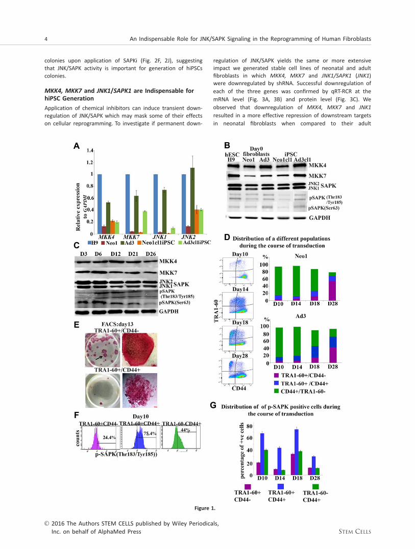
To understand the role of JNK/SAPKs during the generation ofhiPSCs we assessed the expression of JNK1 and JNK2 andtheir upstream activators MKK4 and MKK7 in two differentprimary dermal skin fibroblasts (Neonatal/Neo1 and Adult/Ad3), several hiPSCs clones derived therefrom (Fig. 1A, 1B,Supporting Information Fig. 1B) and hESCs (H9). Human ESCsare characterised by high levels of JNK/SAPK activity whichhas been shown to be important for maintenance of the plu-ripotent stem cell state [19]. In accordance with this, wefound the highest levels of mRNA JNK1 expression in hESCswhen compared to several hiPSCs clones derived from twoadult fibroblast samples (Fig. 1A, 1B, Supporting InformationFig. 1B); however these differences were not maintained atthe protein level across the iPSC clones examined (Fig. 1B).We also observed that neonatal fibroblasts had lower expres-sion of all four kinases examined when compared to adultfibroblasts (Fig. 1A, 1B). These differences were in part main-tained in the respective hiPSC lines with the adult derivedhiPSC clones showing higher expression of JNK1 when com-pared to neonatal derived hiPSCs at both transcript and pro-tein level (Fig. 1A, 1B, Supporting Information Fig. 1A, 1B).
Transduction of OSKM caused a significant increase inJNK1 expression in adult fibroblasts and a dual increase inJNK1 and JNK2 expression in neonatal fibroblasts as early asday 3 of reprogramming (Fig. 1A-1C, Supporting InformationFig. 1A). This was followed by an increase in expression ofpSAPK [Tyr 185/Thr 183SAPK)] from day 6 to day 21 in neona-tal fibroblasts (Fig. 1B, 1C) and from day 12 to day 21 in adultfibroblasts (Fig. 1B, Supporting Information Fig. 1A). Theexpression of pSAPK (Ser63) was increased as early as day 3continuing till day 21 of reprogramming in both neonatal andadult fibroblasts (Fig. 1B, 1C, Supporting Information Fig. 1A).Together these data suggest an increased JNK/SAPK activityduring the initiation and maturation stage of reprogramming.
To determine how the four transcription factors (OSKM)individually contribute to JNK/SAPK activation during reprog-ramming, we performed transduction with each single factorin neonatal fibroblasts and substituted the rest of the factorswith an equivalent number of control-GFP virus particles (Sup-porting Information Fig. 1C). Transduction with OCT4, KLF4
and c-MYC contributed mostly to an increase in JNK2 expres-sion, while introduction of SOX2 increased both JNK1 andJNK2 expression with a preference for JNK1. Transduction withcontrol viral particles alone did not lead to increased JNK1/JNK2 expression or their phosphorylated form (data notshown), indicating that activation of JNK/SAPK pathway duringreprogramming is not related to the transduction event, butspecifically to introduction of OSKM in somatic cells.
To further confirm the increase in p-JNK/SAPK expressionat a cellular level, we used flow cytometric analysis asdescribed previously [20]. This enabled us to follow three cel-lular subpopulations during the course of reprogramming;fully reprogrammed cells (TRA-1-601CD44-), partially reprog-
rammed cells (TRA-1-601CD441) and fibroblasts (TRA-1-60-CD441; Fig. 1D, 1E). pSAPK was expressed in all three subpo-pulations (Fig. 1F, 1G). It is interesting to note that thepartially reprogrammed cells showed the highest percentageof pSAPK1 expressing cells; however this declined toward theend of the reprogramming period (Fig. 1G). Furthermore, thepercentage of pSAPK expressing cells increased in the fullyreprogrammed subpopulation from day 14 to day 18 (Fig. 1G)then decreased by day 28, corroborating the Western blottinganalysis (Fig. 1B, 1C, Supporting Information Fig. 1A). Weobtained the same profile of emergence of the three cellularsubpopulations and the same trend of pSAPK activation (Sup-porting Information Fig. 2A, 2B) upon application of a polycis-tronic Sendai vector (Cytotune 2.0), demonstrating thatactivation of JNK/SAPK is independent of hiPSC transductionprotocol. Together these data suggest an important role forthe activity of JNK/SAPK in fully reprogrammed cells duringthe maturation stage of reprogramming.
Inhibition of JNK/SAPK Activation with ChemicalInhibitors Causes Disaggregation and Loss of hiPSCColonies During Maturation Stage
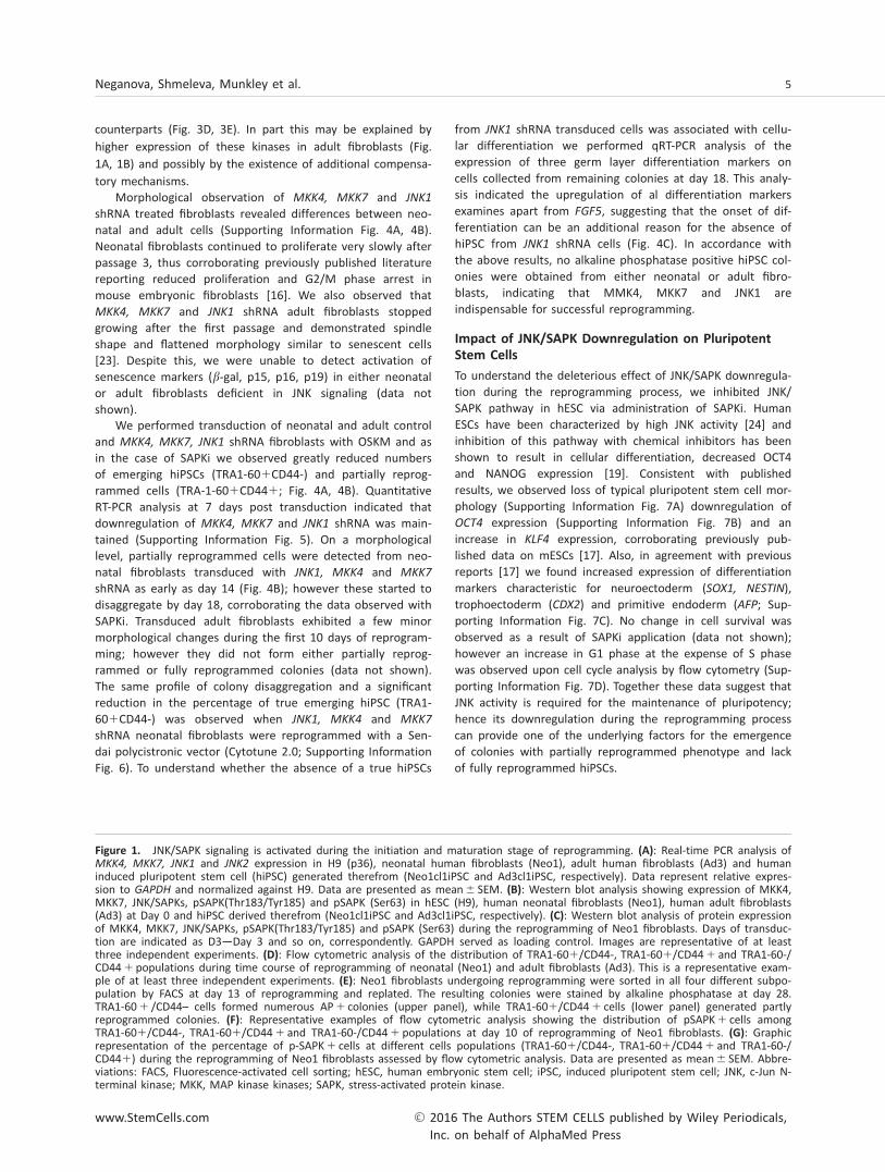
To test the function of JNK/SAPKs in generation of hiPSCs weused a small molecule SP600125, which has been shown tosignificantly inhibit expression of all three JNK genes namelyJNK1, JNK2, and JNK3 [21]. Our data show that 5 mMSP600125 (named SAPKi thereafter) is sufficient to downregu-late the expression of JNK1 and JNK2 in hESCs (Fig. 2A) cor-roborating previous data published in mouse ESCs [22]. Toinvestigate the impact of JNK/SAPK inhibition in the reprog-ramming process, we used SAPKi for 24 hours at differenttime points as summarized in Figure 2B and Supporting Infor-mation Figure 3. Our results indicate that application of SAPKihad a detrimental effect on hiPSC generation regardless ofthe time of application, for no hiPSC colonies were obtainedat the end of the transduction period from either adult orneonatal fibroblasts (Fig. 2F-2J, Supporting Information Fig. 3).In all cases, flow cytometric analysis indicated a significantdecrease in the percentage of emerging hiPSCs (TRA-1-601CD44-; Fig. 2B-2D). Furthermore, SAPKi applicationaffected the TRA1-601 populations specifically (partiallyreprogrammed and fully reprogrammed distinguished by pres-ence or absence of CD44 respectively, Fig. 2C, 2D) as noreduction in the TRA-1-60-CD441 population of dermal skinfibroblasts was observed (Fig. 2D). In control cultures (treatedwith DMSO vehicle only) we observed morphological changes(cells rounding up, starting to group together and showingmorphology typical of pluripotent stem cells) which led to theemergence of hiPSC colonies with clear compact edges asearly as day 16 (Fig. 2E, Supporting Information Fig. 3). InSAPKi treated cultures, we observed formation of colonieswith morphology typical of partially reprogrammed cells; how-ever most of these started to disintegrate as early as day 16(Fig. 2E, 2D) and by day 18 all these partially reprogrammedcolonies were lost from the culture (Fig. 2E).Thus, formationof hiPSCs colonies showing morphological features of pluripo-tent stem cells was not observed in the presence of SAPKi.Assessment of total colony number at the mid-point (day 16)as well as hiPSC colonies at day 28 (identified by alkalinephosphatase staining) corroborated the morphological andflow cytometric analysis and indicated no viable hiPSC
Neganova, Shmeleva, Munkley et al. 3
www.StemCells.com VC 2016 The Authors STEM CELLS published by Wiley Periodicals,Inc. on behalf of AlphaMed Press
colonies upon application of SAPKi (Fig. 2F, 2J), suggestingthat JNK/SAPK activity is important for generation of hiPSCscolonies.
MKK4, MKK7 and JNK1/SAPK1 are Indispensable forhiPSC Generation
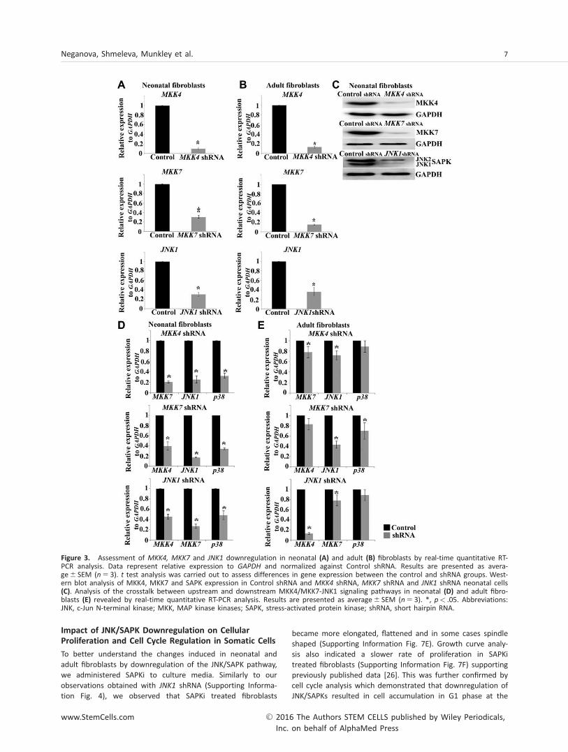
Application of chemical inhibitors can induce transient down-regulation of JNK/SAPK which may mask some of their effectson cellular reprogramming. To investigate if permanent down-
regulation of JNK/SAPK yields the same or more extensiveimpact we generated stable cell lines of neonatal and adultfibroblasts in which MKK4, MKK7 and JNK1/SAPK1 (JNK1)were downregulated by shRNA. Successful downregulation of
each of the three genes was confirmed by qRT-RCR at the
mRNA level (Fig. 3A, 3B) and protein level (Fig. 3C). We
observed that downregulation of MKK4, MKK7 and JNK1
resulted in a more effective repression of downstream targets
in neonatal fibroblasts when compared to their adult
Figure 1.
4 An Indispensable Role for JNK/SAPK Signaling in the Reprogramming of Human Fibroblasts
VC 2016 The Authors STEM CELLS published by Wiley Periodicals,Inc. on behalf of AlphaMed Press STEM CELLS
counterparts (Fig. 3D, 3E). In part this may be explained by
higher expression of these kinases in adult fibroblasts (Fig.
1A, 1B) and possibly by the existence of additional compensa-
tory mechanisms.Morphological observation of MKK4, MKK7 and JNK1
shRNA treated fibroblasts revealed differences between neo-natal and adult cells (Supporting Information Fig. 4A, 4B).Neonatal fibroblasts continued to proliferate very slowly afterpassage 3, thus corroborating previously published literaturereporting reduced proliferation and G2/M phase arrest inmouse embryonic fibroblasts [16]. We also observed thatMKK4, MKK7 and JNK1 shRNA adult fibroblasts stoppedgrowing after the first passage and demonstrated spindleshape and flattened morphology similar to senescent cells[23]. Despite this, we were unable to detect activation ofsenescence markers (b-gal, p15, p16, p19) in either neonatalor adult fibroblasts deficient in JNK signaling (data notshown).
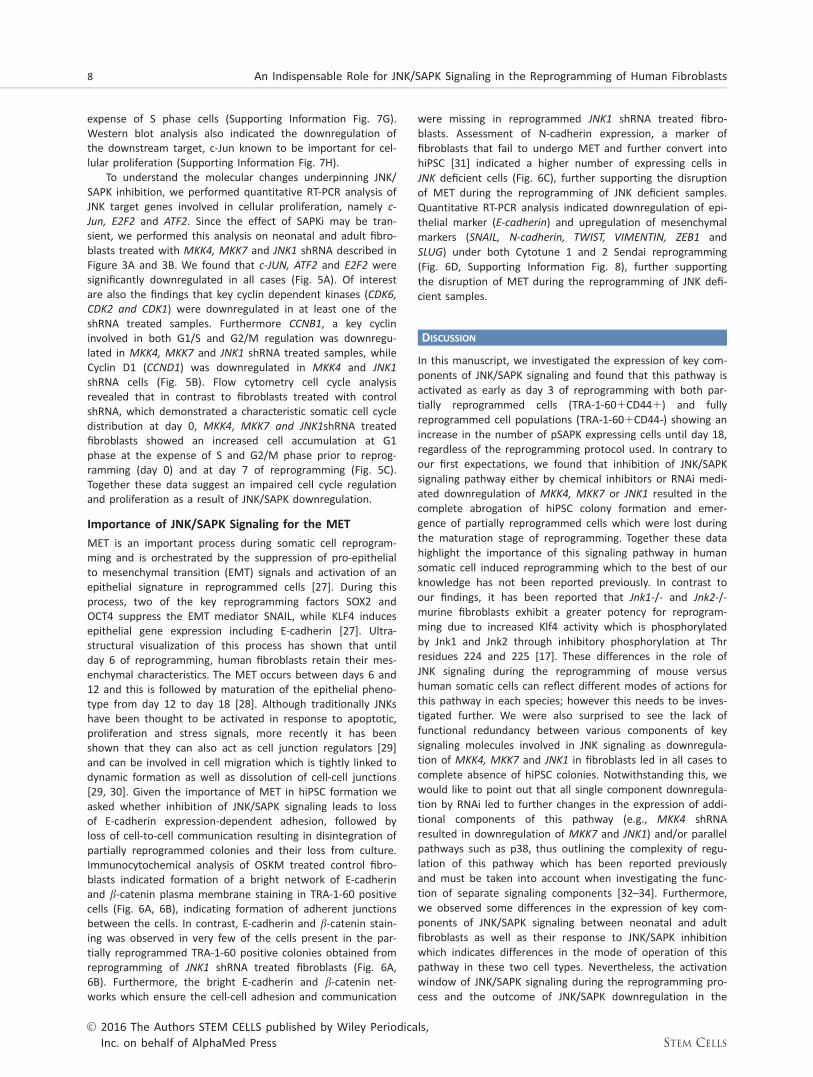
We performed transduction of neonatal and adult controland MKK4, MKK7, JNK1 shRNA fibroblasts with OSKM and asin the case of SAPKi we observed greatly reduced numbersof emerging hiPSCs (TRA1-601CD44-) and partially reprog-rammed cells (TRA-1-601CD441; Fig. 4A, 4B). QuantitativeRT-PCR analysis at 7 days post transduction indicated thatdownregulation of MKK4, MKK7 and JNK1 shRNA was main-tained (Supporting Information Fig. 5). On a morphologicallevel, partially reprogrammed cells were detected from neo-natal fibroblasts transduced with JNK1, MKK4 and MKK7
shRNA as early as day 14 (Fig. 4B); however these started todisaggregate by day 18, corroborating the data observed withSAPKi. Transduced adult fibroblasts exhibited a few minormorphological changes during the first 10 days of reprogram-ming; however they did not form either partially reprog-rammed or fully reprogrammed colonies (data not shown).The same profile of colony disaggregation and a significantreduction in the percentage of true emerging hiPSC (TRA1-601CD44-) was observed when JNK1, MKK4 and MKK7
shRNA neonatal fibroblasts were reprogrammed with a Sen-dai polycistronic vector (Cytotune 2.0; Supporting InformationFig. 6). To understand whether the absence of a true hiPSCs
from JNK1 shRNA transduced cells was associated with cellu-lar differentiation we performed qRT-PCR analysis of theexpression of three germ layer differentiation markers oncells collected from remaining colonies at day 18. This analy-sis indicated the upregulation of al differentiation markersexamines apart from FGF5, suggesting that the onset of dif-ferentiation can be an additional reason for the absence ofhiPSC from JNK1 shRNA cells (Fig. 4C). In accordance withthe above results, no alkaline phosphatase positive hiPSC col-onies were obtained from either neonatal or adult fibro-blasts, indicating that MMK4, MKK7 and JNK1 areindispensable for successful reprogramming.
Impact of JNK/SAPK Downregulation on Pluripotent
Stem Cells
To understand the deleterious effect of JNK/SAPK downregula-tion during the reprogramming process, we inhibited JNK/SAPK pathway in hESC via administration of SAPKi. HumanESCs have been characterized by high JNK activity [24] andinhibition of this pathway with chemical inhibitors has beenshown to result in cellular differentiation, decreased OCT4and NANOG expression [19]. Consistent with publishedresults, we observed loss of typical pluripotent stem cell mor-phology (Supporting Information Fig. 7A) downregulation ofOCT4 expression (Supporting Information Fig. 7B) and anincrease in KLF4 expression, corroborating previously pub-lished data on mESCs [17]. Also, in agreement with previousreports [17] we found increased expression of differentiationmarkers characteristic for neuroectoderm (SOX1, NESTIN),trophoectoderm (CDX2) and primitive endoderm (AFP; Sup-porting Information Fig. 7C). No change in cell survival wasobserved as a result of SAPKi application (data not shown);however an increase in G1 phase at the expense of S phasewas observed upon cell cycle analysis by flow cytometry (Sup-porting Information Fig. 7D). Together these data suggest thatJNK activity is required for the maintenance of pluripotency;hence its downregulation during the reprogramming processcan provide one of the underlying factors for the emergenceof colonies with partially reprogrammed phenotype and lackof fully reprogrammed hiPSCs.
Figure 1. JNK/SAPK signaling is activated during the initiation and maturation stage of reprogramming. (A): Real-time PCR analysis ofMKK4, MKK7, JNK1 and JNK2 expression in H9 (p36), neonatal human fibroblasts (Neo1), adult human fibroblasts (Ad3) and humaninduced pluripotent stem cell (hiPSC) generated therefrom (Neo1cl1iPSC and Ad3cl1iPSC, respectively). Data represent relative expres-sion to GAPDH and normalized against H9. Data are presented as mean6 SEM. (B): Western blot analysis showing expression of MKK4,MKK7, JNK/SAPKs, pSAPK(Thr183/Tyr185) and pSAPK (Ser63) in hESC (H9), human neonatal fibroblasts (Neo1), human adult fibroblasts(Ad3) at Day 0 and hiPSC derived therefrom (Neo1cl1iPSC and Ad3cl1iPSC, respectively). (C): Western blot analysis of protein expressionof MKK4, MKK7, JNK/SAPKs, pSAPK(Thr183/Tyr185) and pSAPK (Ser63) during the reprogramming of Neo1 fibroblasts. Days of transduc-tion are indicated as D3—Day 3 and so on, correspondently. GAPDH served as loading control. Images are representative of at leastthree independent experiments. (D): Flow cytometric analysis of the distribution of TRA1-601/CD44-, TRA1-601/CD441 and TRA1-60-/CD441 populations during time course of reprogramming of neonatal (Neo1) and adult fibroblasts (Ad3). This is a representative exam-ple of at least three independent experiments. (E): Neo1 fibroblasts undergoing reprogramming were sorted in all four different subpo-pulation by FACS at day 13 of reprogramming and replated. The resulting colonies were stained by alkaline phosphatase at day 28.TRA1-601 /CD44– cells formed numerous AP1 colonies (upper panel), while TRA1-601/CD441 cells (lower panel) generated partlyreprogrammed colonies. (F): Representative examples of flow cytometric analysis showing the distribution of pSAPK1 cells amongTRA1-601/CD44-, TRA1-601/CD441 and TRA1-60-/CD441 populations at day 10 of reprogramming of Neo1 fibroblasts. (G): Graphicrepresentation of the percentage of p-SAPK1 cells at different cells populations (TRA1-601/CD44-, TRA1-601/CD441 and TRA1-60-/CD441) during the reprogramming of Neo1 fibroblasts assessed by flow cytometric analysis. Data are presented as mean6 SEM. Abbre-viations: FACS, Fluorescence-activated cell sorting; hESC, human embryonic stem cell; iPSC, induced pluripotent stem cell; JNK, c-Jun N-terminal kinase; MKK, MAP kinase kinases; SAPK, stress-activated protein kinase.
Neganova, Shmeleva, Munkley et al. 5
www.StemCells.com VC 2016 The Authors STEM CELLS published by Wiley Periodicals,Inc. on behalf of AlphaMed Press
A recent report has suggested that heat shock treatmentof hESC results in activation of SAPK/JNK signaling andreduced expression of OCT4 due to increased binding of heatshock factor 1 to the OCT4 promoter [25]. Although this find-ing may appear contradictory to our data presented above,we would like to point out that the difference may be due to
physiological stimuli. While hESC data presented in this workwere collected from normal culture conditions (378C and nor-moxic oxygen levels), the Buyn et al. findings relate to the“heat shock” conditions which represent stress conditions andare likely to affect many other signaling pathways in hESC inaddition to the JNK/SAPK pathway.
Figure 2. Application of JNK/SAPKs inhibitor (SP60015) abrogates human induced pluripotent stem cell generation. (A): Western blotanalysis of JNK/SAPKs downregulation by SP60015 (SAPKi) in hESCs (H9). GAPDH used as a loading control. Images are representative ofat least three independent experiments. (B): Schematic representation of inhibitor application (SAPKi) at day 8 during the reprogram-ming process. (C): Graphic representation of flow cytometric analysis data (day 13) indicating a significant impact of SAPKi application(applied at day 8 for 24 hours) on the percentage of TRA1-601/CD44- cells. Results are presented as mean6 SEM (n 5 3). (D): Graphicrepresentation of flow cytometric analysis data (day 13) demonstrating a significant impact of SAPKi application on TRA1-601/CD44-and TRA1-601/CD441 subpopulations generated during reprogramming of Neo1 fibroblasts. Results are presented as mean6 SEM(n 5 3). (E): Phase–contrast observation showing the morphology of partially reprogrammed colonies arising during the reprogrammingof Neo1 and Ad3 fibroblasts treated with DMSO or SAPKi for 24 hours at day 8, scale bars5 100 mm. (F): Graphic representation of totalcolony numbers at day 16 and 28 of reprogramming in SAPKi and DMSO treated Neo1 and Ad3 fibroblasts. Data are presented as mean-6 SEM (n 5 3). (J): Alkaline phosphatase staining at day 28 confirmed the absence of true AP1 colonies from neonatal and adult fibro-blasts undergoing reprogramming and treated with SAPKi at day 8 of transduction for 24 hours. (C, D, F): *, p< .05. Abbreviations: AP,Alkaline phosphatase; DMSO, Dimethyl sulfoxide; FCM, Flow Cytometric and Morphological Analysis; JNK, c-Jun N-terminal kinase; SAPK,stress-activated protein kinase.
6 An Indispensable Role for JNK/SAPK Signaling in the Reprogramming of Human Fibroblasts
VC 2016 The Authors STEM CELLS published by Wiley Periodicals,Inc. on behalf of AlphaMed Press STEM CELLS
Impact of JNK/SAPK Downregulation on CellularProliferation and Cell Cycle Regulation in Somatic Cells
To better understand the changes induced in neonatal andadult fibroblasts by downregulation of the JNK/SAPK pathway,we administered SAPKi to culture media. Similarly to ourobservations obtained with JNK1 shRNA (Supporting Informa-tion Fig. 4), we observed that SAPKi treated fibroblasts
became more elongated, flattened and in some cases spindleshaped (Supporting Information Fig. 7E). Growth curve analy-sis also indicated a slower rate of proliferation in SAPKitreated fibroblasts (Supporting Information Fig. 7F) supportingpreviously published data [26]. This was further confirmed bycell cycle analysis which demonstrated that downregulation ofJNK/SAPKs resulted in cell accumulation in G1 phase at the
Figure 3. Assessment of MKK4, MKK7 and JNK1 downregulation in neonatal (A) and adult (B) fibroblasts by real-time quantitative RT-PCR analysis. Data represent relative expression to GAPDH and normalized against Control shRNA. Results are presented as avera-ge6 SEM (n 5 3). t test analysis was carried out to assess differences in gene expression between the control and shRNA groups. West-ern blot analysis of MKK4, MKK7 and SAPK expression in Control shRNA and MKK4 shRNA, MKK7 shRNA and JNK1 shRNA neonatal cells(C). Analysis of the crosstalk between upstream and downstream MKK4/MKK7-JNK1 signaling pathways in neonatal (D) and adult fibro-blasts (E) revealed by real-time quantitative RT-PCR analysis. Results are presented as average6 SEM (n 5 3). *, p< .05. Abbreviations:JNK, c-Jun N-terminal kinase; MKK, MAP kinase kinases; SAPK, stress-activated protein kinase; shRNA, short hairpin RNA.
Neganova, Shmeleva, Munkley et al. 7
www.StemCells.com VC 2016 The Authors STEM CELLS published by Wiley Periodicals,Inc. on behalf of AlphaMed Press
expense of S phase cells (Supporting Information Fig. 7G).Western blot analysis also indicated the downregulation ofthe downstream target, c-Jun known to be important for cel-lular proliferation (Supporting Information Fig. 7H).
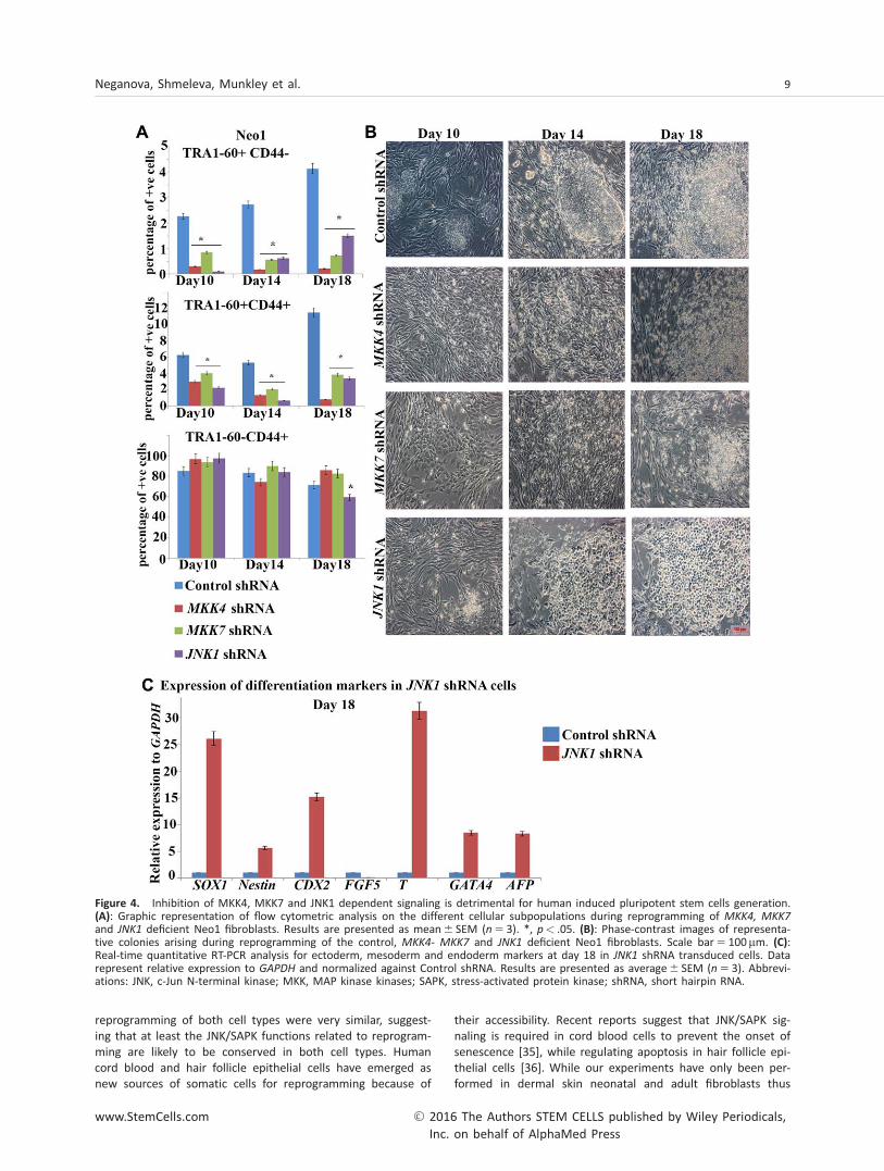
To understand the molecular changes underpinning JNK/SAPK inhibition, we performed quantitative RT-PCR analysis ofJNK target genes involved in cellular proliferation, namely c-
Jun, E2F2 and ATF2. Since the effect of SAPKi may be tran-sient, we performed this analysis on neonatal and adult fibro-blasts treated with MKK4, MKK7 and JNK1 shRNA described inFigure 3A and 3B. We found that c-JUN, ATF2 and E2F2 weresignificantly downregulated in all cases (Fig. 5A). Of interestare also the findings that key cyclin dependent kinases (CDK6,
CDK2 and CDK1) were downregulated in at least one of theshRNA treated samples. Furthermore CCNB1, a key cyclininvolved in both G1/S and G2/M regulation was downregu-lated in MKK4, MKK7 and JNK1 shRNA treated samples, whileCyclin D1 (CCND1) was downregulated in MKK4 and JNK1
shRNA cells (Fig. 5B). Flow cytometry cell cycle analysisrevealed that in contrast to fibroblasts treated with controlshRNA, which demonstrated a characteristic somatic cell cycledistribution at day 0, MKK4, MKK7 and JNK1shRNA treatedfibroblasts showed an increased cell accumulation at G1phase at the expense of S and G2/M phase prior to reprog-ramming (day 0) and at day 7 of reprogramming (Fig. 5C).Together these data suggest an impaired cell cycle regulationand proliferation as a result of JNK/SAPK downregulation.
Importance of JNK/SAPK Signaling for the MET
MET is an important process during somatic cell reprogram-ming and is orchestrated by the suppression of pro-epithelialto mesenchymal transition (EMT) signals and activation of anepithelial signature in reprogrammed cells [27]. During thisprocess, two of the key reprogramming factors SOX2 andOCT4 suppress the EMT mediator SNAIL, while KLF4 inducesepithelial gene expression including E-cadherin [27]. Ultra-structural visualization of this process has shown that untilday 6 of reprogramming, human fibroblasts retain their mes-enchymal characteristics. The MET occurs between days 6 and12 and this is followed by maturation of the epithelial pheno-type from day 12 to day 18 [28]. Although traditionally JNKshave been thought to be activated in response to apoptotic,proliferation and stress signals, more recently it has beenshown that they can also act as cell junction regulators [29]and can be involved in cell migration which is tightly linked todynamic formation as well as dissolution of cell-cell junctions[29, 30]. Given the importance of MET in hiPSC formation weasked whether inhibition of JNK/SAPK signaling leads to lossof E-cadherin expression-dependent adhesion, followed byloss of cell-to-cell communication resulting in disintegration ofpartially reprogrammed colonies and their loss from culture.Immunocytochemical analysis of OSKM treated control fibro-blasts indicated formation of a bright network of E-cadherinand b-catenin plasma membrane staining in TRA-1-60 positivecells (Fig. 6A, 6B), indicating formation of adherent junctionsbetween the cells. In contrast, E-cadherin and b-catenin stain-ing was observed in very few of the cells present in the par-tially reprogrammed TRA-1-60 positive colonies obtained fromreprogramming of JNK1 shRNA treated fibroblasts (Fig. 6A,6B). Furthermore, the bright E-cadherin and b-catenin net-works which ensure the cell-cell adhesion and communication
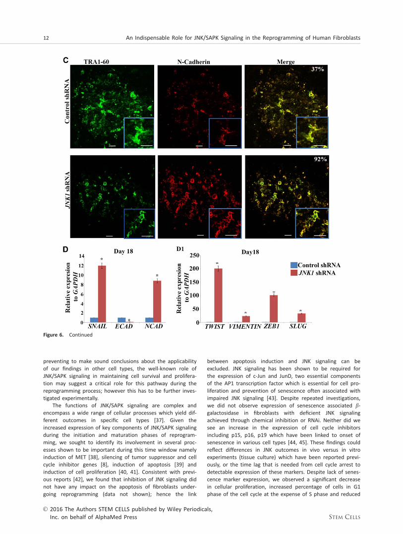
were missing in reprogrammed JNK1 shRNA treated fibro-blasts. Assessment of N-cadherin expression, a marker offibroblasts that fail to undergo MET and further convert intohiPSC [31] indicated a higher number of expressing cells inJNK deficient cells (Fig. 6C), further supporting the disruptionof MET during the reprogramming of JNK deficient samples.Quantitative RT-PCR analysis indicated downregulation of epi-thelial marker (E-cadherin) and upregulation of mesenchymalmarkers (SNAIL, N-cadherin, TWIST, VIMENTIN, ZEB1 andSLUG) under both Cytotune 1 and 2 Sendai reprogramming(Fig. 6D, Supporting Information Fig. 8), further supportingthe disruption of MET during the reprogramming of JNK defi-cient samples.
DISCUSSION
In this manuscript, we investigated the expression of key com-ponents of JNK/SAPK signaling and found that this pathway isactivated as early as day 3 of reprogramming with both par-tially reprogrammed cells (TRA-1-601CD441) and fullyreprogrammed cell populations (TRA-1-601CD44-) showing anincrease in the number of pSAPK expressing cells until day 18,regardless of the reprogramming protocol used. In contrary toour first expectations, we found that inhibition of JNK/SAPKsignaling pathway either by chemical inhibitors or RNAi medi-ated downregulation of MKK4, MKK7 or JNK1 resulted in thecomplete abrogation of hiPSC colony formation and emer-gence of partially reprogrammed cells which were lost duringthe maturation stage of reprogramming. Together these datahighlight the importance of this signaling pathway in humansomatic cell induced reprogramming which to the best of ourknowledge has not been reported previously. In contrast toour findings, it has been reported that Jnk1-/- and Jnk2-/-murine fibroblasts exhibit a greater potency for reprogram-ming due to increased Klf4 activity which is phosphorylatedby Jnk1 and Jnk2 through inhibitory phosphorylation at Thrresidues 224 and 225 [17]. These differences in the role ofJNK signaling during the reprogramming of mouse versushuman somatic cells can reflect different modes of actions forthis pathway in each species; however this needs to be inves-tigated further. We were also surprised to see the lack offunctional redundancy between various components of keysignaling molecules involved in JNK signaling as downregula-tion of MKK4, MKK7 and JNK1 in fibroblasts led in all cases tocomplete absence of hiPSC colonies. Notwithstanding this, wewould like to point out that all single component downregula-tion by RNAi led to further changes in the expression of addi-tional components of this pathway (e.g., MKK4 shRNAresulted in downregulation of MKK7 and JNK1) and/or parallelpathways such as p38, thus outlining the complexity of regu-lation of this pathway which has been reported previouslyand must be taken into account when investigating the func-tion of separate signaling components [32–34]. Furthermore,we observed some differences in the expression of key com-ponents of JNK/SAPK signaling between neonatal and adultfibroblasts as well as their response to JNK/SAPK inhibitionwhich indicates differences in the mode of operation of thispathway in these two cell types. Nevertheless, the activationwindow of JNK/SAPK signaling during the reprogramming pro-cess and the outcome of JNK/SAPK downregulation in the
8 An Indispensable Role for JNK/SAPK Signaling in the Reprogramming of Human Fibroblasts
VC 2016 The Authors STEM CELLS published by Wiley Periodicals,Inc. on behalf of AlphaMed Press STEM CELLS
reprogramming of both cell types were very similar, suggest-ing that at least the JNK/SAPK functions related to reprogram-ming are likely to be conserved in both cell types. Humancord blood and hair follicle epithelial cells have emerged asnew sources of somatic cells for reprogramming because of
their accessibility. Recent reports suggest that JNK/SAPK sig-naling is required in cord blood cells to prevent the onset ofsenescence [35], while regulating apoptosis in hair follicle epi-thelial cells [36]. While our experiments have only been per-formed in dermal skin neonatal and adult fibroblasts thus
Figure 4. Inhibition of MKK4, MKK7 and JNK1 dependent signaling is detrimental for human induced pluripotent stem cells generation.(A): Graphic representation of flow cytometric analysis on the different cellular subpopulations during reprogramming of MKK4, MKK7and JNK1 deficient Neo1 fibroblasts. Results are presented as mean6 SEM (n 5 3). *, p< .05. (B): Phase-contrast images of representa-tive colonies arising during reprogramming of the control, MKK4- MKK7 and JNK1 deficient Neo1 fibroblasts. Scale bar5 100mm. (C):Real-time quantitative RT-PCR analysis for ectoderm, mesoderm and endoderm markers at day 18 in JNK1 shRNA transduced cells. Datarepresent relative expression to GAPDH and normalized against Control shRNA. Results are presented as average6 SEM (n 5 3). Abbrevi-ations: JNK, c-Jun N-terminal kinase; MKK, MAP kinase kinases; SAPK, stress-activated protein kinase; shRNA, short hairpin RNA.
Neganova, Shmeleva, Munkley et al. 9
www.StemCells.com VC 2016 The Authors STEM CELLS published by Wiley Periodicals,Inc. on behalf of AlphaMed Press
Figure 5. MKK4, MKK7 and JNK1 downregulation results in changes in the expression of key genes involved in cellular proliferationand cell cycle progression. (A, B): Real-time quantitative PCR analysis of c-JUN, E2F2, ATF2 (A) and CDK6, CDK2, CDK1, CCND1 andCCNB1 (B) in Control shRNA (Control) and MKK4, MKK7 and JNK1 deficient Neo1 fibroblasts at Day 1 of reprogramming. Results are pre-sented as mean6 SEM (n 5 3). *, p< .05. (C): Flow cytometric cell cycle analysis of MKK4, MKK7 and JNK1 shRNA treated fibroblastsduring the reprogramming process. Results are presented as mean6 SEM (n 5 3). Abbreviations: JNK, c-Jun N-terminal kinase; MKK,MAP kinase kinases; shRNA, short hairpin RNA.
10 An Indispensable Role for JNK/SAPK Signaling in the Reprogramming of Human Fibroblasts
VC 2016 The Authors STEM CELLS published by Wiley Periodicals,Inc. on behalf of AlphaMed Press STEM CELLS
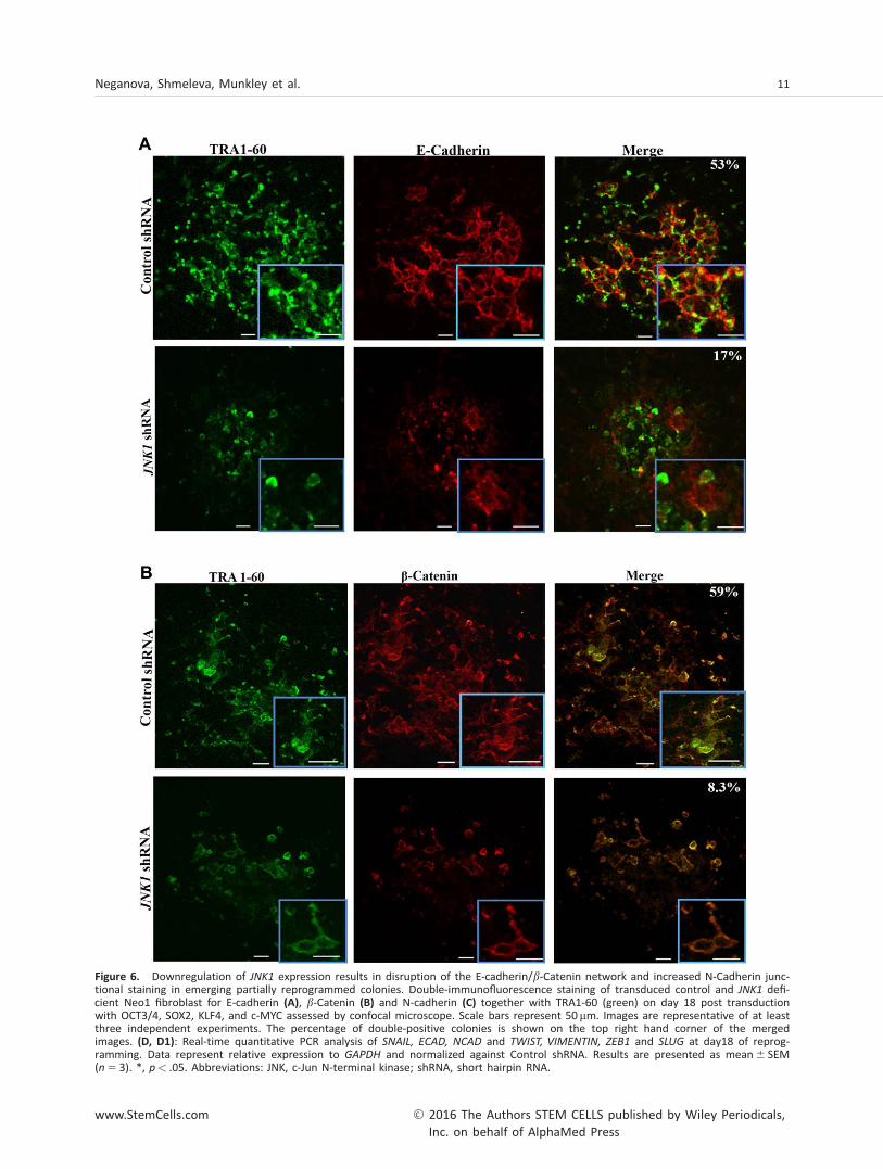
Figure 6. Downregulation of JNK1 expression results in disruption of the E-cadherin/b-Catenin network and increased N-Cadherin junc-tional staining in emerging partially reprogrammed colonies. Double-immunofluorescence staining of transduced control and JNK1 defi-cient Neo1 fibroblast for E-cadherin (A), b-Catenin (B) and N-cadherin (C) together with TRA1-60 (green) on day 18 post transductionwith OCT3/4, SOX2, KLF4, and c-MYC assessed by confocal microscope. Scale bars represent 50 mm. Images are representative of at leastthree independent experiments. The percentage of double-positive colonies is shown on the top right hand corner of the mergedimages. (D, D1): Real-time quantitative PCR analysis of SNAIL, ECAD, NCAD and TWIST, VIMENTIN, ZEB1 and SLUG at day18 of reprog-ramming. Data represent relative expression to GAPDH and normalized against Control shRNA. Results are presented as mean6 SEM(n 5 3). *, p< .05. Abbreviations: JNK, c-Jun N-terminal kinase; shRNA, short hairpin RNA.
Neganova, Shmeleva, Munkley et al. 11
www.StemCells.com VC 2016 The Authors STEM CELLS published by Wiley Periodicals,Inc. on behalf of AlphaMed Press
preventing to make sound conclusions about the applicabilityof our findings in other cell types, the well-known role ofJNK/SAPK signaling in maintaining cell survival and prolifera-tion may suggest a critical role for this pathway during thereprogramming process; however this has to be further inves-tigated experimentally.
The functions of JNK/SAPK signaling are complex andencompass a wide range of cellular processes which yield dif-ferent outcomes in specific cell types [37]. Given theincreased expression of key components of JNK/SAPK signalingduring the initiation and maturation phases of reprogram-ming, we sought to identify its involvement in several proc-esses shown to be important during this time window namelyinduction of MET [38], silencing of tumor suppressor and cellcycle inhibitor genes [8], induction of apoptosis [39] andinduction of cell proliferation [40, 41]. Consistent with previ-ous reports [42], we found that inhibition of JNK signaling didnot have any impact on the apoptosis of fibroblasts under-going reprogramming (data not shown); hence the link
between apoptosis induction and JNK signaling can beexcluded. JNK signaling has been shown to be required forthe expression of c-Jun and JunD, two essential componentsof the AP1 transcription factor which is essential for cell pro-liferation and prevention of senescence often associated withimpaired JNK signaling [43]. Despite repeated investigations,we did not observe expression of senescence associated b-galactosidase in fibroblasts with deficient JNK signalingachieved through chemical inhibition or RNAi. Neither did wesee an increase in the expression of cell cycle inhibitorsincluding p15, p16, p19 which have been linked to onset ofsenescence in various cell types [44, 45]. These findings couldreflect differences in JNK outcomes in vivo versus in vitroexperiments (tissue culture) which have been reported previ-ously, or the time lag that is needed from cell cycle arrest todetectable expression of these markers. Despite lack of senes-cence marker expression, we observed a significant decreasein cellular proliferation, increased percentage of cells in G1phase of the cell cycle at the expense of S phase and reduced
Figure 6. Continued
12 An Indispensable Role for JNK/SAPK Signaling in the Reprogramming of Human Fibroblasts
VC 2016 The Authors STEM CELLS published by Wiley Periodicals,Inc. on behalf of AlphaMed Press STEM CELLS
expression of key cell cycle components involved in G1/S tran-sition (such as CDK6, CDK2, CDK1, CCDN1) and S phase pro-gression and G2/M transition (e.g., CCNB1). It is important tonote that such expression changes were specific for eachcomponent of the JNK signaling pathway downregulated;however in all cases simultaneous downregulation of at leastone CDK and one partnering Cyclin, an event that has beenshown to be important for efficient reprogramming [40] wasobserved. These findings were further corroborated bydecreased expression of cellular proliferation markers c-Jun,
E2F2 and ATF2 in all three shRNA treated samples consistentwith previous reports indicating an important role for MKK4,MKK7 and JNK1 for cell cycle progression [16, 46]. High cellu-lar proliferation rate akin to ESC state is an important earlyevent for cellular reprogramming [40]. This leads us to suggestthat suppressed cellular proliferation and cell accumulation atthe G1 phase (both known as reprogramming barriers; [47])at the onset of reprogramming of JNK1, MKK4 and MKK7 defi-cient neonatal and adult fibroblasts is a key reason for thecomplete lack of hiPSC observed during reprogramming ofthese samples.
The efficiency of reprogramming can be promoted bythe onset of epithelial expression markers concomitantlywith the repression of mesenchymal markers in cells under-going reprogramming during the 6th until the 12th day ofreprogramming, known as initiation phase [48, 49]. This isprecisely the window during which the JNK signaling is acti-vated; hence we asked the question of whether MET transi-tion is affected in JNK deficient cells. Our data suggest thatwhile control cells showed well organized TRA-1-601 colonies characterized by plasma membrane staining ofE-cadherin and b-catenin indicating formation of adherentjunctions typical of epithelial cells, JNK deficient cells dis-played a reduced percentage of cells expressing thesemarkers at both cellular and mRNA levels, which suggeststhat fewer cells are able to undergo MET transition, thusleading to inefficient reprogramming. Furthermore, the cel-
lular networks that guarantee cell-cell communication whilepresent and well organized in control cells were lacking inJNK/SAPK deficient fibroblasts. It has been suggested thatexpression of E-cadherin and the presence of intact adher-ent junctions are essential for maintenance of pluripotencyin hESC by ensuring access to critical autocrine signals andby promoting cell-cell exchange of signals through the gapjunctions [49, 50]. In this context, it is interesting to notethat all colonies that emerged during reprogramming ofJNK/SAPK deficient fibroblasts were partially reprogrammedand completely disaggregated and subsequently lost duringthe maturation stage of reprogramming thus leading us tosuggest that deficient JNK/SAPK signaling followed byimpaired cell to cell contact is likely to be one of the caus-ative factor that counts for the partially reprogrammed phe-notype and for the loss of colonies.
Our inhibition experiments were performed during the ini-tiation stage of reprogramming. In all cases despite the timeof JNK/SAPK downregulation we obtained no hiPSC colonies.However, we did not perform JNK/SAPK downregulation stud-ies at the stabilization stage where hiPSC colonies are presentand continue to grow and develop. There were two main rea-sons behind this decision: (1) JNK/SAPK activation occurs dur-ing the initiation and maturation stage and shows a dropduring the stabilization stage and (2) our data presented inthis manuscript together with published reports indicate lossof pluripotency in hESCs upon JNK downregulation [19], whichwould suggest that downregulation of JNK/SAPK signalingafter the emergence of hiPSC colonies would result in theirdifferentiation and thus provide a further push toward loss oftrue bona fide hiPSC colonies.
CONCLUSION
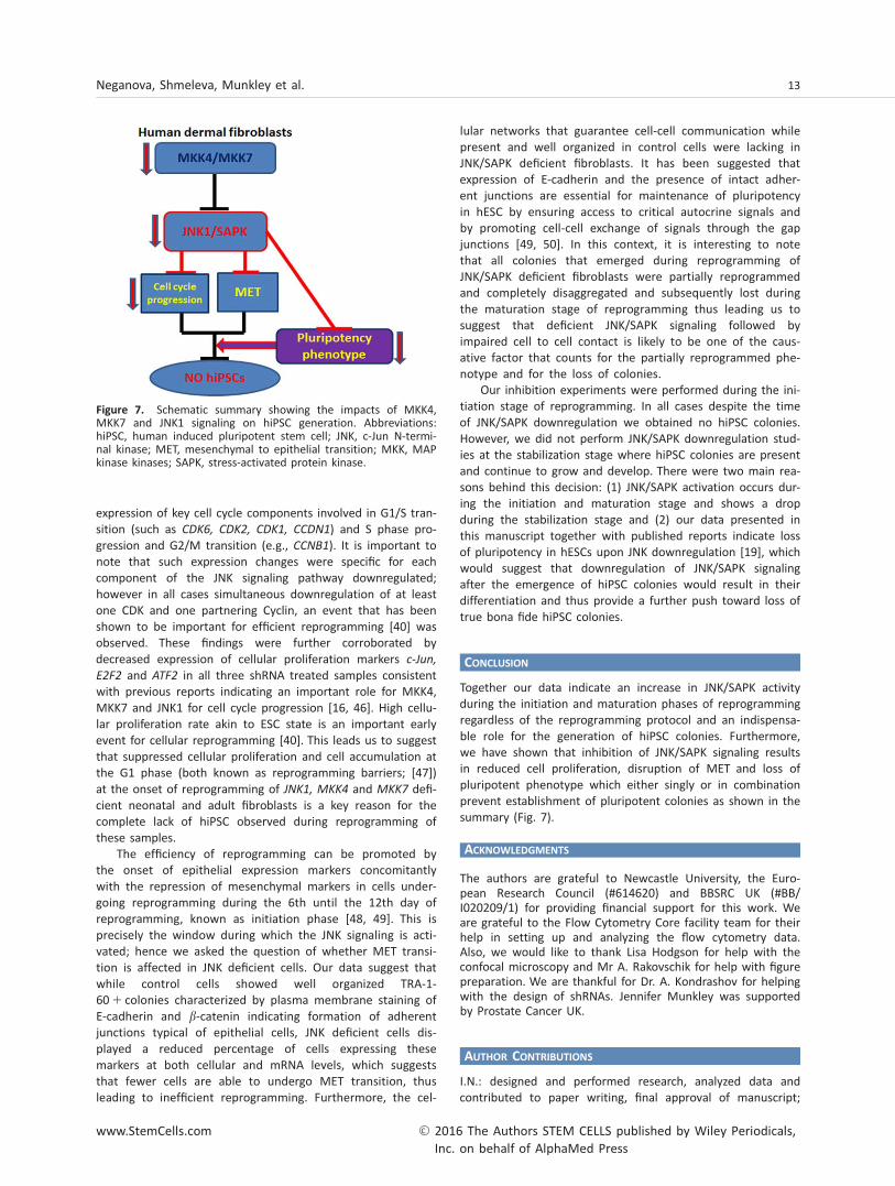
Together our data indicate an increase in JNK/SAPK activityduring the initiation and maturation phases of reprogrammingregardless of the reprogramming protocol and an indispensa-ble role for the generation of hiPSC colonies. Furthermore,we have shown that inhibition of JNK/SAPK signaling resultsin reduced cell proliferation, disruption of MET and loss ofpluripotent phenotype which either singly or in combinationprevent establishment of pluripotent colonies as shown in thesummary (Fig. 7).
ACKNOWLEDGMENTS
The authors are grateful to Newcastle University, the Euro-pean Research Council (#614620) and BBSRC UK (#BB/I020209/1) for providing financial support for this work. Weare grateful to the Flow Cytometry Core facility team for theirhelp in setting up and analyzing the flow cytometry data.Also, we would like to thank Lisa Hodgson for help with theconfocal microscopy and Mr A. Rakovschik for help with figurepreparation. We are thankful for Dr. A. Kondrashov for helpingwith the design of shRNAs. Jennifer Munkley was supportedby Prostate Cancer UK.
AUTHOR CONTRIBUTIONS
I.N.: designed and performed research, analyzed data andcontributed to paper writing, final approval of manuscript;
Figure 7. Schematic summary showing the impacts of MKK4,MKK7 and JNK1 signaling on hiPSC generation. Abbreviations:hiPSC, human induced pluripotent stem cell; JNK, c-Jun N-termi-nal kinase; MET, mesenchymal to epithelial transition; MKK, MAPkinase kinases; SAPK, stress-activated protein kinase.
Neganova, Shmeleva, Munkley et al. 13
www.StemCells.com VC 2016 The Authors STEM CELLS published by Wiley Periodicals,Inc. on behalf of AlphaMed Press
E.S., J.M., R.A., J.P.: performed some research and data analy-sis, final approval of manuscript; V.C.: performed someresearch and data analysis, final approval of manuscript; G.A.:performed some research, final approval of manuscript; D.J.E.:contributed to design of study and paper writing, finalapproval of manuscript; L.A.: designed research, contributedto paper writing, final approval of manuscript and fund rais-
ing; M.L.: designed research, data analysis, wrote the paper,final approval of manuscript and fund raising.
DISCLOSURE
The author(s) indicates no potential conflicts of interest.
REFERENCES
1 Takahashi K, Tanabe K, Ohnuki M et al.Induction of pluripotent stem cells fromadult human fibroblasts by defined factors.Cell 2007;131:861–872.
2 Lako M, Armstrong L, Stojkovic M.Induced pluripotent stem cells: It looks sim-ple but can looks deceive? STEM CELLS 2010;28:845–850.
3 Beltran AS, Rivenbark AG, Richardson BTet al. Generation of tumor-initiating cells byexogenous delivery of OCT4 transcription fac-tor. Breast Cancer Res 2011;13:R94.
4 Liu K, Lin B, Zhao M et al. The multipleroles for Sox2 in stem cell maintenance andtumorigenesis. Cell Signal 2013;25:1264–1271.
5 Yu F, Li J, Chen H et al. Kruppel-like fac-tor 4 (KLF4) is required for maintenance ofbreast cancer stem cells and for cell migra-tion and invasion. Oncogene 2011;30:2161–2172.
6 Haigis KM, Sweet-Cordero A. Newinsights into oncogenic stress. Nat Genet2011;43:177–178.
7 Muller LU, Milsom MD, Harris CE et al.Overcoming reprogramming resistance ofFanconi anemia cells. Blood 2012;119:5449–5457.
8 Banito A, Rashid ST, Acosta JC et al.Senescence impairs successful reprogram-ming to pluripotent stem cells. Genes Dev2009;23:2134–2139.
9 Bao X, Wu H, Zhu X et al. The p53-induced lincRNA-p21 derails somatic cellreprogramming by sustaining H3K9me3 andCpG methylation at pluripotency gene pro-moters. Cell Res 2015;25:80–92.10 Chang L, Karin M. Mammalian MAPkinase signalling cascades. Nature 2001;410:37–40.11 Schramek D, Kotsinas A, Meixner Aet al. The stress kinase MKK7 couples onco-genic stress to p53 stability and tumor sup-pression. Nat Genet 2011;43:212–219.12 Johnson GL, Nakamura K. The c-junkinase/stress-activated pathway: Regulation,function and role in human disease. BiochimBiophys Acta 2007;1773:1341–1348.13 Wang J, Chen L, Ko CI et al. Distinct sig-naling properties of mitogen-activated pro-tein kinase kinases 4 (MKK4) and 7 (MKK7)in embryonic stem cell (ESC) differentiation.J Biol Chem 2012;287:2787–2797.14 Bogoyevitch MA, Kobe B. Uses for JNK:The many and varied substrates of the c-JunN-terminal kinases. Microbiol Mol Biol Rev2006;70:1061–1095.15 Yang D, Tournier C, Wysk M et al. Tar-geted disruption of the MKK4 gene causesembryonic death, inhibition of c-Jun NH2-ter-
minal kinase activation, and defects in AP-1transcriptional activity. Proc Natl Acad SciUSA 1997;94:3004–3009.16 Wada T, Nakagawa K, Watanabe T et al.Impaired synergistic activation of stress-activated protein kinase SAPK/JNK in mouseembryonic stem cells lacking SEK1/MKK4: Dif-ferent contribution of SEK2/MKK7 isoformsto the synergistic activation. J Biol Chem2001;276:30892–30897.17 Yao K, Ki MO, Chen H et al. JNK1 and 2play a negative role in reprogramming to plu-ripotent stem cells by suppressing Klf4 activ-ity. Stem Cell Res 2014;12:139–152.18 Chichagova V, Sanchez-Vera I, ArmstrongL et al. Generation of human induced pluri-potent stem cells using RNA-based sendaivirus system and pluripotency validation ofthe resulting cell population. Methods MolBiol 2015;1353:285–307.19 Brill LM, Xiong W, Lee KB et al. Phos-phoproteomic analysis of human embryonicstem cells. Cell Stem Cell 2009;5:204–213.20 Takahashi K, Tanabe K, Ohnuki M et al.Induction of pluripotency in human somaticcells via a transient state resembling primi-tive streak-like mesendoderm. Nat Commun2014;5:3678.21 Bennett BL, Sasaki DT, Murray BW et al.SP600125, an anthrapyrazolone inhibitor ofJun N-terminal kinase. Proc Natl Acad SciUSA 2001;98:13681–13686.22 Zhou Y, Jiang M, Wang M et al. Effect ofSP600125 on proliferation of embryonic stemcell. AJMB 2013;3:67–71.23 Dimri GP, Lee X, Basile G et al. A bio-marker that identifies senescent human cellsin culture and in aging skin in vivo. Proc NatlAcad Sci USA 1995;92:9363–9367.24 Van Hoof D, Munoz J, Braam SR et al.Phosphorylation dynamics during early differ-entiation of human embryonic stem cells.Cell Stem Cell 2009;5:214–226.25 Byun K, Kim TK, Oh J et al. Heat shockinstructs hESCs to exit from the self-renewalprogram through negative regulation ofOCT4 by SAPK/JNK and HSF1 pathway. StemCell Res 2013;11:1323–1334.26 Tournier C, Dong C, Turner TK et al.MKK7 is an essential component of the JNKsignal transduction pathway activated byproinflammatory cytokines. Genes Dev 2001;15:1419–1426.27 Liu X, Sun H, Qi J et al. Sequential intro-duction of reprogramming factors reveals atime-sensitive requirement for individual fac-tors and a sequential EMT-MET mechanismfor optimal reprogramming. Nat Cell Biol2013;15:829–838.28 Hoffding MK, Hyttel P. Ultrastructuralvisualization of the mesenchymal-to-epithelial transition during reprogramming of
human fibroblasts to induced pluripotentstem cells. Stem Cell Res 2015;14:39–53.29 Lee MH, Koria P, Qu J et al. JNK phos-phorylates beta-catenin and regulates adhe-rens junctions. FASEB J 2009;23:3874–3883.30 You H, Padmashali RM, Ranganathan Aet al. JNK regulates compliance-inducedadherens junctions formation in epithelialcells and tissues. J Cell Sci 2013;126(Pt 12):2718–2729.31 Chen T, Yuan D, Wei B et al. E-cadherin-mediated cell-cell contact is critical forinduced pluripotent stem cell generation.STEM CELLS 2010;28:1315–1325.32 Davis RJ. Signal transduction by the JNKgroup of MAP kinases. Cell 2000;103:239–252.33 Wang X, Destrument A, Tournier C.Physiological roles of MKK4 and MKK7:Insights from animal models. Biochim Bio-phys Acta 2007;1773:1349–1357.34 Whitmarsh AJ. Regulation of gene tran-scription by mitogen-activated protein kinasesignaling pathways. Biochim Biophys Acta2007;1773:1285–1298.35 Spallarossa P, Altieri P, Barisione C et al.p38 MAPK and JNK antagonistically controlsenescence and cytoplasmic p16INK4Aexpression in doxorubicin-treated endothelialprogenitor cells. PLoS One 2010;5:e15583.36 Neisch AL, Speck O, Stronach B et al.Rho1 regulates apoptosis via activation ofthe JNK signaling pathway at the plasmamembrane. J Cell Biol 2010;189:311–323.37 Wagner EF, Nebreda AR. Signal integra-tion by JNK and p38 MAPK pathways in can-cer development. Nat Rev Cancer 2009;9:537–549.38 Polo JM, Hochedlinger K. When fibro-blasts MET iPSCs. Cell Stem Cell 2010;7:5–6.39 Cheung HH, Liu X, Rennert OM. Apopto-sis: Reprogramming and the fate of maturecells. ISRN Cell Biol 2012;2012:1–8.40 Ruiz S, Panopoulos AD, Herrerias A et al.A high proliferation rate is required for cellreprogramming and maintenance of humanembryonic stem cell identity. Curr Biol 2011;21:45–52.41 Kareta MS, Gorges LL, Hafeez S et al.Inhibition of pluripotency networks by theRb tumor suppressor restricts reprogrammingand tumorigenesis. Cell Stem Cell 2015;16:39–50.42 Tournier C, Hess P, Yang DD et al.Requirement of JNK for stress-induced activa-tion of the cytochrome c-mediated deathpathway. Science 2000;288:870–874.43 Das M, Jiang F, Sluss HK et al. Suppres-sion of p53-dependent senescence by theJNK signal transduction pathway. Proc NatlAcad Sci USA 2007;104:15759–15764.
14 An Indispensable Role for JNK/SAPK Signaling in the Reprogramming of Human Fibroblasts
VC 2016 The Authors STEM CELLS published by Wiley Periodicals,Inc. on behalf of AlphaMed Press STEM CELLS
44 Stein GH, Drullinger LF, Soulard A et al.Differential roles for cyclin-dependent kinaseinhibitors p21 and p16 in the mechanisms ofsenescence and differentiation in humanfibroblasts. Mol Cell Biol 1999;19:2109–2117.45 Lin AW, Barradas M, Stone JC et al. Pre-mature senescence involving p53 and p16 isactivated in response to constitutive MEK/MAPK mitogenic signaling. Genes Dev 1998;12:3008–3019.
46 Wang J, Xia Y. Assessing developmentalroles of MKK4 and MKK7 in vitro. CommunIntegr Biol 2012;5:319–324.47 Ramos-Mejia V, Fraga MF, Menendez P.iPSCs from cancer cells: Challenges andopportunities. Trends Mol Med 2012;18:245–247.48 Samavarchi-Tehrani P, Golipour A, DavidL et al. Functional genomics reveals a BMP-driven mesenchymal-to-epithelial transition
in the initiation of somatic cell reprogram-ming. Cell Stem Cell 2010;7:64–77.49 Li R, Liang J, Ni S et al. A mesenchymal-to-epithelial transition initiates and isrequired for the nuclear reprogramming ofmouse fibroblasts. Cell Stem Cell 2010;7:51–63.50 Houghton FD. Role of gap junctions dur-ing early embryo development. Reproduction2005;129:129–135.
See www.StemCells.com for supporting information available online.
Neganova, Shmeleva, Munkley et al. 15
www.StemCells.com VC 2016 The Authors STEM CELLS published by Wiley Periodicals,Inc. on behalf of AlphaMed Press


![Original Article Puerarin modulates autophagy to …signaling pathway with JNK inhibitor SP600125 can produce protective effects against CIRI [18]. Thrombolysis, antiplatelet agents,](https://static.fdocuments.net/doc/165x107/5e39305f90bc3d6d0c3b72a7/original-article-puerarin-modulates-autophagy-to-signaling-pathway-with-jnk-inhibitor.jpg)