Jessica Thesis Update 0723 08.pdf
135
ANTI-VIRAL RNAi AND ITS SUPPRESSION IN PLANTS A Thesis by JESSICA J. CIOMPERLIK Submitted to the Office of Graduate Studies of Texas A&M University in partial fulfillment of the requirements for the degree of MASTER OF SCIENCE August 2008 Major Subject: Plant Pathology
Transcript of Jessica Thesis Update 0723 08.pdf

ANTI-VIRAL RNAi AND ITS SUPPRESSION IN PLANTS
A Thesis
by
JESSICA J. CIOMPERLIK
Submitted to the Office of Graduate Studies of Texas A&M University
in partial fulfillment of the requirements for the degree of
MASTER OF SCIENCE
August 2008
Major Subject: Plant Pathology

ANTI-VIRAL RNAi AND ITS SUPPRESSION IN PLANTS
A Thesis
by
JESSICA J. CIOMPERLIK
Submitted to the Office of Graduate Studies of Texas A&M University
in partial fulfillment of the requirements for the degree of
MASTER OF SCIENCE
Approved by:
Chair of Committee, Herman B. Scholthof Committee Members, Karen-Beth G. Scholthof Hisashi Koiwa Head of Department, Dennis C. Gross
August 2008
Major Subject: Plant Pathology

iii
ABSTRACT
Anti-viral RNAi and Its Suppression in Plants. (August 2008)
Jessica J. Ciomperlik, B.S., Texas A&M University
Chair of Advisory Committee: Dr. Herman B. Scholthof
As a defense against viral infection, plants are thought to use RNA-induced
silencing complexes (RISCs) to target and cleave viral RNA. To counteract this, some
viruses have evolved proteins to inhibit RISC-mediated activity, thus ensuring
continued virulence. This research focused on the study and analysis of the anti-viral
RNAi response to various viruses in plants to gain an understanding of how the plant
defense operates on the molecular and biochemical levels. Nicotiana benthamiana plants
were infected with Tomato bushy stunt virus (TBSV) and Tobacco rattle virus (TRV).
These plants were subjected to column chromatography methods, and fractions
contained a virus-specific ribonuclease activity, co-eluting with small interfering RNAs
(siRNA), that was shown to be sensitive to inhibition with EDTA and enhanced by the
addition of divalent metal cations. This ribonuclease activity co-purified with proteins
that contained a domain from the hallmark RISC protein Argonaute family. To further
study host responses to viral infection, monocots were infected with Panicum mosaic
virus (PMV) and satellite panicum mosaic virus (SPMV) and also were subjected to
column chromatography following infection. Preliminary studies show that fractions
contained ribonuclease activity as well as siRNAs and proteins containing an Argonaute
domain. Additionally, silencing suppressors have been directly implicated in interfering
with RNAi pathways in plants. Studies involving Agrobacterium- and virus-vectored
cDNA to express green fluorescent protein (GFP) were used to establish that co-
introduced suppressors of RNAi can extend the production of a foreign protein for
enhancement of biotechnological applications. It was found that the hordeivirus protein
γb contributes to enhancement of expression for the foreign protein GFP early in the

iv
infection, while the potyvirus protein HcPro and tombusvirus protein P19 enhance and
extend protein production later in the infection.

v
ACKNOWLEDGEMENTS
In working toward my master’s degree, I am fortunate to have several people
whom I would like to acknowledge here.
Many, many thanks to Dr. Herman Scholthof, my advisor, for allowing me to
work in his laboratory, as well as for his encouragement, advice, and especially patience.
My appreciation goes to Dr. Karen-Beth Scholthof for her support, discussions, and
literature recommendations. Thank you to Dr. Hisashi Koiwa for experimental design
advice and manuscript comments.
I would like to convey my gratitude to my labmates. Thank you to Rustem
Omarov for his mentorship, advice, and for teaching me most of the laboratory
techniques used here - without him this thesis would not have been possible. Many
thanks to Yoshimi Yamamura, Yi-cheng Hsieh (John) and Dong Qi (Tony) for their
encouragement, general lab advice, and leadership by example (and a special thanks to
John for the healthy plants!). I am grateful to Bonnie Seaberg, Anthany Everett, Christa
Chavez, Kristina Twigg, Malika Shamekova, Shuga Manabayeva, Veria Alvarado, and
Vanessa Vaughn for their support and conversations. I would also like to express my
appreciation to the faculty, staff, and students of Plant Pathology for their
encouragement.
Additionally, I thank my parents, brother, family, and friends - Kelly, Karla,
Cass, Zachary, Frankie, Sonia, Brandon, Shaw, and others - for their unfailing support
and enthusiasm.
Thanks, you guys!

vi
TABLE OF CONTENTS
Page
ABSTRACT .............................................................................................................. iii
ACKNOWLEDGEMENTS ...................................................................................... v
TABLE OF CONTENTS .......................................................................................... vi
LIST OF FIGURES................................................................................................... vii
LIST OF TABLES .................................................................................................... x
CHAPTER
I INTRODUCTION AND SYSTEMS ................................................... 1
Introduction……………………………………………………….. 1 Systems........................................................................................... 6 Objectives....................................................................................... 15
II BIOCHEMICAL CHARACTERIZATION OF AN RNAi
RESPONSE AGAINST TBSV IN N. BENTHAMIANA ...................... 17
Introduction .................................................................................... 17 Materials and Methods ................................................................... 23 Results ............................................................................................ 28 Discussion ...................................................................................... 39 III DETERMINATION OF AN ANTI-VIRAL RESPONSE
FOLLOWING INFECTION OF PLANTS WITH TRV, PMV AND
SPMV ................................................................................................... 45
Introduction .................................................................................... 45 Materials and Methods ................................................................... 48 Results ............................................................................................ 52 Discussion ...................................................................................... 68

vii
CHAPTER Page
IV USE OF SILENCING SUPPRESSORS TO EXTEND AND
ENHANCE THE LENGTH OF TIME A FOREIGN PROTEIN IS
PRODUCED VIA AGROBACTERIUM TUMAFACIENS AND A
VIRAL VECTOR................................................................................. 75
Introduction .................................................................................... 75 Materials and Methods ................................................................... 81 Results ............................................................................................ 85 Discussion ...................................................................................... 94 V FINAL SUMMARY AND DIRECTIONS .......................................... 102
REFERENCES ........................................................................................................ 106
APPENDIX: TBSV VIRION PURIFICATION BY COLUMN
CHROMATOGRAPHY ..................................................................... 115
Introduction .................................................................................... 115 Materials and Methods ................................................................... 116 Results ............................................................................................ 118 Discussion ...................................................................................... 122
VITA ......................................................................................................................... 125

viii
LIST OF FIGURES
Page Fig. 1.1 Proposed model of anti-viral RNAi in plants………………….………….… 3
Fig. 1.2 Tomato bushy stunt virus (TBSV) genome and infected plants…. ...………. 8
Fig. 1.3 Tobacco rattle virus (TRV) genome…………………………………….….. 10
Fig. 1.4 Panicum mosaic virus (PMV) and satellite panicum mosaic virus (SPMV)
genomes…………………………….…………............................................ 13
Fig. 2.1 Sephacryl S200 gel filtration column chromatography of TBSV ∆19-
infected plants……..……………………………………………………….. 30
Fig. 2.2 Sephacryl S200 fractions following anion exchange chromatography……... 31
Fig. 2.3 Proteins present in anion exchange fractions containing ribonuclease
activity……………. …………………………………….…………………. 33
Fig. 2.4 Ribonuclease activity test of TBSV ∆19 fractions……..………………….... 34
Fig. 2.5 siRNAs and proteins detection in hydroxyapatite fractions from TBSV
∆ P19………………………………………………………………………. 34
Fig. 2.6 DEAE chromatography after hydroxyapatite chromatography of TBSV
∆19- infected plant tissue……………………………………...…………... 35
Fig. 2.7 Fractions from the hydroxyapatite chromatography of wt TBSV-infected
N. benthamiana ….………………..………………………………….……. 37
Fig. 2.8 Detection of siRNA from wt TBSV-infected tissue following
hydroxyapatite column chromatography…………………..………….……. 38
Fig. 3.1 Tobacco rattle virus (TRV) agro-infiltration construct and primers….…….. 47
Fig. 3.2 TRV-infected plants….……………………………………………………... 54
Fig. 3.3 DEAE ion exchange chromatography was performed on N. benthamiana 8
weeks post infiltration…………………………………………….…………. 55
Fig. 3.4 Hydroxyapatite fractions collected from 14 dpi TRV-infected N.
benthamiana plants were characterized….. ……………………………...…. 56
Fig. 3.5 siRNA assay for TRV-infected plant tissue extract chromatography
fractions……………………………………………………………………... 57

ix
Page
Fig. 3.6 Analysis of siRNAs and Ago-associated proteins present in fractions
collected from hydroxyapatite fractionation of TRV-infected N.
benthamiana plants, 5dpi..………………...……………………….……..... 59
Fig. 3.7 Sephacryl S200 chromatography of TRV hydroxyapatite fractions…..…… 60
Fig. 3.8 Further characterization of ribonuclease activity following S200 and
hydroxyapatite chromatography of 5 dpi TRV-infected plant tissue…..…... 62
Fig. 3.9 Ribonuclease test for NaCl gel filtration of TRV hydroxyapatite fractions... 64
Fig. 3.10 Westerns for NaCl gel filtration of TRV hydroxyapatite fractions..……...... 65
Fig. 3.11 Further characterization of NaCl gel filtration of TRV hydroxyapatite
fractions.....……………….…………………………...……………………. 67
Fig. 3.12 Hydroxyapatite fractions from PMV/SPMV infected plants, tested for
ribonuclease activity…..…………………………………………………… 69
Fig. 3.13 Preliminary characterization of hydroxyapatite fractions from PMV/SPMV-
infected millet plants.....……………...……………………………………. 69
Fig. 4.1 RNAi silencing suppressors……………………………………………….. 76
Fig. 4.2 TBSV vectors expressing GFP……………………………………………... 76
Fig. 4.3 Silencing suppressors infiltrated with high optical density Agrobacterium
cultures……………………...……………………….……………………… 87
Fig. 4.4 Western blots from protein extractions of plants infiltrated with high OD
cultures of Agrobacterium…………………….………………………….… 89
Fig. 4.5 Silencing suppressors infiltrated with low OD Agrobacterium cultures….... 90
Fig. 4.6 Agrobacterium-vectored silencing suppressors and virus vectors.......…….. 95
Fig. 4.7 Western blots from protein extractions of plants infiltrated with low OD
cultures of Agrobacterium..……………………..….…………………………… 97
Fig. A.1 Visualization of TBSV (virion) proteins…………………………………. 119
Fig. A.2 Virion-inoculated N. benthamiana...….…………………………………... 119
Fig. A.3 Blots from plants infected with dilutions of the virions…………………... 120
Fig. A.4 Virions visualized by electron microscopy…………………..…………… 121

x
LIST OF TABLES
Page
Table 4.2 Additions of GFP and silencing suppressors, following
standardization of optical density..................................................... 86
Table A.1 Virion dilution for N. benthamiana plant assays............................... 117

1
CHAPTER I
INTRODUCTION AND SYSTEMS
Introduction
Following infection with a virus, some plants can ‘clear’ viral material from
upper, new plant tissue, and remain resistant to a second infection. This phenomenon was
first observed in a Tobacco ringspot virus infection of tobacco (Wingard, 1928), but only
recently ascribed to RNA interference (RNAi) (Baulcombe, 2004). RNAi is a conserved
pathway that post-transcriptionally silences RNA by recognition of target RNA. Double-
stranded RNA (dsRNA) serves as the trigger for the RNAi pathway upon cleavage into
duplexed small interfering RNAs (siRNAs) or hairpin microRNAs (miRNAs), which act
in either a sequence-specific manner to target and degrade ssRNAs, or to guide
methylation of specific nucleotide sequences. Fire, Mello and colleagues first described
the RNAi pathway using Caennorhabditis elegans (Fire et al., 1998; Baulcombe, 2004).
Due to wide conservation across many species, RNAi has also been described as co-
suppression of homologous genes in petunia plants, quelling in the fungus Neurospora
crassa, and as RNAi in Drosophila melanogaster, mammalian and human cells (Romano
and Macino, 1992; Hammond et al., 2001; Liu et al., 2004).
Virus-infected plants form a convenient platform to elucidate the so far
incomplete understanding of the biochemical complexities of RNA effector complexes
(Omarov et al., 2007; Pantaleo et al., 2007). This is particularly important in studying
plant-microbe interactions, especially while considering the frequently-used technique of
virus-induced gene silencing (VIGS) (Burch-Smith et al., 2004). This work will center
on the siRNA branch of the RNAi pathway, as all viruses produce RNA within plants,
rather than the endogenous miRNA pathway in plants.
Because D. melanogaster and human RNAi pathways have been studied the most
intensely, models are based on what is known from these systems. The most commonly
accepted RNAi model is described in Fig. 1.1. For this, dsRNA in the cell is either
transcribed directly from DNA by RNA synthesis from complementary strands, or
___________
This thesis follows the style of The Plant Cell.

2
can accumulate in the cell via viral infection or by artificial introduction (Filipowicz,
2005). These dsRNAs are cleaved into smaller segments by a member of the Dicer
protein family before being loaded into a RNA-induced silencing complex (RISC) (Fig.
1.1), which is postulated to be a high-molecular weight complex composed of several
proteins. Proteins described in other systems as contributing to RISC include one or more
loading proteins, proteins from the Argonaute (Ago) family, and possibly a protein from
the Dicer family (Song and Joshua-Tor, 2006; MacRae et al., 2007; Tomari et al., 2007).
Ago proteins are the catalytic effector unit of RISC, and as such, is a signature protein of
this pathway (Hammond et al., 2001; Baumberger and Baulcombe, 2005). It must be
mentioned that while there are similarities, RNAi processes are not identical in different
species. Along these same lines, RNAi and RISCs for plants might share some common
elements with other systems, but may also have specific properties.
For the general postulated pathway, there are several concepts regarding how a
duplexed siRNA is loaded onto the RISC and converted into an ssRNA able to associate
with long ssRNA for targeting. Possibilities include interaction of the siRNAs with the
RISC proteins themselves forcing apart the siRNA duplex to allow association with the
Ago protein (Tomari and Zamore, 2005), or that RNA helicase A might be involved to
unwind the siRNA duplex, rendering it an active siRNA, as seen in the human cell model
(Robb and Rana, 2007). Yet other theories speculate that the spare siRNA strand may be
discharged from the RISC in a manner similar to that of the later cleavage of long
ssRNA, leaving 11 and 12 nucleotide (nt) strands (Leuschner et al., 2006). While it was
previously thought that this process required ATP, this is not the case (Hannon, 2002).
The Ago family of proteins can be divided into two subgroups based on their
similarity to Ago1 or Piwi found in Arabidopsis thaliana or Drosophila, respectively.
Two protein domains, Piwi-Argonaute-Zwille (PAZ) and Piwi, are always found
associated with this family of proteins, in addition to the N-terminal domain and middle
domain (Song and Joshua-Tor, 2006). The Ago Piwi domain is thought to have an RNase
H-type fold (Liu et al, 2004) containing an Mg2+ ion to catalyze the cleavage of target
RNA (Tomari and Zamore, 2005). The PAZ domain (Baulcombe, 2004), also found in

3
Fig. 1.1 Proposed model of anti-viral RNAi in plants, based on the previously acknowledged proteins from other model systems. Long dsRNAs are recognized by Dicer and cleaved into 21-nt duplexed siRNAs. These siRNA duplexes are loaded onto RISCs, and the passenger strands are released. The active RISC then targets ssRNA in the host homologous to the siRNA for cleavage.
Dicer Duplexed siRNAs RISC loading Targeting ssRNA Cleavage

4
Dicer, recognizes the 2-nt overhangs on duplex siRNAs (Meister et al., 2004) and is a
highly-conserved 130 amino acid sequence (Carmell and Hannon, 2004). The crystal
structure for an Ago protein from Pyrococcus furiosus, an archeabacterium, shows that
the PAZ domain is located across a positively charged ‘groove’ in the protein. It binds the
3’ end of the guide siRNA, and the position of the PAZ domain facilitates cleavage via
Piwi domain of the long ssRNA upon presumable association with the bound guide
siRNA (Song and Joshua-Tor, 2006). As there are several Ago proteins contributing to
different types of RNAi, the specific role that the many Ago proteins fulfill is still under
investigation (Meister et al., 2004; Toila and Joshua-Tor, 2007).
Once RISC is loaded, the incorporated siRNA allows for sequence-specific
binding to a target ssRNA (Fig. 1.1). Cleavage of the target RNA then occurs in a manner
similar to that of RNase H, 10-nt in from the 5’- end of the bound siRNA (Ameres et al.,
2007). An Ago protein of about 150 kDa has been isolated from Arabidopsis thaliana
chromatography fractions possibly indicating that only the Ago protein and associated
siRNA are required for ribonuclease activity (Baumberger and Baulcombe, 2005). This,
considered with other data, suggests that while the holoRISC (before activity) may
contain multiple proteins, only the Ago protein is necessary for cleavage activity and that
this exact protein varies between species and possibly even functions of RNAi.
During a viral infection, it is hypothesized that viral RNAs are cleaved into
duplex siRNAs by a Dicer-like protein. Following this, these siRNAs then associate with
a RISC-like complex to form an active, anti-viral RISC that can subsequently be purified
using column chromatography methods and studied in vitro. Experiments toward the
elucidation of the proteins involved in the RNAi pathway following viral infection of
Nicotiana benthamiana with Tomato bushy stunt virus (TBSV) have shown evidence of
RISC-like activity (Omarov et al., 2007). For these experiments, proteins from infected
plant tissue are separated using column chromatography and subsequently analyzed for
ribonuclease activity. For instance, viral RNA transcripts are added to plant fractions to
assay for activity; fractions exhibiting such activity will degrade the exogenously added
RNA. Currently, the composition of the anti-TBSV RISC is not known, nor is it clear
whether other viruses activate a similar anti-viral RISC. Therefore, a major aim of this
project was to further purify the anti-viral RISC and better characterize potential RISC-

5
contributing proteins. TBSV, Tobacco rattle virus (TRV), and the monocot-infecting
Panicum mosaic virus (PMV) and satellite panicum mosaic virus (SPMV) (Scholthof et
al., 1999b) will be compared to examine how different plant viruses might affect an
RNAi pathway.
Interestingly, viruses have evolved mechanisms that overcome or impede the
RNAi pathway by encoding silencing suppressor proteins, though these proteins often
have other functions in addition to their roles in silencing suppression (Scholthof, 2005).
This is a widely used manner of host defense evasion, and there are myriad suppressors
and modes of action (Silhavy and Burgyan, 2004; Voinnet, 2005). For instance, some
silencing suppressors produce proteins to interact with the siRNAs after generation by
Dicer, before the duplex is incorporated into the RISC. This method is used by the P19
protein from tombusviruses, where dimers interact with the sugar-phosphate backbone on
the siRNAs in a sequence unspecific manner to sequester the siRNAs away from RISCs
(Fig. 1.1). HcPro is a silencing suppressor encoded by potyviruses. It has been suggested
that HcPro possibly modifies the function of plant Dicer-like enzymes that generate
duplexed siRNAs (Mlotshwa et al., 2005) due to accumulation of long dsRNAs in the
plant. However, HcPro has been shown to associate with duplexed siRNAs (Lakatos et
al., 2006), indicating that it might also function at that step in the RNAi pathway. The γb
protein, from hordeiviruses, displays a cysteine-rich motif at the C-terminal region, to
which RNA binding and anti-silencing actions are attributed (Yelina et al., 2002; Bragg
and Jackson, 2004). The veritable arms-race between the host defense proteins and
viruses is well established with silencing suppressor proteins encoded not only from plant
viruses, but also animal and insect viruses (Chao et al., 2005; Bennasser and Jeang, 2006;
Hemmes et al., 2007).
It has been shown that suppression of RNA silencing can increase the yield of co-
introduced foreign gene expression because the suppressors protect all mRNA, including
foreign mRNA, from silencing (Voinnet et al., 2003). To explore the interference of
RNAi by suppressors for potential use for biotechnology, part of this work sought to
extend non-native protein production in plants. This included the examination of the
effect of silencing suppressors singly and in combination, on the expression of a co-
introduced green fluorescent protein (GFP) cDNA. For this, I used the well-characterized

6
silencing suppressors P19 from TBSV, HcPro from the potyvirus Tobacco etch virus
(TEV), and the γb protein from Barley stripe mosaic virus (BSMV), with the goal of
maximizing the length of time that GFP is produced either from a co-inoculated T-DNA,
or expressed by a virus vector. My hypothesis was that as the silencing suppressors act at
different steps in the RNAi pathway, their use in combination will provide expression of
GFP for a longer length of time than inoculation with a single silencing suppressor or
with GFP alone.
Systems
The following section provides background and details on the techniques used in
the research Chapters II, III, and IV.
Viruses used
TBSV is a positive-sense, single stranded RNA virus, and the type member of the
Tombusviridae family. An icosahedral capsid of T=3 is made up of 180 coat protein (CP)
subunits, and particles are about 33 nm in diameter. These hold a 4.8 kb genome with 5
open reading frames (ORFs) that do not have a 5’-cap or 3’ -poly-A tail (Fields et al.,
2007) (Fig. 1.2A). The 5’ proximal genes p33 and p92 encode the replicase proteins
through direct genomic translation. The p33 ORF has an amber (UGA) stop codon which
can be readthrough for production of P92, though a 20-fold greater amount of P33 is
present in infected cells (Scholthof et al., 1995b). Subgenomic RNAs (sgRNA) are used
to produce the remaining 3 proteins (Fig. 1.2A). sgRNA1 contains the p41 ORF for
production of the viral coat protein. The coat protein (P41) of TBSV is not required for
virus movement through certain hosts (Scholthof et al., 1993), and can be replaced or
dispensed with to utilize TBSV as a virus vector (Scholthof et al., 1996). However, it has
been determined that CP is required for systemic virus spread in pepper, and contributes
to systemic infections even in hosts where it is not required (Desvoyes and Scholthof,
2002; Qu and Morris, 2002; Turina et al., 2003).
P22 and P19 are both produced from sgRNA2, albeit from separate ORFs. P22 is
the cell-to-cell movement protein of TBSV in compatible hosts, and has been shown to
be phosphorylated, membrane- and cell-wall associated, bind to RNA, and most likely

7
assist in the formation of a ribonucleoprotein complex that moves a replicating virus
through plasmodesmata without the need for encapsidation (Desvoyes et al., 2002;
Scholthof, 2005). Additionally, it is thought that P22 interacts with specific host proteins
to facilitate this movement, as shown via association with HiFi22, a host factor, in a
yeast-two hybrid screen (Desvoyes et al., 2002).
P19 is translated from the nested ORF on sgRNA2 by a leaky scanning
mechanism, where the ribosome preferentially initiates translation of P19 versus P22 due
to the optimal start codon context (Scholthof et al., 1999a; Scholthof, 2006). While P19 is
not required for infection of all hosts (some experimental Nicotiana species), it is
necessary in others for a systemic infection for instance, Capsicum annuum (pepper), and
Spinacia oleracea, (spinach) (Scholthof et al., 1995a), and has been implicated as a
pathogenicity factor with involvement in local and systemic infections (Turina et al.,
2003). It is now known that P19 acts as a suppressor of gene silencing by dimerizing and
isolating duplexed siRNAs following their production by Dicer, preventing their
incorporation into RISCs (Voinnet et al., 1999; Qiu et al., 2002; Park et al., 2004;
Omarov et al., 2006; Scholthof, 2006).
While the natural host range of TBSV is limited to a few dicotyledonous plant
species, the experimental host range is very broad though infection is sometimes limited
to the site of entry (Yamamura and Scholthof, 2005). TBSV does not infect Arabidopsis.
The virus is transmitted mechanically, for instance, by wounding or rub-inoculation, and
through the soil. Systemic infections typically consist of stunted growth of the infected
plant, followed by a general wilting phenotype, resulting in death of susceptible hosts
within a week of infection (Fig 1.2B). Hosts with non-systemic infections display local
lesions and necrotic tissue. When passaged several times using infected tissue,
particularly in a laboratory setting, it is not uncommon for RNA viruses like TBSV to
form defective interfering RNAs (DIs), which consist of short segments of internal and
terminal, usually distant, segments of the viral genome. For TBSV, DIs usually consist of
4 segments from conserved regions of the genome. DIs are thought to be generated as the
viral replicase complex ‘skips’ and consequently does not replicate certain regions of the
genome (Scholthof et al., 1995c; White, 1996; Yamamura and Scholthof, 2005).

8
Fig 1.2 Tomato bushy stunt virus (TBSV) genome and infected plants. A.) TBSV is a positive-sense, single-stranded RNA virus that encodes 5 ORFs and produces 5 proteins. P33 and P92, expressed from the genomic RNA directly, encode for replicase proteins. P41 is the capsid protein, expressed from subgenomic (sgRNA)1. sgRNA2 encodes P22, the virus movement protein, and P19, a protein shown to have multiple functions, though the best known is as a silencing suppressor. B.) TBSV-infected N. benthamiana infected with TBSV and a TBSV mutant (∆19), which does not produce the silencing suppressor. Following infection with wild type (wt) TBSV (left), plants succumb to vascular wilting symptoms, followed by a lethal necrosis about a week post inoculation. When plants are infected with the mutant deficient for the silencing suppressor protein P19 (right), the plant displays a ‘recovery’ phenotype, and eventually clears the infection. The center plant is a healthy control. (Plant panel used with permission of Dr.Yi-Cheng (John) Hsieh, TAMU)
MP Replicase Capsid TBSV RNA
p22p22 p41
p22
p19 p92 p33 gRNA
sgRNA1
sgRNA2
TBSV wt
TBSV ∆P19 Healthy
A
B

9
TBSV offers an unparalleled model for the study of RNAi in plants. The virus is
well adapted to a laboratory setting, with infectious cDNA clones available (Yamamura
and Scholthof, 2005; Qiu and Scholthof, 2007), and symptoms are readily visible in
hosts. A large amount of ss- and ds- RNAs are produced in planta, to the level of
visibility when separated by agarose gel electrophoresis followed by staining with
ethidium bromide after total RNA extraction. These dsRNAs would provide ample
substrate for Dicer-generation of siRNAs. TBSV virus mutants not producing the
silencing suppressor P19 (TBSV ∆P19) (Omarov et al., 2006; Qiu and Scholthof, 2007),
instead of causing a lethal infection in the plant, display minor symptoms culminating in
eventual clearance of the infection (Fig 1.1B). This recovery is due to RNAi (Omarov et
al., 2006), and it was shown that this TBSV mutant is a useful tool for studying anti-
viral RNAi-effector complexes (Omarov et al., 2007).
Tobacco rattle virus (TRV) is the type member of the Tobravirus genus. It has a
bipartite, positive-sense ss-RNA genome (Fig 1.3A). The positive-sense RNAs are
encapsidated in rod-shaped particles. RNA1 is about 6.8 kb, with 4 ORFs, and a tRNA-
like 3’-terminus. There is a 5’ non-coding region of about 255 nts directly upstream of
the 5’ proximal gene, which encodes a 134 kDa protein with methlytransferase and
nucleotide-binding/helicase characteristics (Hull, 2002). ORF1 also has a leaky stop
codon to produce a 194 kDa replicase protein. The TRV MP is encoded by gene 1a,
which produces a 29 kDa protein, probably via a subgenomic RNA. The final gene on
RNA1, 1b, produces a 16 kDa protein with a cysteine-rich N-terminal region from ORF
4. The 16 kDa protein can be incorporated into a complex, possibly with host proteins,
which has a high molecular mass when detected by western blotting and localizes to the
nucleus. Additionally, deletion of 1b (the 16kDa protein) decreases viral accumulation in
N. benthamiana protoplasts and whole plants for the TRV isolate PpK20, while having
no effect for another isolate, SYM (MacFarlane, 1999). RNA 1 can replicate and move
through the plant without RNA 2, though virions will not be formed. The 16 kDa protein
encoded by RNA1 is thought to act as a weak silencing suppressor, implicated in both
TRV infections and as expressed from a Potato virus X viral vector (Martin-Hernandez
and Baulcombe, 2008). As TRV invades and infects meristematic tissue, it is speculated

10
Fig. 1.3 Tobacco rattle virus (TRV) genome. TRV is a bipartite, positive sense single-strand RNA virus. TRV RNA1 encodes 4 ORFs, the first and second of which is expressed from the genomic RNA, and produce the 134 kDa and 194 kDa replicase proteins. The third ORF is expressed from a subgenomic RNA, and encodes a 29kDa MP, and the fourth ORF, on subgenomic RNA 1b, is a 16 kDa protein that has recently been implicated as a silencing suppressor. TRV RNA II has 3 ORFs, the first of which produces a 23 kDa coat protein (CP), and the other two produce 29kDa and 32 kDa nematode transmission (NT) factors.
MP
Replicase
TRV RNA1 cap
Capsid cap TRV RNA2
p13 p19 p29
P16
p29 p32NT factors
p23

11
that P16 allows for build up of viral RNAs for infection of seeds, though it does not
suppress silencing to the level that would interfere with RNAi-sensitive tissue and seed
generation, as has been shown for stronger silencing suppressors like TBSV P19 (Martin-
Hernandez and Baulcombe, 2008). The exact mechanism of P16-mediated suppression is
not known at this time.
TRV RNA2 differs between various isolates of the virus due to the presence of
additional genes as well as specific 3’-regions homologous to those found on RNA1. The
only gene present in all isolates is the 5’-proximal CP gene, which usually encodes a 22-
24 kDa protein (Hull, 2002). This CP is said to strongly resemble the CPs of
tobamoviruses and hordeiviruses, though with a larger, ‘protruding’ C-terminal region,
possibly involved in the interaction of viral particles with nematodes during transmission
(MacFarlane, 1999). Other genes present are non-structural, transcribed from sgRNAs,
and can include proteins necessary for nematode transmission. Further sources of
variation between strains of TRV are attributed to the high rate of recombination, both
from the 3’homologous region of RNA1, as well as between the 3’ regions of RNA2 in
different strains of TRV. RNA2 from TRV strain pPk20 encodes two nematode
transmission factors, the 29.4 k protein and 32.8 k proteins, and the CP (MacFarlane,
1999). TRV is vectored by nematodes, utilizing proteins encoded by RNA2 for
transmission. TRV can also be spread through mechanical inoculation or by vegetative
propagation of bulbs. (MacFarlane, 1999; Hull, 2002). It has a wide host range spanning
12 plant families, with over 60 different species reported as hosts. These include
important agricultural crops including oats, potato, and tomato (Ratcliff et al., 2001).
TRV is commonly used as a viral vector to stimulate VIGS. These vectors are
found to be very stable, able to spread through the entire plant including meristematic
tissue, and cause very mild host symptoms (Ratcliff et al., 2001; Burch-Smith et al.,
2004). Again, it has a wide host infection range, including plants in the Solanaceae
family, like tomato, pepper, Petunia hybrida (petunia), N. benthamiana, and Solanum
tuberosum (potato), and has been shown to infect Arabidopsis (Burch-Smith et al., 2006),
usually vectored in with Agrobacterium tumefaciens. It is for this reason that TRV is
studied here for isolation of a RISC complex; it stimulates VIGS, therefore must induce
an RNAi response with detectable characteristics.

12
Panicum mosaic virus (PMV) is the type member of the genus Panicovirus. It is a
member of the Tombusviridae family, and has a narrow host range limited to a few
species of Poaceae. PMV is a positive-sense, ss-RNA virus encapsidated in a T=3
iscosahedral capsid. The genomic RNA is 4.3 kb, contains 5 ORFs, and RNAs require no
capping or polyadenylation, though the 3’ untranslated region of both the genomic RNA
and subgenomic RNA contain a translational enhancer (Batten et al., 2006) (Fig. 1.4).
The 5’ proximal genes are p48 and p112, which are expressed from the genomic RNA
directly, and encode replicase proteins (Batten et al., 2006). P112 is produced by
readthrough of the amber stop codon of the first ORF (Turina et al., 1998). One sgRNA
containing 4 ORFs is also produced, and proteins encoded include P8, P6, the CP (P26),
and P15. P8 is produced from the first ORF (Turina et al., 2000). P6 is probably produced
by a noncanonical start codon of GUG, as is the CP from a start codon of CUG, possibly
mediated by an internal ribosomal entry site (Turina et al., 2000; Batten et al., 2006). P8,
P6, and P15 localize to the cell wall, and are thought to be involved in viral movement
(Turina et al., 2000).
Satellite panicum mosaic virus (SPMV) is a positive-sense ss-RNA of
approximately 824 nts, encapsidated in a T=1 icosahedral particle. It relies on PMV for
movement and replication. It contains one ORF with four start codons, at least two of
which are used (Omarov et al., 2005); to yield a 17 kDa CP and a truncated 9.4 kDa
product (Fig. 1.3B) (Qi and Scholthof, 2008). The CP has been shown to localize to both
the cell wall and cytosol, while P9.4 localizes to only the cell wall. P9.4 has been shown
to increase host symptom severity for a mixed PMV/SPMV infection (Omarov et al.,
2005). While CP is not required for replication and movement of SPMV through the
plant, constructs not producing the CP rapidly succumb to defective interfering RNAs
(DIs), which inhibit replication of SPMV (Qi and Scholthof, 2008). Mutations to the N-
terminal region, rich in arginine, affect RNA binding and virion assembly in addition to
generation of DIs. This and other evidence suggest that CP binds PMV and SPMV RNA
to form non-virion complexes which move cell-to-cell (Scholthof, 1999; Qi and
Scholthof, 2008). Additionally, the CP might interfere with viral silencing suppressors or
slow effects of RNAi, as demonstrated by Qiu and Scholthof for Potato virus X (PVX)

13
Fig. 1.4 Panicum mosaic virus (PMV) and satellite panicum mosaic virus (SPMV) genomes. A.) The PMV genes p48 and p112 are expressed from the genomic RNA directly and encode replicase proteins. One subgenomic RNA containing 4 ORFs is also produced, and proteins encoded include P8, P6, the CP, and P15. B.) SPMV produces a 17 kDa protein encoding a CP from genomic RNA directly, although there are 4 ORFs present.
p26
p112 p48 p15
PMV
p17 SPMV
A
B
Replicase Capsid
Capsid
p6.6 p8

14
p25, when SPMV is co-expressed from a PVX viral vector. This also enhanced host
symptoms caused by that same vector (Qiu and Scholthof, 2004).
Column chromatographic systems
Separation of biological moieties in the crude extracts of virus-infected plants is
accomplished for this project utilizing column chromatography. This technique involves
the use of a stationary phase media, a column, through which a mobile phase (typically
the sample of interest) is applied. Molecules in the mobile phase interact with sites of the
stationary phase in various manners, usually by means of characteristic groups present on
mobile molecules (Nelson and Cox, 2005), and these interactions are reversible to allow
elution, usually in a gradient-related fashion. Several types of chromatography were used
in this project. One of these is ion exchange chromatography, in which charged
functional groups of mobile phase (sample) molecules interact with oppositely charged
groups on the stationary phase – the greater the charge, the more tightly interacting. Gel
filtration, or size separation, is another method used; larger molecules in the mobile phase
move more slowly through the gel matrix, and subsequently elute in later fractions.
Hydroxyapatite chromatography, using a calcium phosphate ceramic, is a ‘mixed-media’
or ‘pseudo-affinity’ stationary phase that interacts with both ionic and anionic groups; for
instance, the positively charged functional groups of some amino acids, as well
carboxylate residues of various proteins. Elution occurs with an increasing gradient of
phosphates (Gagnon et al., 1996; Schroder et al., 2005).
The experiments outlined in Chapters II and III use all three described
chromatography methods. Early experiments used Sephacryl S200 gel filtration
chromatography, and then DEAE anion exchange chromatography was used for the
characterization of TBSV and TRV –infected plant antiviral complexes. Hydroxyapatite
chromatography was substituted for DEAE anion exchange chromatography with the
discovery that fractions containing activity following anion exchange chromatography
co-eluted with ribulose 1,5-bisphosphatase (Rubisco). Therefore, hydroxyapatite was
used to separate samples and obtain a different elution profile. Gel filtration was used
often as a second step of chromatography to further separate proteins present in fractions

15
containing activity; this type of chromatography had the additional benefit of removing
salt and phosphates from the fractions.
Objectives
This work aimed to examine the RNAi pathway in plants for defense against
viruses. The objectives included 1) biochemical characterization of an RNAi response
against TBSV in N. benthamiana, 2) determination of an antiviral response following
infection of N. benthamiana with TRV, as well as examination of defense elements
present following PMV and SPMV infection of a monocot model system, and 3) using
agro-infiltration of silencing suppressors HcPro, P19, and γb to measure their effects on
the performance of a virus-vectored GFP gene. Supplementary data complementing the
first objective also address the rapid method of TBSV virion purification using
hydroxyapatite column chromatography. The research presented in the first chapter of this thesis was undertaken with the
intent of biochemically characterizing the antiviral plant defense response in N.
benthamiana against the plant virus TBSV. To do this, N. benthamiana was infected with
either wt TBSV or its derivative, TBSV ∆P19, which does not produce the silencing
suppressor P19. Following establishment of infection, the plant tissue was harvested,
proteins separated using 3 different types of column chromatography, and the fractions
assayed for ribonuclease activity, and presence of siRNAs, plus the ribonuclease activity
was further characterized by examining the effect of divalent metal cations, and EDTA.
Subsequently, active fractions were subjected to further steps of column chromatography
to improve protein separation, and those proteins were visualized by staining.
Once these were established, the second chapter sought to characterize N. benthamiana
responses to another virus, TRV, and further more, to examine if a virus similar to TBSV,
PMV together with its satellite SPMV, produced a similar response in a monocot host,
proso millet. These responses were examined using the same methods as in Chapter II,
namely, biochemical analysis of the proteins present following column chromatography
of virus-infected plants. Because RNAi is thought to be a cellular pathway conserved
across 3 kingdoms, it was hypothesized that the anti-viral RNAi response for plants
would be identical for different viruses, in both dicots and monocot hosts.

16
The supplementary material for Chapter II found in the appendix characterizes a
new method of virion purification by column chromatography. Following hydroxyapatite
chromatography of TBSV-infected plants, a distinct band was seen upon electrophoresis
of the flowthrough wash from the column. This band was determined to represent the
TBSV coat protein, and visualization with electron microscopy showed that this fraction
in fact contains a relatively pure high titer of virions that can be used to readily infect
plants.
The third chapter aimed to use three RNAi silencing suppressors from viruses, -
carried by Agrobacterium, to measure the effect on a virus-vectored GFP gene. Silencing
suppressors are encoded by viruses to evade the RNAi defense in plants. The goal of the
third chapter was to use these silencing suppressors for biotechnology; to enhance and
extend the length of production that a virus-vectored foreign gene was produced in
planta. Three silencing suppressors, the hordeivirus protein γb, the potyvirus protein
HcPro, and the tombusvirus protein P19, which are thought to act at different steps in the
RNAi pathway, were expressed singly as well as in combination, vectored by
Agrobacterium. To establish the system, initial experiments used co-infiltrated
Agrobacterium-vectored silencing suppressors with an Agrobacterium-vectored foreign
gene (GFP, here) for production of a visible green signal under UV light. When the
system was operational, plants were agro-infiltrated with the silencing suppressors, then
infected with a TBSV-derived vector expressing GFP. Expression of GFP was
determined both with observation of the visible GFP signal, as well as with western blots
for detection of the GFP protein.
The in vitro system for RNAi analysis described above is unique because it has
not yet been described for any virus-host system, and offers the advantage of isolating
and determining the composition of RISC. Determining the specifics of the RNAi
pathway in plants is necessary in order to better understand how this process correlates to
described model systems, as well as designing better strategies of protecting plants
against viruses, and for the exploitation of the pathway for biotechnology. Additionally,
the method of virion purification is much quicker than those traditionally used, and
presents a reliable, attractive alternative.

17
CHAPTER II
BIOCHEMICAL CHARACTERIZATION OF AN RNAi RESPONSE
AGAINST TBSV IN N. BENTHAMIANA
Introduction
RNAi
RNAi is a conserved pathway that silences RNA by recognition of target RNA.
This pathway can be divided into two separate mechanisms. For both, double-stranded
RNA (dsRNA) serves as the trigger for the RNAi pathway upon cleavage into duplexed
small interfering RNAs (siRNAs) or (usually) host-encoded hairpin microRNAs
(miRNAs), which can act in a sequence-specific manner to target and degrade ssRNAs,
called post-transcriptional gene silencing (PTGS) (Baulcombe, 2004). Alternatively,
these small RNAs guide methylation of specific nucleotide sequences (transcriptional
gene silencing, TGS) (Brodersen and Voinnet, 2006). PTGS acts during the growth and
development of an organism using endogenous miRNAs or other species of short RNAs,
or during the defense of an organism against viral infections, using siRNAs (Baulcombe,
2004). The two pathways (PTGS and TGS) coalesce to use the same cellular machinery
to target mRNA for degradation (Filipowicz, 2005).
As mentioned in Chapter I, plants can ‘clear’ viral material from upper, new plant
tissue following infection and remain resistant to a second infection. This phenomenon
was first observed in a Tobacco ringspot virus-infected tobacco plant in 1928 (Wingard,
1928; Baulcombe, 2004), but the occurrence was only recently attributed to RNA
interference (RNAi). This is widely conserved across many species/kingdoms; early
observations were made in a Caennorhabditis elegans system (Fire et al., 1998), and in
plants, the RNAi pathway was first termed co-suppression of homologous genes
following studies in petunia plants (Napoli et al., 1990). Virus-infected plants form a
convenient platform to elucidate the so far incomplete understanding of the biochemical
complexities of RNA effector complexes (Omarov et al., 2007; Pantaleo et al., 2007).
While the details can vary between organisms, RNAi is thought to occur as
described in Chapter I (Fig. 1). As illustrated in the figure, a member of the Dicer protein

18
family cleaves dsRNA in the cell. This dsRNA originally is either transcribed directly
from DNA by RNA synthesis from complementary strands, or can accumulate in the cell
via viral infection as well as by artificial introduction (Filipowicz, 2005). Following
cleavage, the dsRNA segments are loaded into a RNA-induced silencing complex
(RISC). The following section will describe in some detail what is known about the
pathway and mechanisms of RNAi with regard to siRNAs, Dicer, RISC, and how this
might be relevant to an anti-viral RNAi.
Key molecules for RNAi are short RNAs, but recent literature suggest that
different types of these short RNAs are used toward different ends of the RNAi pathway
and as such, are generated in various ways. For production of anti-viral siRNAs, viral
double-stranded RNA is produced in the cytoplasm directly, where it is acted upon by a
Dicer-type protein. Genes inserted into a viral vector to direct the silencing of
endogenous genes are typically designed as inverted repeats which form secondary
structures of hairpins, for recognition by a Dicer and subsequent processing into siRNAs.
RNA-dependent RNA polymerase 6 (RDRP6) has been implicated in siRNA generation
for transitivity in Arabidopsis, using RNA templates without 5’ caps to generate
transcripts (Brodersen and Voinnet, 2006; Moissiard et al., 2007). Transitivity uses single
stranded (ss) siRNAs as primers for RNA dependent RNA polymerases, to yield
secondary siRNAs up- and downstream from the initial siRNA site, and is said to be
responsible for the spread of the systemic silencing signal in plants (Moissiard et al.,
2007). Regarding endogenous siRNAs, these are thought to originate from small RNAs
produced from hairpins from Dicer like proteins (DCL) other than DCL1, such as DCL2,
DCL3, or DCL4 (Deleris et al., 2006; Moissiard et al., 2007). ‘True’ microRNAs
(miRNAs) are thought to be processed by DCL1, and again, act in development and
regulation of the plant, specifically in Arabidopsis (Zhang et al., 2007).
Plant genome siRNAs are thought to be important in protecting the plant from
transposons, and possibly viruses. The particular DCL involved is thought to direct the
length of the siRNA (from 21-24 nts) and indirectly specify the Argonaute (Ago) protein
with which the siRNAs interacts. This might have significance in instances where certain
DCL or Ago proteins are inhibited. Other examples of endogenous siRNAs include
transacting siRNAs (ta-siRNAs), which are produced from non-coding regions of the

19
genome, similar to the mechanism of transitivity, as seen in studies with Arabidopsis,
though it is likely that other species produce these as well (Brodersen and Voinnet, 2006).
These ta-siRNAs are thought to silence genes in trans (Vasquez et al., 2004), with targets
separate from the locus of origin (Jones-Rhoades et al., 2006). Similar to ta-siRNAs are
natural anti-sense siRNAs (nat-siRNAs), which are produced from overlapping regions of
neighboring genes on opposite DNA strands, and target parental gene products, possibly
for stress adaptation in the plant (Borsani et al., 2005; Brodersen and Voinnet, 2006).
Recent studies suggest that a nat-siRNA is induced by plant infection with the pathogenic
bacteria Pseudomonas syringae, to target a negative regulator of the plant resistance
pathway (Katiyar-Agarwal et al., 2006). This has implications in that not only can viral
pathogens trigger RNAi, but the pathway has significance for other types of plant
pathogens. Other small RNAs are directed against repeat-associated elements, repeat-
associated siRNAs (ra-siRNAs) (Vagin et al., 2006; Gunawardane et al., 2007), as well
as against transposons, piwi- interacting RNAs (pi-RNAs), as seen in Drosophila (Vagin
et al., 2006).
Dicer-type enzymes belong to the ribonuclease III family of endo-ribonucleases,
along with eubacteria RNaseIIIs and Drosha proteins. Enzymes in this family range in
size from 200 to about 2000 amino acid residues, function in the processing of dsRNA,
have characteristic catalytic sites, and leave 2-nt overhangs on their target RNAs
(Bernstein et al., 2001; Jaskiewicz and Filipowicz, 2008; Ji, 2008). Dicers are multi-
domain enzymes found in nearly all eukaryotes, with varying degrees of complexity
determined by the domains present (Jaskiewicz and Filipowicz, 2008). These domains
always include the RNase III catalytic domain containing divalent metal ion co-factors
and conserved amino acids as well as a dsRNA binding domain, for minimum
ribonuclease activity (Ji, 2008). Other domains present can include the PAZ domain
(Piwi-Argonaute-Zwille, also found in Argonaute proteins (Cerutti et al., 2000), a
helicase/ATPase for some Dicer-type proteins, as well as others. The PAZ domain is
shown bind to the 2’ nt overhangs of the duplex RNAs, and possibly contributes to
transferring siRNAs to Argonaut (Cerutti et al., 2000; Carmell and Hannon, 2004; Ji,
2008). The endonuclease domain dimerizes to form a catalytic groove in the enzyme to
hold a dsRNA substrate, as well as having two catalytic sites containing divalent metal

20
ions, usually Mg2+ and Mn2+. These cofactors interact with conserved amino acids and a
water molecule to cleave a single strand of duplexed RNA (MacRae et al., 2006; Ji,
2008).
Because Dicer cleaves a dsRNA into an siRNA, two catalytic sites and a total of
four Mg2+ metal ions are necessary. The distance of the catalytic sites directly influences
the size of the small RNA that is produced (MacRae et al., 2006). The dsRNA binding
site, while not completely necessary, increases the activity of the enzyme (MacRae et al.,
2006). Dicer is common in nearly every organism, in varying forms. Arabidopsis has 4
Dicer-like proteins (DCLs), and these are used for production of different small RNAs;
DCL1 for miRNAs, DCL2 for siRNAs, DCL3 to generate siRNAs involved in chromatin
RNA modification, and DCL4 to produce ta-siRNAs (Xie et al., 2004; Jaskiewicz and
Filipowicz, 2008). For activity in the cell, Dicer-type proteins have been shown to
associate with other proteins. For humans, this is the TRBP (Chendrimada et al., 2005),
Prbp in mice, Drosophila’s R2D2 (Liu et al., 2003), and HYL1/DRB proteins in
Arabidopsis (Hiraguri et al., 2005).
A RISC is postulated to be a high-molecular weight complex composed of several
proteins. RISC elements have been described in other systems to include one or more
loading proteins, proteins from the Ago family, and possibly a protein from the Dicer
family (Song and Joshua-Tor, 2006; MacRae et al., 2007; Tomari et al., 2007). The
model pathway is based on D. melanogaster and human RNAi pathways, because these
are the most comprehensively studied. RNAi processes are similar but not identical in
various species. Along these same lines, RNAi and RISCs for plants likely share some
common elements with other systems, but may also have specific properties.
How do these different components operate together? There are several concepts
regarding how a duplexed siRNA or miRNA, upon export from the nucleus, is loaded
onto the RISC and converted into an ssRNA able to associate with long ssRNA for
targeting. Some model systems implicate Dicer-type proteins as a sort of shuttle for
siRNAs, usually called a RISC loading complex (Tomari et al., 2004; Liu et al., 2006),
with interacting proteins like Arabidopsis’ HYL1 from Arabidopsis, RDE-4 from C.
elegans, and R2D2 from Drosophila serve to facilitate the loading of siRNAs (Tabara et
al., 2002; Liu et al., 2003; Hiraguri et al., 2005; Liu et al., 2006; Tomari et al., 2007).

21
A RISC without a bound small RNA is usually referred to as inactivated, or a
holoRISC. The orientation in which the small RNA is associated to the Dicer protein
might govern the direction that RISC is loaded (Tomari et al., 2004). For Drosophila,
R2D2 seems to bind to the more thermodynamically stable 5’ end of the siRNA, leaving
Dicer at the less stable end; this manner of protein/siRNA orientation would allow a sort
of directionality for small RNA loading (Schwartz et al., 2003; Tomari et al., 2004; Liu et
al., 2006), though other work suggests this might not be a conserved property in other
systems (Hong et al., 2008). It is possible that the small RNA binds externally to the Ago
protein, then is internalized, and that this might be facilitated by interactions between
Dicer and the Piwi domain of Ago (Yuan et al., 2006). Once the small RNA is associated
with RISC, several possibilities exist to explain how the duplexed RNA unwinds. Among
the earliest theories was that small ssRNAs associate with the Ago protein, without an
‘unwinding’ step after association. Current theories involve the interaction of small
RNAs with the RISC proteins themselves forcing the siRNA duplex apart to allow
association with the Ago protein (Tomari and Zamore, 2005) or that RNA helicase A
might become involved to unwind the siRNA duplex and render it an active siRNA,
shown for the human model (Robb and Rana, 2007). Yet other theories speculate that the
spare siRNA strand must be discharged from the RISC in a manner similar to that of the
later cleavage of long ssRNA, leaving 11- and 12- nt strands (Leuschner et al., 2006).
This process does not require ATP (Hannon, 2002), though siRNA initial binding to
RISC is facilitated by phosphorylation (Schwarz et al., 2002).
The catalytic unit of RNAi in all cases is thought to be an Ago protein (Hammond
et al., 2001; Song et al., 2004). The Ago family of proteins can be divided into two
subgroups based on their similarity to Ago1 or Piwi found in Arabidopsis or Drosophila,
respectively. Two protein domains, Piwi-Argonaute-Zwille (PAZ) and Piwi, are always
found associated with this family of proteins, in addition to the N-terminal domain and
middle domain. (Song and Joshua-Tor, 2006) In human cell lines, the Piwi domain has
been shown to be involved in loading the RISC complex, via a protein-protein interaction
between Ago and Dicer (Meister et al., 2004), although this might not be the case for
other RISCs. The Ago Piwi domain is thought to have an RNase H-type fold, with a Asp-
Asp-Glu/His/Lys amino acid catalytic region (Liu et al., 2004; Hutvagner and Simard,

22
2008) containing an Mg2+ ion to catalyze the cleavage of target RNA (Tomari and
Zamore, 2005), rendering products with a 3’-OH and 5’-phosphate (Schwarz et al.,
2004). Non-catalytic Piwi domains have a catalytic region with amino acids different
than those seen in cleavage-capable Agos (Toila and Joshua-Tor, 2007; Hutvagner and
Simard, 2008), though gene repression with animal miRNAs involves binding of
miRNAs with non-perfect sequence complementarities (Hutvagner and Simard, 2008).
The PAZ domain, also found in Dicer (Cerutti et al., 2000; Baulcombe, 2004),
recognizes the 2-nt overhangs on duplex siRNAs (Meister et al., 2004) and is a highly-
conserved 130 amino acid sequence (Carmell and Hannon, 2004). The crystal structure
for an Ago protein from Pyrococcus furiosus, an archeabacteria, shows that the PAZ
domain binds the 3’ end of siRNAs, and is located across a positively charged ‘groove’ in
the protein, holding the siRNA for cleavage by the Piwi domain (Song and Joshua-Tor,
2006). As there are several Ago proteins contributing to different types of RNAi, the
many roles that these proteins fulfill is still under investigation (Meister et al., 2004;
Toila and Joshua-Tor, 2007). It has been shown that in mammalian cell lines, Ago-2
functions as the RNAi endonuclease, and there may be potential differences in the
amount of each Ago species per cells (Meister et al., 2004). For Arabidopsis, there are 10
known Ago proteins (Hutvagner and Simard, 2008). Ago-1 is said to be the ribonuclease
associated with RNAi (Baumberger and Baulcombe, 2005), while studies suggest that
Ago-1 and Ago-4 fill this role in N. benthamiana (Jones et al., 2006).
Once RISC is loaded, the incorporated siRNA allows for sequence-specific
binding to a target ssRNA. Cleavage of the target RNA then occurs in a manner similar to
that of RNase H, 10-nt in from the 5’ end of the bound siRNA (Ameres et al., 2007). An
Ago protein of about 150 kDa has been isolated from A. thaliana chromatography
fractions, suggesting that only the presence of the Ago protein and associated siRNA
(Baumberger and Baulcombe, 2005) are required for activity, while other RISCs range in
size from 70 kDa to 500 kDa (Nykanen et al., 2001; Martinez et al., 2002). This,
considered with other data, suggests that while the holoRISC (before activity) may
contain multiple proteins, only the Ago protein is necessary for cleavage activity and that
this exact protein varies between species and possibly even between functions of RNAi,
though the human RISC is composed of Ago-2, TRBP, and Dicer (MacRae et al., 2006).

23
It remains to be determined if Ago-1 is the common plant RNAi ribonuclease (Jones et
al., 2006), and if any other proteins are required for minimal RISC ribonuclease activity.
Considering the literature on silencing pathways as it is described in previous
papers, my hypothesis is that the anti-viral RISC in plants should be a high molecular
weight complex that can be isolated using chromatography procedures. Furthermore, the
isolated complex should specifically cleave viral RNA when tested in vitro, contain
virus-derived ss-siRNAs, and has biochemical properties and protein composition
(including Ago proteins) consistent with RISC. To test this, Tomato bushy stunt virus
(TBSV) was used to infect N. benthamiana. The plant defense response was observed
following infection with both wildtype (wt) TBSV, as well a TBSV derivative (TBSV
∆P19) that is deficient for the silencing suppressor protein P19.
Materials and Methods
Inoculation of plants with TBSV and TBSV ∆P19
TBSV and TBSV ∆P19 cDNA were available in plasmids with resistance to
Ampicillin for selection purposes, and these were grown overnight in a 37° C incubator,
in a broth containing yeast extract, tryptone, salt, and dextrose (Luria broth) (Sambrook et
al., 1989) until turbid. The plasmids were then isolated, according to manufacturer’s
directions (Qaigen, Valencia, CA), and linearized with Sma1 (20 µl DNA plasmid, 5 µl
5X Buffer 4, 2 µl Sma1, 23 µl sterile ddH2O at 25°). The linearized DNA was then
extracted with phenol/chloroform (1:1 vol/vol), vortexed and centrifuged at 10,000 rpm,
4° C in a Beckman F4180 rotor for 20 min. The aqueous layer was removed, 1/10 vol
sodium acetate added, plus 2 vol 800 µl ice cold absolute ethanol, and the mixture placed
at - 80°C for at least an hour. This was centrifuged to precipitate the linearized DNA. The
DNA pellet was then rinsed with 500 µl 70% ethanol, and dried briefly before re-
suspending in 50 µl a/c ddH2O. Infectious RNA transcripts were then made using the
linearized DNA as a template [1 µl linearized DNA was added to 16 µl did-water, 5 µl
5X transcript buffer, 2.5 µl 5 mM rNTP mix, 2 µl 0.1 mM DTT, 0.25 µl Ribolock RNase
inhibitor, and 0.5 µl T7 RNA polymerase (Fermentas, Glen Burnie, MD)]. These
transcripts were used to rub-inoculate N. benthamiana with RNA-inoculation buffer (50
mM KH2PO4, 50 mM Glycine, pH 9.0, 1% celite, 1% bentonite) by lightly rubbing

24
approximately 20 µl on a leaf.
Column chromatography
For Sephacryl S200 (gel filtration) column chromatography, columns were
packed using Sephacryl S200 high resolution resin (Amersham Piscataway, NJ), with 200
mM Tris-HCL, pH 7.4, 5 mM DTT, and the indicated concentration of NaCl. This
chapter uses 150 mM NaCl. With DEAE anion exchange column chromatography, about
50 ml MacroPrep DEAE Support (Bio-Rad, Hercules, CA) was packed using 50 mM
sodium phosphate buffer, pH7.4. Fractions were eluted off with a NaCl gradient of 0.1 –
1 M NaCl after application of the clarified plant extract, and about 200 ml wash. To pack
a hydroxyapatite column for chromatography, about 40 ml hydroxyapatite bio-gel HT
(Bio-Rad) in 10 mM sodium phosphate buffer, pH 6.8 was poured into a clean glass
column. After the column was loaded and washed extensively with this buffer, the
fractions were eluted using a 10 mM - 200 mM or -400 mM (as indicated) increasing
sodium phosphate gradient, pH 6.8.
Plants were harvested about 1 week post inoculation. About 40 grams of infected
plant tissue was ground with a mortar and pestle in 50 ml of the buffer appropriate for the
column specified, and further processed in a blender with 50 ml more buffer. This crude
extract was filtered through cheesecloth and centrifuged at 4000 rpm with a Beckman
S4180 rotor for 20 min. at 4° C. The supernatant was then filtered through cheesecloth
into round-bottomed tubes for centrifugation at 10,000 rpm for 20 min. at 4° C in a
Beckman F0630 rotor, when the supernatant was removed and placed on ice until it was
loaded on the column.
Once all plant extract had been applied to the column (about 100 ml), it was
washed thoroughly, and then proteins eluted off as described above. These fractions were
then combined (1 and 2, 3 and 4, 5 and 6, etc.) for ease of manipulation, and stored at -
20° C until needed.
Extraction of siRNAs from chromatography fractions
Analysis of 300 µl of each combined fraction was added to a 1.5 ml micro-
centrifuge tube, and 30 µl of 10% SDS was added. These fractions were incubated at 60°

25
C for 20 min. The volume of the sample was brought to 500 µl with sterile dd H2O and
500 µl 1:1 phenol/chloroform was added to each tube. The mix was vortexed, then
centrifuged at 10,000 rpm, 4° C in a Beckman F4180 rotor for 20 min. to separate
aqueous and inorganic phases. The upper aqueous phase (~350 µl) was removed to a
separate mini-fuge tube for each fraction, and 35 µl sodium acetate plus 800 µl ice cold
ethanol was added. These were inverted to mix, and incubated at -56° C for at least an
hour (preferably overnight). Fractions were then centrifuged at 10,000 rpm, 4° C, for 20
min. to pellet the siRNAs. The supernatant was decanted, and the pellet washed with cold
70% ethanol by centrifuging for 10 min. at 10,000 rpm and 4° C. The supernatant was
discarded, and the pellet dried briefly to evaporate the remaining ethanol. The pellet was
then re-suspended in siRNA loading dye (1 ml formamide, 500 µl of agarose
electrophoresis loading mix), and boiled for 3 min. before being immediately iced. These
siRNAs were then loaded into a 17 % acrylamide gel containing 8 M urea, and run at 30-
45 volts in 0.5 X TBE (45 mM Tris, 45 mM Boric acid, 1 mM EDTA) until adequate
separation occurred. The gel was removed from the SDS-PAGE electrophoresis
apparatus (Bio-Rad Mini-PROTEAN tetra cell), and stained with ethidium bromide
before visualization with UV. The siRNAs were then transferred to a nylon membrane
using a western blot apparatus and 0.5X TBE, at 150 mA for 1 hour. The membrane was
subsequently crosslinked with UV (twice on each side, turning with a 90° angle between)
using the autolink setting, and the blot stored at 4° before hybridization probing. For
extraction of siRNAs from other sources, the sample was brought to a total volume of
300 µl before the addition of 30 µl of 10% SDS, then the extraction proceeded as
described above.
Assays for the presence and characterization of ribonuclease activity
Fractions from column chromatography were mixed with either total RNA
extracted from virus-infected plants, or with transcripts generated in vitro from linearized
viral cDNA as outlined above. To test for activity of ribonucleases, 5 µl of each
combined fraction was incubated at room temperature (about 25°C) with 1.5-2 µl RNA
for 20 min. Then, 2 µl DNA loading dye was added, and samples were run on a 1%
agarose gel, 120 volts, in 1X TBE (90 mM Tris, 90 mM Boric acid, 2 mM EDTA) until

26
the lower dye band was about ¾ of the way from the front of the gel. These gels were
then stained with ethidium bromide for 15 min., and viewed with a UV light box.
To test for inhibition by EDTA or NaCl, the indicated amount of 50 mM or 100
mM EDTA or 5 M NaCl stock solution was added to each fraction before the addition of
RNA. To stop a reaction, EDTA was added at the time detailed in the assay, or after 20
min. To determine the effect of divalent metal cations on ribonuclease activity present in
the fractions, 50 mM MgCl2 and 50 mM MnSO4 were used in the amount specified by
the assay.
Northern blotting and hybridization with radioactive DNA probes
After visualization with UV, 1% agarose gels were usually blotted to a nylon
membrane (Osmotics, Westborough, MA) for northern blot analysis using capillary
transfer with 10X SCC (1.5 M NaCl and 150 mM sodium citrate, pH 7.0). After
transferring at least 12 hours, the membrane was crosslinked with UV light, twice on
each side, turning the blot 90° between each repetition. The blot was then incubated 4
hours - overnight in 2X SSPE + 1% SDS (20X SSPE stock contains 3M NaCl, 0.2 M
NaH2PO4, 26 mM EDTA, pH7.4). The membranes were incubated at 65° for regular
RNA assays and 41°C for siRNA blots, on a standard drum rotator. Hybridization probes
were made using the appropriate DNA plasmid. For this, 1 µl plasmid or linearized DNA
was added to 12 µl a/c ddH20 and 3 µl random primers, boiled for 3 min. and cooled on
ice. Then, 2.5 µl EcoPol 5X buffer, 2.5 µl 12 mM mixed dNTPs (without C), 2 µl 32P-
dCTP (10 µCi/ µl) and 1 µl Klenow (5,000 U/ml) was added to the cooled probe, and this
was incubated for at least an hour at 25°C before boiling again for 3 min. and cooled
again on ice. This mixture (about 25 µl) was added to the blot in 2X SSPE+1% SDS, and
incubated at the suitable temperature overnight. The blot was then removed from the
radio-isotope mixture and washed with about 50 ml 2X SSPE +1% SDS for 20 min.,
three times, or until ‘cool’ when signal strength was tested with a Geiger counter
(Ludlum Measurements, Sweetwater, TX). The blot was then dried briefly, wrapped in
plastic wrap, and exposed to Kodak BioMax X-ray film. The length of exposure varied
with radio-isotope signal strength, but typically was done overnight.
27
SDS-PAGE analysis and western analysis
For SDS-PAGE electrophoresis, samples were typically boiled with a 5X
cracking buffer containing SDS, glycerol, and the reducing agent ß-mercaptoethanol,
usually with a 3:1 ratio, for 3 min. Then, 30 µl of these boiled samples were loaded into
5% acrylamide SDS-PAGE gels (gel consisting of 3 ml 30% acrylamide stock, 3 ml
water, 3.8 ml 1.5 Tris pH 8.8, 100 µl 10% ammonium persulfate, 100 µl 10% SDS and
30 µl TEMED; stacking gel with 600 µl 30% acrylamide, 500 µl Tris pH 6.8, 2.7 ml
water, 100 µl 10% ammonium persulfate, 40 µl 10% SDS, and 3.2 µl Temed), and
electrophoreised at 90 and 120 volts for 2 hours in 1X running buffer (24.8 mM Tris, 192
mM glycine, 3.5 mM SDS). The gel was then either transferred to a nitrocellulose
membrane for western blot analysis, or stained with Coomassie Brilliant Blue R
according to standard methods, (Sambrook et al., 1989).
For western blotting, the proteins on the SDS-PAGE gel were transferred to
nitrocellulose membrane (Osmotics, Westborough, MA) at 300 mA for an hour, and
transfer was verified by staining of the membrane with Ponceau S (Sigma, St. Louis,
MO). The membrane was then blocked with 7.5% milk solution ( 7.5 grams skim milk
powder, 1X TBS/Tween-20; 50 mM Tris, 200 mM NaCl, 500 µl Tween-20) for an hour.
This was then rinsed for 15 min., 3 times, with about 20 ml TBS-Tween, and the primary
antibody added at 1:2,000 dilutions for at least 2 hours. The secondary antibody in 7.5%
milk solution was added to each blot following three 15-min. 20 ml TBS-Tween-20
washes, and the blots were developed with 5-Bromo-4-chloro-3-indoyl phosphate p-
toludine (BCIP) (66 µl) and Nitrotetrazolium blue chloride (NTB) (33 µl) (Sigma-
Aldrich, St. Louis, MO) in alkaline phosphate buffer. The reaction was stopped by
rinsing blot with ddH20.
Silver staining (AgNO3) of proteins
Following SDS-PAGE, the gel was removed to a clean dish. The gel was then
immersed in fixative solution (30% ethanol with 10% acetic acid) for at least an hour,
then rinsed with for 20 min. with water followed by 10 min. in 20% ethanol. Sensitizer
solution (0.02% sodium thiosulfate) was then added to the dish for 1 min., and the gel
was rinsed with ddH2O 3 times for 20 seconds apiece. Silver nitrate solution ( 0.2%) was

28
then used to stain the gel for at least an hour, after which the gel was rinsed for 15
seconds with ddH2O, and incubated in the developer solution [3% sodium carbonate, 2.5
ml sodium thiosulfate stock (1 mg in 100 ml ddH2O) , and 40 µl Formaldehyde] until
desired band intensity was observed. At that time, the developer was removed and a stop
solution was added for 15 min. (Tris, 25% acetic acid). The stained gel was stored in dd
H2O.
Protein immunoprecipitation
About 1 ml of the fractions of interest were incubated with 2 µl P19-specific
antiserum for 2 hours, and shaken at room temperature. Then, 30 µl of well-mixed
agarose beads with IgG was added, and samples were shaken at room temperature for 2
hours. Samples were then spun down in a table top centrifuge (10,000 rpm, 10 min.,
4°C), the supernatant removed, and 1 ml immunoprecipitation buffer (150 mM HEPES,
pH 7.5, 200 mM NaCl, 1 mM EDTA) added; great care was taken to not disturb the
pellet. The sample was re-suspended, and rinsing process repeated 6 times.
For western detection of the immunoprecipitated P19, the agarose bead pellet was
re-suspended in 35 µl cracking buffer (50 mM Tris, pH 6.8, 100 mM Dithiothreitol, 0.1%
bromophenol blue, 10% glycerol, 2% SDS), boiled 3 min., and loaded onto a 5%
acrylamide gel for SDS-PAGE. For 17% acrylamide gel with 8M urea, to detect siRNAs
bound to the immunoprecipitated P19, the pellet was re-suspended in siRNA loading dye,
boiled 3 min., and set on ice immediately before loading on gel.
Results
The experiments highlighted in this chapter were done in collaboration with Dr. Rustem
Omarov.
Biochemical characterization of TBSV ∆19 infected plant tissue with Sephacryl S200 or
anion exchange chromatography
To determine if any ribonuclease activity could be detected from virus-infected
plants, TBSV-infected plant tissue was first subjected to Sephacryl S200 gel filtration
column chromatography to separate out macromolecules. For this purpose, N.

29
benthamiana plants were infected with the P19 deficient mutant, TBSV ∆19, and the
infection was allowed to progress for one week. Following establishment of infection,
plants were homogenized, and the crude extract was applied to a S200 gel filtration
chromatography column. The resultant fractions were then tested for ribonuclease
activity with the addition of TBSV transcripts generated in vitro, and the results verified
by northern blotting (Fig. 2.1A) The results show that TBSV RNA transcripts were
degraded predominately in fractions 6-9. Based on size markers for this column, the
complex would be approximately 500 kDa. This same experiment was performed for
plants infected with wtTBSV, and no ribonuclease activity was detected in those same
fractions (data not shown, see Chapter V), which is consistent with the effect of a
silencing suppressor protein on the RNAi pathway; P19 binds to siRNAs to prevent their
loading onto RISCs, and subsequently, no RNA targeting occurs.
Fractions exhibiting ribonuclease activity were combined. Because RISC contains
a divalent metal ion as part of Piwi domain catalytic site (Tomari and Zamore, 2005), the
fractions were tested with the addition of two types of divalent metal cations, Mg2+ and
Mn2+, as well as a metal chelator, EDTA, to examine the effects these would have on the
ribonuclease present in fractions active against RNA (active fractions) (Fig 2.1B). Results
showed that with the addition of EDTA, activity is inhibited (Fig. 2.1B, lane ‘E’)
Moreover, both Mn2+ and Mg2+ seemed to increase the ribonuclease activity (Fig 2.1B,
lanes Mn and Mg, compared to lane A), though Mn2+ had a greater effect. Since the DNA
template for transcription was present in high quantities in Fig. 2.1B, this verified that the
nuclease was RNA-specific.
Using S200 column chromatography, there were still many proteins present in
these gel filtration fractions which would make it very hard to correlate any proteins with
the measured activity. For better separation of the proteins present, TBSV ∆19 infected
plant tissue was also applied to a DEAE anion exchange chromatography column for
separation by charge. These fractions were silver stained to visualize the proteins present
that might contribute to the ribonuclease activity (Fig. 2.2A). Fractions with RNase

30
Fig 2.1 Sephacryl S200 gel filtration column chromatography of TBSV ∆19-infected plants. A.) Fractions were analyzed for ribonuclease activity against added TBSV RNA transcripts, as indicated by the arrow. Fractions 5-8 show degradation of virus transcripts. B.) Due to the capacity to degrade TBSV transcripts, fraction 7 was further tested for ribonuclease activity with the addition of EDTA and divalent metal cations, in addition to these in combination. T = only transcripts, A is transcripts plus the active fraction and H20, E is the fractions and with the addition of EDTA. The next two lanes each have the reaction mixture of fractions and transcripts, plus the addition Mg2+ and Mn2+, respectively. The last two reactions have Mg2+ and Mn2+ in combination with EDTA.
T A E Mn Mg Mn Mg +EDTA
TBSV RNA transcripts
TBSV RNA transcripts
1 2 3 4 5 6 7 8 9 10 11 12 13 14 15 16 17 18 19 20
A
B TBSV DNA template

31
Fig 2.2 Sephacryl S200 fractions following anion exchange chromatography. Fractions were assayed for A.) proteins present in fractions visualized by silver staining, and B.) siRNAs extracted from the anion exchange chromatography fractions, then visualized after northern hybridization for TBSV. Fractions displaying ribonuclease activity also had detectible levels of siRNAs (19-21). The size of siRNAs is approximately between 20-25 nts based on similar analyses that incorporated size markers.
siRNAs
3 5 7 9 11 13 15 17 19 21 23 25 27 29 31 33 35 37
3 5 7 9 11 13 15 17 19 21 23 25 27 29 31 33 35
A
B
~200kDa ~ 50 kDa

32
activity had a range of proteins present, and fractions 17-19 have a band at about 200-100
kDa (indicated in Fig. 2.2A) not present in other fractions which might correspond to an
Ago protein. The siRNAs were extracted from all DEAE anion exchange
chromatography fractions, and northern blotted on a 17% acrylamide gel with 8M urea.
Fraction 19 and those following it contained small RNAs of about 20-25 nt (Fig. 2.2b)
and had RNase activity (not shown). Because of the multitude of proteins present in these
fractions, further protein purification was performed by combining fractions containing
ribonuclease activity after DEAE anion exchange chromatography, and subsequent
separation with S200 gel filtration. The fractions containing activity after this additional
step were then denatured on an SDS-PAGE gel, and silver-stained to reveal the protein
content (Fig 2.3). These fractions all contain similarly sized proteins, from about 200
kDa to about 75 kDa, as best seen for the silver stained fraction from gel filtration after
anion exchange chromatography (Fig. 2.2a-b). The densest of these bands were sent in
for analysis by mass spectrometry (Yale University’s Keck Center), though no proteins
are known to be involved in the RNAi pathway (i.e, Dicer, Ago, heatshock, loading
proteins, etc.) were represented. Instead, the proteins were found to share identity with
Phosphoenolpyruvate or Rubisco.
Hydroxyapatite chromatography
Because the proteins analyzed failed to yield a protein that was a signature for the
RNAi pathway, such as Ago, additional separation techniques were needed. Fractions
from TBSV ∆19-infected tissue were collected using hydroxyapatite column
chromatography, and again first tested for ribonuclease activity against TBSV RNA
transcripts (Fig 2.4). Fraction 7 and those following displayed ribonuclease activity.
Then, 300 µl of these fractions were also assayed for siRNAs. Fraction 7 and, even more
so later fractions, were shown to contain siRNAs (Fig 2.5A). Because of the mixed-
affinity aspect of hydroxyapatite chromatography, a silver stain was also done for these
fractions to examine protein (Fig 2.5B). Again, this single type of chromatography
yielded multiple proteins present. To further separate fractions containing RISC-like
proteins, these active hydroxyapatite fractions were loaded onto a DEAE anion exchange
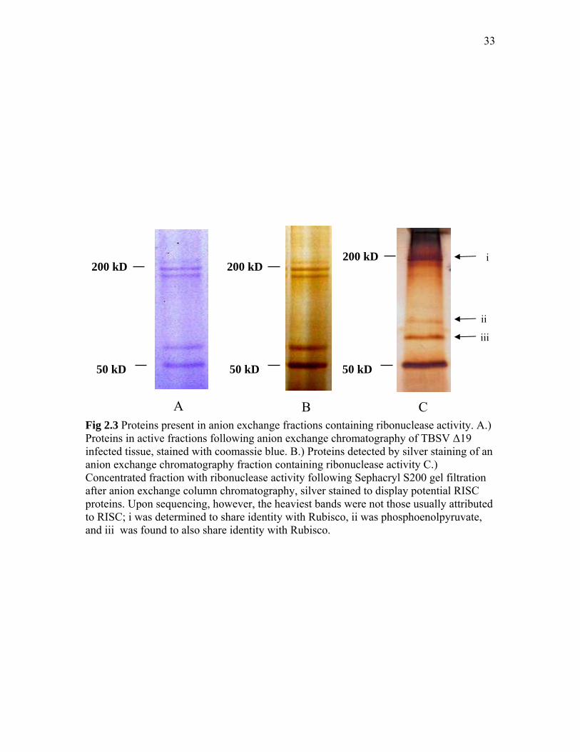
33
Fig 2.3 Proteins present in anion exchange fractions containing ribonuclease activity. A.) Proteins in active fractions following anion exchange chromatography of TBSV ∆19 infected tissue, stained with coomassie blue. B.) Proteins detected by silver staining of an anion exchange chromatography fraction containing ribonuclease activity C.) Concentrated fraction with ribonuclease activity following Sephacryl S200 gel filtration after anion exchange column chromatography, silver stained to display potential RISC proteins. Upon sequencing, however, the heaviest bands were not those usually attributed to RISC; i was determined to share identity with Rubisco, ii was phosphoenolpyruvate, and iii was found to also share identity with Rubisco.
200 kD
50 kD
200 kD200 kD
50 kD 50 kD
i
ii
iii
A B C

34
Fig 2.4 Ribonuclease activity test of TBSV ∆19 fractions. TBSV ∆19 infected plant tissue applied to a hydroxyapatite chromatography column and eluted with a 10 mM to 400 mM sodium phosphate gradient: 5 µl of the resultant combined fractions were tested for ribonuclease activity with the addition of 2 µl full length TBSV transcripts. The samples were incubated for 20 min., and viewed with a UV light after gel electrophoresis on a 1% agarose gel and staining with EtBr. Fractions with ribonuclease activity begin around fraction 5. T is transcripts without addition of fractions (volume equilibrated with sterile ddH2) I is column crude input, F is column flowthrough.
Fig 2.5 siRNAs and proteins detection in hydroxyapatite fractions from TBSV ∆ P19. A.) siRNAs were extracted following hydroxyapatite column chromatography of TBSV ∆19–infected plants. These siRNAs were separated on a 17% acrylamide gel with 8 M urea, and blotted to a nylon membrane for northern blotting. The hybridization probe was specific for TBSV, and siRNAs are present starting around fraction 7. B.) Fractions were run on 15% acrylamide SDS-PAGE, and silver stained. I is column crude input, F is column flowthrough.
TBSV siRNAs
I F 1 3 5 7 9 11 13 15 17 19 21 23 25 27 29 31 33 35
I F 1 3 5 7 9 11 13 15 17 19 21 23 25 27 29 31 33 35
kDa 230 72 56 36 28 17
A
B
TBSV RNA transcripts
T I F 1 3 5 7 9 11 13 15 17 19 21 23 25 27 29 31 33
TBSV DNA template

35
Fig. 2.6 DEAE chromatography after hydroxyapatite chromatography of TBSV ∆19-infected plant tissue. Following hydroxyapatite chromatography of TBSV ∆19-infected plant tissue, fractions with ribonuclease activity were pooled and subjected to DEAE anion exchange chromatography. These fractions then were tested for ribonuclease activity with the addition of TBSV RNA transcripts, and the resultant blot was probed with a TBSV-specific hybridization probe. Fraction 5/6 demonstrates the best activity. B.) This fraction was concentrated by spin-filtration in a centrifuge, and the resultant sample was loaded onto a 7.5% acrylamide SDS-PAGE gel, which was silver stained to detect proteins. Bands are apparent at about 100 kDa, appropriately the expected size for Ago. C.) siRNAs were also extracted from the concentrated fraction, and run on a 17% acrylamide gel with 8 M urea alongside a ~21 nt marker, indicated by an arrow. This is indicative of siRNAs, and that the complex has the hallmarks of a potential RISC. I is column crude input, F is column flowthrough.
I F 1 3 5 7 9 11 13 15 17 19 21 23 25 27 29 31 33 35 37
A
B C
[5/6] M
TBSV transcripts
~100 kDa
[5/6] M
kDa 230 130
56
36
28
95 72

36
chromatography column. When tested for ribonuclease activity against TBSV RNA
transcripts, fractions 3-11 degraded transcripts (Fig 2.6A). Fractions 5 and 6 were then
concentrated for analysis of proteins present via silver staining (Fig. 2.6B). There was a
100 kDa protein present, which is approximately the size for Ago. Moreover, when this
concentrated sample was assayed for siRNAs (Fig. 2.6C) alongside a known 21 nt RNA
marker, it shows a fairly clean, readily detectable 21 nt siRNA signal.
Biochemical characterization of TBSV-wt infected plant tissue
The hypothesis was that if the complex that is under investigation represents
RISC, it should not become programmed against TBSV in the presence of P19.
Therefore, to further characterize the RNAi response as seen in a natural virus infection,
wt TBSV (containing p19) -infected plant tissue extract was fractionated with
hydroxyapatite chromatography. Resultant fractions were tested for ribonuclease activity
(Fig 2.7) against TBSV RNA transcripts, as done in previous experiments. While some
activity was observed (fractions 5-23), it was to a much lower intensity that that seen for
the TBSV ∆19, with a minor band of transcripts remaining instead of the typical ‘wash’
of RNA and complete degradation of the TBSV RNA transcripts; the arrow indicates
partial RNA transcript bands that remained. Additionally, for hydroxyapatite fractions
collected from TBSV ∆19-infected tissue, the input lane usually shows degradation of
transcripts (Fig 2.4, lane ‘I’), though this was not the case for the wt TBSV-infected
tissue hydroxyapatite fractions – the transcripts added to the input lane remained intact
(Fig 2.7A, lane ‘I’).
Western blotting with antibodies against TBSV P19 and CP was performed for
the wt TBSV-infected tissue hydroxyapatite fractions (Fig 2.7B, C, respectively). P19
proteins accumulate in fractions that show inhibited ribonuclease activity. For assurance
that P19 is present in these fractions and binds siRNAs, IP for P19 was performed on the
input, flowthrough, and fraction 9, which showed both activity and an intense siRNA
signal, and fraction 33, which had unspecific activity and low siRNAs signal (included as
a (-) control). The IP successfully precipitated P19, as shown by western blot. However,
when these same IP samples were assayed for siRNAs, only the denser band from the

37
Fig 2.7 Fractions from the hydroxyapatite chromatography of wt TBSV-infected N. benthamiana. These were tested for A.) ribonuclease activity with the addition of TBSV full length transcripts, then blotted to a nylon membrane for hybridization with a TBSV-specific probe. Fractions were assayed for B.) TBSV P19 and C.) CP proteins after running on a 15% acrylamide gel, and transfer to nitrocellulose membrane for protein detection by western blotting with appropriate antibodies.

38
Fig 2.8 Detection of siRNA from wtTBSV-infected tissue following hydroxyapatite column chromatography. A.) Northern blotting of fractions with a hybridization probe for TBSV. These siRNAs appear in about fraction 5, and angle of the bands is due to the increasing amount of sodium phosphate present following chromatography. B.) Fraction 9, which shows a very strong siRNA signal, and 33, which shows a much weaker signal were subjected to immunoprecipitation (IP) with P19 antibodies to correlate presence of P19 and siRNAs with inhibited ribonuclease activity (fraction 33 was included as a control) and subjected to a western blot for P19 following IP. Input and fraction 9 show the presence of P19. (i indicates the dimerized P19, ii indicates P19 monomer C.) siRNAs are evident in the input, but undetectable following IP. At this time, it is hypothesized that reduced amounts of P19 in fraction 9 is the reason these levels are undetectable.
I F 9 33 I F 9 33
I F 1 3 5 7 9 11 13 15
i
ii
kDa 230 56 36 28 17 11
72
A
C B
siRNAs
siRNAs

39
column input showed a band on the western blot. Nevertheless, the results indicate that
even though siRNAs are abundantly present in the plant, the presence of P19 has
diminished the programming of the anti-TBSV RNA ribonuclease activity.
Discussion
RISC-like anti-viral ribonuclease activity
As described in the Introduction, there is a lot of evidence for RNAi in plants. At
this time, however, much of this has been determined using forward or reverse genetics,
like knocking out or mutating DCLs and Ago genes to determine effects on the RNAi
pathway, both for the endogenous regulatory pathway, as well as for a defensive, anti-
viral pathway (for example, Jones et al, 2006) The majority of this research focuses on
the model plant Arabidopsis thaliana (as illustrated by Baumberger and Baulcombe,
2005). However, little work has concentrated on the direct biochemical isolation of RNAi
pathway proteins in plants toward determining which proteins contribute to an anti-viral
defense. To be brief, it is expected that the anti-viral response in plants, specifically N.
benthamiana here, will be comprised of a Dicer or DCL protein (Xie et al., 2004), which
cleaves specifically viral RNA into about 21 nt duplexes (Baulcombe, 2004). These
duplexed siRNAs will associate with a protein complex, RISC, possibly through the use
of some Dicer-type loading protein for association with a protein complex. The complex
itself would contain an Ago-family protein, which are hallmarks of RNAi (Song and
Joshua-Tor, 2006). Silencing has implicated Ago1 and Ago4 for this in N. benthaminana
(Jones et al., 2006), though other studies indicate that Ago1 is involved in anti-viral
RISCs for Arabidopsis (Baumberger and Baulcombe, 2005). Therefore, to biochemically
determine if these virus-infected plants launch a RNAi anti-viral response, siRNAs and
Ago proteins should be present in these plants, ‘programmed’ against viral RNAs.
To examine the antiviral plant defense following a viral infection of plants, 2
week old N. benthamiana plants were inoculated with a plant virus, TBSV, which has
been shown to produce a large amount of dsRNA in the plant (serving as a substrate for
Dicer), and encodes a known silencing suppressor, P19 that was shown to bind siRNAs
(Omarov et al., 2006). The plants were first infected with mutants of TBSV that do not
produce functional P19 (TBSV∆P19), to ensure that an optimal RNAi response is

40
stimulated without the hindrance of a silencing suppressor. Infected plants were then
harvested upon display of obvious viral infection symptoms, and the first experiments
used Sephacryl S200 gel filtration to separate out proteins present that would contribute
to the plant defense. To test for fractions that might contain RNAi associated
ribonuclease activity, TBSV RNA transcripts, generated in vitro from cDNA with a T7
polymerase promoter, was added to each of the fractions. Fractions containing
ribonuclease activity demonstrated this activity in vitro against the TBSV RNA
transcripts, resulting in degradation (Fig. 2.1A). This fits with the current literature;
plants infected with a virus should result in the generation of siRNAs which are then
loaded onto a RISC, to further target viral RNA (Baulcombe, 2004). These anti-viral
ribonucleases are separated out by column chromatography, and target TBSV transcripts
in vitro.
In addition, these results suggested that upon gel filtration, at approximately 500
kDa anti-viral RISC-like complex could be isolated. To confirm that this was a real
complex and that results were not due to the co-incidental co-fractionation of elements,
other separation techniques were incorporated. Other types of column chromatography
were also used to improve protein separation, with the intent to isolate protein complexes
associated with activity. Towards this, TBSV plants infected with TBSV∆P19 were
subjected to anion exchange chromatography. After ribonuclease activity was
determined, divalent metal cations were added to the active fractions to determine the
effects. Current literature states that Piwi and PAZ domains contain divalent metal
cations; the Piwi domain uses these for catalytic activity (Song et al., 2004), and the PAZ
domain is thought to hold a Mg2+ to anchor the RNA molecule in a conserved
hydrophobic pocket by interacting with the phosphates present on the RNA (Parker et al.,
2005). Therefore, the addition of divalent metal ions might stimulate activity following
column chromatography, or prevent activity loss due to ion dissociation. As a metal
chelator, EDTA would bind to the metal ion present and prevents ribonuclease activity, if
indeed the metal ion is contributing to it. This is precisely what was observed in the
present study, EDTA inhibited ribonuclease activity. When surplus divalent metal ions
were added to active fractions in which activity was inhibited by EDTA, the degradative
activity was partially rescued. This supports data that the divalent metal ions contribute to

41
RISC activity, most likely to the catalysis mechanism of RISC; if the addition of divalent
metal ions in this experiment affected the binding of siRNAs, it is unlikely that activity
could be stimulated. When the divalent metal cations were added at the same time as
EDTA, RNA was still degraded, though not to the same extent as that of Mn2+ and Mg2+
alone.
siRNA and protein components of RISC
Following DEAE anion exchange chromatography, siRNAs were extracted from
all fractions to further confirm the presence of active, genuine RISC; a true RISC carries
a ss-siRNA molecule to use as a template for target mRNA (Baulcombe, 2004). The
siRNAs were extracted from all DEAE anion exchange chromatography fractions;
fractions found to have ribonuclease activity also were shown to contain siRNAs,
indicated by the arrow in Fig. 1.3B. Again, this is strongly indicative of a RISC. After
checking potential protein content of the fractions by silver staining (Fig. 2.2A), a
multitude of proteins were shown. Again, the Ago protein family is widely conserved
across many species, though the actual Ago proteins themselves vary in size and precise
function (Song and Joshua-Tor, 2006). The Ago that contributes to RISC in Drosophila
(Ago2) was predicted to be ~130 kDa in size, based on the coding sequence (Hammond
et al., 2001), though that same paper cites smaller bands visualized by SDS-PAGE
corresponding to the same ago gene, predicted to be products of protein proteolysis.
Other papers place the Drosophila Ago2 at about 235-500 kDa (Nykanen et al., 2001),
and the human RISC proteins at 90-160 kDa after isolation by affinity purification
(Martinez et al., 2002).
There are several proteins present in Fig 1.3A that might match the expected size.
However, because of the multitude of proteins present in these fractions, further protein
purification was performed by combining fractions containing ribonuclease activity after
DEAE anion exchange chromatography, then additional purification with S200 gel
filtration. When these fractions were stained for protein content (Fig 1.4), several
prospective bands remained, and these were sent for protein identification by MS/MS.
These did not turn up any matches to known RISC proteins; they were found to be
proteins found in energy synthesis of the plant. Because these proteins are among the

42
most abundant in plant tissue, this result is not illogical, though this does not reveal
anything about the anti-viral RNAi response, such as Ago, Dicer, or perhaps an additional
protein. However, it was decided at this point that a different method of column
chromatography might allow for differential separation, without co-elution of these
proteins.
The next type of column chromatography used was hydroxyapatite, which was
selected as the media interacts with both ionic and anionic groups. This would give a very
different protein elution profile than those previously seen. Additionally, it has been seen
that hydroxyapatite chromatography does not inhibit ribonuclease activity in other
systems (Hammond et al., 2001). When TBSV ∆19 infected plant tissue was separated by
this method and tested for ribonuclease activity as well as the presence of siRNAs, again,
these were found to co-elute. Following hydroxyapatite fractionation, the fractions were
tested for ribonuclease activity with the addition of EDTA and divalent metal cations
(data not shown). The effect was found to be similar to that seen upon gel filtration in
Fig. 2.1B. Additionally, these fractions displayed activity against genomic RNA, though
the activity was limited to only TBSV RNA. When studies were done with the addition of
viral transcripts for another virus (PMV), fractions did not target and degrade the PMV
RNA (unpublished data). This indicates that the ribonuclease activity is specific for
TBSV RNA, which is consistent for real RISC; only viral RNA for which the RISC has
been programmed is targeted for degradation (Baulcombe, 2004). This further validates
the presence of a complex that is in agreement with RISC; siRNAs (Fig 2.7A) and
fractions that target RNA transcripts for degradation (Fig. 2.4A) are the same for three
different types of chromatography.
After an additional step of DEAE chromatography, fractions demonstrating
activity (Fig. 2.6A) were concentrated, checked for siRNAs (Fig. 2.6C) alongside a 21 nt
marker, as well as tested for protein content by silver-staining. When the proteins in this
concentrated sample were visualized, a reasonably clear set of bands were detected
around 100 kDa, roughly the size of Ago determined for other organisms. Ago alone
displays minimal RISC activity for some systems (Baumberger and Baulcombe, 2005),
so it is possible that this protein is the anti-viral protein present in plants, although other
data, like the gel filtration experiments, where fractions with ribonuclease activity eluted

43
from the column in fractions corresponding to about 500 kDa, suggest that this protein
might act in combination with other proteins. At this time, a reverse approach was
undertaken by generating Ago-specific antibodies. These did detect bands in the fractions
containing activity. This assay was done for this project by Dr. R. Omarov. I will
incorporate this technique into Chapter III.
Effect of P19 on RISC-like activity
The work described above was performed with TBSV ∆19 to examine RNAi
silencing without inhibition by the viral suppressor. However, most natural viral
infections of plants occur for viruses that encode silencing suppressors. Earlier work had
been performed with Sephacryl S200 that suggested for plant tissue infected with wt
TBSV, the RNAi response is inhibited (Omarov et al., 2007). The presented study
showed that following hydroxyapatite column chromatography of wt-TBSV-infected
plants, fractions still contained activity in a manner similar to that seem for TBSV ∆19,
though to a much lower extent; partial RNA bands remained intact, and the input lane did
not seem to degrade transcripts at all. This helps to illustrate the protective function of a
silencing suppressor for the virus in the RNAi pathway. P19 has been shown to bind
duplexed siRNAs for sequestration before they are loaded onto a RISC (Voinnet et al.,
1999; Qiu et al., 2002; Park et al., 2004; Omarov et al., 2006; Scholthof, 2006).
Degradation of transcripts in crude extract from TBSV ∆19 –infected tissue, and lack
thereof in the crude extract of wt-TBSV infected tissue can be interpreted to mean that
due to the presence of the silencing suppressor P19, presumably isolating siRNAs,
RISCs are inefficiently loaded with viral siRNAs in wt-TBSV infected tissue and do not
target transcripts. Likewise, this interpretation can be extended to the fractions
themselves. Because of the much lower amount of anti-viral programmed RISCs in wt-
TBSV fractions, TBSV RNA transcripts remain. The minor amount of ribonuclease
activity present can be attributed to the concentrating effect of a chromatography column;
fractions contain much more of the same anti-viral protein than does an equal amount of
the crude plant extract.
To determine if the P19 shown to accumulate in the same fractions (Fig 2.7B) as
those containing ribonuclease activity bind siRNAs as predicted (Qiu et al., 2002; Ye et

44
al., 2003), immunoprecipitation was preformed with P19 antibodies. When siRNAs were
extracted from the immunoprecipitated P19, the fraction with input showed a very minor
band, though this was not detected for a fraction with slight ribonucleic activity. This
might be related to the amount of P19 that was pulled down; a western for the IP shows a
reduced amount of P19 for that fraction than for the input (Fig. 2.8C). Additionally, this
also might be related to the amount of ribonuclease activity present; the input fraction
contained more P19 with bound siRNAs than did the fraction with slight ribonuclease
activity; more siRNAs bound to P19 indicate less siRNAs that can be bound to RISC.
Fraction 9 shows slight activity; the amount of siRNAs bound to RISC would imply less
siRNAs bound to P19, and less to be detected by immunoprecipitation. Regardless, this
further substantiates the claim that the ribonuclease activity seen in these fractions can be
attributed to RNAi, as it is inhibited, at least partially, by a silencing suppressor.
In conclusion, the data indicate that fractions from TBSV-infected plants contain
a TBSV-specific ribonuclease complex with characteristics shared by RISC seen in the
RNAi pathway. The complex can be isolated with three independent separation
techniques indicating that the residual activity and siRNAs are associated with the same
complex. This ribonuclease activity is enhanced by the addition of divalent metal cations,
inhibited by EDTA, is specific for TBSV, and affected by a silencing suppressor. This
work documents one of the first successful attempts for any virus-host system towards
isolation of a high molecular-weight protein complex that is programmed against a
specific virus, and displays properties consistent with RISC.

45
CHAPTER III
DETERMINATION OF AN ANTI-VIRAL RESPONSE FOLLOWING
INFECTION OF PLANTS WITH TRV, PMV AND SPMV
Introduction
RNAi has been shown to be strongly conserved across multiple species, as a
method to regulate gene function in everything from plants (Baulcombe, 2004) to fungi
(Romano and Macino, 1992), insects (Hammond et al., 2001) and mammals (Liu et al.,
2004). Chapter II and recent papers (Omarov et al., 2007; Pantaleo et al., 2007)
demonstrated RISC-like antiviral ribonuclease activity specific for Tombusviruses. There
is evidence that other viruses trigger a virus-specific ribonuclease that is attributed to an
RNAi response, specifically illustrated by those used to trigger virus induced gene
silencing (VIGS) (Ratcliff et al., 2001; Batten et al., 2003; Burch-Smith et al., 2006).
Furthermore, many viruses encode silencing suppressors (Voinnet, 2005; Li and Ding,
2006); viruses favor the smallest functional genome size, and it is reasonable to assume
that the silencing suppressors would not be encoded without purpose. To strengthen the
model for a conserved anti-viral silencing pathway, and to determine if it corresponds to
the postulated models currently based on other systems like Drosophila, it is necessary to
biochemically analyze the defensive response of plants to viruses other than TBSV.
This work attempted to determine how similar these plant responses are for
Tobacco rattle virus (TRV), which is not related to TBSV, and identify possible common
elements that delineate a conserved RNAi pathway in plants by in vitro examination of
subcellular elements. Furthermore, an antiviral defense was studied for a monocot plant
model system, versus the dicot system outlined above. Panicum mosaic virus (PMV) and
satellite panicum mosaic virus (SPMV) were used to study antiviral elements induced in
the monocot Panicum miliaceum (proso millet) plants upon infection, for comparison to
those seen in N. benthamiana.
Tobacco rattle virus (TRV) is a viral vector commonly used to silence genes
(VIGS) in a wide range of plants (Ratcliff et al., 2001; Burch-Smith et al., 2006). The
form of TRV used in this study is a TRV virus vector, constructed for infiltration into the
host plant using Agrobacterium, based on a construct originally generated by Ratcliff and

46
colleagues (Fig. 3.1) (Ratcliff et al., 2001). Our T-DNA constructs consist of cDNAs of
the TRV strain Ppk20 as constructed by by Liu and colleagues; viral cDNAs were
inserted behind CaMV 35-S promoters, with a self-cleaving ribozyme from the satellite
viroid of Subterranean clover mottle virus at the 3’ end (Liu et al., 2002b). RNA1
remains pretty much intact as described above for the wild type virus, with only minor
alterations before insertion into the pBIN19 binary vector T-DNA plasmid (Liu et al.,
2002b). The RNA2 CP gene remains intact, followed by a multiple cloning site (MCS).
The MCS carries a 369-nt segment of a conserved region of the plant phytoene
desaturase (pds) gene (Fig. 3.1A). This gene is a precursor of the plant carotenoid
pathway, and when targeted for silencing by VIGS, the plant tissues turn white.
SPMV is not required for systemic infection of PMV in a host, though a tandem
infection has a synergistic effect on host symptom severity (Scholthof, 1999). PMV alone
will give a mild mottled phenotype, with slight stunting in millet. Co-infection with
SPMV results in much more severe symptoms, including streaked, chlorotic leaves with
occasionally necrotic regions (Qiu and Scholthof, 2004). This virus system was chosen
for study because it infects monocots. RNAi silencing suppressors have been discovered
for viruses that infect monocots (Hemmes et al., 2007), leading to the possibility that
RNAi occurs in monocots in a manner similar to that of dicots, though studies have yet to
determine whether this occurs. Part of this objective aims to determine if RNAi elements
similar to those seen for virus-infected dicots are present in virus-infected monocots. As
PMV is classified within the Tombusviridae, a virus family with a known RNAi response
(Omarov et al., 2007; Pantaleo et al., 2007), PMV stands as a good choice for this work.
The system used to study the RNAi defensive response is essentially the same as
that used in Chapter II for the characterization of an RNAi response against TBSV. Plants
were inoculated with viruses, and tissue was harvested once the infection was established.
Column chromatography was used to separate proteins present, and these fractions were
tested for virus-specific ribonuclease activity. Fractions containing ribonuclease activity

47
Fig. 3.1 Tobacco rattle virus (TRV) agro-infiltration construct and primers. A.) TRV is commonly used as a virus vector, as TRV RNA2 is not required for a systemic infection. This particular construct is used for Agrobacterium infiltration, and has cDNA for TRV RNA1 between the left border (Lb) and right border (Rb) of the Agrobacterium Ti plasmid, behind a CaMV 35S promoter (35S) and upstream of a ribozyme (Rb) and nopaline synthase terminator (poly A) signal (T) for expression in planta. TRV RNA2 has a similar design, but with a multiple cloning site (MCS) replacing the nematode transmission factors. For this project, a portion of the N. benthamiana PDS gene, with high sequence homology to other plant PDS genes, has been inserted (PDS). C.) Primers used to generate TRV-RNA2.PDS transcripts used later in this chapter. These primers encompass part of the TRV RNA2.PDS cp and pds. The forward primer has a sequence for the T7 polymerase promoter to allow synthesis of RNA directly from PCR products.

48
were tested for the presence of small RNAs, specifically those corresponding to the virus
used to infect the plant, as the presence of siRNAs is a hallmark of RNAi. These fractions
were then assayed for the presence of potential Ago proteins with western blotting with
antibodies against the conserved region of the Piwi domain. Fractions containing both
siRNAs and proteins with a Piwi domain were then tested to determine if the
ribonuclease activity was inhibited by the addition of EDTA, which is a metal chelator,
and high concentrations of NaCl. The hypothesis was that if the plant defensive response
is RNAi-based, EDTA would act as an inhibitor because the Ago protein of RISC uses
metal ions for catalysis of the ribonuclease activity (Tomari and Zamore, 2005).
NaCl has been shown to have an impact of RISC activity (Rand et al., 2004),
though it might inhibit it in a different way; siRNAs are shown to be associated with the
PAZ domain using ionic interactions (Song and Joshua-Tor, 2006). It might be that an
increase of salt would remove siRNAs from RISC, though this has not been observed for
other systems; treatment with high concentrations of salt has been used to as part of the
Ago purification process (Hammond et al., 2001). Additionally, NaCl would prohibit
unspecific binding of siRNAs by causing the associated guide siRNAs to bind more
tightly to the target long ssRNA. Because RISC has been shown to use divalent metal
ions in catalysis, as mentioned above, Mg2+ and Mn2+ were also used for stimulation of
RISC activity in ribonuclease-containing fractions. The coincidence of these factors
would indicate with high likelihood the presence of a genuine RISC-type protein.
The results show that TRV and PMV/SPMV -induced RNAi responses share key
features with those observed for TBSV Chapter II, but some unique features were
observed as well.
Materials and Methods Infection of plants with TRV and PMV/SPMV
Agrobacterium-mediated infection of plants using T-DNA plasmids that express
TRV RNA1 and the RNA2 vector with a PDS gene (Burch-Smith et al., 2004) were used
to examine a potential RNAi plant response. Overnight cultures of Agrobacterium were
added to 50 ml of Luria broth with 1 ml 10 mM 2-(4-Morpholino)-ethanesulfonic Acid
(MES) and MgCl2, 50 µl kanamycin, 50 µl tetracyline, 25 µl rifampicin and 20 mM

49
acetosyringone for 12 hours. These cultures were centrifuged for 30 min. at 4000 rpm
with a Beckman S4180 rotor, the bacterial pellet re-suspended in 10 ml of 10 mM MES
and 10 mM MgCl2+, plus 150 µl 150 mM acetosyringone, and incubated at room
temperature following a 20 min. agitation period. Equal amounts of these cultures for
TRV RNA1 and TRV RNA2 were combined, and 3 leaves of 3 wk old N. benthamiana
plants were infiltrated with 1 ml per plant using a needle-less syringe. These plants were
grown at 25° C until infection was established, after which tissue was harvested.
As the project progressed, plants infiltrated with the TRV constructs were
harvested at different timepoints. The earliest experiments used obviously silenced
(whitened) tissue harvested about 8 weeks post inoculation. As the project progressed, it
became obvious that fractions from this older tissue gave a very unspecific ribonuclease
response, and it was thought that tissue collected from TRV-infiltrated plants earlier in
the infection (2 weeks, 1 week) would give a more virus-specific defense response. Thus,
TRV-infected plant tissue was harvested at these times.
Three-week old proso millet (Panicum miliaceum L.) plants were rub-inoculated
with infected tissue. This tissue was obtained from millet that had been inoculated with
infectious PMV and SPMV in vitro-generated infectious transcripts, made via a T7 RNA
polymerase system (Turina et al., 1998), as done with TBSV RNA transcripts in Chapter
II. These transcripts were then used to rub-inoculate three week-old proso millet plants
using RNA-inoculation buffer (50 mM KH2PO4, 50 mM Glycine, pH 9.0, 1% celite, 1%
bentonite).
Infected tissue was processed by grinding in a chilled mortar and pestle with 10
mM sodium phosphate buffer, with sea sand as added abrasive. This crude extract was
then further homogenized with a blender, and clarified using centrifugation, as described
in Chapter II. The plant extract was filtered through cheesecloth into falcon tubes, and
placed on ice until it was applied to the hydroxyapatite chromatographic column. For
column packing, please refer to Chapter II and the appendix. Again, fractions from
hydroxyapatite chromatography were combined for easier manipulation, mixing equal
amounts of the fractions, 2 at a time (for instance, fractions 1 and 2, 3 and 4, 5 and 6,
etc.), before storing at - 20° C.

50
Extraction of siRNAs from chromatographic fractions
This was done as described in the Material and Methods section of Chapter II.
SDS-PAGE and Western analysis of samples for Piwi-containing proteins.
The peptide “kivegqryskrlnerq” was used for production of the antibody as
BLAST analysis of plant Ago proteins show this to be a conserved plant-Ago specific
domain, specifically for the Piwi-domain of Ago1 and 2 from N. benthamiana. The
antibodies were raised in rabbits (SigmaAldrich, St. Louis, MO). SDS-PAGE was
performed as described in the Chapter II Materials and Methods section.
Upon protein gel electrophoresis with very small amounts of protein/samples,
these proteins were detected using antibodies and chemiluminesence. After rinsing the
blot following addition of primary antibodies, the horseradish-peroxidase anti-rabbit
secondary antibody (Pierce) was added to 10 ml of 7.5% milk solution. This was then
incubated at least 2 hours, then the blot was rinsed extensively with TBS-Tween (20 ml
for 15 min., 4-6 times). The blot was then developed with the chemiluminesence western
detection kit (Pierce), immediately exposed to Kodak BioMax X-ray film (Bio-rad) for 1
min., then the film was developed.
Assays to further characterize ribonuclease activity.
Total RNA extraction from infected tissue
For TRV, many assays were carried out using total RNA extracted 1 week post
inoculation from TRV-RNA1 and RNA2-PDS infected plants. About 1 gram of infected
tissue was ground with 1 ml 2X STE+1% SDS (2 mM Tris, 20 mM NaCl, 0.002 mM
EDTA, and 1% SDS) with chilled mortars and pestles. Then, 1 ml 1:1 phenol/chloroform
was added, and tissue was reground to mix. This was centrifuged at 10,000 rpm, at 4° C
using a Beckman F2402 rotor for 20 min. The upper aqueous phase was removed to a
separate 1.5 ml microfuge tube, 1 ml of 1:1 phenol:chloroform was added, the sample
vortexed, then centrifuged at 10,000 rpm again for 20 min. at 4°C. The upper aqueous
phase (~400 µl) was again placed in a new microfuge tube, and 40 µl 8M lithium chloride
was added to precipitate RNA while the sample was placed on ice for 15 min. The RNA

51
was then pelleted by centrifugation at 10,000 rpm in a Beckman F2402 for 20 min. at
4°C, and rinsed with ice cold 70% ethanol, centrifuging again at 10,000 rpm for 10 min.
at 4°C. The 70% ethanol was discarded, and the pellet dried very briefly by vacuum
centrifugation (no more than 5 min. to completely remove the ethanol). The RNA was re-
suspended in a/c ddH2O plus RNasin and DTT [100 µl sterile ddH20, 1 µl Ribolock
RNase inhibitor, and 2 µl 0.1 mM DTT (Fermentas, Glen Burnie, MD)], depending upon
the final concentration as detected by nanodrop. The assays typically used ~250 ng/ µl
RNA/fraction sample, unless otherwise noted.
TRV RNA2.PDS PCR and RNA transcripts
Additionally, ribonuclease activity was assayed using RNA transcripts generated
in vitro from linearized cDNA. For TRV, a PCR reaction to amplify a 2 kb segment
TRV-RNA2.PDS was made using primers that overlap the PDS-region of the TRV-
RNA2 vector carrying a segment of the N. benthamiana pds gene (TRV-RNA2.PDS)
(Fig. 3.1B), designed to add a T7 polymerase promoter sequence for in vitro
transcription. For PCR, TRV-RNA2.PDS was amplified in overnight cultures consisting
of 2 mls LB and Kan50. DNA was extracted using Qiagen Mini-prep kits (Valencia, CA),
and stored at - 20°C until needed. For PCR, primers were diluted to about 50 ρm; 1 µl of
the DNA was mixed with 1 µl each primer, 1 µl 12.5 mM dNTP mix, 1 µl MgSO4, 5 µl
Thermopol buffer, 40 µl a/c ddH20, and 1 µl Vent DNA polymerase (2000 U/ml) (New
England Biolabs) in bubble-topped tubes. PCR settings used were 3 min. pre-denature at
93° C, 1 min. denature at 93° C, 1 min. re-annealing at 55 ° C, and 2 min. 15 seconds
extension at 72 ° C, for 35 cycles. The resultant DNA was checked by ethidium bromide
staining after electrophoresis on a 1% agarose gel, and purified with phenol-cholorform
as described above. RNA transcripts were then made from this PCR template as
described above. PMV transcripts were generated as outlined in a previous section.
Assays for the presence and characterization of ribonuclease activity
Fractions from column chromatography were mixed with either total RNA
extracted from virus-infected plants, or with transcripts generated in vitro from linearized
viral cDNA as outlined above. For the 5 dpi TRV and PMV experiments, the

52
concentration of RNA was determined using a nanodrop, and standardized to ~250 ng/µl
to ensure uniformity of ribonuclease substrate. In other experiments, RNA was assayed
by electrophorsis with a 1% agarose gel, stained with ethidium bromide, and checked for
quality using a UV light.
To test for activity of ribonucleases, 5 µl of each combined fraction was incubated
at room temperature (about 25°C) with 1.5-2 µl RNA for 20 min. with 1.5 µl RNasin-
treated ddH2O [100 µl sterile ddH2O, 2 µl DTT, 1 µl Ribolock RNase inhibitor
(Fermentas, Glen Burnie, MD)]. Then, 2 µl DNA loading dye was added, and samples
were run on a 1% agarose gel, 120 volts, in 1X TBE until the lower dye band was about
¾ of the way from the front of the gel. These gels were then stained with ethidium
bromide for 15 min., and viewed with a UV light box. To test for inhibition by EDTA or
NaCl, the indicated amount of 50 mM or 100 mM EDTA or 5 M NaCl stock solution was
added to each fraction before the addition of RNA. To stop a reaction, EDTA was added
at the time detailed in the assay, or after 20 min.. Determination of the effect of divalent
metal cations on ribonuclease activity present in the fractions, 50 mM MgCl2 and 50 mM
MnSO4 were used in the amount specified by the assay.
Northern blotting and hybridization with radioactive DNA probes
Northern hybridizations were performed essentially as described in Chapter II.
Results Determination of an anti-viral plant defense response against TRV.
N. benthamiana plants infected with TRV RNA1 plus RNA2.PDS displayed a
‘silenced’ phenotype of whitened tissue. This photo-bleached phenotype was readily
visible after about 5-7 days post-infiltration for N. benthamiana (Fig. 3.2A). The region
of the inserted gene is conserved not only in N. benthamiana, but shares homology to the
tomato pds gene (Liu et al., 2002b), and also triggers a white phenotype in pepper plants
(Fig. 3.2.B). Interestingly, manifestation of the photo-bleached phenotype took much
longer in pepper, up to 3 weeks post-infiltration (Fig 3.2B), versus 5-7 days in N.
benthamiana.

53
The first experiments for characterization of a TRV RNAi-associated
ribonuclease used 8 week post infiltrated N. benthamiana plants, particularly the upper,
whitened tissue. This tissue was applied to both DEAE anion exchange chromatography
columns (Fig. 3.3), and later, hydroxyapatite chromatography (not shown). The resultant
fractions were tested for degradation of TRV RNA2.PDS RNA transcripts. The northern
blot from the 1% agarose gel shows degradation of RNA transcripts beginning in fraction
5, showing complete degradation in fractions 7 as well as later fractions (Fig. 3.3A). This
activity was inhibited with the addition of EDTA (Fig. 3.3B), and a kinetics study for
ribonuclease activity showed that RNA was nearly totally degraded at about 5 and a half
min. (data not shown). However, fractions also displayed activity against TBSV RNA
transcripts. No siRNAs were detected upon extraction from the fractions, and with an
additional chromatography step of S200 gel filtration, no ribonuclease activity against
TRV RNA2.PDS RNA transcripts was observed (data not shown).
With the idea that perhaps the infection was established too long, more N.
benthamiana plants infiltrated with TRV RNA1 and RNA2.PDS were harvested
for hydroxyapatite chromatography at 14 dpi. These fractions displayed ribonuclease
activity, against TRV RAN2.PDS RNA transcripts (Fig. 3.4A), which appeared to begin
in fraction 11. Fractions were also tested for the specificity of this degradation with the
addition of TBSV wt RNA transcripts, alongside degradation of TRV RNA2.PDS RNA
transcripts (Fig. 3.4B). Activity corresponds in fraction 11, again, for both sets of
transcripts, indicating unspecificity. This activity was inhibited by the addition of 1.5M
NaCl as well as EDTA (data not shown). A small Sephacryl S200 gel column was used to
separate out fractions 7-13. RNA from the fractions was electrophoresed on a 17%
acrylamide gel with 8M urea, blotted to a nylon membrane, and probed with a TRV
RNA2.PDS hybridization probe. After the membrane had been exposed to film for over 2
weeks, the film was developed. A small RNA signal can be seen in active hydroxyapatite
fractions collected from both 2 mts and (very faint for) 2 wks –pi plant
tissue, but none was seen for the fractions collected after hydroxyapatite plus gel
filtration (Fig. 3.5).

54
Fig. 3.2 TRV-infected plants. A.) N. benthamiana and B.) C. annuum (pepper) were agro-infiltrated with the TRV RNA1 and RNA2.PDS constructs. Picture was taken 2 weeks post infiltration for N. benthamiana, and about 4 weeks post infiltration for pepper. Visible silencing of the plant PDS gene is illustrated by the white tissue of the upper leaves.
A B

55
Fig. 3.3 DEAE ion exchange chromatography was performed on N. benthamiana 8 weeks post infiltration. A.) Transcripts generated in vitro for TRV-RNA2.PDS were added to fractions, incubated for 20 min., and electrophoresed on a 1% agarose gel, then followed by northern hybridization against TRV-RNA2. (T= 2 µl transcript only, plus 5 µl a/c ddH2O; 2 µl transcripts were added to 5 µl combined fractions). These fractions show ribonuclease activity, starting in fraction 7. B.) hydroxyapatite fraction 7 was assayed for inhibition of ribonuclease activity with the addition of increasing amounts of 100 mM EDTA. For each sample, 5µl fraction 7 (H2O for T) + 2 µl transcript were combined and incubated for the amount of EDTA indicated, then samples were analyzed by ethidium bromide staining after gel electrophoresis. (i:TBSV DNA, ii: TBSV RNA transcripts, iii: TRV RNA2.PDS transcripts)

56
Fig 3.4 Hydroxyapatite fractions collected from 14 dpi TRV-infected N. benthamiana plants were characterized. A.) Fractions were tested for ribonuclease activity against A.) TRV-RNA2.PDS transcripts; 5 µl of each combined fraction (T with a/c ddH20) was combined RNA. B.) Test for specificity of RNA degradation, as indicated. 5 µl of each combined fraction (T with a/c ddH20 ) was combined with either 2 µl TBSV or TRV RNA2 transcripts, prepared in vitro. These fractions and transcripts were then subjected to gel electrophoresis, and stained with ethidium bromide.

57
Fig 3.5 siRNA assay for TRV-infected plant tissue extract chromatography fractions. Combined fractions indicated, taken from hydroxyapatite chromatography fractions 8 weeks post infiltration, 2 weeks post infiltration, and the 2 week post infiltration hydroxyapatite chromatography fractions 7-13 following Sephacryl S200 column chromatography fractions were collected and used for siRNA extraction. Resultant samples were then separated by 17% acrylamide, 8 M urea gel electrophoresis, and blotted to a nylon membrane for northern detection of TRV-RNA2.PDS. Visible potential siRNAs are apparent for fractions containing activity 8 weeks and 2 weeks post inoculation following hydroxyapatite fractionation steps, but unapparent following an additional gel filtration step.
7 9 10 7 9 11 8 10
8 wks pi 2 wks pi 2 wks pi +S200

58
Despite numerous tests, the above summarizes results showing that fractions did
not display TRV-specific ribonuclease activity, nor were siRNAs detected reliably. It was
suggested that plants be harvested even earlier in the course of infection. For this
purpose, tissue from TRV RNA1/RNA2.PDS infected plants was harvested 5-7 dpi, and
applied to a hydroxyapatite chromatography column. Fractions were tested for
degradation of RNA this time using total RNA extracted from TRV-infected plants,
instead of using TRV RNA2.PDS transcripts. Unspecific ribonuclease activity was seen
beginning in fraction 3 (data not shown). From this fraction to fraction 15, siRNAs were
extracted, run on a 17% acrylamide SDS-PAGE with 8 M urea, and blotted to a
membrane to probe with a hybridization probe using a TRV RNA2 vector without a gene
insert in the MCS. The resultant northern blot showed a clear signal for siRNAs in
fraction 9 and later fractions (Fig 3.6A), though it is possible that siRNAs are present in
undetectable levels in earlier fractions.
These siRNA-containing fractions were then subjected to western blotting with
antibodies generated against a conserved region of the Ago Piwi domain, which is a
hallmark of RNAi pathway. Fractions showed several bands (about 130 kDa, 95 kDa, 60
kDa, 50 kDa, and faint others), with those of an appropriate size (around 100 kDa or
larger) (Hammond et al., 2001). Beginning around fraction 9, corresponding to the first
siRNAs detected by northern analysis, the presence of a much smaller band was detected
(about 17 kDa) (Fig 3.6B).
To possibly increase specificity, these fractions with detectable siRNAs and
potential Ago proteins were concentrated and further separated using a Sephacryl S200
gel column. Fractions were again analyzed with total RNA extracted from TRV-infected
plants, and northerns probed with TRV RNA2 showed ribonuclease activity in fractions 8
– 10 (Fig 3.7A), though these same fractions did not degrade TBSV RNA transcripts (Fig
3.7B). This result verifies that the ribonuclease activity displayed in these fractions is
virus-specific. The experiment was repeated for another set of TRV- infected plants,
these were harvested 5 dpi and subjected to hydroxyapatite and gel chromatography. The
second set of fractions also displays ribonuclease activity for TRV total RNA as
illustrated by northern blotting, but not for TBSV RNA transcripts, also detected by
northern blotting (Fig 3.7 C, D). The exact fraction with activity varies between these two

59
Fig. 3.6 Analysis of siRNAs and Ago-associated proteins present in fractions collected from hydroxyapatite fractionation of TRV-infected N. benthamiana plants, 5 dpi. A.) siRNAs extracted from hydroxyapatite fractions visualized by northern hybridization for TRV-RNA2. Arrow indicates siRNAs. B.) Western blot for Piwi domain of Ago protein in hydroxyapatite fractions of TRV-infected plant extract. Fractions 9-17, which show ribonuclease activity, show a smaller band that might possibly be associated with RISC activity. The + lane is a positive control consisting of TBSV crude extract, previously demonstrated to have a positive response with the Piwi antibody, which also has the smaller band.

60
Fig 3.7 Sephacryl S200 chromatography of TRV hydroxyapatite fractions. Following hydroxyapatite chromatography, fractions containing ribonuclease activity and potential Ago proteins were subjected to Sephacryl S200 gel filtration to test for activity (A and B are from same fractionation, C and D are from corresponding fractions). A.) and C.) Fractions were mixed with total RNA from TRV-infected followed by northern hybridization with TRV-RNA2. B.) and D.) Ribonuclease activity with TBSV RNA full length transcripts, generated in vitro. C.) Ethidium bromide stained agarose gel, and D.) Northern blot probed with TBSV hybridization probe. (Lane T is RNA without addition of fractions, lane I (input) consists of hydroxyapatite fractions showing activity and presence of siRNAs before S200 chromatography, plus addition of TRV RNA. M is a DNA ladder. Lanes 1-18 are S200 fractions with the addition of RNA. Activity against TRV seen in different lanes for different fractionations, due to variability in collecting fractions following elution. This variability does not affect activity; it only changes the fraction number.

61
iterations; this is due to normal column variability and the collection of eluted fractions.
Protein elution is detected by spectroscopy as fractions are collected, and minor
variances in the timing lead to differences in fraction number, though not elution profile
of proteins.
These results indicate that at early timepoints following infection, a true anti-TRV
ribonuclease was programmed in virus-infected plants. To obtain additional evidence that
this ribonuclease acts similarly to that seen in the previous chapter for TBSV, as well as
to determine how it fits the model RNAi pathway, the fractions containing activity were
then subjected to a battery of biochemical analyses.
Characterization of TRV-specific ribonuclease activity
Fractions from gel filtration after hydroxyapatite were tested with differing
amounts of 100 mM EDTA, to establish the amount of EDTA required for inhibition
following another chromatography separation step (Fig 3.8A); 10 mM EDTA was
determined adequate for inhibition of activity. Active fractions were also assayed with
increasing amounts of total RNA from TRV-infected tissue, to determine how the
ribonuclease activity would be affected with the addition of more substrate (Fig. 3.8B).
For this purpose, the standard amount of RNA (1.5 µl RNA) was compared to fractions
that had 2, 3, and 4 µl of RNA added (a/c ddH2O was added to all reactions to bring the
total volume to 16 µl). Fractions demonstrating ribonuclease activity continued to target
TRV RNA2, though to a level proportionate to the amount of RNA added. TRV
RNA2.PDS transcripts were also added to the fractions alongside total RNA extracted
from TRV-infected plant tissue. Fraction 9 displayed ribonuclease activity against the in
vitro generated transcripts, and increased activity against plant-extracted TRV RNA,
though fraction 8 also degraded plant-extracted TRV RNA, to a lesser extent than
fraction 9 (Fig 3.8C). TRV RNA I was also degraded, as determined by northern blotting
(data not shown).
Collectively, this and other tests showing that the anti-TRV RISC-like complex is
active against in vitro and in planta generated TRV RNA in an EDTA and substrate
dosage dependent manner.

62
Fig. 3.8 Further characterization of ribonuclease activity following S200 and hydroxyapatite chromatography of 5 dpi TRV-infected plant tissue. Active fractions (7-10) were mixed with RNA extracted from plants infected with TRV (all volumes standardized with the addition of a/c ddH2O), incubated for 20 min., and visualized by northern blotting of the resultant 1% agarose gel. A.) 5 µl of the active fractions were tested for inhibition of ribonuclease activity for 1.5 µl RNA with increasing concentrations of EDTA, as indicated. Ribonuclease activity is inhibited by addition of 10 mM EDTA. B.) Five µl of each active fraction was mixed with amount of RNA indicated. Ribonuclease activity still functions with the addition of ~1000 ng/ µl RNA, though proportionately decreased. C.) Five µl fractions were mix with 1.5 µl RNA from TRV-RNA2.PDS transcripts (left), or total RNA extracted from TRV-infected plants (right). It is not known what is the significance of the RNA II duplex band.

63
Effects of NaCl on ribonuclease activity for hydroxyapatite fractions with S200 gel
filtration
To determine the influence of NaCl on (possibly inhibiting) ribonuclease activity,
hydroxyapatite fractions with ribonuclease activity were also applied to a Sephacryl S200
gel filtration column that had been equilibrated with 250 mM NaCl (as added to the 50
mM Tris buffer, pH 7, used for gel filtration). Fractions were then assayed as described
above. For detection of ribonucleic activity, fractions were assayed with total RNA
extracted from TRV-infected plants (Fig 3.9A). Fractions 11 and 12 displayed definite
activity, though to a lesser extent than fractions collected following gel filtration without
NaCl, which suggests siRNA dissociation. Intriguingly, when these fractions were
assayed with total RNA collected from plants infected with TBSV ∆P19 (see Chapter II),
fractions did demonstrate activity against the TBSV genomic RNA though not against
subgenomic RNA, a finding that is in agreement with the ribonuclease activity seen for
Sephacryl S200 fractions after hydroxyapatite chromatography of TBSV-infected plant
extracts (unpublished data). Moreover, based on the northern blot hybridization data from
this experiment, it appears that this activity against TBSV gRNA is even more vigorous
than that observed with the addition of TRV-infected plant total RNA (Fig. 3.9B).
Fractions, following gel filtration with salt, do not target TBSV RNA full length
transcripts generated in vitro (data not shown), suggesting that the total RNA extracted
from infected plant tissue carries with it some element that re-programs the ribonuclease
activity. These are very exciting observations, and will be addressed in the discussion.
Considering the strong ribonuclease activity displayed by the NaCl-gel filtration
fractions 10-12, these were tested for the presence of potential Ago proteins by western
blotting with antibodies against the Ago Piwi-domain (Fig 3.10A). It was nearly
impossible to detect any proteins at all in fractions using colorimetric detection with
alkaline phosphatase, as has been done in previous westerns, though the gel column input
positive control showed a positive signal. Therefore, a more sensitive assay was
preformed with secondary antibody conjugated to horseradish peroxidase to detect Piwi-
containing proteins. Positive signals were observed for fractions 10 and 11, estimated to
represent approximately 90 and 60-80 kDa proteins, when compared to the low weight
molecular markers by film overlay on the membrane.

64
Fig. 3.9 Ribonuclease test for NaCl gel filtration of TRV hydroxyapatite fractions. Fractions containing ribonuclease activity following hydroxyapatite column chromatography were fractionated on a Sephacryl S200 gel chromatography column equilibrated with 250 mM NaCl, to test for increased specificity of ribonuclease activity and potential dissociation of siRNAs. Two µl ( ~250 ng/µl) total RNA was added to 5 µl fractions in the presence of an RNase inhibitor, and incubated for 20 min., then separated on a 1% agarose gel, and blotted to a nylon membrane for northern blotting. A.) Fractions were assayed for TRV ribonuclease activity with the addition of total RNA extracted from TRV-infected plants. The resultant northern blot was probed with a TRV-RNA2 hybridization probe. Fractions 11 and 12 display residual ribonuclease activity. B.) Same fractions plus the addition of total RNA extracted from a TBSV P19 deficient-infected plant. Northern hybridization using the TSBV P19-deficient cDNA probe. The arrows indicate i.) TBSV gRNA; ii.) TBSV sgRNA1; iii.) TBSV sgRNA 2.

65
Fig 3.10 Westerns for NaCl gel filtration of TRV hydroxyapatite fractions. Following hydroxyapatite column chromatography fractionation and further protein purification by gel filtration with a Sephacryl S200 column with 250 mM NaCl, fractions were analyzed by western blotting with antibodies generated against a conserved region of the Piwi domain from Ago. A.) Thirty µl of each fraction (I = input) were boiled with cracking buffer and loaded onto 7.5% acrylamide SDS-PAGE gels, then blotted to a nitrocellulose membrane for detection of proteins present by western analysis using chemiluminescence. Fractions displaying activity (namely, 10, 11 and 12) displayed bands, red box indicated region highlighted for B. B.) Bands present for input, and Fractions 10 and 11 were enlarged for illustration. C.) Fractions 8-13 were visualized on a 5% SDS-PAGE gel with silver staining.

66
To better visualize this, the bands were compared to that seen in the column input
lane in Fig. 3.10B. These fractions were separated by 5% SDS-PAGE followed by silver
staining (Fig. 3.10C), showing several likely bands present in the silver stained gel. As
expected following S200 chromatography, later fractions do not contain larger proteins in
large quantities (judged by band density), through these do contain ribonuclease activity.
There are 2-3 bands present in the size (Fig. 3.10C) range corresponding to the bands
seen with western blotting for a Piwi-domain containing protein (Fig. 3.10B), but most
likely do not represent this protein as a positive signal required chemiluminescense to
detect. It is possible that these are proteolyic products of RISC, as was observed for
Drosophila RISC purifications (Hammond et al., 2001), or components of RISC. It is also
possible that the ribonuclease proteins are present in quantities undetectable by silver
staining.
These fractions displaying activity after hydroxyapatite plus gel filtration with
NaCl were then tested with the addition of divalent metal cations and 50 mM EDTA (Fig
3.11). Because TBSV fractions showed an increase in activity following addition of very
minute amounts of metal ions, these were added to NaCl-gel filtration fractions with 1.5
mM and 3 mM concentrations of Mg2+ and Mn2+, and then with the addition of these two
concentrations plus varying concentrations of EDTA to test for re-stimulation of activity.
Mn2+ appears to enhance ribonuclease activity better than Mg2+, though both enhance
activity in 3 mM concentrations. Where EDTA, when added to ribonuclease-containing
fractions in 12.5 mM and 25 mM concentrations, interferes with this RNA degradation,
the addition of metals seems to refresh activity. Despite the notable ribonuclease activity,
the presence of potential Ago proteins, and predicted response to the exogenous addition
of EDTA and divalent metal cations, numerous attempts to detect siRNAs from these
fractions have failed. This is likely due to a dilution factor imposed by additional steps of
column chromatography, and will be addressed in the discussion.

67
Fig 3.11 Further characterization of NaCl gel filtration of TRV hydroxyapatite fractions. Characterization assays of ribonuclease activity following hydroxyapatite and Sephacryl S200 column chromatography (with 250 mM NaCl). For this, 1.5 µl RNA extracted from TRV-infected plants was analyzed in the presence of RNasin and fractions containing activity. Fractions 9, 10, 11 and 12 (Fig. 3.9) were combined, and tested with the addition of 1.5 mM (1 µl) and 3 mM (2 µl), and for inhibition with 12.5 mM (2 µl) and 25 mM (4 µl) EDTA. These were also done in combination, to determine if activity could be re-stimulated following treatment with EDTA. All were equilibrated to 16 µl with sterile ddH2O.
TRV RNA2

68
Preliminary determination of an anti-viral plant defense response against PMV and
SPMV
To test for RNAi-like ribonuclease activity in a monocot-virus system, 2 week old
Proso millet plants were infected with PMV and SPMV. The infection was allowed to
establish for about two and half weeks, until systemic chlorotic symptoms were very
obvious. Extracts from these plants were then applied to a hydroxyapatite
chromatography column, and fractions were collected following elution via a gradient of
10 mM to 400 mM, in a manner similar to that done for TBSV- and TRV- infected
plants.
For characterization of PMV/SPMV infected tissues, CP for both viruses was
detected using western blotting (data not shown), showing plants were infected. When
PMV transcripts, generated in vitro, were added to the fractions, degradation was
observed (Fig. 3.12) for fractions 11 and beyond. siRNAs were also extracted from these
fractions, and small PMV/SPMV RNAs seem to be present for fraction 7 and thereafter
(data not shown). Likewise, when Piwi-containing proteins were detected in fractions 7-
19 with western blotting, these blots displayed a very interesting, very prominent band at
aboutut 100 kDa, as well as the usual bands around 60 kDa and one at 11 kDa (Fig. 3.13).
These preliminary tests indicate that in this monocot virus-host model system, an
antiviral RISC is present potentially with features similar to those described for the dicot
system.
Discussion
Based on current literature and work described in the previous chapter for TBSV,
it was expected that a similar RISC-like anti-viral plant defense response could be
isolated from TRV and PMV/SPMV -infected plant tissue using column chromatography.
Properties would include ribonuclease activity against viral RNA, presence of siRNAs,
inhibition with EDTA and high concentrations of salt, and possibly proteins that correlate
to those expected for Ago.

69
Fig 3.12 Hydroxyapatite fractions from PMV/SPMV infected plants, tested for ribonuclease activity. Five µl fractions were mixed with 2 µl PMV RNA full length RNA transcripts generated in vitro, with 1.5 µl a/c ddH20 treated with RNasin. These samples were incubated for 20 min., and visualized after electrophoresis on 1% agarose and staining with ethidium bromide, and blotting to a nylon membrane for hybridization probing with PMV cDNA.
Fig. 3.13 Preliminary characterization of hydroxyapatite fractions from PMV/SPMV-infected millet plants. Fractions were boiled with cracking buffer, and loaded on 15% acrylamide SDS-PAGE gels. These were blotted to a nitrocellulose membrane for western detection of a Piwi-containing protein. The blot looks very similar to those seen for TRV and TBSV.

70
Early experiments to characterize the RNAi-associated anti-viral response in
TRV-infected tissue was done using 2 mt. post infiltrated plant tissue, with the thought
that this tissue, being obviously silenced, would give the best RNAi- associated
characterization. This was performed using both DEAE as well as hydroxyapatite column
chromatography; hydroxyapatite chromatography was chosen for use in later experiments based on results from results determined in Chapter II. With both types of
chromatography, fractions contained very intense activity against TRV RNA2.PDS
transcripts, though these fractions also targeted TBSV RNA transcripts, indicating
unspecific activity. Also, I was unable to detect siRNAs from these fractions. When these
results were compared to those seen for TBSV-infected tissue fractions undergoing the
same chromatography methods (Fig. 2.5B, 2.7A, and 2.9, and (Omarov et al., 2007)), the
fractions derived from TRV-infected plant extracts contained much more intense
ribonuclease activity.
Several conclusions were reached at that point. The ribonuclease activity was
unspecific, leading to the thought that potentially other anti-viral pathways might be
triggered in these plants, as RNAi should be specific for the RNA used to program RISC
(Fire et al., 1998; Baulcombe, 2004). For example, current literature describes a link
between the plant defense salicylic acid pathway, usually involved in hypersensitive
responses, and the RNAi pathway via the Tobacco mosaic virus RNA-dependent RNA
polymerase. This suggests the possibility that other plant defense response pathways may
be triggered following a long infection, to degrade viral RNA indiscriminately (Singh et
al., 2004). In hydroxyapatite fractions from infected plants collected after a prolonged
infection, it is possible that other plant defense responses are mounted. Plants infected
with a P19-deficient mutant of TBSV have been shown to eventually ‘clear’ the viral
infection (Omarov et al., 2006); the infection is much shorter lived, so the RNAi response
is adequate. Because TRV is introduced into the host plant with agro-infiltration, this
means that TRV is inserted into the host genome and as such, would be constitutively
expressed. While typically Agrobacterium-mediated infections are silenced, as these
obviously are (exhibited by the whitening phenotype), TRV RNA is still produced.
Additionally, other recent literature reports that for yeast and Drosophila RISCs
induce the formation of P-bodies, discrete foci within the cell. These are not static in size,

71
and contain enzymes involved in RNA degradation (Sheth and Parker, 2003; Rossi, 2005;
Eulalio et al., 2007). It is logical that these P-bodies grow to accommodate the amount of
RNA being degraded; not only are RISC elements present, but other RNA degradation
enzymes also accumulate to process RISC targets. Components of these P-bodies might
co-elute in true RISC-containing fractions following chromatography and demonstrate
unspecific degradation. It has also been shown that the addition of a high concentration of
siRNAs leads to unspecific activity (data not shown), but that idea is at odds with the lack
of siRNA detection.
Further experiments were designed to take plant material at an earlier time post
infiltration, before potential stimulation of unspecific ribonucleases or P-body formation
(Eulalio et al., 2007), and to test for ribonuclease activity with a different TRV RNA
substrate. Detached leaf assays show that the silencing signal (possibly siRNAs) is
transmitted into the rest of the plant by 3 dpi (data not shown). Considering this, it was
hypothesized that RISC was programmed at this time, and these fractions were collected
at 5 dpi. An additional modification was that ribonuclease activity was tested with TRV
RNA present in total RNA isolated from infected plants. This was done to address the
concern that while RISCs loaded with siRNAs targeting PDS, comprising a major part of
the in vitro transcripts, might be present in infected tissues, it is more likely that a greater
amount ribonuclease activity will be demonstrated against a larger substrate, the TRV
RNA itself. Moreover, for TRV, it has recently been shown that siRNAs are produced
heterogeneously along the full length RNA instead of a particular region of RNA
(Donaire et al., 2008), and the silencing signal is thought to involve ds-siRNAs (Lecellier
and Voinnet, 2004). To take advantage of this theory, a hybridization probe against TRV
RNA2, as a whole, might detect a larger amount of siRNAs (generated against TRV at
random instead of specifically at the PDS insert). This was tested and performed
successfully. While fractions tested for activity against total RNA from TRV infected
plants demonstrated degradation of not only TRV but also ribosomal RNAs (it has been
shown that RISC is loaded with siRNAs against ribosomal RNA), fractions demonstrated
the presence of TRV-specific siRNAs. As seen for a TBSV-specific, high molecular
weight ribonuclease (Omarov et al., 2007), ribonuclease activity co-eluted with siRNAs,
indicative of a RISC (Baulcombe, 2004).

72
Another hallmark of RISC is the presence of Ago proteins (Hammond et al.,
2001; Song et al., 2004). To determine if these were present, antibodies against a
conserved region of the Ago-protein Piwi domain were used. Hydroxyapatite fractions,
collected at 5 dpi, were western blotted to a nitrocellulose membrane and analyzed with
these antibodies. Bands corresponding to potential Ago proteins were observed. Fractions
containing both TRV-specific siRNAs and potential Ago proteins were combined,
concentrated from about 5 ml to 1 ml, and applied to Sephacryl S200 gel filtration
columns, equilibrated with and without a high concentration of salt. Again, these
fractions were assayed for TRV-specific ribonuclease activity (Fig 3.7). Specific fractions
demonstrated ribonuclease activity against total RNA extracted from TRV-infected
plants, but not TBSV transcripts, indicating the presence of an Ago-protein containing
high molecular weight TRV-specific RISC-like ribonuclease.
For fractions applied to a Sephacryl S200 gel filtration column equilibrated with
250 mM NaCl, total RNA from TRV-infected plants was added to the fractions. These
fractions exhibited RNase activity in fractions 10, 11, and 12, though to a much
decreased extent than activity observed against fractions applied to a S200 gel column
without salt. When TRV RNA2.PDS transcripts were added to these fractions, the
amount of ribonuclease activity was even lower than that seen for total RNA from TRV-
infected plants, a result that is in agreement with gel-filtration studies done without NaCl
(Fig. 3.9C). While this might be related to the concentration of the actual TRV RNA
added, it is also very likely that total RNA extracted from TRV-infected plants brings
with it several species of RNAs, including siRNAs, while TRV RNA2.PDS transcripts
are only TRV RNA. To further explore this possibility, total RNA extracted from TBSV-
infected plants was added to these fractions. Ribonuclease activity, apparently directed
against genomic RNA as determined by Northern blot hybridization, was observed to be
even more intense than the RNase activity seen for total RNA from TRV-infected tissue.
When TBSV RNA transcripts were added to these fractions, no ribonuclease activity was
observed.
This has several interesting implications. An early working hypothesis to explain
this result was that possibly high concentrations of NaCl (250 mM) dissociate the
siRNAs from RISCs, which were then ‘re-programmed’ by the siRNAs that are

73
presumably brought in by the total RNA extracted from virus-infected plant tissue, due to
reports that siRNAs associate to the PAZ domain of Ago using base-stacking and ionic
interactions (Toila and Joshua-Tor, 2007). However, further delving into the literature
disagrees with this model of siRNA dissociation; other systems report that RISCs
containing siRNAs can be purified using affinity chromatography and then washing with
high concentrations of salt (Hammond et al., 2001). Additional reports exist of high salt
concentrations affecting RISC activity itself as well as the loading of siRNAs onto
holoRISCs, but having no effect on programmed RISCs once the salt is removed (Rand et
al., 2004). At this time, it is unclear why exogenous addition of viral RNA present in total
RNA from virus-infected plants, used at the same concentration as full length RNA
transcripts generated in vitro, are targeted for degradation whereas the exogenously added
transcripts remain intact.
It is possible that because of the early time point post-infiltration that the samples
were taken that some tissue was included to which the virus had not yet located, nor the
postulated RNAi systemic signal had spread (Hannon, 2002). This raises the likelihood
that the fractions containing loaded RISCs also contain holoRISCs, which can then be
loaded in vitro with the addition of total RNA from infected plants to result in
ribonuclease activity against that virus RNA. This might contribute to the effect seen with
the addition of RNA taken from TBSV-infected plants (Fig. 3.10B).
Upon viral infection and subsequent generation of anti-viral siRNAs, it is possible
that these anti-viral siRNAs out-compete endogenous small RNAs to load RISCs,
eventually saturating the RNAi system. This leads to not only targeting of viral ssRNA
for degradation, but also unspecific activity against host mRNA, plus perturbation of
normal host functions usually controlled via miRNA pathways (as all RISCs are loaded
with siRNAs and not host miRNAs). This is supported by the unspecific activity of
fractions collect from infected plant tissue following prolonged infection, as well as the
interesting phenotypes seen in virus-infected younger plants, like severe deformation of
leaves. It is also possible that there is a timeframe for the amount of RISCs present in
cells – younger plants have many more RISCs to help regulate cell development, whereas
older plant cells might have down regulated RISC production - less RISCs mean more
virus accumulation, and greater infection.

74
The data presented for monocots infected with PMV and SPMV is still a work in
progress. It is interesting to note that while fractions demonstrate ribonuclease activity for
PMV transcripts, activity seems to begin in later fractions than those displaying
ribonuclease activity for the earliest experiments with TRV. Preliminary data suggests
that siRNAs are present in fraction 7 and later fractions, also agreeing with data observed
for TBSV and TRV- originating siRNAs following hydroxyapatite chromatography; this
suggests that the anti-viral RISC-like plant defense is present across plant species. This
supports the current theory that RNAi is conserved across kingdoms (Hannon, 2002;
Baulcombe, 2004). The western blot for Piwi-containing proteins seems to be the most
promising piece of data for this particular experiment. A distinct band at a little over 100
kDa is present (Fig 3.13B), which is what would be expected for a plant RISC-associated
Ago protein (Martinez et al., 2002; Baumberger and Baulcombe, 2005).
In summary, it was demonstrated that TRV-infected N. benthamiana plants mount
an anti-viral RISC-like response, as indicated by: the presence of virus-specific
ribonuclease activity following hydroxyapatite and Sephacryl S200 gel filtration column
chromatography; inhibition with EDTA and high concentrations of NaCl; the presence of
Piwi-containing proteins suggestive of the signature RISC Ago protein; and siRNAs in
hydroxyapatite fractions with activity. Likewise, hydroxyapatite column chromatography
of fractions from PMV and SPMV yield fractions containing siRNAs, ribonuclease
activity, and Piwi-containing proteins.
Based on the observations reported above, it seems very likely that the
ribonuclease activity demonstrated can indeed be attributed to a genuine RNAi RISC
response. These observations also agree with those shown for corresponding fractions
taken from TBSV-infected N. benthamiana tissue after column chromatography (Chapter
II). Therefore, the RISC-like complex shows similar characteristics for different virus-
host combinations. This lends support to a common anti-viral RNAi defense in plants
irrespective of the particular virus or host, signifying a conserved RNAi pathway as
denoted by the current model (Baulcombe, 2004). .

75
CHAPTER IV
USE OF SILENCING SUPPRESSORS TO EXTEND AND ENHANCE THE
LENGTH OF TIME A FOREIGN PROTEIN IS PRODUCED VIA
AGROBACTERIUM TUMAFACIENS AND A VIRAL VECTOR
Introduction
Silencing suppressors
Viruses have evolved mechanisms to overcome or impede the RNAi pathway by
encoding silencing suppressor proteins (Voinnet et al., 1999), though these proteins often
have other functions in addition to their roles in silencing suppression, including acting in
viral movement, as transcriptional activators or replication enhancers (Voinnet, 2005).
Furthermore, many proteins that are now known as silencing suppressors were previously
determined to be pathogenicity or virulence factors, as their presence enhance symptom
severity in viral infections (Brigneti et al., 1998). Suppression of RNAi is a widely used
manner of host defense evasion, and there are myriad suppressors and modes of action
(Silhavy and Burgyan, 2004; Voinnet, 2005).
The veritable arms-race between the host defense proteins and viruses is well
established with silencing suppressor proteins encoded not only from plant viruses, but
also animal and insect viruses (Li and Ding, 2006; Hemmes et al., 2007). These silencing
suppressors have been shown to act at nearly every step of the RNAi model pathway, a
brief example of suppressors known to act at each step is outlined here (Fig. 4.1).
Following viral replication, the Flock house virus protein B2 has been shown to bind to
dsRNA and prevent cleavage by Dicer into 21-nt duplexed siRNAs (Chao et al., 2005)
(Fig. 4.1B), Other proteins have been shown to target Dicer directly, as seen with HIV
Tat protein (Bennasser and Jeang, 2006). HcPro is a silencing suppressor encoded by
potyviruses, and possibly modifies the function of plant Dicer-like enzymes that generate
duplexed siRNAs (Mlotshwa et al., 2005), as illustrated by the observed accumulation of

76
Fig. 4.1 RNAi silencing suppressors. Suppressors act to inhibit different stages of the RNAi pathway. This is illustrated to the right of the figure. A.) The HIV tat protein and potyvirus protein HcPro might act to modify Dicer. B.) Flock house virus 2B binds dsRNAs, so Dicer is unable to generate siRNAs. C.) Many silencing suppressors bind to siRNAs (the tombusvirus protein P19 is pictured), to prevent the loading and activation of RISCs. D.) The hordeivirus Barley stripe mosaic virus protein γb binds to ssRNA, which might prevent RISC targeting. E.) Proteins like the Cucumber mosaic virus protein 2b and the P0 protein from poleroviruses inhibit RISC from cleaving by binding to the PAZ or Piwi domains. (Original figure provided by Dr. Rustem Omarov; 2b from Chao et al, 2005, HcPro from http://www.cib.csic.es/es/ detalle_linea_investigacion.php?idlinea_ investigacion=43 ; P19 from Ye et al, 2003; and γb based on structure in Rakitina et al, 2006.) Fig. 4.2 TBSV vectors expressing GFP. A.) RMJ-1, TBSV with only about 75 nt of the CP fused with GFP. B.) RMJ-3 is identical to RMJ-1, except modified so that the P19 protein is not expressed. (RMJ-1 and RMJ-3 constructed by M. Shamekova)
Dicer Duplexed siRNAs RISC loading Targeting ssRNA Cleavage
A
B
C
D
E
MP Replicase CP/GFP
RMJ-1
p22p22
p22 p19 p92 p33 gRNA
MP Replicase CP/GFP p22
p22 p22 p92 p33 gRNA
A
B RMJ-3

77
long dsRNAs in the plant (Fig. 4.1A). However, HcPro has also been shown to associate
with duplexed siRNAs (Lakatos et al., 2006), indicating that it might also function at that
step in the RNAi pathway. Multiple silencing suppressors act by binding dsRNA,
including those shown to work across species (Lakatos et al., 2006). These silencing
suppressors interact with the siRNAs following generation by Dicer, before the duplex is
incorporated into the RISC. This method is used by the P19 protein from Tombusviruses,
where dimers interact with the sugar-phosphate backbone on the siRNAs in a sequence
unspecific manner to sequester the siRNAs away from RISCs (Fig. 4.1C).
The Barley stripe mosaic virus γb protein, from displays a cysteine-rich motif at
the C-terminal region, to which RNA binding (Donald and Jackson, 1996) and anti-
silencing actions are attributed (Yelina et al., 2002; Bragg and Jackson, 2004). The γb
protein likely binds in a sequence unspecific manner to ssRNA via a coiled-coiled
domain with zinc binding sites, to prevent it’s degradation by RISC (Donald and Jackson,
1996; Rakitina et al., 2006) (Fig. 4.1D). Other silencing suppressors act on Ago proteins,
like the Cucumber mosaic virus 2b protein, which has been shown to bind to the interface
of the Piwi and PAZ domains of Ago to inhibit slicing (Voinnet et al., 2003; Zhang et al.,
2006; Ruiz-Ferrer and Voinnet, 2007) (Fig. 4.1E). Another silencing suppressor that acts
at this step is the F-box protein P0 from polerovirus, which adds polyubiquitins to
proteins. This stimulates their degradation by proteosomes, though P0 also seems to act
in another manner when functioning as a silencing suppressor, targeting the Piwi domain
of Ago1 and preventing ribonuclease activity (Baumberger et al., 2007; Bortolamiol et
al., 2007).
Virus vectors
Traditionally-produced transgenic plants employ genetics to clone in a gene for
production of a protein. However, this requires a large amount of time and resources. The
use of Agrobacterium tumafaciens also provides a transient method (as opposed to
transgenic) for expression of foreign proteins in plants, but limitations for this system
include the host range for Agrobacterium, public concerns about genetically modified
organisms (GMOs) localization of protein expression to only the tissue treated with

78
Agrobacterium, plus occasional loss of expression due to random insertion of the gene
into the host genome as well as stimulation of RNAi, which promptly silences expression
of the protein of interest (Ratcliff et al., 2001).
Viruses provide an extremely efficient manner of expressing foreign proteins in
plants (Scholthof et al., 2002). Plant virus gene vectors offer a very high level of
expression potentially for a broad host range, ease of transmission to a large amount of
plants, quick genetic manipulation, and systemic infections for high levels of protein
expression. However, limitations involve vector instability and induction of RNAi
(Scholthof, 2007). This chapter offers a perspective on how to overcome the induction of
RNAi by viral vectors with the use of viral silencing suppressors, in this case using
TBSV derived vectors carrying a gfp gene (Fig. 4.2).
There are several types of strategies to use viral vectors available, pending
desired result (Scholthof et al., 2002). These can include the simple insertion of a gene of
interest into the virus genome, as done best with rod-shaped viruses, as there are space
constraints associated with icosahedral viruses. To address space constraints, it is
sometimes possible to delete a viral gene that is dispensable for replication or systemic
infection in a host, and insert the gene of interest in its place. Epitope display involves
engineering fusions of gene segments into the cp gene so that the protein(s) of interest are
displayed to the exterior of the particle (Scholthof et al., 1996). Complementation is used
for multi-partite viral genomes or helper-virus systems; one genome segment can be used
for genes required for replication, while another genome segment can carry gene(s) for
production of the protein(s) of interest. These methods can be used in combination with
others, like gene replacement and complementation. In all viral vectors, the gene of
interest is placed behind promoter elements for expression or are expressed as read-
through proteins, with suitable processing elements in place (Scholthof et al., 1996; Qiu
and Scholthof, 2007).
It has been shown in several instances that viral vectors stimulate the RNAi
pathway; indeed, this response is used in many instances to transiently silence host genes
in plants, as referred to above in the previous chapter on TRV (Liu et al., 2002a; Liu et
al., 2002b; Burch-Smith et al., 2004). However, viral vectors in plants also provide an
elegant system for amplification or over-expression of native proteins to discern their

79
effect on the host. Virus vectors also can be used to generate non-native proteins, usually
for pharmaceutical purposes, recently shown with antibodies, human cytokines, and
allergens (McCormick et al., 2003; Wagner et al., 2004; Matsuo et al., 2007). These
therapeutic proteins can be used for oral delivery of the therapeutic proteins, or proteins
can be recovered from plant tissue for further purification and processing (Matsuo et al.,
2007). Unfortunately, plants do not distinguish between genes to be silenced and proteins
to be expressed; RNAi is activated in both cases. This often limits the optimal use of
virus vectors.
To avoid the negative effects of silencing, suppressors obviously present a set of
useful tools. However, they are not without limitation either. As addressed in recent
studies (Siddiqui et al., 2008), silencing suppressors seem to have different efficiencies in
inhibiting RNAi. Additionally, certain silencing suppressors affect plant tissue
development by interfering with miRNA steps (Chapman et al., 2004), particularly those
suppressors which act by binding small RNAs. To explore the effects that these silencing
suppressors have on developing plant tissues, experiments were conducted where 6
suppressors from different virus genera were expressed in different species of Nicotiana
transgenic plants and the phenotypes were observed. Results indicated that the severity of
deformation induced by the silencing suppressor varied with the strain of virus from
which that particular suppressor was isolated, as well as which species of Nicotiana
served as the host (Siddiqui et al., 2008). This might be due to the expression strategy of
the virus normally encoding the suppressor, as hypothesized for TRV, that encodes as
weak suppressor for infection in meristematic tissue, where a stronger suppressor would
affect tissue development and subsequent virus dissemination (Martin-Hernandez and
Baulcombe, 2008). Other explanations include at which step of the RNAi pathway that
the suppressors act; for instance, if the suppressor binds small RNAs, then miRNA and
plant developmental pathways would be affected. Furthermore, viruses adapt to different
hosts in numerous ways, which might also have an impact on involvement in the miRNA
pathway.
This has bearing on this work in that the choice of silencing suppressors can have
a large influence on the host-virus vector model system. By expressing different

80
suppressors transiently instead of transgenically, and separate from the viral genome,
undesired secondary effects may be minimized.
To explore the inhibition of RNAi by suppressors for potential use for
biotechnology, part of this work sought to extend non-native protein production in plants.
The use of silencing suppressors to extend the length of time that proteins of interest,
vectored by Agrobacterium tumafaciens, has been the subject of several recent papers
(Voinnet et al., 1999; Voinnet et al., 2003; Chiba et al., 2005; Scholthof, 2007; Shams-
Bakhsh et al., 2007). Based on this, I hypothesize that by using silencing suppressors
vectored in by Agrobacterium, proteins of interest carried by viral vectors will also have
an extended time of expression. The present study includes the examination of the effect
of silencing suppressors singly and in combination, on the expression of a co-introduced
green fluorescent protein (GFP) cDNA. I have used the well-characterized silencing
suppressors P19 from TBSV, HcPro from the potyvirus Tobacco etch virus (TEV), and
the γb protein from Barley stripe mosaic virus (BSMV), with the goal of maximizing the
length of time that GFP is produced either from a co-inoculated T-DNA, or expressed by
a virus vector. It is hypothesized that as the silencing suppressors act at different steps in
the RNAi pathway, their use in combination will provide expression of GFP for a longer
length of time than inoculation with a single silencing suppressor or with GFP alone.
Agrobacterium infiltration
Agrobacterium tumafaciens (Agrobacterium) is a gram-negative plant bacterium
that naturally causes uncontrolled proliferation of infected cells in many plants, usually
resulting in tumors for natural infections. It has been shown to transform many types of
cells, including fungal and human cell lines (Tzfira and Citovsky, 2006). It acts by
transferring its DNA (T-DNA, or transfer DNA) into the host genome where it is
integrated at a random location, and expressed along with host genes using host cell
machinery. This system can be used to insert any DNA for expression into a plant cell,
and the result varies depending upon the contents of the T-DNA (Grimsley et al., 1986;
Tzfira and Citovsky, 2002; Lacroix et al., 2006) T-DNA plasmids have vir genes, some
of which are known to induce opines, to enhance the cellular environment for
proliferation of Agrobacterium, and enzymes for the control of tissue proliferation. These

81
are carried by Ti (tumor-inducing) plasmids. Phenolic compounds and sugars produced
by wounded cell walls stimulate the Ti plasmid with VirA/VirG 2 component regulatory
system. T-DNA is excised from the plasmid using a VirD2/VirD1 endonuclease complex,
generating a ss-T-DNA defined by two borders of 25-bp repeats (Tzfira and Citovsky,
2002). The T-DNA is then injected into the plant cell using a type IV secretion system
originating from the bacterium along with other Agrobacterium-related virulence
proteins. Once the T-DNA is inside the cell, it forms a complex with other
Agrobacterium virulence proteins that is then imported into the nucleus. On gaining entry
into the nucleus, the associated virulence proteins are removed from the T-DNA, which is
then converted to a ds-DNA molecule, and is integrated into the host genome (Lacroix et
al., 2006; Tzfira and Citovsky, 2006). Because it is the T-DNA 25-nt borders that specify
integration into the host genome, and not the DNA between them, the DNA that is
integrated into the host genome can be replaced with DNA for genes of interest very
successfully (Hooykaas and Schilperoort, 1992). This system can be used to great effect
for stimulation of RNAi as well as a vector for proteins (Grimsley et al., 1986; Ryu et al.,
2004). Agrobacterium carrying the gene of interest can be injected into plants using a
needle-less syringe (agroinfiltration), integrate into the host genome, and the protein of
interest will be expressed very quickly, usually within a week.
The work described in this chapter means to establish if agroinfiltration of
silencing suppressors can be used singly and in combination to enhance and extend the
length of time that a foreign gene is transiently expressed, particularly from a viral vector.
Materials and Methods
Agroinfiltration of N. benthamiana with silencing suppressors and GFP
The plasmids carrying the silencing suppressors Hc-Pro and γb are pGD binary
vectors, specifically created to be used with Agrobacterium, modified from the binary
vector pCAMBIA-1303, with a multiple cloning site downstream of a CaMV 35S
promoter, and upstream of a nopaline synthase polyA terminal (Goodin et al., 2002;
Bragg and Jackson, 2004). P19 is expressed from the binary vector pCass4N (provided
by S. Gowda), a derivative of a PBin19 binary vector. The plasmid carrying the gfp gene
is 35S-gfp (provided by D. Baulcombe) (Voinnet and Baulcombe, 1997). All of the

82
silencing suppressors are infiltrated into N. benthamiana using the Agrobacterium strain
EHA, for consistency (Hood et al., 1986). This strain has a less virulent host response
than C58C1, the strain that was used in the earliest experiments and caused necrosis of
infiltrated tissue (data not shown).
To insert the plasmids into Agrobacterium, cell electroporation was used. For this,
1 µl (about 400 ng/µl) of each plasmid was mixed with 50 µl of Agrobacterium strain
EHA, thawed on ice. This mixture was inserted between the metal hubs of a chilled
GIBCO-BRL electroporation cuvette (Cat. No. 11608-031), and subjected to
electroporation with a GIBCO-BRL Cell-porator system (LCT) as follows: the chamber
of the cell-porator system was packed with ice and water, and the loaded cuvette set in
the tray. The cover was fastened, and settings adjusted to a capacitance of 330 µF, Low Ω
DC volts, Fast Charge rate, and the voltage booster set at 4KΩ . Once the machine
charged up to 400, the cell-porator was armed and cells were electroporated for about 2
seconds. The cells were then transferred into a 1.5 ml microfuge tube with 1 ml of a
tryptone/yeast extract containing (LB) broth, and shaken at 28° C for about 2 hours. This
culture was then centrifuged at 10,000 rpm for one minute, and the supernatant discarded.
The pellet was plated out onto a kanamycin-containing LB plate, and incubated at 28°
overnight, or until colonies were established.
A single colony was added to 3 ml of Kanamycin-containing LB broth, and grown
to turbidity in a 28° C shaker overnight. Then, 500 µl of this culture were added to 50 ml
of LB broth containing kanamycin (Kan50), 1 ml of 10 mM MES buffer [2-(4-
Morpholino)-ethanesulfonic acid] and 6.5 µl of wound-inducing 150 mM acetosyringone
(gallacetophenone 3’-4’-dimethyl ether in DMSO), and incubated at 28° C for at least 12
hours. The culture was pelleted by centrifugation at 4000 rpm with a Beckman S4180
rotor, and the supernatant discarded. To re-suspend the pellet, 10 ml of 10 mM MES plus
10 mM MgCl2 and 150 µl 150 mM acetosyringone were used, and the re-suspension was
shaken for 20 min. then allowed to incubate at room temperature for at least 8 hours.
In order to establish that the system was functioning, preliminary experiments
were arranged so that the silencing suppressors were co-infiltrated with Agrobacterium
carrying a gfp gene as originally described (Voinnet et al., 2003). This system worked
very well; once the infection was established and GFP was visible, gene expression was

83
monitored visually. Later experiments refined this system; bacterium concentrations were
determined by finding the optical density (OD) with spectrophotrometry. ODs of the
Agrobacterium cultures were found using a 1/10, 1/100, and 1/1000 dilutions of each
Agrobacterium/silencing suppressor, Agrobacterium/GFP and Agrobacterium strain EHA
with a Spectronic20 spectrophotometer (Molton Roy). The spectrophotometer was
calibrated (‘blanked’) with 10 mM MES/MgCl2 at 425 nm, the wavelength appropriate
for cultures in clear medium, and between each set of samples. Each dilution was then
plated out on LB plates (Kan50) to establish colony forming units. Once the optical
densities were known, the cultures were adjusted to the specified OD (0.6 or 0.8) by
dilution, and these were verified again by spectrophotometry.
The cultures were then mixed (Table 4.2), and infiltrated into 3 week-old N.
benthamiana plants. Agrobacterium EHA was added to standardize the amount of
bacteria infiltrated into a plant; each leaf was infiltrated with 1 ml total of a combination
of either Agrobacterium with GFP, one of the silencing suppressors, and/or the silencing
suppressors in combination and the remainder was brought up to volume with the
untransformed strain EHA. This was to ensure that expression did not vary due to amount
of bacteria infiltrated into the leaf, which might alter the resultant amount of protein
expression. Infiltration occurred by filling a needle-less syringe with the mixed cultures
and carefully injecting 1 ml of the cultures into the underside of the leaf (once on each
side of the leaf mid-rib). This was typically sufficient to infiltrate an entire leaf.
Verification of protein expression by western blotting
Expression of the silencing suppressors and GFP was verified with western blots
following extraction of proteins on days 3 and 12 following infiltration. For this, areas
infiltrated with GFP/silencing suppressor Agrobacterium (1-cm2) sections were removed
with a razor blade. These sections were homogenized in 300 µl 2X STE+1% SDS (2 mM
Tris, 20 mM NaCl, 2 nM EDTA, and 1% SDS) with chilled mortars and pestles. Then,
200 µl of the crude extract was then added to 1.5 microfuge tubes containing 60 µl 5X
cracking buffer, and boiled for 3 min. These samples were centrifuged for 1 min. at 10K
rpm to pellet cellular debris, and 30 µl of each sample was loaded onto a 15 % SDS-poly
acrylamide gel (gel consisting of 5ml 30% acrylamide stock, 2 ml water, 3.8 ml 1.5 Tris

84
pH 8.8, 100 µl 10% ammonium persulfate, 100 µl 10% SDS and 30 µl Temed; stacking
gel with 600 µl 30% acrylamide, 500 µl Tris pH 6.8, 2.7 ml water, 100 µl 10%
ammonium persulfate, 40 µl 10% SDS, and 3.2 µl Temed), and electrophoreised at 90
and 120 volts for 2 hours in 1X running buffer (24.8 mM Tris, 192 mM glycine, 3.5 mM
SDS). The SDS-PAGE gel was then transferred to nitrocellulose membrane (300 mA, 1
hour), and the membrane was then blocked with 7.5% milk solution ( 7.5 grams skim
milk power, 1X TBS/Tween-20; 50 mM Tris, 200 mM NaCl, 500 µl Tween-20) for an
hour. This was then rinsed 3 times for 15 min. apiece with about 20 ml TBS-Tween-20,
and the primary antibody added (GFP B2 monoclonal antibodies generated in mice, Santa
Cruz Biotechnology; HcPro generated in rabbits, a gift from M. Goodin; γb generated in
mice, kindly provided by A. Jackson; and anti-rabbit P19, Scholthof lab) in 1:2,000
dilutions for at least 2 hours. The secondary antibody in 7.5% milk solution was added to
each blot following 3 15-min. 20 ml TBS-Tween washes, and the blots were developed
with BCIP (66 µl) and NBT (33 µl) in alkaline phosphatate buffer.
Visualization of GFP
The agro-infiltrated plants were inspected under a UV light for visualization of
GFP expression, to track the length of time that GFP protein was produced before
silencing. Pictures were taken of these plants under 488 nm-emitting UV light with a 4
second exposure, no flash, over a time period of 3 weeks, beginning 3 dpi and occurring
every second day or until silencing/tissue necrosis occurred.
Infection of N. benthamiana with silencing suppressors and GFP expressed from a virus
vector.
Later experiments involved the use of TBSV-based virus vectors carrying gfp
instead of the Agrobacterium-vectored gfp. The virus vector used is a TBSV-GFP
modified vector, which fuses the 5’ 75 nt of the CP in frame to GFP (Fig. 4.2). This
vector was modified into two versions, one producing the TBSV silencing suppressor
P19 (TBSV-RMJ-1, Fig. 4.2A) and the other deficient of P19.

85
Half-leaf assays to examine the effects of the silencing suppressors on the virus vector
Half-leaf assays were used to compare the TBSV-P19 expressing- and deficient-
virus vectors. Because it has been shown that agroinfiltrated proteins reach maximum
expression 3 days post inoculation (Shamekova, unpublished data), 2 leaves (the 2nd and
3rd true leaves) of 3 wk-old N. benthamiana were inoculated with 1 ml of the silencing
suppressor/Agrobacterium culture, and allowed to recover for 24 hours before rub-
inoculation of each leaf with TBSV-RMJ1 RNA transcripts on the left half of the leaf,
and TBSV-RMJ-3 RNA transcripts on the right half of the leaf, with great caution taken
to not cross the boundary formed by the mid-vein. RNA transcripts were generated in
vitro using a Fermentas T7 transcription kit as described in Chapter II. These transcripts
were then mixed in a 1:4 dilution with cold RNA inoculation buffer (50 mM KH2PO4, 50
mM Glycine, pH 9.0, 1% celite, 1% bentonite), and each leaf mechanically inoculated
with 10 µl of each transcript. The recovery time between agro-infiltration and rub-
inoculation was designed to allow for maximum expression of the viral-vector proteins
and agro-infiltrated silencing suppressors coincidentally.
About an hour prior to first assaying plants by UV light, the inoculated leaves
were very carefully rinsed with 5 ml A/C ddH2O (from a squirt bottle), each leaf half
separately, as celite and bentonite fluoresce as white under UV, and interfere with the
GFP signal visibility. Leaves were allowed to dry, the plants were assayed visually for
virus expression by GFP signal under a 488 nm UV-light, and pictures were taken with 4
second exposures, no flash. Again, initial silencing suppressor protein expression was
verified by western blotting of extracted proteins with silencing suppressor-specific
antibodies.
Results
System set-up
The earliest experiments were performed for the purpose of determining the best
experimental design for later experiment iterations. Three week-old N. benthamiana
plants were infiltrated with Agrobacterium carrying the silencing suppressors and with
those expressing GFP. Three days were allowed for protein expression to become
established, and then plants were viewed under a 488 nm black light (data not shown).

86
Later experiments involved the infiltration of the silencing suppressors in
combination (Table 4.2), along with GFP (Figs. 4.3, 4.4, 4.5). This was repeated twice,
though the optical density of infiltrated Agrobacterium cultures was adjusted to avoid
tissue necrosis. The first incarnation was performed with an OD of 1.2. Plants infiltrated
with multiple silencing suppressors and GFP displayed heavy yellowing and some tissue
necrosis (Fig. 4.3b). The second set of plants were infiltrated with Agrobacterium
cultures at ODs of 0.8, and avoided the previous issue in all but the very last days of the
timecourse (Fig. 4.5). Then, the lower OD was preferred in subsequent experiments.
Table 4.1 Additions of GFP and silencing suppressors, following standardization of
optical density.
GFP* HcPro γb P19 Untransformed
Agrobacterium
Strain EHA
GFP only 0.5 ml 1.5ml
Single suppressors 0.5 ml 0.5 ml 1 ml
0.5 ml 0.5 ml 1 ml
0.5 ml 0.5 ml 1 ml
Double
suppressors
0.5 ml 0.5 ml 0.5 ml 0.5 ml
0.5 ml 0.5 ml 0.5 ml 0.5 ml
0.5 ml 0.5 ml 0.5 ml 0.5 ml
All 0.5 ml 0.5 ml 0.5 ml 0.5 ml
* for the experiments involving the RMJ virus vectors, Agrobacterium-GFP is omitted.
Additionally, proteins were extracted from these plants co-infiltrated with
silencing suppressors and Agrobacterium-gfp for western blot analysis, to ensure that
proteins were being expressed (Fig. 4.4A). This was done an additional time at 12 dpi, to
see if protein expression might persist (Fig 4.4B). For the proteins tested both times, both
GFP and P19 seem to be expressed in greater amounts in Fig 4.4B that that seen on the

87
Fig. 4.3 Silencing suppressors infiltrated with high optical density Agrobacterium cultures. Three week-old N. benthamiana plants were infiltrated with Agrobacterium- mediated GFP and silencing suppressors at an optical density of 1.2, mixed as indicated. The plants were then photographed at each time point indicated under a 488nm wavelength UV light with a 4 second exposure, no flash, to monitor levels of visible GFP expressed. A.)The plants in the first column, labeled EHA, are those infiltrated with untransformed Agrobacterium, as a negative control. The plants in the second column are those infiltrated with 0.125 ml Agrobacterium-gfp per leaf, plus Agrobacterium EHA to ensure each leaf was inoculated with 0.5 mls of the culture. The plants in later lanes were infiltrated with 0.15 ml Agrobacterium-gfp plus 0.125 ml of one of the silencing suppressor cultures.
A

88
Fig. 4.3 Continued B.) The plants in the first column, again, are infiltrated with 0.125 ml Agrobacterium-gfp per leaf, plus Agrobacterium EHA to ensure each leaf was inoculated with 0.5 ml of the culture. The plants in columns 2-4 are infiltrated with 0.125 ml Agrobacterium-gfp, 0.125 ml each of the other silencing suppressors, and 0.125 Agrobacterium EHA. The plants in the 5th column are infiltrated with 0.125 ml Agrobacterium-gfp, and 0.125 ml each of Agrobacterium-hcpro, Agrobacterium-p19, and Agrobacterium-γb. These combinations were mixed in this manner to ensure each leaf was infiltrated with 0.5 ml Agrobacterium.
B

89
Fig 4.4 Western blots from protein extractions of plants infiltrated with high OD cultures of Agrobacterium. These were probed with antibodies against the silencing suppressors indicated. Centimeter-square sections were taken from 1.2 starting OD infiltrated plant tissue, and the proteins extracted by homogenization, boiled with cracking buffer, and 30 µl of each sample was loaded, along with 3 µl of the low weight protein ladder. These westerns were then probed with antibodies against the proteins indication, and visualized with alkaline phosphatase reactions. Lanes are marked as follows: M- low molecular weight protein marker, A – healthy, B – Agrobacterium EHA, C – Agrobacterium-gfp, D - Agrobacterium-gfp and -γb, E – Agrobacterium-gfp and -hcpro, F – Agrobacterium-gfp and - p19, G – Agrobacterium- gfp, - p19, -hcpro, H – Agrobacterium-gfp, -p19 and -hcpro, I – Agrobacterium-gfp, -γb, and –hcp, and J – Agrobacterium-gfp, -hcpro, -p19, and γb. For unknown reasons, γb was not detected in the indicated blot. (lane 6) A.) These samples were taken from infiltrated plants at 3 dpi. B.) These samples were taken 12 dpi, to check for continued protein expression.
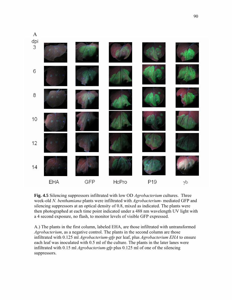
90
Fig. 4.5 Silencing suppressors infiltrated with low OD Agrobacterium cultures. Three week-old N. benthamiana plants were infiltrated with Agrobacterium- mediated GFP and silencing suppressors at an optical density of 0.8, mixed as indicated. The plants were then photographed at each time point indicated under a 488 nm wavelength UV light with a 4 second exposure, no flash, to monitor levels of visible GFP expressed. A.) The plants in the first column, labeled EHA, are those infiltrated with untransformed Agrobacterium, as a negative control. The plants in the second column are those infiltrated with 0.125 ml Agrobacterium-gfp per leaf, plus Agrobacterium EHA to ensure each leaf was inoculated with 0.5 ml of the culture. The plants in the later lanes were infiltrated with 0.15 ml Agrobacterium-gfp plus 0.125 ml of one of the silencing suppressors.
A

91
Fig. 4.5 Continued B.) The plants in the first column, again, are infiltrated with 0.125 ml Agrobacterium-gfp per leaf, plus Agrobacterium EHA to ensure each leaf was inoculated with 0.5 ml of the culture. The plants in columns 2-4 are infiltrated with 0.125 ml Agrobacterium-gfp, 0.125 mls each of the other silencing suppressors, and 0.125 Agrobacterium EHA. The plants in the 5th column are infiltrated with 0.125 ml Agrobacterium-gfp, and 0.125 ml each of Agrobacterium-hcpro, Agrobacterium-p19, and Agrobacterium-γb. These combinations were mixed in this manner to ensure each leaf was infiltrated with 0.5 ml Agrobacterium.
B

92
prior blot in Fig. 4.4A. This might indicate that as the infection progressed, proteins
accumulated, which supports the aims of this chapter. For the plant tissue initially
infiltrated with Agrobacterium with the low OD of 0.8, proteins were extracted at 3 dpi to
ensure proteins were expressed (Fig. 4.7).
Effect of silencing suppressors on GFP expression as vectored by Agrobacterium.
For plants infiltrated with Agrobacterium strain EHA alone, slight non-green
(grayish) fluorescence was seen as the Agrobacterium established a very mild infection,
usually seen as a slight chlorosis of the infiltrated tissue that progressively becomes more
yellowed over time. Agrobacterium carrying the gfp gene behind a CaMV 35S promoter
showed a definite green signal peaking around day 6, and declining thereafter to a level
nearly indistinguishable from the Agrobacterium strain EHA- infiltrated tissue around 12
days post inoculation. This was consistent in all repetitions of the experiments, following
both infiltrations with ODs of 1.2, and 0.8. For each of the silencing suppressors
infiltrated singly with GFP, the GFP signal was not only enhanced on the 3rd day
following infiltration, but the length of time that GFP was detected visually was increased
(Fig. 4.3A), the GFP signal itself enhanced, and the density of the band yielded by
western blotting with GFP antibodies was more prominent with tissue taken at 12 dpi
than at 3 dpi (Fig. 4.4).
The silencing suppressors themselves varied in extension and enhancement of the
GFP signal (Fig. 4.3A). In general, HcPro and P19 extended the length of time that GFP
was visible more so than γb, for which extension seemed to peak around 8 days post
inoculation and then decline. P19 and HcPro took a longer length of time for maximum
expression of the GFP visual signal, as seen with the timecourse data from plants
infiltrated with a starting ODs of 0.8 (Fig. 4.5). Agrobacterium-gfp by itself, the
maximum level of visible GFP expression, did not reach the level of visible signal
comparable to that oberserved with the co-infiltrated silencing suppressors/GFPs at their
peak GFP expression. While P19 enhanced the GFP signal to the level of HcPro, it did
not lengthen the expression of the signal for as long (12 dpi for P19 versus 14 dpi for
HcPro, Fig. 4.5A).

93
GFP visual expression was monitored for the silencing suppressors infiltrated in
dual combinations and then all together. Expression was certainly enhanced when
compared to tissue infiltrated with only Agrobacterium-gfp, as well as certain instances
with combined suppressors when compared to the suppressors alone. Visual levels of
GFP expression were comparable at 3, 6, and 8 dpi time points for the silencing
suppressors expressed in singly, in tandem, and in triplicate, though HcPro- and P19-
mixtures co-infiltrated with γb gave an enhanced signal at these points when compared to
HcPro- and P19- infiltrated with Agrobacterium-gfp alone (Fig 4.3, 4.5). Between 8 and
12 dpi, the silencing suppressors expressed dually and in triplicate enhanced signals when
compared to the singly- infiltrated silencing suppressors. For plants infiltrated with a
higher starting OD (1.2), however, this extended time of protein expression is offset by
necrosis in the infiltrated tissue (Fig 4.3B, lower rows). What tissue remained intact still
gave a vivid GFP signal, particularly for the HcPro/P19 combination as well as with all of
the suppressors co-infiltrated (Fig. 4.3B). For tissue infiltrated with a lower OD (0.8), this
necrosis was circumvented, as expected (Fig. 4.5B). At 12 dpi, constructs containing
HcPro seem to exhibit the most obvious GFP signal, though this was diminished by 14
dpi, when all three silencing suppressors co-infiltrated with Agrobacterium-gfp was the
most vivid (Fig. 4.5A). This agrees with the hypothesis, and will be addressed in the
discussion section.
Effect of silencing suppressors on GFP expressed from a virus vector.
Due to necrosis observed at later time-points when plants were infiltrated with a
culture OD of 1.2, the starting OD was diluted to 0.8 for plants to be infiltrated with
Agrobacterium-silencing suppressor constructs followed by inoculation with the virus
vectors (RMJ-1 and RMJ-3). Plants infected with only the RMJ-1 and RMJ-3 virus
vectors displayed differing phenotypes on each of the comparable ½ leaf assays. RMJ-1
displayed vivid, broad GFP lesions, with the lesions growing in size over time to nearly
confluence (Fig. 4.6). RMJ-3 yielded very weak GFP-expression lesions under UV light,
and these lesions decreased in size and intensity until no longer displaying a visible GFP
signal at 10 dpi, similar to the loss of signal seen with Agrobacterium-gfp (Fig 4.6). The
resultant phenotypes for Agrobacterium-mediated silencing suppressors inoculated with
94
either form of the viral construct was in agreement. RMJ-1, expressing P19 itself, gives a
nearly overwhelming GFP signal when inoculated with the silencing suppressors. For
RMJ-1 half leaf assays, the GFP lesions were larger overall, and the Agrobacterium-
silencing suppressors had generally the same effect on GFP expression as that
demonstrated with RMJ-3. There was a slight increase in the amount of necrotic tissue
present in RMJ-1 assays (compared to RMJ-3).
Because RMJ-3 lacks P19 expression, the resultant phenotypes had greater
variance than those for RMJ-1, and so will be described here in greater detail. RMJ-3
lesions had a much stronger GFP signal when co-expressed with silencing suppressors
(Fig. 4.6). While GFP was visible at 3 dpi for the tissues expressing a single silencing
suppressor, it increased with progression of the time-course. At 3 dpi, the visible GFP
signal was strongest for plants infiltrated with Agrobacterium-γb, and the lesions
themselves larger, though this expression was lost by 12 dpi in the plant co-infiltrated
with that suppressor alone. For plants infiltrated with Agrobacterium-hcpro, the lesions
remained vivid in infiltrated tissue through 14 dpi, though there was heavy tissue
necrosis. Agrobacterium-p19 gave enhanced GFP expression with increasing lesion size
as the timecourse progressed, and the lesions were similar in appearance to those seen on
RMJ-1 only inoculated plant. For plants infected with RMJ-3 and combinations of the
Agrobacterium- silencing suppressors, plants infiltrated with γb in combination with
another suppressor displayed a visible GFP signal at an earlier time-point, though this
treatment seemed to display the most tissue necrosis (Fig. 4.6B). The HcPro/P19
combination and all the silencing suppressors co-expressed gave the most vivid signal
and largest lesions (Fig 4.6B). This experiment is currently undergoing more repetitions.
Discussion
The goal of this work was to extend and enhance the amount of foreign protein
transiently produced in a plant using RNAi silencing suppressors. The titer and
expression of transiently expressed proteins has been shown to decrease over time due to
the silencing of mRNA (Voinnet et al., 2003). The main hypothesis of the present study
was that as silencing suppressors operate at different steps in the RNAi pathway
(Mlotshwa et al., 2005; Rakitina et al., 2006; Scholthof, 2006), their combined use allows

95
Fig. 4.6 Agrobacterium-vectored silencing suppressors and virus vectors. TBSV vector RMJ-1 (expressing P19) and RMJ-3 (lacking P19 expression) both carry gfp. Three week-old N. benthamiana plants were infiltrated with Agrobacterium-mediated GFP and silencing suppressors at an optical density of 0.8, mixed as indicated. The plants were then photographed at each time point indicated under a 488 nm wavelength UV light with a 4 second exposure, no flash, to monitor levels of visible GFP expressed. A.) The plants in the first column, labeled EHA, are those infiltrated with untransformed Agrobacterium, as a negative control. The plants in the second column are those inoculated with RMJ-1 on the left half and RMJ-3 on the right half. The plants in the later lanes were inoculated with RMJ-1 and RMJ-3 plus 0.125 ml of one of the silencing suppressors, plus A. tumafaciens EHA to raise the correct volume each leaf was inoculated with to 0.5 ml Agrobacterium culture.
A

96
Fig. 4.6 Continued B.) The plants in the first column, labeled EHA, are those infiltrated with untransformed Agrobacterium, as a negative control. The plants in columns 2-4 are inoculated with RMJ-1 and RMJ-3, plus 0.125 mls each of the other silencing suppressors, and 0.125 Agrobacterium EHA. The plants in the 5th column are inoculated with 0.125 ml each of Agrobacterium-hcpro, Agrobacterium-p19, and Agrobacterium-γb. These combinations were mixed in this manner to ensure each leaf was infiltrated with 0.5 ml Agrobacterium.
B

97
Fig. 4.7 Western blots from protein extractions of plants infiltrated with low OD cultures of Agrobacterium .The blots were probed with antibodies against the silencing suppressors indicated. Centimeter-square sections were processed as for 4.4. Lanes are marked as follows: M- low molecular weight protein marker, A – healthy, B – Agrobacterium EHA, C – Agrobacterium-gfp, D - Agrobacterium-gfp and -γb, E – Agrobacterium-gfp and -hcpro, F – Agrobacterium-gfp and - p19, G – Agrobacterium- gfp, - p19, -hcpro, H – Agrobacterium-gfp, -p19 and -hcpro, I – Agrobacterium-gfp, -γb, and –hcp, and J – Agrobacterium-gfp, -hcpro, -p19, and γb. These samples were taken from infiltrated plants 3 dpi, to establish that the proteins were being produced at the start of the time course. Proteins were not extracted from later time points as their expression was correlated with enhanced and extended virus-vector related expression. The western blot for γb displayed only unspecific binding and was not shown, though plants infiltrated with the γb culture display evidence of silencing suppression (Fig. 4.5-6).

98
GFP to be produced for a longer length of time by synergistic hampering of the pathway
when this foreign gene is expressed by a virus vector. The results are fairly straightforward, and consistent for silencing suppressors
expressed by Agrobacterium regardless of the GFP source, shown previously for agro-
GFP but here demonstrated successfully for a gfp-TBSV viral vector. Visual signals of
GFP alone are lost by 8 dpi, and the use of silencing suppressors extends both the length
of time that these signals are visible, as well as enhancing the amount of GFP expressed
(Fig. 4.3, 4.5). Agrobacterium-γb seems to enhance GFP expression early in the time-
course. Agrobacterium-p19 and Agrobacterium-hcpro seem to enhance GFP expression
later in the time-course, and extend the length of time that GFP is produced. When
infiltrated in combination, the actions of the silencing suppressors in the RNAi pathway
complement each other, and the GFP signal is correspondingly affected. For instance, γb
affects GFP expression earlier in the time-course. This early activity is complemented by
other suppressors and their action at later time points, resulting in the γb enhanced
expression extending longer. Furthermore, co-infiltrations involving P19 and HcPro show
an extended GFP visible signal, though the co-infiltration of HcPro and P19, sans γb, do
not display as visible a GFP signal at earlier time points as do those in presence of γb.
The results are consistent with my hypothesis. These silencing suppressors are
thought to act at different steps in the RNAi pathway; γb is thought to bind to RNA and
interfere with silencing (Fig. 4.1D), HcPro possibly modifies Dicer and binds to dsRNAs
(Fig. 4.1A, C), and P19 has been shown to sequester siRNA duplexes before they are
loaded onto RISC (Fig. 4.1C). In the context of this experiment, the results are semi-
logical. While unexpected, early enhancement of the GFP signal by γb might be caused
by binding of the silencing suppressor to ssRNA, which would protect it from elements
of the RNAi pathway. However, while it does enhance the GFP signal, this activity seems
temporally out of place. Previous characterization of γb suggest that an RNA-binding
domain contributes to its function as a silencing suppressor (Donald and Jackson, 1996;
Yelina et al., 2002; Rakitina et al., 2006), as γb has been shown to bind to ssRNA in a
sequence unspecific manner. This mode of silencing suppression would place it
downstream of the pathway where P19, which binds dsRNAs, and HcPro, which is
thought to both bind siRNAs and modify Dicer (Mlotshwa et al., 2005; Lakatos et al.,

99
2006). The data indicate that γb acts earlier in the pathway than HcPro and P19. This
might be due more to the known role of this protein as a replication enhancer; it is
possible that the early enhancement is due to the fact that γb does not have to compete
with plant proteins and can act immediately upon expression to protect the co-expressed
ssRNA, and this effect is not seen later in the infection because ssRNA is being targeted
heavily by RISCs. The later suppression activities by HcPro and P19 might require the
presence of siRNAs in the cell, and lag of silencing suppression activity, as compared to
that of γb, would be explained by necessity of siRNAs accumulation, which would take
more time. The combined synergistic effect of all three silencing suppressors, early in the
time course, plus extending the length of time that GFP is produced by about a week,
indicates that the suppressors constitute a valuable addition to transient protein
expression designs.
In the design and execution of these experiments, there are several issues to take
into consideration. It is exceedingly difficult to quantify levels of GFP expression
visually, and nearly as difficult to accomplish this using other protein-assaying
techniques while conducting a time-course due to the destruction of the GFP-expressing
tissue. This study aimed to make conditions for the plants as identical as possible to avoid
introducing any other outside variables that would interfere with protein expression. This
was at odds with collection of tissue for western assays, as removal of leaves or
infiltrated tissue from the plants might stimulate plant defenses that could affect protein
expression and accumulation. Thus, visualization of GFP and detection of protein levels
at only 2 points in each experiment seemed the best method to determine extension and
enhancement of GFP expression.
Other experimental aspects were at odds during this study. Agrobacterium has
been shown to elicit a necrotic response in certain hosts, with some hosts more sensitive
than others to the bacterium. This has been shown for agroinfiltration of tomato, as well
as members of the Solanaceous species (Wroblewski et al., 2005; Lindbo, 2007). As
illustrated with varying the starting OD of the infiltrated Agrobacterium constructs, more
turbid ODs negatively impacted the experiments with the stimulation of leaf chlorosis.
However, in theory and as determined experimentally based on comparison of GFP
pictures as well as GFP detection by western blotting, that more Agrobacterium used to

100
infiltrate the plant gave a higher expression of GFP, earlier in the time-course. This
observation would have resonance based on the reason necessitating protein expression in
an industrial setting. If the goal is to produce as much protein in a short amount of time, a
higher infiltration OD would be appropriate. If extension of the length of time that the
protein is expressed is the objective, I would recommend using a lower infiltration OD to
avoid negative effects on the infiltrated tissue. These two factors would need to be
adjusted in each case to best fit needs.
Additionally, while plants infiltrated with low starting ODs of the Agrobacterium
constructs (including GFP) did not display tissue necrosis, the added stress of a virus
vector rendered a necrotic response in co-infiltrated and rub-inoculated tissues at about 2-
weeks post inoculation. There are several possible reasons for this. It has been shown that
silencing suppressors interfere with regular RNAi pathways in plants (Chapman et al.,
2004). This might interfere with the way proteins are processed, as mRNAs accumulate
or regular plant cellular activities are interrupted, or it is possible that the combination
Agrobacterium/virus infections might trigger an overwhelming defensive response within
the cell, resulting in necrosis. To counter such a response, this study aimed to minimize
necrosis by using a less virulent Agrobacterium, as well as diluting the optical density of
the infiltrate. It may be speculated that the necrosis is a result of harsh inoculation
practices; however, the entire leaf was rub-inoculated and the only necrotic tissue was in
areas in which the silencing suppressor and virus-vector were both present, ruling out
inoculation as the cause of necrosis.
An observation not addressed above was that plant age seemed to play a role for
protein expression with agro-infiltration. N. benthamiana that were inoculated 1 week
later, at 4 weeks, as opposed to 3 wks, did not seem to experience the same amount of
chlorosis that the 3-wk old plants did following agro-infiltration. However, this is offset
by the fact that older plants are much less susceptible to infection by a virus (Hull, 2002),
and their use in this experiment would be deleterious in that regard.
Furthermore, silencing suppressors have been shown to act at different
efficiencies in the RNAi pathway, and interact with potential hosts differently (Voinnet,
2005; Martin-Hernandez and Baulcombe, 2008). This has implications while comparing
the response of HcPro and P19. While HcPro seemed to have a enhanced effect of GFP

101
when compared to P19, it is important to consider the toxic effect of P19 in N.
benthamiana (Chapman et al., 2004; Siddiqui et al., 2008), in addition to the fact that the
virus vector used expresses P19 as the native silencing suppressor. This may impact
comparisons of the responses of P19 and HcPro, and so this was avoided here. However,
it is important to note that HcPro, when used as a heterologous suppressor in a TBSV
virus vector, acts to enhance foreign protein production as well, if not better, than TBSV
P19, when both were expressed by agro-infiltration. This is consistent with the roles that
suppressors work on conserved, virus non-specific steps, and furthermore, that it is
certain that the use of a heterologous suppressor such as HcPro will not result in a
recombined, complete virus vector.
To summarize the main points of this study, silencing suppressors can be used to
extend and enhance a virus-vectored foreign protein in a plant host. The step at which the
silencing suppressor works in the RNAi pathway impacts functionality; silencing
suppressors which bind to dsRNAs or interfere with Ago might have a delayed response
when compared to silencing suppressors that act in earlier in the RNAi pathway, like
binding to RNA to prevent cleavage by Dicer. Other factors to consider include the
starting optical density of the Agrobacterium culture; a greater starting OD produces a
greater amount of protein more quickly, but also causes plant physiological issues and
might impact protein accumulation. A lower starting OD extends the length of time that
the protein is expressed by avoiding overload of the cell, though the initial amount of
protein expressed is less. Collectively, the data suggest that early expression of any
silencing suppressor combination may subsequently improve the performance of viral
vectors for expression of value-added proteins in plants.

102
CHAPTER V
FINAL SUMMARY AND DIRECTIONS
In objective 1, biochemical characterization of an RNAi response against TBSV in
N. benthamiana, TBSV-infected plant tissue was fractionated by column
chromatography, and the resultant fractions were tested for anti-viral RNAi
ribonucleases. These ribonucleases were present in the same fractions as viral siRNAs,
were inhibited by treatment with EDTA, enhanced by the addition of Mn2+ and Mg2+, and
stable through further steps of column chromatography. When plants were inoculated
with wt TBSV encoding a silencing suppressor, activity was inhibited. Additionally,
ribonuclease activity eluted in Sephacryl S200 gel filtration fractions that would represent
molecules or moieties of 500 kDa, possibly denoting a protein complex (RISC). The
results support the model that following viral infection, a RNAi response is triggered
which subsequently targets TBSV for degradation and eventual virus clearance.
Following other methods of column chromatography, individual proteins were
found to be in the 100 kDa range (perhaps representing Ago). However, after isolation
and sequencing of proteins after visualization by silver staining, those proteins known to
be associated with RNAi were not detected. RISC is thought to be a multiple turnover
complex, thus it would not be unreasonable to attribute difficulty in isolating the
responsible catalytic proteins to the hypothesis that they are present in such low
quantities. It was also shown that ribonuclease activity was inhibited by the addition of
NaCl in work done after the conclusion of this chapter; because siRNAs are known to
interact with the RISC Ago protein through ionic interactions, this finding was also in
agreement with the proposed RNAi pathway.
To further identify the proteins present that might contribute to RNAi, antibodies
were generated against a conserved region of the Piwi domain, a domain unique to the
Ago family of proteins. Fractions displaying RNase activity and siRNAs were also shown
to contain proteins with a Piwi-domain (unpublished data). Further characterization of the
RISC proteins might include some sort of antibody-affinity column, or possibly a north-
western (for antibodies and siRNAs) to help directly determine potential proteins.
Preliminary data indicate that the RISCs can be reloaded with siRNAs, therefore it might

103
be possible to label these siRNAs before they are loaded onto the RISCs perhaps using a
biotin molecule, crosslink the RNA to the ribonuclease once it associates, then use a
detection method for the siRNAs to find the proteins responsible.
The material presented in the appendix, a description of a novel method of TBSV
virion purification, is very straightforward. Hydroxyapatite column chromatography was
used to isolate virions from TBSV-infected N. benthamiana plant tissue as well as TBSV-
infected pepper plant tissue. The true identity of the virions was confirmed by re-
inoculation of N. benthamiana by western blotting with antibodies for the CP of TBSV,
by northern blotting of both the virions themselves and virion-infected plant tissue with a
TBSV hybridization probe, and upon electron microscopy of the virions. Exploration of
this virion purification method might involve the isolation of viruses with different capsid
morphologies, to see if these also can be purified by this method. Regardless, this is a
very useful method of isolating virions and maintaining stock for experimental use.
For objective 2, determination of the antiviral response following infection of N.
benthamiana with TRV, as well as examination of defense elements present following
PMV and SPMV infection of monocots, again, the results of these experiments are
consistent with those seen for TBSV; an anti-viral ribonuclease with RISC-like
characteristics, targeting only TRV-virus RNA, was isolated. To further clarify the TRV
studies, it is unknown at this point whether the ribonucleases present in TRV fractions
following hydroxyapatite and S200 gel filtration with 250 mM NaCl are targeting the
total RNA from TBSV-infected plants due to the presence of siRNAs brought in with the
total RNA. This might be resolved by somehow adding only genomic and subgenomic
TBSV RNA, without small RNA fragments/siRNAs, to the fractions and then observing
if degradation occurs.
Along with this, TRV infections might also stimulate other antiviral defenses,
which then target and degrade all RNAs. An alternative theory is that the ribonuclease
present, instead of being programmed with siRNAs against a particular virus, simply
targets all RNA of a particular size. To support this theory, in many of the experiments
above, it appears that the ribonuclease seems to prefer TBSV genomic RNA (Fig 2.13B,
particularly), and for total RNA extracted from plants, ribosomal RNAs around 2000 bp
are also targeted. Other literature has shown that RISC is programmed with ribosomal

104
RNA in some instances, but it is interesting that the ribonuclease seems to prefer larger
strands of ssRNA. At this time, it is unknown how RISC finds viral RNA in the cell; the
target ssRNA might need to be a certain length for recognition, as was also found for the
anti-TBSV RISC.
Other interesting results not addressed in the chapter indicate that it is difficult to
‘reprogram’ RISCs from TRV-infected tissue with siRNAs purified from the
fractionation of TBSV-infected plants. This might be due to some sort of modification
required upon binding to RISC, and following dissociation, these siRNAs can no longer
re-associate. To address this, viral siRNAs might need to be purified directly from
infected plants, and used for ‘reprogramming’.
It is also hypothesized that TRV produces less siRNAs than does TBSV; TRV
does not seem to produce nearly as much viral dsRNA intermediates as TBSV. This
might help explain why detection of siRNAs following gel filtration after hydroxyapatite
is a challenge. At this time, I suggest this might be due to RNA used as a target for the
hybridization probe. Current efforts are investigating this by using hybridization probes
against TRV-RNA1 in addition to TRV-RNA2 to probe these membranes, and finally tie
siRNAs directly to the fractions containing ribonuclease activity and Piwi-containing
proteins.
The PMV and SPMV-infected plant tissue hydroxyapatite fractions need to be
subjected to another type of column chromatography, probably gel filtration, to further
study the anti-viral defense in monocots more in depth. This set of experiments also
needs to be repeated an additional time.
In conclusion, this work further supports the RNAi model that an anti-viral RISC
is triggered upon virus infection; TRV-infected plant extracts and possibly PMV/SPMV
infected plant tissues harbor a virus-specific ribonuclease with properties that agree with
the RISC model.
In objective 3, determining the effect of silencing suppressors HcPro, P19, and γb
on the performance of a virus-vectored GFP gene plants, it was found that the silencing
suppressors did enhance GFP signal and extend the length of time that the signal was
visible. Furthermore, it was hypothesized that as these silencing suppressors are thought
to act in different steps of the RNAi pathway, their use in combination would have a

105
synergistic effect. This hypothesis was upheld by the data to an extent; the use of γb
enhanced the signal earlier in the infection, but did not seem to have any effect of the
length of time that GFP is produced. HcPro and P19 did not enhance GFP expression
until later in infection, but co-infection with these extended the length of GFP signal
visibility by about a week for both the Agrobacterium-vectored GFP, and the RMJ-gfp
viral vectors. However, plants inoculated with the Agrobacterium-silencing suppressors
and the virus vectors resulted in necrotic tissue, seen in two iterations of the project.
Studies are ongoing to examine if the necrotic leaves are due to an overwhelming amount
of silencing suppressors, or simple damage to the plant. Other projects might examine if
these silencing suppressors have a similar effect on other foreign proteins. Additionally, it
was observed that older plants do not display quite the same tissue chlorosis seen for
younger plants; the age of plants might be adjusted to manipulate the system for maximal
protein expression.
In summary, silencing suppressors can be used to enhance and extend the length
of time a foreign gene is expressed from a virus vector. This has valuable implications for
use in pharming, and expression of proteins in plants for use in biotechnology.

106
REFERENCES Ameres, S.L., Martinez, J., and Schroeder, R. (2007). Molecular basis for target RNA
recognition and cleavage by human RISC. Cell 130, 110-112. Batten, J., Desvoyes, B., Yamamura, Y., and Scholthof, K.-B.G. (2006). A
translational enhancer element on the 3′-proximal end of the Panicum mosaic virus genome FEBS Lett. 580, 2591-2597.
Batten, J.S., Yoshinari, S., and Hemenway, C. (2003). Potato virus X; a model system for virus replication, movement, and gene expression. Mol. Plant Path. 4, 125-131.
Baulcombe, D. (2004). RNA silencing in plants. Nature 431, 356-363. Baumberger, N., and Baulcombe, D.C. (2005). Arabidopsis ARGONAUTE1 is an
RNA slicer that selectively recruits microRNAs and short interfering RNAs. Proc. Natl. Acad. Sci. USA 102, 11928-11933.
Baumberger, N., Tsai, C.-H., Lie, M., Havecker, E., and Baulcombe, D.C. (2007). The polerovirus silencing suppressor P0 targets Argonaute proteins for degradation. Curr. Biol. 17, 1609-1614.
Bennasser, Y., and Jeang, K.-T. (2006). HIV-I Tat interaction with Dicer: requirement for RNA. Retrovirology 3.
Bernstein, E., Caudy, A.A., Hammond, S.M., and Hannon, G.J. (2001). Role for a bidentate ribonuclease in the initiation step of RNA interference. Nature 409, 363-366.
Borsani, O., Zhu, J., Verslues, P.E., Sunkar, R., and Zhu, J.K. (2005). Endogenous siRNAs derived from a pair of natural cis-antisense transcripts regulate salt tolerance in Arabidopsis. Cell 123, 1279-1291.
Bortolamiol, D., Pazhouhandeh, M., Marrocco, K., Genschik, P., and Ziegler-Graff, V. (2007). The poleovirus F Box protein P0 targets Argonaute1 to suppress RNA silencing. Curr. Biol. 17, 1615-1621.
Bragg, J.N., and Jackson, A.O. (2004). The C-terminal region of the Barley stripe mosaic virus gamma b protein participates in homologous interactions and is required for suppression of RNA silencing. Mol. Plant Pathol. 5, 465-482.
Brigneti, G., Voinnet, O., Li, W.-X., Ji, L.-H., Ding, S.-W., and Baulcombe, D.C. (1998). Viral pathogenicity determinants are suppressors of transgene silencing in Nicotiana benthamiana. EMBO J. 17, 6739-6746.
Brodersen, P., and Voinnet, O. (2006). The diversity of RNA silencing pathways in plants. Trends Genet. 22, 268-280.
Burch-Smith, T.M., Anderson, J.C., Martin, G.B., and Dinesh-Kumar, S.P. (2004). Applications and advantages of virus-induced gene silencing for gene function studies in plants. Plant J. 39, 734-746.
Burch-Smith, T.M., Schiff, M., Liu Y., and Dinesh-Kumar, S.P. (2006). Efficient virus-induced gene silencing in Arabidopsis. Plant Physiol. 142, 21-27.
Carmell, M.A., and Hannon, G.J. (2004). RNase III enzymes and the initiation of gene silencing. Nat. Struct. Mol. Bio. 11, 214-218.
Cerutti, L., Mian, N., and Bateman, A. (2000). Domains in gene silencing and cell differentiation proteins: the novel PAZ domain and redefinition of the Piwi domain. Trends Biochem. Sci. 25, 481-482.

107
Chao, J.A., Lee, J.H., Chapados, B.R., Debler, E.W., Schneemann, A., and Williamson, J.R. (2005). Dual roles of RNA-silencing suppression by Flock House virus protein B2. Nature Struct. Biol. 12, 952-957.
Chapman, E., Prokhnevsky, A.I., Gopinath, K., Dolja, V.V., and Carrington, J.C. (2004). Viral RNA silencing suppressors inhibit the microRNA pathway at an intermediate step. Genes Dev. 18, 1179-1186.
Chendrimada, T.P., Gregory, R.I., Kumaraswamy, E., Norman, J., Cooch, N., Nishikura, K., and Shiekhattar, R. (2005). TRBP recruits the Dicer complex to Ago2 for microRNA processing amd gene silencing. Nature 436, 740-744.
Chiba, M., Reed, J.C., Prokhnevsky, A.I., Chapman, E.J., Mawassi, M., Koonin, E.V., Carrington, J.C., and Dolja, V.V. (2005). Diverse suppressors of RNA silencing enhance agroinfection by a viral replicon. Virology 346, 7-14.
Deleris, A., Gallego-Bartolome, J., Bao, J., Kasschau, K.D., Carrington, J.C., and Voinnet, O. (2006). Hierarchical action and inhibition of plant dicer-like proteins in antiviral defense. Science 313, 68-71.
Desvoyes, B., Faure-Rabasse, S., Chen, M.-H., Park, J.-W., and Scholthof, H.B. (2002). A novel plant homeodomain protein interacts in a functionally relevant manner with a virus movement protein. Plant Physiol. 129, 1521-1532.
Desvoyes, B., and Scholthof, H.B. (2002). Host-dependent recombination of a Tomato bushy stunty virus coat protein mutant yields truncated capsid subunits that form virus-like complexes which benefit systemic spread. Virology 304, 434-442.
Donaire, L., Barajas, D., Martinez-Garcia, B., Martinez-Priego, L., Pagan, I., and Llave, C. (2008). Structural and genetic requirements for the biogenesis of Tobacco rattle virus-derived small interfering RNAs. J. Virol. 82, 5167-5177.
Donald, R.G.K., and Jackson, A.O. (1996). RNA-binding activities of barley stripe mosaic virus γb fusion proteins. J. Gen. Virol. 77, 879-888.
Eulalio, A., Behm-Ansmant, I., Schweizer, D., and Izaurralde, E. (2007). P-body formation is a consequence, not the cause of RNA-mediated gene silencing. Mole. Cell Biol. 27, 3970-3981.
Fields, B.N., Knipe, D.M., Howley, P.M., Griffin, D.E., Lamb, R.A., Martin, M.A., Roizman, B., and Straus, S.E. (2007). Fields Virology. (Boston, MA: Lippincott Williams and Wilkins).
Filipowicz, W. (2005). RNAi: the nuts and bolts of the RISC machine. Cell 122, 17-20. Fire, A., Xu, S., Montgomery, M.K., Kostas, S.A., Driver, S.E., and Mello, C.C.
(1998). Potent and specific genetic interference by double-stranded RNA in Caenorhabditis elegans. Nature 391, 806-811.
Gagnon, P., Frost, R., Tunon, P., and Ogawa, T. (1996). CHT Ceramic hydroxyapatite - a new dimension in chromatography of biological molecules. (Technote: Bio-Rad).
Goodin, M.M., Dietzgen, R.G., Schichnes, D., Ruzin, S., and Jackson, A.O. (2002). pGD vectors: versatile tools for the expression of green and red fluorescent protein fusions in agroinfiltrated plant leaves. Plant J. 31, 375-383.
Grimsley, N., Hohn, B., Hohn, T., and Walden, R.M. (1986). "Agroinfection", an alternative route for viral infection of plants by using the Ti plasmid. Proc. Nat. Acad. Sci. USA 83, 3282-3286.

108
Gunawardane, L.S., Saito, K., Nishida, K.M., Miyoshi, K., Kawamura, Y., Nagami, T., Siomi, H., and Siomi, M.C. (2007). A slicer-mediated mechanism for repeat-associated siRNA 5' end formation in Drosophila. Science 315, 1587-1590.
Hammond, S.M., Boettcher, G., Caudy, A.A., Kobayashi, T., and Hannon, G.J. (2001). Argonaute2, a link between genetic and biochemical analyses of RNAi. Science 293, 1146-1150.
Hannon, G.J. (2002). RNA interference. Nature 418, 244-251. Heaton, L.A. (1992). Use of agarose gel electrophoresis to monitor conformational
changes of some small, spherical plant viruses. Phytopathology 82, 803-807. Hemmes, H., Lakatos, L., Goldbach, R., Burgyan, J., and Prins, M. (2007). The NS3
protein of Rice hoja blanca tenuivirus suppresses RNA silencing in plant and insect hosts by efficiently binding both siRNAs and miRNAs. RNA 13, 1079-1089.
Hiraguri, A., Itoh, R., Kondo, N., Nomura, Y., Aizawa, D., Murai, Y., Koiwa, H., Shinozaki, K., and Fukuhara, T. (2005). Specific interactions between Dicer-like proteins and HYL1/DRB-family of dsRNA-binding proteins in Arabidopsis thaliana. Plant Mol. Biol. 57, 173-188.
Hong, J., Na, W., Chalk, A., Wang, J., Song, Y., Yi, F., Qiao, R.P., Sonnhammer, E.L.L., Wahlestedt, C., Liang, Z., and Du, Q. (2008). Focusing on RISC assembly in mammalian cells. Biochem. Biophys. Res. Commun. 368, 703-708.
Hood, E.E., Helmer, G.L., Fraley, R.T., and Chilton, M.-D. (1986). The hypervirulence of Agrobacterium tumefaciens A281 is encoded in a region of pTiBo542 outside of T-DNA. J. Bacteriol. 168, 1291-1301.
Hooykaas, P.J.J., and Schilperoort, R.A. (1992). Agrobacterium and plant genetic engineering. Plant Mol. Biol. 19, 15-38.
Hull, R. (2002). Matthews' Plant Virology. (London: Academic Press). Hutvagner, G., and Simard, M.J. (2008). Argonaute proteins: key players in RNA
silencing. Nature Rev. Mol. Cell Biol. 9, 22-32. Jaskiewicz, L., and Filipowicz, W. (2008). Role of Dicer in posttranscriptional RNA
silencing. In RNA interference. Current Topics in Microbiology and Immunology., P.J. Paddison, and P.K.Vogt, eds. (Berlin: Springer-Verlag.) pp.77-97.
Ji, X. (2008). The mechanism of RNase III action: how Dicer dices. In RNA interference. Current topics in Microbiology and Immunology., P.J. Paddison, and P.K. Vogt, eds. (Berlin: Springer-Verlag.) pp. 99-116.
Jones, L., Keining, T., Eamens, A., and Vaistij, F.E. (2006). Virus-induced gene silencing of Argonaute genes in Nicotiana benthamiana demonstrates that extensive systemis silencing requires Argonaute1-like and Argonaute4-like genes. Plant Physiol. 141, 598-606.
Jones-Rhoades, M.W., Bartel, D.P., and Bartel, B. (2006). MicroRNAs and their regulatory roles in plants. Annu. Rev. Plant Biol. 57, 19-53.
Katiyar-Agarwal, S., Morgan, R., Dahlbeck, D., Borsani, O., Villegas Jr., A., Zhu, J.K., Staskawicz, B., and Jin, H. (2006). A pathogen-inducible endogenous siRNA in plant immunity. Proc. Nat. Acad. Sci. USA 103, 18002-18007.
Lacroix, B., Tzfira, T., and Vainstein, A.C., V. (2006). A case of promiscuity: Agrobacterium's endless hunt for new partners. Trends Genet. 22, 29-37.
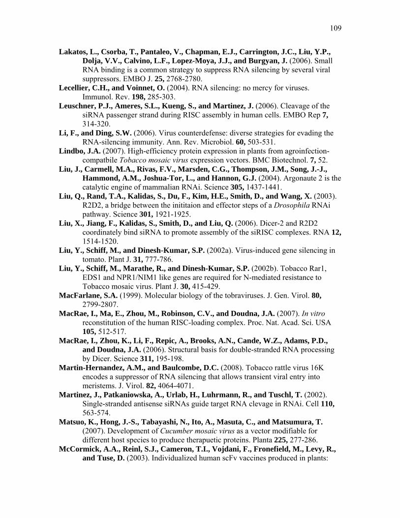
109
Lakatos, L., Csorba, T., Pantaleo, V., Chapman, E.J., Carrington, J.C., Liu, Y.P., Dolja, V.V., Calvino, L.F., Lopez-Moya, J.J., and Burgyan, J. (2006). Small RNA binding is a common strategy to suppress RNA silencing by several viral suppressors. EMBO J. 25, 2768-2780.
Lecellier, C.H., and Voinnet, O. (2004). RNA silencing: no mercy for viruses. Immunol. Rev. 198, 285-303.
Leuschner, P.J., Ameres, S.L., Kueng, S., and Martinez, J. (2006). Cleavage of the siRNA passenger strand during RISC assembly in human cells. EMBO Rep 7, 314-320.
Li, F., and Ding, S.W. (2006). Virus counterdefense: diverse strategies for evading the RNA-silencing immunity. Ann. Rev. Microbiol. 60, 503-531.
Lindbo, J.A. (2007). High-efficiency protein expression in plants from agroinfection-compatbile Tobacco mosaic virus expression vectors. BMC Biotechnol. 7, 52.
Liu, J., Carmell, M.A., Rivas, F.V., Marsden, C.G., Thompson, J.M., Song, J.-J., Hammond, A.M., Joshua-Tor, L., and Hannon, G.J. (2004). Argonaute 2 is the catalytic engine of mammalian RNAi. Science 305, 1437-1441.
Liu, Q., Rand, T.A., Kalidas, S., Du, F., Kim, H.E., Smith, D., and Wang, X. (2003). R2D2, a bridge between the inititaion and effector steps of a Drosophila RNAi pathway. Science 301, 1921-1925.
Liu, X., Jiang, F., Kalidas, S., Smith, D., and Liu, Q. (2006). Dicer-2 and R2D2 coordinately bind siRNA to promote assembly of the siRISC complexes. RNA 12, 1514-1520.
Liu, Y., Schiff, M., and Dinesh-Kumar, S.P. (2002a). Virus-induced gene silencing in tomato. Plant J. 31, 777-786.
Liu, Y., Schiff, M., Marathe, R., and Dinesh-Kumar, S.P. (2002b). Tobacco Rar1, EDS1 and NPR1/NIM1 like genes are required for N-mediated resistance to Tobacco mosaic virus. Plant J. 30, 415-429.
MacFarlane, S.A. (1999). Molecular biology of the tobraviruses. J. Gen. Virol. 80, 2799-2807.
MacRae, I., Ma, E., Zhou, M., Robinson, C.V., and Doudna, J.A. (2007). In vitro reconstitution of the human RISC-loading complex. Proc. Nat. Acad. Sci. USA 105, 512-517.
MacRae, I., Zhou, K., Li, F., Repic, A., Brooks, A.N., Cande, W.Z., Adams, P.D., and Doudna, J.A. (2006). Structural basis for double-stranded RNA processing by Dicer. Science 311, 195-198.
Martin-Hernandez, A.M., and Baulcombe, D.C. (2008). Tobacco rattle virus 16K encodes a suppressor of RNA silencing that allows transient viral entry into meristems. J. Virol. 82, 4064-4071.
Martinez, J., Patkaniowska, A., Urlab, H., Luhrmann, R., and Tuschl, T. (2002). Single-stranded antisense siRNAs guide target RNA clevage in RNAi. Cell 110, 563-574.
Matsuo, K., Hong, J.-S., Tabayashi, N., Ito, A., Masuta, C., and Matsumura, T. (2007). Development of Cucumber mosaic virus as a vector modifiable for different host species to produce therapuetic proteins. Planta 225, 277-286.
McCormick, A.A., Reinl, S.J., Cameron, T.I., Vojdani, F., Fronefield, M., Levy, R., and Tuse, D. (2003). Individualized human scFv vaccines produced in plants:

110
humoral anti-idiotype responses in vaccinated mice confirm relevance to the tumor Ig. J. Immunol. Methods 278, 95-104.
Meister, G., Landthaler, M., Patkaniowska, A., Dorsett, Y., Teng, G., and Tuschl, T. (2004). Human Argonaute2 mediates RNA cleavage targeted by miRNAs and siRNAs. Mol. Cell 15, 185-197.
Mlotshwa, S., Schauer, S.E., Smith, T.H., Mallory, A., Herr-Jr., J.M., Roth, B.M., Merchant, D.D., Ray, A., Bowman, L.H., and Vance, V. (2005). Ectopic DICER-LIKE1 expression in P1/HC-Pro Arabidopsis rescues the phenotypic anomalies but not the defects in microRNA and silencing pathways. Plant Cell 17, 2873-2885.
Moissiard, G., Parizotto, E.A., Himber, C., and Voinnet, O. (2007). Transitivity in Arabidopsis can be primed, requires the redundant action of the antiviral Dicer-like 4 and Dicer-like 2, and is compromised by viral-encoded suppressor proteins. RNA 13, 1268-1278.
Napoli, C., Lemieux, C., and Jorgensen, R.A. (1990). Introduction of a chimeric chalcone synthase gene into petunia results in reversible co-suppression of homologous genes in trans. Plant Cell 2, 279-289.
Nelson, D.L., and Cox, M.M. (2005). Lehninger’s Principles of Biochemistry. (New York: Freeman and Company).
Nykanen, A., Haley, B., and Zamore, P.D. (2001). ATP requirements and small interfering RNA structure in the RNA interference pathway. Cell 107, 309-321.
Omarov, R.T., Ciomperlik, J.J., and Scholthof, H.B. (2007). RNAi-associated ssRNA-specific ribonucleases in Tombusvirus P19 mutant-infected plants and evidence for a discrete siRNA-containing effector complex. Proc. Natl. Acad. Sci. USA 104, 1714-1719.
Omarov, R., Qi, D., and Scholthof, K.-B.G. (2005). The capsid protein of satellite panicum mosaic virus contributes to systemic invasion and interacts with its helper virus. J. Virol. 79, 9756–9764.
Omarov, R., Sparks, K., Smith, L., Zindovic, J., and Scholthof, H.B. (2006). Biological relevance of a stable interaction between the tombusvirus-encoded P19 and siRNAs. J. Virol. 80, 3000-3008.
Pantaleo, V., Szittya, G., and Burgyan, J. (2007). Molecular bases of viral RNA targeting by viral small interfering RNA-programmed RISC. J. Virol. 81, 3797-3806.
Park, J.-W., Faure-Rabasse, S., Robinson, M.A., Desvoyes, B., and Scholthof, H.B. (2004). The multifunctional plant viral suppressor of gene silencing P19 interacts with itself and an RNA binding host protein. Virology 323, 49-58.
Parker, J.S., Roe, S.M., and Barford, D. (2005). Structural insights into mRNA recognition from a PIWI domain-siRNA guide complex. Nature 434, 663-666.
Qi, D., and Scholthof, K.-B.G. (2008). Multiple activities associated with the capsid protein of satellite panicum mosaic virus are controlled separately by the N- and C-terminal regions. Mol. Plant-Microbe Interact. 21, 613-621.
Qiu, W., and Scholthof, K.-B.G. (2004). Satellite panicum mosaic virus capsid protein elicits symptoms on a nonhost plant and interferes with a suppressor of virus-induced gene silencing. Mol. Plant-Microbe Interact. 17, 263-271.

111
Qiu, W., and Scholthof, H.B. (2007). Tomato bushy stunt virus derived gene vectors. Curr. Prot. Microbiol. 7, 16In14.12-16In14.16.
Qiu, W.P., Park, J.-W., and Scholthof, H.B. (2002). Tombusvirus P19-mediated suppression of virus induced gene silencing is controlled by genetic and dosage features that influence pathogenicity. Mol. Plant-Microbe Interact. 15, 269-280.
Qu, F., and Morris, T.J. (2002). Efficient infection of Nicotiana benthamiana by Tomato bushy stunt virus is facilitated by the coat protein and maintained by p19 through suppression of gene silencing. Mol. Plant-Microbe Interact. 15, 193-202.
Rakitina, D.V., Yelina, N.E., and Kalinina, N.O. (2006). Zinc ions stimulate the cooperative RNA binding of hordeiviral γb protein. FEBS Lett. 580, 5077-5083.
Rand, T.A., Ginalski, K., Grishin, N.V., and Wang, X. (2004). Biochemical identification of Argonaute 2 as the sole protein required for RNA-induced silencing complex. Proc. Natl. Acad. Sci. USA 101, 14385-14389.
Ratcliff, F., Martin-Hernandez, A.M., and Baulcombe, D. (2001). Tobacco rattle virus as a vector for anaylsis of gene function by silencing. Plant J. 25, 237-245.
Robb, G.B., and Rana, T.M. (2007). RNA helicase A interacts with RISC in human cells and functions in RISC loading. Mol. Cell 26, 523-527.
Romano, N., and Macino, G. (1992). Quelling: transient inactivation of gene expression in Neurospora crassa by transformation with homologous sequences. Mol. Microb. 6, 3343-3353.
Rossi, J.J. (2005). RNAi and the P-body connection. Nat. Cell Biol. 7, 643-644. Ruiz-Ferrer, V., and Voinnet, O. (2007). Viral suppression of RNA silencing: 2b wins
the golden fleece by defeating Argonaute. BioEssays 29, 319-323. Ryu, C.-M., Anand, A., Kang, L., and Mysore, S. (2004). Agrodrence: a novel and
effective agroinoculation method for virus-induced gene silencing in roots and diverse Solanaceous species. Plant J. 40, 322-331.
Sambrook, J., Fritsch, E.F., and Maniatis, T. (1989). Molecular Cloning: A Laboratory Manual, 2nd ed. (Cold Spring Harbor, N.Y.: Cold Spring Harbor Laboratory Press).
Scholthof, H.B. (2005). Plant virus transport: motions of functional equivalence. Trends Plant Sci. 10, 376-382.
Scholthof, H.B. (2006). The Tombusvirus-encoded P19: from irrelevance to elegance. Nature Rev. Microbiol. 4, 405-411.
Scholthof, H.B. (2007). Heterologous expression of viral RNA interference suppressors; RISC management. Plant Physiol. 145, 1110-1117.
Scholthof, H.B., Desvoyes, B., Kuecker, J., and Whitehead, E. (1999a). The biological activity of two tombusvirus proteins translated from nested genes is influenced by dosage control via context-dependent leaky scanning. Mol. Plant-Microbe Interact. 12, 670-679.
Scholthof, H.B., Morris, T.J., and Jackson, A.O. (1993). The capsid protein gene of tomato bushy stunt virus is dispensable for systemic movement and can be replaced for localized expression of foreign genes. Mol. Plant-Microbe Interact. 6, 309-322.
Scholthof, H.B., Scholthof, K.-B.G., and Jackson, A.O. (1996). Plant virus gene vectors for transient expression of foreign proteins in plants. Annu. Rev. Phytopathol. 34, 299-323.

112
Scholthof, H.B., Scholthof, K.-B.G., Kikkert, M., and Jackson, A.O. (1995a). Tomato bushy stunt virus spread is regulated by two nested genes that function in cell-to-cell movement and host-dependent systemic invasion. Virology 213, 425-438.
Scholthof, K.-B.G. (1999). A synergism induced by satellite panicum mosaic virus. Mol. Plant-Microbe Interact. 12, 163-166.
Scholthof, K.-B.G., Jones, R.W., and Jackson, A.O. (1999b). Biology and structure of plant satellite viruses activated by icosahedral helper viruses. Curr. Top. Microbiol. Immunol. 239, 123-143.
Scholthof, K.-B.G., Mirkov, T.E., and Scholthof, H.B. (2002). Plant viral gene vectors: biotechnology applications in agriculture and medicine. Genet. Eng. 24, 67-86.
Scholthof, K.-B.G., Scholthof, H.B., and Jackson, A.O. (1995b). The tomato bushy stunt virus replicase proteins are coordinately expressed and membrane associated. Virology 208, 365-369.
Scholthof, K.-B.G., Scholthof, H.B., and Jackson, A.O. (1995c). The effect of defective interfering RNAs on the accumulation of tomato bushy stunt virus proteins and implications for disease attenuation. Virology 211, 324-328.
Schroder, E., Jonsson, T., and Poole, L. (2005). Hydroxyapatite chromatography: altering the phosphate-dependent elution profile of protein as a function of pH. Anal. Biochem. 313, 176-178.
Schwartz, D.S., Hutvagner, G., Du, T., Xu, Z., Aronin, N., and Zamore, P.D. (2003). Assymmetry in the assembly of the RNAi enzyme complexes. Cell 115, 199-208.
Schwarz, D.S., Hutvagner, G., Haley, B., and Zamore, P.D. (2002). Evidence that siRNAs function as guides, not primers, in the Drosophila and human RNAi pathway. Mol. Cell 10, 537-548.
Schwarz, D.S., Tomari, Y., and Zamore, P.D. (2004). The RNA-induced silencing complex is a Mg2+-dependent endonuclease. Curr. Biol. 14, 787-791.
Shams-Bakhsh, M., Canto, T., and Palukaitis, P. (2007). Enhanced resistance and neutralization of defense responses by suppressors of RNA silencing. Virus Res. 130, 103-109.
Sheth, U., and Parker, R. (2003). Decapping and decay of messanger RNA occur in cytoplasmic processing bodies. Science 300, 805-808.
Siddiqui, S.A., Sarmiento, C., Trueve, C., Lehto, H., and Lehto, K. (2008). Phenotypes and functioanl effects caused by various viral RNA silencing suppressors in transgenic Nicotiana benthamiana and N. tabacum. Molec. Plant Microbe Interact. 21, 178-187.
Silhavy, D., and Burgyan, J. (2004). Effects and side-effects of viral RNA silencing suppressors on short RNAs. Trends Plant Sci. 9, 76-83.
Singh, D.P., Moore, C.A., Gilliland, A., and Carr, J.P. (2004). Activation of multiple antiviral defense mechanisms by salicylic acid. Mol. Plant Pathol. 5, 57-63.
Song, J.J., and Joshua-Tor, L. (2006). Ago and RNA - getting into the groove. Curr. Opin. Biol. Rev. 16, 5-11.
Song, J.-J., Smith, S.K., Hannon, G.J., and Joshua-Tor, L. (2004). Crystal structure of Argonaute and its implications for RISC slicer activity. Science 305, 1434-1437.
Tabara, H., Yigit, E., Siomi, H., and Mello, C.C. (2002). The dsRNA binding protein RDE-4 interacts with RDE-1, DCR-1, and a DExH-box helicase to direct RNAi in C. elegans. Cell 109, 861-871.

113
Toila, N.H., and Joshua-Tor, L. (2007). Slicer and the Argonauts. Nat. Chem. Biol. Rev. 3, 36-43.
Tomari, Y., Du, T., and Zamore, P.D. (2007). Sorting of Drosophila small silencing RNAs. Cell 130, 299-308.
Tomari, T.Y., Matranga, C., Haley, B., Martinez, N., and Zamore, P.D. (2004). A protein sensor for siRNA assymetry. Science 306, 1377-1380.
Tomari, Y., and Zamore, P.D. (2005). Perspective: machines for RNAi. Gene Dev. 19, 517-529.
Turina, M., Desvoyes, B., and Scholthof, K.-B.G. (2000). A gene cluster encoded by Panicum mosaic virus is associated with virus movement. Virology 266, 120-128.
Turina, M., Maruoka, M., Monis, J., Jackson, A.O., and Scholthof, K.-B.G. (1998). Nucleotide sequence and infectivity of a full-length cDNA clone of panicum mosaic virus. Virology 241, 141-155.
Turina, M., Omarov, R., Murphy, J.F., Bazaldua-Hernandez, C., Desvoyes, B., and Scholthof, H.B. (2003). A newly identified role for the Tomato bushy stunt virus P19 in short distance spread. Mol. Plant Pathol. 4, 67-72.
Tzfira, T., and Citovsky, V. (2002). Partners-in-infection: host proteins involved in transformation of plant cells by Agrobacterium. Trends Cell Biol. 12, 121-129.
Tzfira, T., and Citovsky, V. (2006). Agrobacterium-mediated genetic transformation of plants: biology and biotechnology. Curr. Opin. Biotechnol. 17, 147-154.
Vagin, V.A., Sigova, A., Li, C., Seitz, H., Gvozdev, V., and Zamore, P.D. (2006). A distinct small RNA pathway silences selfish genetic elements in the germline. Science 313, 320-324.
Vasquez, F., Vaucheret, H., Rajagopalan, R., Lepers, C., Gasciolli, V., Mallory, A., Hilbert, J., Bartel, D.P., and Crete, P. (2004). Endogenous trans-acting siRNAs regulate the accumulation of Arabidopsis mRNAs. Mole. Cell 16, 69-79.
Voinnet, O. (2005). Induction and suppression of RNA silencing: Insights from viral infections. Nat Rev Genet. 6, 206-220.
Voinnet, O., and Baulcombe, D.C. (1997). Systemic signaling in gene silencing. Nature 389, 553.
Voinnet, O., Pinto, Y.M., and Baulcombe, D.C. (1999). Suppression of gene silencing: a general strategy used by diverse DNA and RNA viruses of plants. Proc. Natl. Acad. Sci. USA 96, 14147-14152.
Voinnet, O., Rivas, S., Mestre, P., and Baulcombe, D. (2003). An enhanced transient expression system in plants based on suppression of gene silencing by the p19 protein of tomato bushy stunt virus. Plant J. 33, 949-956.
Wagner, B., Fuchs, H., Adhami, F., Ma, Y., Scheiner, O., and Breiteneder, H. (2004). Plant virus expression systems for transient production of recombinant allergens in Nicotiana benthamiana. Methods 32, 227-234.
White, K.A. (1996). Formation and evolution of Tombusvirus defective interfering RNAs. Sem. Virol. 7, 409-416.
Wingard, S.A. (1928). Hosts and symptoms of ringspot, a virus disease of plants. J. Agric. Res. 37, 127-153.
Wroblewski, T., Tomczak, A., and Michelmore, R. (2005). Optimization of Agrobacterium-mediated transient assays of gene expression in lettuce, tomato and Arabidopsis. Plant Biotechnol. J. 3, 259-273.

114
Xie, Z., Johansen, L.K., Gustafson, A.M., Kasschau, K.D., Ellis, A.D., Zilberman, D., Jacobsen, S.E., and Carrington, J.C. (2004). Genetic and functional diversification of small RNA pathways in plants. PLoS Biol. 2, E104.
Yamamura, Y., and Scholthof, H.B. (2005). Pathogen profile: Tomato bushy stunt virus: A resilient model system for studying virus-plant interactions. Mol. Plant Pathol. 6, 491-502.
Ye, K., Malinina, L., and Patel, D. (2003). Recognition of small interfering RNA by a viral suppressor of RNA silencing. Nature 426, 874-878.
Yelina, N.E., Savenkov, E.I., Solovyev, A.G., Morozov, S.Y., and Valkonen, J.P. (2002). Long-distance movement, virulence, and RNA silencing suppression controlled by a single protein in hordei- and potyviruses: complementary functions between virus families. J. Virol. 76, 12981-12991.
Yuan, Y.R., Pei, Y., Chen, H.Y., Tuschl, T., and Patel, D. (2006). A potential protein-RNA recognition event along the RISC-loading pathway from the structure of A. aeolicus Ago with the externally bound siRNA. Structure 14, 1557-1565.
Zaitlin, M. (1998). The discovery of the causal agent of the Tobacco mosaic disease. In Discoveries in Plant Biology, S.D. Kung and S.F. Yang, eds (Hong Kong: World Publishing Co., Ltd), pp. 105-110.
Zhang, X., Henderson, I.R., Lu, C., Green, P.J., and Jacobsen, S.E. (2007). Role of RNA polymerase IV in plant small RNA metabolism. Proc. Nat. Acad. Sci. USA 104, 4536-4541.
Zhang, X., Yuan, Y.R., Pei, Y., Lin, S.S., Tuschl, T., Patel, D.J., and Chua, N.H. (2006). Cucumber mosaic virus-encoded 2b suppressor inhibits Arabidopsis Argonaute1 cleavage activity to counter plant defense. Genes Dev. 20, 3255-3268.

115
APPENDIX
TBSV VIRION PURIFICATION BY COLUMN CHROMATOGRAPHY
Introduction
Tomato bushy stunt virus (TBSV) serves as a common model virus for plant
systems. It is a single-stranded, positive-sense RNA virus with an iscosahedral capsid
structure. The particles are reported to be 33 nm in diameter (Hull, 2002; Yamamura and
Scholthof, 2005). TBSV is also of research importance as it produces the highly effective
silencing suppressor P19, routinely used in RNAi studies (Scholthof, 2006). Traditional
methods of purification for Tomato bushy stunt virus (TBSV) and other viruses involve
isolation of the virions by centrifugation gradients with sucrose and cesium chloride
(Fields et al., 2007). These protocols are time consuming and require multiple steps. In
this section, I address the possibility for the use of hydroxyapatite as an alternative to
purify virions; it is rapid, inexpensive to assemble, and economical as the column media
can be regenerated for multiple uses.
Hydroxyapatite is a calcium phosphate ceramic, Ca10(PO4)6(OH)2 (Gagnon et al.,
1996), coincidentally used in the medical field as a biomaterial filler for teeth and bones
and as a covering to promote growth of bones into prosthetic implants. As a column
chromatography medium, it separates by a ‘mixed-modes’ or pseudo-affinity means of
ion exchange. The positively-charged calcium ions and negatively-charged phosphate
ions allow for interaction with both positively charged amino acids of proteins, and
negatively charged carbonyls of DNA groups, or carboxylate residues on protein surfaces
(Schroder et al., 2005). This allows for differential separation via functional group
electrostatic interactions with the column media, and elution through application of buffer
with an increasing gradient of phosphates (Gagnon et al., 1996; Schroder et al., 2005).
Here, a method is reported to quickly purify TBSV and possibly other virions
from infected plant crude extract following centrifugation and column application. When
crude plant extract was applied to the column, it is demonstrated that virions comprise the
only contents of the flowthrough buffer, without further purification steps. These virions
are present in high concentration and purity, and are suitable for rub inoculation with up
to a thousand-fold dilution. Resultant infections are identical to those of plants that have

116
been inoculated with TBSV RNA-transcripts, or inoculated with infected tissue. Virion
purification via hydroxyapatite chromatography may be preferable to other methods in
that it is more economical than generating RNA-transcripts in vitro, and deleterious
effects of defective interfering RNAs, seen in passaged TBSV infections (Scholthof et al.,
1995c; White, 1996), are not a concern.
Materials and Methods
Infection of plants with TBSV.
Infectious TBSV RNA transcripts were generated in vitro using a Fermentas T7
transcription kit (1 µl linearized DNA, 16 µl dd-water, 5 µl 5X transcript buffer, 2.5 µl 5
mM rNTP mix, 2 µl 0.1 mM DTT, 0.25 µl Ribolock RNase inhibitor, and 0.5 µl T7 RNA
polymerase (Fermentas, Glen Burnie, MD). To confirm transcription, 3 µl of transcripts
were mixed with 1 µl loading dye and electrophoresied on a 1% agarose gel and stained
with ethidium bromide. These transcripts were then mixed in a 1:4 dilution with cold
RNA inoculation buffer (50 mM KH2PO4, 50 mM Glycine, pH 9.0, 1% celite, 1%
bentonite), and 3 leaves of 3 week-old N. benthamiana plants were rub-inoculated with
20 µl of the dilution apiece. A week was allowed for establishment of the infection, then
plant tissue was harvested by grinding upper leaves with 50 ml of 10 mM sodium
phosphate buffer, pH 6.8, by mortar and pestle. This crude extract was then centrifuged at
4000 rpm, 4°C, for 20 min. with a Beckman S4180 rotor to remove insoluble proteins.
The supernatant was filtered through cheesecloth into conical-bottomed tubes, and further
clarified by centrifugation at 10,000 rpm, 4°C, for 20 min. in a Beckman F0630 rotor,
and the supernatant placed on ice for application to the column.
Hydroxyapatite column preparation and virion purification
The chromatographic arrangement involved a 30 x 2.5 cm chromatographic
column attached to a peristaltic pump for elution purposes. The column was packed with
40 ml of CHT ceramic hydroxyapatite media (Bio-Rad, Hercules, CA) according to
manufacturer’s directions, briefly reviewed as follows: 80 ml of 200 mM dibasic sodium
phosphate buffer was applied to the column, followed by 80 ml 200 mM sodium
phosphate buffer, pH 6.8, to rapidly lower the pH. A 10 mM sodium phosphate buffer,

117
pH 6.8, was used to equilibrate the column and as running buffer; the column was
washed extensively (~200 ml) with this buffer before application of the plant extract.
Once the column was packed, the extract was applied to the column at a flow rate of
about 1.3 ml/min. Flowthrough containing virions was collected (usually about 50-100
ml). This preparation was either used immediately to inoculate plants, or frozen at (-) 20°
C for later use.
Additionally, Capsicum annuum (pepper) plants were inoculated with wt TBSV,
and a small (about 10 ml) hydroxyapatite column was used for chromatography of 5
grams of plant tissue. The flowthrough was collected, and concentrated using a vacuum
dehydrator. The concentration of potential virions was determined by staining a 1%
agarose electrophoresis gel before submitting the sample for electron microscopy.
Verification of virions
Column flowthrough was used to inoculate three week-old N. benthamiana plants.
For this, 100 µl of the flowthrough was mixed with RNA inoculation buffer as described
in Table A.1. Plants were incubated at room temperature, and checked for infection.
Table A.1 Virion dilution for N. benthamiana plant assays.
I Ii Iii iv V
Virions soln. 0.1 µl 1.0 µl 10 µl 50 µl 100 µl
Virus
inoculation
buffer
200 µl 199 µl 190 µl 150 µl 100 µl
Additionally, flowthrough was inspected by transmission electron microscopy. A
sample (2 µl) was stained with uranyl acetate, and viewed at high magnification.
(Electron microscopy performed by E. Ann Ellis, Microscopy and Imaging Center, Texas
A&M University).

118
Results
Upon column chromatography of extracts from TBSV ∆P19 infected plants,
proteins can be visualized as a single band at about 41 kDa with Coomassie and silver
staining (AgNO3) techniques (Fig. A.1A). Using an antibody generated against TBSV
CP, western blot analysis (Fig. A.1B) verified the presence of TBSV CP, as well as
another band around 85 kDa (possibly CP aggregates). Following northern blotting of
SDS-treated crude extracts and virion-containing column flowthrough (Fig. A.1C),
hybridization probes specific for TBSV demonstrate the presence of RNA. Additionally,
a native (whole virus) gel displays the presence of a discrete band in the flowthrough
column, slightly smaller than that in the crude extracts column, representative of virions
(Fig. A.1D).
The column flowthrough was diluted out in 0.25, 0.05, 0.005, and 0.0005
dilutions (Table A.1), and all were used to rub inoculate 3 week-old N. benthamiana
plants. Plants infected with virion-containing column flow-through displayed typical
TBSV symptoms (Fig. A.2), with curled, crinkled upper leaves. Following one week post
inoculation, all plants showed evidence of infection, indicating a very high concentration.
To determine if the symptoms present were the result of a true TBSV infection, about 0.5
grams (1 leaf) infected tissue was removed from the plants. RNA was extracted from the
tissue, and analyzed by northern blotting with a TBSV-specific probe. These northern
blots from virion-infected plants displayed ample genomic and subgenomic RNA signals
when probed with a TBSV hybridization probe (Fig. A.3), indicating a TBSV infection
results upon inoculation of N. benthamiana with the virions purified with hydroxyapatite.
It is interesting to note that it appears that equal amounts of TBSV RNA are present for
all dilutions.
Virions were also visualized with transmission electron microscopy. Following
staining with uranyl acetate and magnification, spherical virions about 35 nm in size were
seen isolated from TBSV and TBSV ∆P19 infected plants (Fig. 2.S.4A- B). These were
present in what appeared to be a high titer, and the sample looked clear of any cellular

119
Fig. A.1 Visualization of TBSV virions. M is Protein ladder, I indicates crude extract prior to application to column (the input), F is flow-through elution from column during washing with 10 mM sodium phosphate buffer. A.) The single band present in F (flowthrough) lane is TBSV coat protein (CP). B.) Western blot probed with antibodies against TBSV coat protein. C.) Northern blot hybridization following treatment of samples with 10% SDS to disrupt capsid and release nucleic acid, probed for TBSV; D. northern blot of a native gel, probed for TBSV. (lines indicate marker size, as denoted on the left side, and boxes enclose the ~ 41 kDa CP bands).
Fig. A.2 Virion-inoculated N. benthamiana. N. benthamiana inoculated with a thousand-fold dilution of hydroxyapatite column flowthrough containing only virions for a TBSV 19-defective mutant. Infection demonstrates typical TBSV symptoms for the TBSV mutant.
230 M I F M I F I F I F
C D BA
72 56
3628

120
i ii iii iv v i ii iii iv v Fig. A.3 Blots from plants infected with dilutions of the virions. These were assayed for A.) TBSV RNA (indicated as genomic or subgenomic) by a northern blot with a hybridization probe for TBSV, and B.) CP with a western blot. Dilutions of virions are i.) 0.1 / 200; ii.) 1/200; iii.) 1/20; iv.) 0.25; and v.) 0.5.
A B
72563628
kDagRNA sgRNA1 sgRNA2
121
Fig. A.4 Virions visualized by electron microscopy. A.) wt TBSV virions and B.) TBSV ∆ P19 virions, isolated from N. benthamiana. C.) wt TBSV virions isolated from C. annuum (pepper) plants, and concentrated prior to visualization. (Bar indicates approx. size of 100 nm) .
A B C

122
Slightly smaller virions, ~33 nm in size, were found in the flowthrough of wt TBSV
infected peppers plants (Fig. 2.S.4C). These virions were present in a much more dilute
quantity, and required concentration by vacuum centrifugation before visualization. The
sample from infected pepper tissue also contains what appears to be slight cellular debris,
most likely due to the necessary concentration, or possibly effects of a different host on
hydroxyapatite chromatography.
Discussion
This set of experiments was performed following the observation of a discrete
protein band for about 42 kDa in the flowthrough of TBSV-infected plant tissue crude
extract after hydroxyapatite chromatography. Following silver staining, northern blotting,
and detection of CP with western blotting, it was shown that the protein band represents
CP and that the flowthough contains complete virions. However, following a native
whole gel analysis by northern blotting, the flowthrough bands were smaller than
that observed for the crude plant extract; this might be interpreted as the virions not being
associated with any cellular elements after purification.
Based on EM visualization, it appears that the method yields a remarkably pure
isolation of virions, eliminating need of further purification steps. This method also
works for various TBSV mutants in the lab, provided that they form complete virions.
One of the few limitations to this method of virion purification seem to be due to storage.
It has been observed that following several rounds of thawing and refreezing, virions
stored at - 20°C lose infectivity. This is most likely due to the damage associated with
freezing and thawing, and virions viewed by EM seem to exhibit capsid damage.
Different methods of storage are recommended, such as in a 25% glycerol buffer.
Tissue from proso millet (Panicum miliaceum L.) plants infected with a
PMV/SPMV mixed infection were also subjected to hydroxyapatite column
chromatography, and the flowthrough was examined for the presence of virions. Virions
were not detected in the flowthrough (data not shown). There are several possible
explanations for this; virions might be associated with different cellular components, and
not as easily purified by grinding tissue. There might be less virions present, leading to
undetectable amount by staining. To test this, these samples would need to be inoculated

123
on proso millet, and see if an infection is established. Alternately, the flowthrough could
be concentrated, and re-assayed.
Additionally, to further explore if virions can only be purified in this manner due
to host specificity, a 2 week-old pepper plant was inoculated with TBSV. Following
establishment of infection, extract from the upper leaves was subjected to column
chromatography. There appears to be a band present at about the correct size after
staining with ethidium bromide (data not shown), and electron microscopy was done to
further verify that virions were present (Fig. 2.S.4). The presence of virions in
flowthrough from pepper indicate that this method of TBSV virion purification is not
host specific. This might imply that it is possible to purify virions in this manner for other
icosahedral viruses of a similar size. Future experiments to develop this method of virion
purification would include purifying virions from plant tissue infected with viruses that
have differently shaped capsids; ideal for this would be Tobacco mosaic virus with a rigid
rod shape, or the flexuous rod-shaped Potato virus X, as well as possibly determining if
viruses can be purified for insect or animal systems.
The concept of using hydroxyapatite to isolate virions from infected plant tissue is
not necessarily that surprising; Tobacco mosaic virus was originally purified from
infected plant tissue using porcelain filters (Zaitlin, 1998), and hydroxyapatite medium
forms a ceramic column. However, cellular debris is removed by centrifugation before
column application, and all other cellular and unassociated viral elements most likely
remain bound to the mixed affinity matrix of the column before elution with a higher
concentration of sodium phosphate, leaving only virions to pass through.
The size difference between virions isolated from the tissues of different plants
(Fig. 2.S.4) might be due to swelling of the virus particle. This has been observed upon
removal of divalent cations like calcium from the particles of several different viruses,
such as Brome mosaic virus, Turnip crinkle virus, and Cowpea chlorotic mottle virus,
(Heaton, 1992). It is reasonable to assume something similar might occur during
purification (as illustrated in the differences between band sizes as observed in Fig.
2.S.1.D, with a native gel assay). However, this has no bearing on infectivity, as seen in
Fig. 2.S.2.

124
In conclusion, this report shows that TBSV virions can be quickly and simply
purified from infected plant tissue using a hydroxyapatite chromatography column.
Following centrifugation to remove insoluble plant tissue, crude plant extract can be
applied to a column, and virions are present in the flowthrough wash. This produces a
clear source of inoculum that might be preferable to current methods. For instance,
sucrose density centrifugation is time consuming and requires multiple steps, the in vitro
generation of TBSV RNA transcripts using T7 polymerase is expensive, and using crude
extract from infected plants has detrimental effects to the infection due to the co-infection
with TBSV DIs (Scholthof et al., 1995c; White, 1996). The method reported here yields
virions that are the correct size and morphology, and are readily infectious, and therefore
provides a rapid, inexpensive, and reliable alternative.

125
VITA
Jessica J. Ciomperlik
2132 TAMU College Station, TX 77843-2132 Telephone: (979) 845-7831 Fax: 979-845-6483 email: [email protected] Education 2006 Texas A&M University, College Station Bachelor of Science – Biology 2008 Texas A&M University, College Station Master of Science – Plant Pathology and Microbiology Publications, Presentations and Abstracts Omarov, R.T., Ciomperlik, J., and Scholthof, H.B. (2007) An RNAi-associated siRNA-
containing ribonuclease complex in Tombusvirus P19 mutant-infected plants Annual Meeting of American Society for Virology, Corvallis, OR, July, 2007.
Omarov, R.T., Ciomperlik, J.J., and Scholthof, H.B. (2007) RNAi-associated ssRNA-
specific ribonucleases in Tombusvirus P19 mutant-infected plants and evidence for a discrete siRNA-containing effector complex. Proc. Natl. Acad. Sci USA. 104, 1714-1719.
Ciomperlik, J., and Scholthof, H.B. (2007) Characterization of siRNA-associated complexes
generated by different viruses and their contribution potential anti-viral RNAi. Annual Plant Virology meeting, Noble Foundation, Ardmore, OK, April, 2007.
Ciomperlik, J. (2006) Isolation and Characterization of an anti-viral RISC in plants.
Senior thesis, Texas A&M University, May 2006.http://handle.tamu.edu/1969.1/3653 Ciomperlik, J., Omarov, R., and Scholthof, H.B. (2006). A virus-activated RNA-induced
silencing complex (RISC) in plants. Annual Plant Virology meeting, Noble Foundation, Ardmore, OK, April 2006.
Ciomperlik, J., Omarov, R., and Scholthof, H.B. (2006). A virus-activated RNA-induced
silencing complex (RISC) in plants. Texas A&M University student research week, March 2006.