Jaypee Brothers -...
20
Jaypee Brothers TEXTBOOK of RADIOLOGY Musculoskeletal Radiology
Transcript of Jaypee Brothers -...
Jayp
ee B
rothe
rs
TEXTBOOK ofRADIOLOGY
Musculoskeletal Radiology
Jayp
ee B
rothe
rs
TEXTBOOK of
New Delhi | London | Philadelphia | PanamaThe Health Sciences Publisher
Editors
Hariqbal Singh MD DMRD
Professor and Head Department of Radiology
Shrimati Kashibai Navale Medical College and General Hospital Pune, Maharashtra, India
Shrikant Nagare DNB (Radiology)
Consultant Department of Radiology
Shrimati Kashibai Navale Medical College and General Hospital Pune, Maharashtra, India
RADIOLOGYMusculoskeletal Radiology
Jayp
ee B
rothe
rs
Jaypee Brothers Medical Publishers (P) Ltd.
HeadquartersJaypee Brothers Medical Publishers (P) Ltd4838/24, Ansari Road, DaryaganjNew Delhi 110 002, IndiaPhone: +91-11-43574357Fax: +91-11-43574314E-mail: [email protected]
Overseas Offices
J.P. Medical Ltd83, Victoria Street, LondonSW1H 0HW (UK)Phone: +44 20 3170 8910Fax: +44 (0)20 3008 6180E-mail: [email protected]
Jaypee-Highlights Medical Publishers Inc.City of Knowledge, Building 235, 2nd Floor Clayton, Panama City, PanamaPhone: +1 507-301-0496Fax: +1 507-301-0499E-mail: [email protected]
Jaypee Medical Inc.325, Chestnut StreetSuite 412, Philadelphia, PA 19106, USAPhone: +1 267-519-9789E-mail: [email protected]
Jaypee Brothers Medical Publishers (P) Ltd17/1-B, Babar Road, Block-B, ShaymaliMohammadpur, Dhaka-1207BangladeshMobile: +08801912003485E-mail: [email protected]
Jaypee Brothers Medical Publishers (P) LtdBhotahity, Kathmandu, NepalPhone: +977-9741283608E-mail: [email protected]
Website: www.jaypeebrothers.comWebsite: www.jaypeedigital.com
© 2016, Jaypee Brothers Medical PublishersThe views and opinions expressed in this book are solely those of the original contributor(s)/author(s) and do not necessarily represent those of editor(s) of the book.All rights reserved. No part of this publication may be reproduced, stored or transmitted in any form or by any means, electronic, mechanical, photocopying, recording or otherwise, without the prior permission in writing of the publishers. All brand names and product names used in this book are trade names, service marks, trademarks or registered trademarks of their respective owners. The publisher is not associated with any product or vendor mentioned in this book.Medical knowledge and practice change constantly. This book is designed to provide accurate, authoritative information about the subject matter in question. However, readers are advised to check the most current information available on procedures included and check information from the manufacturer of each product to be administered, to verify the recommended dose, formula, method and duration of administration, adverse effects and contraindications. It is the responsibility of the practitioner to take all appropriate safety precautions. Neither the publisher nor the author(s)/editor(s) assume any liability for any injury and/or damage to persons or property arising from or related to use of material in this book.This book is sold on the understanding that the publisher is not engaged in providing professional medical services. If such advice or services are required, the services of a competent medical professional should be sought.Every effort has been made where necessary to contact holders of copyright to obtain permission to reproduce copyright material. If any have been inadvertently overlooked, the publisher will be pleased to make the necessary arrangements at the first opportunity.
Inquiries for bulk sales may be solicited at: [email protected]
Textbook of Radiology: Musculoskeletal Radiology
First Edition: 2016
ISBN: 978-93-86056-73-3
Printed at

Jayp
ee B
rothe
rsDedicated to
Arvind V Bhore
Director Shrimati Kashibai Navale Medical College and General Hospital
Pune, Maharashtra, India An ardent, zealous and holistic educationalist and administrator,
guru and mentor to many scholars.
SayingRadiology is a kindergarten
of logical rational coherent exploration and balanced learning and not dexterous adroit smugness or learning egotism
cultivated by fake self-centeredness and egoism.
—Hariqbal Singh
Jayp
ee B
rothe
rs
Aditi Dongre MD (Radiology) Associate Professor Department of Radiology Shrimati Kashibai Navale Medical College and General Hospital Pune, Maharashtra, India
Hariqbal Singh MD DMRD Professor and Head Department of Radiology Shrimati Kashibai Navale Medical College and General Hospital Pune, Maharashtra, India
Santosh Konde MD (Radiology) Associate Professor Department of Radiology Shrimati Kashibai Navale Medical College and General Hospital Pune, Maharashtra, India
Shrikant Nagare DNB (Radiology) Consultant Department of Radiology Shrimati Kashibai Navale Medical College and General Hospital Pune, Maharashtra, India
Sikandar Shaikh DMRD DNB EDiR (European Board of Radiology) Consultant Department of Radiology and PET-CT Yashoda Hospital Hyderabad, Telangana, India
Subodh Laul DNB (Radiology) Consultant Shrimati Kashibai Navale Medical College and General Hospital Pune, Maharashtra, India
Varsha Rangankar MD (Radiology) Professor Shrimati Kashibai Navale Medical College and General Hospital Pune, Maharashtra, India
Contributors

Jayp
ee B
rothe
rs
Textbook of Radiology: Musculoskeletal Radiology is a succinct, concise, short and snappy, laconic, and to-the-point book, which provides the most imaging solutions on musculoskeletal system routinely required at any imaging establishment, and will be a handy reference.
The book does not cover the entire musculoskeletal system but has been structured such that the student is adequately prepared to undertake the postgraduate examination, in a relatively short timeframe; a considerable number of images have been incorporated to give a better grasp over the subject. It covers a few number of questions asked or likely to be asked during the examination.
As today’s radiology occupies an important place in concluding the diagnosis, it will be a great advantage for postgraduate students to undertake the examination not only in radiology but also for other clinical subjects. It will be a valuable order for all residents and general practitioners as well as for medical colleges, institutional and departmental libraries.
Hariqbal Singh
Shrikant Nagare
Preface

Jayp
ee B
rothe
rs
Jayp
ee B
rothe
rs
We thank Professor MN Navale, Founder President, Sinhgad Technical Educational Society, and Dr Arvind V Bhore, Director, Shrimati Kashibai Navale Medical College and General Hospital, Pune, Maharashtra, India, for their kind acceptance in this endeavor.
We profusely extend our gratefulness to the radiology consultants (Shrimati Kashibai Navale Medical College and General Hospital), Anand Kamat, Prashant Naik, Manisha Hadgaonkar, Pooja Shah, Rajlaxmi Sharma, Yasmeen Khan, and Vivek Chaudhari, for their genuine help in building up this educational entity.
We are obliged and appreciate, Swati Shah (PC Resident) of this institute for helping in development of the chapter on ‘Breast’.
We thank the postgraduate residents, Vikram Shende, Jarvis Pereira, Punit Agrawal, Prasad Patil, Swapnil Raut, Priya Bhole, Amar Sangapwad, and Prajakta Jagtap, for their hand which facilitated especially during correction phase of the manuscript.
Our gratitude to Anna Bansode, Sachin Babar, Deepak Shinde, Adinath Sonawane, Shankar Gopale, and Tushar Girme for their clerical help.
We are thankful and grateful to God Almighty and mankind, who have allowed us to have this wonderful experience.
Acknowledgments
Jayp
ee B
rothe
rs
1. Congenital Skeletal Anomalies and Dysplasia 1 Preaxial and Postaxial Polydactyly 1; Ectrodactyly Ectodermal Dysplasia Cleft
Lip Syndrome 1; Madelung’s Deformity 1; Congenital Hip Dislocation 2; Osteopetrosis 2; Sprengel Deformity 3; Macrodystrophia Lipomatosa 3; Multiple Epiphyseal Dysplasia 3; Cleidocranial Dysostosis 4; Holt-Oram Syndrome 5; Fibrous Dysplasia 6; Fibrodysplasia Ossificans Progressiva 7; Mucopolysaccharidoses 7; Perthes Disease 10; Congenital Bifid Sternum 10; Bone Island or Enostosis 11
2. Trauma 12 Patterns of Fracture 12; Healing of Fracture 12; Fall on Outstretched Hand 14;
Fracture 16; Stress Fracture 18; Segond Fracture 19; Avulsion Fracture 20; Fracture Scaphoid 21
3. Metabolic and Endocrine Disorders 22 Rickets 22; Renal Rickets 23; Osteoporosis and Osteomalacia 24;
Acromegaly 25; Fluorosis 26; Resorption of Terminal Tufts 26; Expansile Lesions of Metaphysis 27; Hand as an Index of Disease 29
4. Infections 32 Periosteal Reaction (Periostitis) 32; Osteomyelitis 32; Sequelae to Septic
Arthritis 36; Tuberculous Arthritis 36; Congenital Syphilis 37; Infectious Arthritis 37; Sacroiliitis 38
5. Noninfective Inflammatory Arthritis 41 Seronegative Spondyloarthropathy 41; Hypertrophic Osteoarthropathy 42;
Ankylosing Spondylitis 44; Psoriatic Arthritis 44
6. Joints 46 Anatomy Shoulder Joint 46; Avulsion of Greater Tuberosity 47; Acromioclavicular
Degeneration 48; Spinoglenoid Cyst 49; Glenoid Labrum Tear 50; Tuberculous Arthritis Right Shoulder 50; Rotator Cuff Tears 52; Anatomy Hip Joint 52; Osteoid Osteoma 54; Avascular Necrosis 56; Septic Arthritis 58; Anatomy Knee Joint 58; Cruciate Ligaments 63; Collateral Ligaments 63; Menisci 65; Pigmented Villonodular Synovitis 69; Osteomyelitis 69; Rheumatoid Arthritis 69; Neuropathic Arthropathy 71
7. Bone Tumors 72 WHO Classification of Bone Tumors 72; Aneurysmal Bone Cyst 73; Osteoid
Osteoma 74; Osteochondroma 75; Diaphyseal Aclasis (Chondromatosis) 76; Tallus Chondroblastoma 77; Giant Cell Tumor 77; Hemangioma 78; Lipoma 78; Adamantinoma 79; Fibrolipomatous Hamartoma Median Nerve 80; Plasma Cell Tumor 81; Plasmacytoma 82; Multiple Myeloma 83; Pigmented Villonodular Synovitis 84; Fibrous Dysplasia 86; Polyostotic Fibrous Dysplasia 87; Ameloblastoma 89; Loose Bodies in Shoulder Joint 90; Osteochondritis Dissecans 90; Odontogenic Fibromyxoma of the Maxilla 90; Osteosarcoma 90; Ewing’s Sarcoma 96; Osseous Lymphoma 96; Liposarcomas 98; Sacrococcygeal Teratoma 99; Skeletal Metastasis 99
Contents
Jayp
ee B
rothe
rs
xiv v Textbook of Radiology: Musculoskeletal Radiology
8. Soft Tissues 101 Classification of Soft Tissue Tumors 101; Subcutaneous Lipoma 101;
Macrodystrophia Lipomatosa 103; Liposarcoma 105; Fibrolipomatous Hamartomas of Median Nerve 105; Baker’s Cyst 106; Complete Tear Tendo-Achilles 106; Neurogenic Tumors 106; Bilateral Nasolabial Cyst 109; Soft-tissue Calcifications 109
9. Breast 116 Breast Anatomy 116; Breast Cysts 117; Antibioma 118; Intramammary Lymph
Node 119; Fibroadenomas 119; Gynecomastia 120; Phyllodes Tumor 120; Carcinoma Breast 120; Carcinoma Breast with Metastases 120; Carcinoma In Situ 121
10. Miscellaneous 124 Imaging in Sickle Cell Anemia 124; Pycnodysostosis 125; Solitary Dense
Vertebra 125; Rib Notching 125; Bone Infarct 125; Fibrous Dysplasia 126; Kienböck’s Disease 127; Avascular Necrosis Scaphoid 127; Hyperostosis Frontalis Interna 129; Renal Osteodystrophy 129; Ozone Therapy 130; Geode 131; Magnetic Susceptibility Artifact 132; Cloud Computing 132; Metallic Foreign Bodies 132
11. Ossification Centers 134
Index 139
Jayp
ee B
rothe
rs
3Metabolic and Endocrine
Disorders
• Malabsorptionduetoceliacdisease,biliaryatresia,hepaticosteodystrophyandsmallintestinebypasssurgery,
• Anticonvulsanttherapy.Thecharacteristicchangesareseeninthegrowth
platesprior to closure.Zoneofmaturation is grosslyabnormal. There is overall diminished quantity ofcalcifiedosteoidandincreaseinuncalcifiedosteoid.
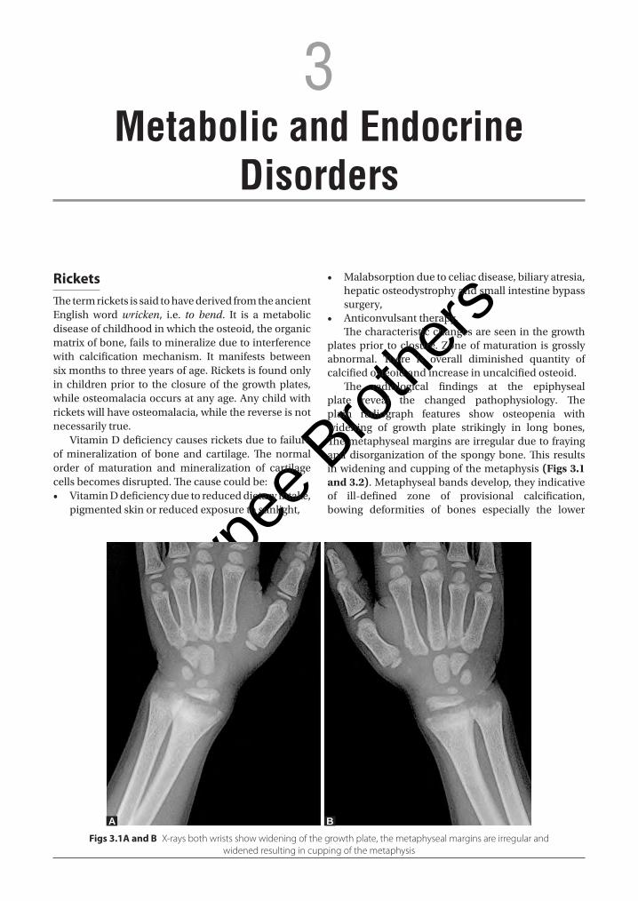
The radiological findings at the epiphysealplate reveal the changed pathophysiology. Theplain radiograph features show osteopenia withwidening of growth plate strikingly in long bones,Themetaphysealmarginsareirregularduetofrayinganddisorganizationof the spongybone.This resultsinwideningandcuppingofthemetaphysis(Figs 3.1 and 3.2).Metaphysealbandsdevelop,theyindicativeof ill-defined zone of provisional calcification,bowing deformities of bones especially the lower
Rickets ThetermricketsissaidtohavederivedfromtheancientEnglish word wricken, i.e. to bend. It is a metabolicdiseaseofchildhoodinwhichtheosteoid,theorganicmatrixofbone,failstomineralizeduetointerferencewith calcification mechanism. It manifests betweensixmonthstothreeyearsofage.Ricketsisfoundonlyin childrenprior to the closure of the growthplates,whileosteomalaciaoccursatanyage.Anychildwithricketswillhaveosteomalacia,whilethereverseisnotnecessarilytrue.
VitaminDdeficiencycausesricketsduetofailureofmineralization of bone and cartilage.The normalorder of maturation and mineralization of cartilagecellsbecomesdisrupted.Thecausecouldbe:• VitaminDdeficiencyduetoreduceddietaryintake,
pigmentedskinorreducedexposuretosunlight,
Figs 3.1A and B X-rays both wrists show widening of the growth plate, the metaphyseal margins are irregular and widened resulting in cupping of the metaphysis
A B
Jayp
ee B
rothe
rs
Metabolic and Endocrine Disorders v 23
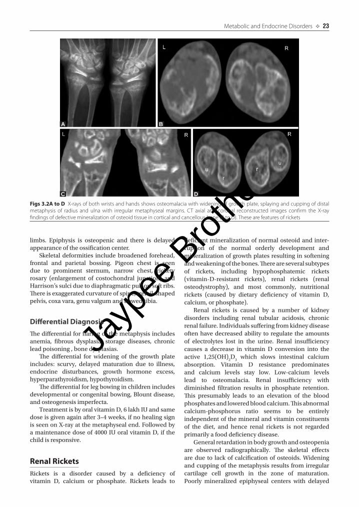
Figs 3.2A to D X-rays of both wrists and hands shows osteo malacia with widening of growth plate, splaying and cupping of distal metaphysis of radius and ulna with irregular metaphyseal margins. CT axial and coronal reconstructed images confirm the X-ray findings of defective mineralization of osteoid tissue in cortical and cancellous bone (B to D). These are features of rickets
A
C D
B
limbs. Epiphysis is osteopenic and there is delayedappearanceoftheossificationcenter.
Skeletaldeformities includebroadened forehead,frontal and parietal bossing. Pigeon chest is seendue to prominent sternum, narrow chest, ricketyrosary (enlargement of costochondral junction) andHarrison’ssulciduetodiaphragmaticpullonsoftribs.Thereisexaggeratedcurvatureofspine,trefoil-shapedpelvis,coxavara,genuvalgumandbowedtibia.
Differential DiagnosisThedifferentialforflaringofthemetaphysisincludesanemia, fibrous dysplasia, storage diseases, chronicleadpoisoning,bonedysplasias.
The differential for widening of the growth plateincludes: scurvy, delayed maturation due to illness,endocrine disturbances, growth hormone excess,hyperparathyroidism,hypothyroidism.
Thedifferentialforlegbowinginchildrenincludesdevelopmentalorcongenitalbowing,Blountdisease,andosteogenesisimperfecta.
TreatmentisbyoralvitaminD,6lakhIUandsamedoseisgivenagainafter3–4weeks,ifnohealingsignisseenonX-rayatthemetaphysealend.Followedbyamaintenancedoseof4000 IUoralvitaminD, if thechildisresponsive.
Renal RicketsRickets is a disorder caused by a deficiency ofvitamin D, calcium or phosphate. Rickets leads to
deficientmineralizationofnormalosteoidand inter-ruption of the normal orderly development andmineralizationofgrowthplatesresultinginsofteningandweakeningofthebones.Thereareseveralsubtypesof rickets, including hypophosphatemic rickets(vitamin-D-resistant rickets), renal rickets (renalosteodystrophy), and most commonly, nutritionalrickets (caused by dietary deficiency of vitamin D,calcium,orphosphate).
Renal rickets is caused by a number of kidneydisorders including renal tubular acidosis, chronicrenalfailure.Individualssufferingfromkidneydiseaseoftenhavedecreasedability to regulate theamountsof electrolytes lost in the urine. Renal insufficiencycauses a decrease in vitamin D conversion into theactive 1,25(OH)
2D
3 which slows intestinal calcium
absorption. Vitamin D resistance predominatesand calcium levels stay low. Low-calcium levelslead to osteomalacia. Renal insufficiency withdiminished filtration results in phosphate retention.This presumably leads to an elevation of the bloodphosphatesandloweredbloodcalcium.Thisabnormalcalcium-phosphorus ratio seems to be entirelyindependentofthemineralandvitaminconstituentsof the diet, and hence renal rickets is not regardedprimarilyafooddeficiencydisease.
Generalretardationinbodygrowthandosteopeniaare observed radiographically. The skeletal effectsaredue to lackof calcificationofosteoids.Wideningandcuppingof themetaphysis results fromirregularcartilage cell growth in the zone of maturation.Poorly mineralized epiphyseal centers with delayed
Jayp
ee B
rothe
rs
24 v Textbook of Radiology: Musculoskeletal Radiology
appearance. Epiphyseal plates appear widened anddistancebetweenendofshaftandepiphysealcenterisincreased.Thepresenceofbulkygrowthplatesattheshaftbonecartilage junctionsof longbonesandribsleadstoarachiticrosaryatthecostochondraljunctionsof themiddle ribs.Bowingdeformityand sabre-shindeformity of the tibia also are typicalmanifestationsof rickets. With increasing age scoliosis develops.Corticalspursprojectingatrightanglestometaphysisareobserved.Thecommondeformitiesassociatedarebowingoflongbones,moldingofepiphysis,fractures,frontalbossing.
Osteoporosis and Osteomalacia (Table 3.1)Osteoporosis is decreased osteoid production, ametabolic disorder characterized by decreasedmassperunitvolumeofanormallymineralizedboneduetolossofboneproteins.Whereasosteomalaciainadultsand rickets in children is under mineralization ofosteoid,isametabolicdisordercharacterizedbyfailureof mineralization and excess of the unmineralizedosteoidduetoaderangementincalcification.
Both osteoporosis and osteomalacia result ingeneralized decrease in bone density, known asosteopenia which means decrease in the bonemineralizationquantity.Otherconditionswhichleadtogeneralizedosteopeniaare:• Hyperparathyroidism• Multiplemyelomaordiffusemetastases• Drugs• Osteogenesisimperfecta
OsteoporosisIt means reduced bone mass with normal bonecomposition secondary to either osteoclasticresorptionofboneorosteocyticresorption. It isverycommonly seen in women above 60 years of age.Various diseases can lead to osteoporosis. Thosediseasesare:• Congenital: Osteogenesis imperfecta, homocysti-
nuria• Idiopathic:Juvenile,adult,postmenopausal,senile
osteoporosis• Nutritional disorders:Scurvy, calciumdeficiency,
proteindeficiency• Endocrinopathies: Cushing’s syndrome, hyperpara-
thyroidism,hyperthyroidism,acromegaly,Addison’sdisease, diabetes, pregnancy, paraneoplastic syn-dromes
• Renalosteodystrophy• Immobilization• Collagendisease• Rheumatoidarthritis• Bonemarrowreplacement
• Drugs such as heparin, methotrexate, cortico-steroids,excessivealcoholconsumption,smoking,dilantin
• Radiationtherapy• Localized due to Sudeck dystrophy, transient
osteoporosisofhip.Osteoporosis canbediagnosedonX-rays andby
calculating bonemineral density. X-ray features canfurtherbeevaluatedwiththehelpofCT,however,bone
Table 3.1 Difference between osteoporosis and osteomalacia
Osteoporosis Osteomalacia
Etiology
• Primary osteoporosis-senile or postmenopausal
• Immobilization due to pro-longed bed rest or paralysis
• Endocrine-glucocorticoid excess, thyrotoxicosis, hypogonadism, hyperprol-actinemia, diabetes mellitus, hyperparathyroidism
• Diet—chronic alcoholism, anorexia nervosa.
• Prolonged use of drugs such as heparin, ethanol.
• Chronic illness like rheuma-toid arthritis, cirrhosis, renal tubular acidosis
• Neoplasm-multiple myelo-ma, leukemia, lymphoma, mastocytosis
• Genetic abnormalities-Ho-mocystinuria, Ehler-Danlos syndrome, osteogenesis im-perfect, Marfan’s syndrome
• Hepatic disease
• Lack of dietary intake of vitamin D
• Decreased absorption of vitamin D-malabsorption syndrome, partial gastrec-tomy
• Deficiency of vitamin D metabolism—chronic renal tubular disease, anticonvul-sant therapy
• Decreased deposition of calcium in the bone due to drugs like diphosphonates
Imaging
Ground glass appearance due to generalized rarefaction
Generalized osteopenia
Loss of vertebral body height due to symmetric transverse compression
Loss of transverse trabeculae
No Looser’s zones Looser’s zones at axillary mar-gin of scapula, ramus of pubis or ischium, femur neck, ribs,
Protrusio acetabuli and triradi-ate pelvis
Pathological fractures at wrist, hip
Pathological fractures
Anterior wedge compression and Codfish vertebrae
Codfish vertebral bodies due to biconcave vertebral bodies
Treatment
Calcitonin, sodium fluoride, diaphosphonates, estrogen replacement
Calcium, vitamin D, high- protein diet
Jayp
ee B
rothe
rs
Metabolic and Endocrine Disorders v 25
mineraldensitypredictsosteoporosisquiteaccuratelyevenbeforeonsetofsymptoms.X-rayfeaturesare:• Decreaseinnumberandthicknessoftrabeculae• Corticalthinningduetoendostealandintracortical
resorption• Juxta-articular-reducedbonedensitypredominantly
affectingtrabecularbone• X-rayfeaturesinosteoporosisofspine:
– Diminishedradiographicdensity– Verticalstriationsduetothinningoftransverse
trabeculaewithrelativeaccentuationofverticaltrabeculae
– Accentuationofendplates– ‘Picture-frame’ vertebrae due to preservation
ofoutercortexwithintracorticalandendostealresorption
– Biconcavevertebrae– Schmorl’snodes– Wedging– Decreasedheightofvertebrae– Absenceofosteophytes.
OsteomalaciaIt is characterized by accumulation of excessiveamounts of uncalcified osteoid with bone softeningand insufficient mineralization of osteoid due toeither high remodeling rate which is excessiveosteoid formationwithnormalmineralizationor lowremodeling ratewhich isnormal osteoidproductionwith diminished mineralization. It occurs in adultpatients due to deficiency of vitamin D. Etiology ofosteomalacia:• Dietary deficiency of vitaminD and lack of solar
exposure,• DeficientmetabolismofvitaminDduetochronic
renalinsufficiency,• DecreasedabsorptionofvitaminDdue topartial
gastrectomyormalabsorptionsyndrome,• Decreaseddepositionofcalciuminbone.
X-rayfeaturesofosteomalaciaare:• Uniformosteopenia• Fuzzy indistinct trabecular details, of endosteal
surface• Coarsened frayed trabeculaewhicharedecreases
innumberandsize• Corticalthinninginlongbones• Bone deformities due to softening such as
hourglassthorax,bowingoflongbones,acetabularprotrusion, buckled or compressed pelvis,biconcavevertebralbodies
• Insufficiencyfractures• PseudofracturesalsoknownasLooser’szones• Mottledappearanceofskull.
Osteomalaciaduringendochondralbonegrowthinchildhoodistermedasrickets.Radiologicalfeaturesare:• Poorly mineralized irregular epiphyseal centers
withdelayedappearance
• Axialwideningof growthplate that is increase indistance between end of shaft and epiphysealcenterduetoincreasedosteoidproduction
• Cupping and fraying of metaphysis with thread-like shadows into epiphyseal cartilage of weightbearingbones
• Corticalspursprojectingatrightanglestometaphysis• Coarsetrabeculations• Periostealreactionmaybepresent• Deformitiesoflongboneswithbowing• Frontalbossingofskull.
Bone densitometry helps in diagnosing osteo-porosis. Bone densitometry is best performed byusing dual energy X-ray absorptiometry (DEXA)scan.This examination is done on outpatient basis.DEXAexamination generallymeasures bonedensityin the hip and spine, the patient lies on the table,X-ray generator is located below the patient and animaging device, or detector, is positioned above andthe detector slowly moves over the area of interest,generatingimagesonamonitor(Table 3.1).
Interpretation of ResultsA bone density test result shows a T-score and a Z-score. A T-score is a number derived by comparingpatients BMD tests results to an average score fora healthy adult of same gender and race who hasreachedtheirpeakbonemassat25yearsofage.
TheT-scoresignifiesvariationfrom“normal.”It isthedifferencebetweenpatientsBMDandtheBMDofapersonatpeakbonemass.T-scorescanbeaslowasonestandarddeviation(SD)belownormalandstillbeconsidered healthy. Patients with T-scores between–1SDand–2.5SDhaveosteopeniaandareconsideredat high-risk for developing osteoporosis. Patientswith T-scores lower than –2.5 SD have osteoporosis(Table 3.2).
Whereas Z-score which is patients bone densitycompared with a person of same age group andsex and interpreted to determine whether patienthas osteoporosis or not. However, T-score is mostcommonlyusedfordiagnosis.
AcromegalyAcromegalyistheresultofexcessivegrowthhormoneproduction,mostcommonlyfromanadenomaofthepituitary. Itmost commonly affects adults inmiddleage and can result in severe disfigurement, serious
Table 3.2 Interpretation of T-score
Bone density T-score Diagnosis
Normal +1.0 to –1.0 Normal
Low –1.0 to –2.5 Osteopenia
High-risk –2.5 or lower Osteoporosis
Jayp
ee B
rothe
rs
26 v Textbook of Radiology: Musculoskeletal Radiology
complicatingconditions,andprematuredeath.Ithasbothaninsidiousonsetandslowprogressionandmaybedifficulttodiagnoseintheearlystages,onlybeingdiagnosedwhentheexternalfeatures,especiallyoftheface,becomenoticeable.
Radiologicalchangesareseen inskull,mandible,paranasalsinusesextremitiesandvertebrae.Skull and mandible show:• Enlargedoccipitalprotuberance• Prognathism• Enlargedparanasalsinuses• EnlargementSellaanditserosion• Calvarialhyperostosis• Vertebralscalloping,newboneformationandloss
ofdiscspace.Extremities show:• Flaredendsoflongbones• Cysticchangesincarpalsandfemoraltrochanters• Osteoporosis• Spade-likehand• Heelpadthickness>25mm.
Majority of cases are the result of a pituitarymacroadenoma. Expansion into the sella turcicamayresultincompressionofsurroundingstructures,most importantly, the optic chiasm. The majorityof pituitary tumors are incidental and do not havea genetic component. MRI detects the presence ofpituitary tumors and its complications like opticchiasma compression. Pituitarymacroadenomas arebydefinitionmorethan10mmmassarisingfromthepituitarygland,andusuallyextendsuperiorly.
Indentation at the diaphragma sellae pituitarymacroadenoma can give a snowman or figure eight configuration on CT. Contrast attenuation can varydepending on hemorrhagic, cystic and necroticcomponents. Adenomas which are solid, withouthemorrhage, typically have attenuation similar tobrain(30-50HU)anddemonstratesmoderatecontrastenhancement.Calcificationisrare.
T1W and T2W MR images show the lesion astypicallyisointensetogreymatterandmayshowareasofnecrosisandhemorrhage.Onpostcontrastimages,they show moderate to bright enhancement. T2WIgradientimagesaresensitiveindetectinghemorrhage.
The differential diagnosis includes cranio-pharyngioma,meningioma, pituitary carcinoma andmetastases.
FluorosisAcute high-level exposure to fluoride is rare andcauses immediate abdominal pain, excessive saliva,nausea and vomiting, seizures and muscle spasms.Acute high-level exposure to fluoride is usually duetoaccidentalcontaminationofdrinking-waterordueto fires or explosions. Chronic fluorosis is exposureto excess of fluorine or its compounds. Sources ofexposurecanbefrom:
• Drinking of water containing concentrations offluorinegreaterthan4partspermillion;
• Industrial exposure to fluorine-containing com-poundsoveralongperiodoftime
• Treatmentofosteoporosiswithsodiumfluoride;• Consumptionoffluorine-containingwine.
Ingestionofexcessoffluorineleadsdentalfluorosisin children less than 8 years of age and presents aspittingandstainingof the teeth,and inseverecases,all the enamel may be damaged. Children above8 years and adults cannot develop dental fluorosis.However,lowlevelsoffluorideintakehelptopreventdentalcaries.Thecontrolofdrinking-waterqualityisthereforecriticalinpreventingfluorosis.
Othermanifestationsoffluorosis includenausea,vomiting,constipation,lossofappetite,toxicnephritis,joint pain and restriction of motion, back stiffness,restriction of respiratory movements, functionaldyspnea,andparaplegia.
Skeleton abnormalities include hypoplasia andirregularity of dental structures, osteosclerosis, ver-tebral osteophytosis, calcification of ligaments andperiostitis. Increasing trabecular condensation leadstotheeventualappearanceofchalkyareasthroughoutthethorax,vertebralcolumnandpelvis.Hyperostosisandboneexcrescencesdevelopatsitesofligamentousattachment, especially in the iliac crests, ischialtuberosities and inferior margins of the ribs. Inadvanced stages, fluorosis can lead to contracturesand deformities of extraspinal joints, kyphosis,restricted spinal and chest motion, and neurologiccomplications. Normally, the abnormalities arereversibleaftercessationofexposure,butacoarsenedtrabecular pattern without increased radiodensitymayremain.
Resorption of Terminal TuftsResorption of terminal tufts commonly related tooccupational conditions. It is also called as acro-osteolysis, i.e. destruction of bone of the acral areas(extremities). Osteolysis may be severe in personsexposed toanumberof industrialmaterials, suchaspolyvinylchloride. The radiographic hallmark of thedisorder is osteolysis, which occurs predominantlyin the terminal phalanges of the hands. Band-likeradiolucent areas across the waist of one or moreterminal phalanges may occur along with tuftalresorption. The thumb is affected more commonlythan the other digits.The sacroiliac joints, the foot,andsometimesother skeletal structuresmayalsobeinvolvedincertaintypesofacro-osteolysis.
Various Conditions Leading to Acro-osteolysis• Traumatic causes: Amputation, burns, electric
injury,frostbite,andvinylchloridepoisoning
Jayp
ee B
rothe
rs
Metabolic and Endocrine Disorders v 27
• Neuropathic causes: Congenital indifference topain, syringomyelia, diabetes mellitus, myelo-meningoceleandleprosy.
• Collagen vascular disease:Scleroderma,dermato-myositis,andRaynaud’sdisease.
• Metabolic causes:Hyperparathyroidism.• Inherited conditions: Familial acro-osteolysis,
pyknodysostosis,andpachydermoperiostosis.• Other conditions:Sarcoidosis,psoriaticarthropathy
andepidermolysisbullosa.
FrostbiteItisthermalinjurytothebody,usuallytheextremitiesorface.Asthetissuesfreeze,sodothebloodvessels,withvascular thrombosis interrupting circulation. Injury isfollowedbyedema.Ifthecirculationisnotrestored,thereisacro-osteolysiswitheitherauto-orsurgicalamputationofthefingers.Theinitialradiologicalchangeissofttissueswelling at the finger tips followed by osteoporosisand periosteal new bone formation, followed by boneresorptionwithlossoftheterminaltufts.
Vinyl Chloride PoisoningIt is a polymerizing agentused industrially thatmayproduce cutaneous abnormalities resemble thoseof scleroderma. It may cause occupational acro-osteolysisinworkersexposedtothissubstanceduringitsmanufacture.Adrum-stickfingerisanabnormalityofthefingersoccurringinpatientswithoccupationalacro-osteolysis.
Diabetes MellitusInthis, thereispencil-and-cupappearanceonXray,where thebaseof theproximalphalanxbroadens toform a cupwithwhich the tapered phalangeal shaftbecomesassociated.
SclerodermaItisaconnectivetissuediseasewhichresultsinfibrosisand sclerosis of the skin andmucosa, subcutaneoustissues and submucosal tissues of internal organs.Skeletal manifestations include absorption of thedistal phalanges of the hands, acro-osteolysis withperiarticular soft tissue swelling, joint destruction,calcificationinthesofttissueandosteopenia.
HyperparathyroidismIn patients with primary or secondary hyperpara-thyroidism, bone resorption is evident radiologicalexamination,especiallyinthehandsintheearlystagesofthedisease.Theresorptioncanbecategorizedinto
various types. Subperiosteal resorption of corticalboneisvirtuallydiagnosticofhyperparathyroidbonedisease. In this form, a lace-like appearance of thephalangealbonemayprogresstoaspiculatedcontourand to complete resorption of the entire cortex.Other sites of subperiosteal resorption include thephalangealtufts.
PyknodysostosisIt is a dysplasia manifested clinically by dwarfism,increased bone fragility and sclerotic bones.Inheritanceisautosomalrecessive.Radiologically,theskullbase isdense.There isawidelyopenpersistentanterior fontanelle and multiple wormian bones.Theangleof themandible isveryobtusewithseveremicrognathia.Thevertebralbodiesandlongbonesaresclerotic, and there is under modeling of the boneswithnarrowingofthemedullarycanals.Fracturingisfrequent.Theremaybeacro-osteolysis.
PachydermoperiostitisIt is anautosomaldominant conditioncausing largeskin foldsof the faceandscalp thatoccurspredomi-nantly in males aged 3 to 38 years old, although itusuallystartsatpuberty.There is irregularperiostealproliferation of the phalanges anddistal third of thelong bones of legs and forearms beginning in theepiphyseal region at tendon or ligament insertions.Thedistalphalangesarerarelyinvolved.Thecortexisthickened but there is no narrowing of themedulla.Acro-osteolysis may be present. Enlargement of theparanasalsinusesandfingerclubbingisseen.
Psoriatic ArthritisIt is a seronegative spondyloarthropathy occurringinsomepatientswithpsoriasis.Thearticulardiseasemaybemonoarticular,pauciarticularorpolyarticularin its distribution, and virtually any joint can beaffected. Classic radiographic features of psoriaticarthritisareinvolvementofsynovialandcartilaginousjoints and entheses, involvement of interphalangealjointsofthehandsandfeet,sacroiliitisandspondylitiswith paravertebral ossification, bone erosion withadjacent proliferation, intra-articular bone ankylosisanddestructionofphalangealtufts.
Expansile Lesions of MetaphysisExpansile lesions affecting the metaphyses of longbonesincluderickets,hypophosphatasia,metaphysealchondroplasia,enchondroma,non-ossifyingfibroma,aneurysmal bone cyst, chondromyxoid fibroma, andgiantcelltumor.
Jayp
ee B
rothe
rs
28 v Textbook of Radiology: Musculoskeletal Radiology
RicketsPlainradiographicandCTfindingsarewideningandcupping of the metaphyseal regions, fraying of themetaphysis, bowing of long bones, development ofknock-knees,orgenuvalgum.
HypophosphatasiaHypophosphatasia is a rare and fatal metabolicbone disease. In the perinatal period, it is themostpernicious form of hypophosphatasia. The infantilesubtype presents in the first 6 months of life. Inchildhood, hypophosphatasia’s clinical expression isextremelyvariable.Asa resultofaplasia,hypoplasia,or dysplasia of dental cementum, premature loss ofdeciduous teeth (i.e. before the age of 5) occurs. Inadult years, hypophosphatasia can present duringmiddleage.X-raysshowhypomineralization,rachiticchanges, and incomplete vertebrate ossification,lateralbonyspursontheulnaeandfibulaeandtongue-likeradiolucentareasprotrudingfromthemetaphysesintotheboneshaft.
Metaphyseal ChondroplasiaIt is a heterogeneous group of intrinsic dysplasiascausingchangesinthemetaphysesoftubularbones.Metaphyseal chondrodysplasia is associated withneutropenia, lymphopenia, immune deficiency,pancreatic exocrine insufficiency, Hirschsprung’sdisease, and intestinalmalabsorption.Theconditionis first recognized in early childhoodwhen childrenpresent with a waddling gait, exaggerated lumbarlordosis,genuvarum,andshortstature.Radiographsshow appearance of an enlarged metaphysis andwidened-cupped physis similar to rickets, coxa varaoccurswithoutanassociatedbowedfemur.
Types
1. Schmidt’s type: THis disorder may arise fromdefectivetypeXcollagen,whichistypicallyfoundinthehypertrophiczoneofthephysis.Patientsshowmildshortstature,legpains,bowedlegs,increasedlordosis,andwaddlinggait.Upperextremityshowsmild wrist swelling, flexion contractures of theelbows. Lower extremities are more significantlyinvolved than upper extremities and show varusdeformities of the knees and ankles are present,withbowingvisibleinthethighandtheleg,severegenuvarum.
2. Jansen’s type: It is a rare autosomal dominantdisordercharacterizedbyshortlimbdwarfismwithsevere hypercalcemia and hypophosphatemia.X-rays show rachitic changes of metaphysiscommonlyaffectingknee jointswithpathologicalfractures.Metatarsals,metacarpalsandskullbasealsoshowscleroticlesions.
EnchondromaItisabenigncartilaginousgrowthinmedullarycavity,usuallysolitaryandisseenwithinmedullarycanalandmetaphysis.X-raysshowoval/roundareaofgeographicdestructionwithlobulatedcontourandfinemarginalline, cortical endosteal scalloping, ground-glassappearance, dystrophic calcifications within smallcartilage nodules/fragments of lamellar bone whichcanbepinhead,stippled,flocculentorarcsandringspattern,bulbousexpansionofbonewith thinningofcortexinsmalltubularbonesofphalanx,ribandfibulaisseenwithMadelungdeformity=bowingdeformitiesof limb,discrepant length.Nocorticalbreakthrough/periostealreaction.MRIshows low-to intermediate-signal intensity on T1WI and high-signal intensityon T2WI, low-signal intensity matrix calcifications,normal fat marrow interspersed between cartilagenodulesandperipheralenhancementpatternonpost-gadoliniumimages.
Non-ossifying FibromaIt is a lesion resulting from proliferative activityof a fibrous cortical defect that has expanded intomedullary cavity shaft of longbonewhich is seen inbonesoflowerextremity.Radiographsshoweccentricmetaphyseal, multilocular ovoid bubbly osteolyticarea with alignment along long axis of bone, about2cminlength,densescleroticbordertowardmedulla,V-orU-shapedatoneend,endostealscallopingwiththinning and overlying bulge. Nuclear scan showsminimal/mild uptake on bone scan. MR showshypointenselesiononT1WIandT2WIorhypointenseonT1WI andhyperintense onT2WIwith peripheralhypointense rim and internal intense contrastenhancement.
Aneurysmal Bone CystIt is an expansile pathologically benign lytic lesionof bone containing thin-walled cystic cavities filledwith chronic blood products with its name derivedfrom roentgen appearance. X-rays show purely lyticeccentric radiolucency with aggressive expansileballooning lesioncalledas soap-bubblepatternwiththininternaltrabeculations.Thereisrapidprogressionwithin6weeksto3monthswithscleroticinnerportion,almostinvisiblethincortex,noperiostealreaction.CTshows blood-filled sponge-like lesion due to fluid-fluid/hematocrit levels due to blood sedimentation.MR showsmultiple cysts of different signal intensityrepresentingdifferentstagesofbloodbyproductslikeheterogeneous fluid-fluid levels within loculationsreflecting hemorrhage with sedimentation andlow-signal intensity rim and shows heterogeneousenhancement.Nuclear scintigraphyshowsdoughnutsignduetoperipheralincreaseduptake.Angiographyshowshypervascularityinlesionperiphery.
Jayp
ee B
rothe
rs
Metabolic and Endocrine Disorders v 29
Chondromyxoid FibromaRare benign cartilaginous tumor which initiallyarises in cortex and is an eccentric, metaphyseallesion. Radiographs show expansile ovoid lesionwith radiolucent center and oval shape at each endof lesion, longaxisparallel to longaxisofhostbone,geographic bone destruction, well-defined scleroticmargin, expanded shell with bulged and thinnedoverlying cortex, partial cortical erosion, scallopedmargin, septationswhichmaymimic trabeculations,stippled calcifications within tumor in advancedlesionsandnoperiostealreaction.
Giant Cell TumorIt is also known as osteoclastoma. Radiographsshow a well-circumscribed expansile solitary lyticbone lesion with a narrow zone of transition, soap-bubbleappearanceduetoexpansileremodelingwithmultiloculated appearance, no internal mineraliza-tion of tumor matrix, prominent trabeculation, nosclerosis/periosteal reaction due to aggressive rapidgrowth, cortical penetration, cortical thinning, soft-tissue invasion, complete/incomplete pathologicfracture,destructionofvertebralbodywithsecondaryinvasionofposteriorelementsandvertebralcollapse.Nuclearscintigraphyshowsdiffuselyincreaseduptakewithcentralphotopeniaondelayedbonescintigraphy.Angiographyshowsahypervascularlesion.CTshowstumorofsoft-tissueattenuationsimilartomusclewithfocioflowattenuation,nomatrixmineralization,andwell-definedmarginswith thin rimof sclerosis.Soft-tissue extension at metaphyseal end of tumor withsignificant enhancement. MR shows well-definedlesionofheterogeneous signal intensitywith low-to-intermediate intensity on T1WI and T2WIwith low-signal-intensity margin significant enhancement ofsolid-tissuecomponent.
Hand as an Index of DiseaseVariety of disorders affects the bones and joints ofthe hand and thus makes it an index to diagnoseassociationsofsystemicdiseasesleadingtoarthritis.Articulardisordersofhandare:
OsteoarthritisItisalsoknownasdegenerativejointdiseaseandoccursduetoabnormalstressonthebone.Sitescommonlyaffectedareproximalanddistalinterphalangealjoints,1stcarpometacarpaljoint,andtrapezioscaphoidjoint.It usually shows bilateral involvementwhich can besymmetricorasymmetric.
Itcanbeclassifiedas:• Primary osteoarthritis:Mostcommonintheolder
agegroupastheresultofwearandtearonarticularcartilageovertime.
• Secondary osteoarthritis: Results from a previousprocessthatdamagedcartilagesuchastrauma,orinflammatoryarthritis.Diagnosis is essentially done with the help of
plainX-rays.CTandMRIcanbeusedasanadjunctto diagnose, if required. Plain X-ray features are:joint space narrowing, subchondral eburnation,radial subluxation of 1st metacarpal base, marginalosteophyteswithsmallossicles.
Inflammatory ArthritisItpredominantlyaffectspostmenopausalormiddle-agedwomenduetorepeated infectionsor inflammations. Itcommonly affects proximal and distal interphalangealjoints,1stcarpometacarpal joint,and trapezioscaphoidjoint.Itusuallyshowsbilateralinvolvementwhichcanbesymmetricorasymmetric.
PlainX-rayfeaturesarecentralerosionscombinedwith osteophytes known as subchondral ‘gull-wing’erosions, joint space narrowing, sclerosis, rarelyankylosiscanoccur.
Rheumatoid ArthritisAninflammatoryprocesswiththetargetorganbeingthesynovialmembrane leading topannus formation(inflammatory exudates in the lining of the synovialcells).Areasaffectedareproximalinterphalangealandmiddleinterphalangealjointswithearlieraffectionof2ndand3rdfingers,allwristjoints,andstyloidprocessofulna. It affectsbothhands in relative symmetricalfashion.
X-ray Features
• Osteopenia: Demineralization of the bone is theresultofincreasedbloodflow,duetoinflammation,which washes out the calcium. Early on in theinflammatory process, only the periarticularportion of the bones is affected. Over time, theinflammatorypaincausesdisuseofaffectedjointsleadingtogeneralizedosteopeniaofwholebones.
• Uniform joint space narrowing: A feature whichhelps differentiates rheumatoid arthritis fromosteoarthritis.
• Marginal erosions at bare areas where synoviumliesonbone.
• Subluxationduetoligamentousorcapsularlaxity.• Fusiformsofttissueswellingandjointdeformities.
GoutIt occurs due to deposition of monosodium uratecrystals in synovial fluid, and usually it remainsasymptomatic from months to years. It commonlytargetscarpo-metacarpaljointsbutcanalsoaffectalljointsofhand.

Jayp
ee B
rothe
rs
30 v Textbook of Radiology: Musculoskeletal Radiology
Features on X-rays or CT are development ofchronictophaceousgoutwhichappearsas lobulatedsoft tissuemasses,well-defined, periarticular eccen-tric erosions with overhanging edge and scleroticmargins,preservationof joint spacesandabsenceofosteoporosis.Mostextensivechangesareseen in thecommoncarpometacarpalcompartmentwhichshowsscallopederosionsofthebasesofulnarmetacarpals.
PsoriasisItisalsoknownasrheumatoidvariantorseronegativespondyloarthropathy. It showsperipheralmanifesta-tion inmonoarthritis or asymmetric oligoarthritis orsymmetricpolyarthritis.Targetareasareallhandandwristjointspredominantlydistal.
X-ray features are ‘Mouseear’marginal erosions,intra-articularosseousexcrescences,newboneforma-tionwithfusionandabsenceofosteoporosis.
Calcium Pyrophosphate Dehydrate: Crystal Deposition DiseaseCalcium pyrophosphate dehydrate (CPPD) alsoknown as pseudogout, is a very common entity.Radiographically, the presence of chondrocalcinosisis typical of this entity. Common locations forchondrocalcinosisincludethetriangularfibrocartilageat the wrist. Chondrocalcinosis in the setting ofCPPD is commonly associated with calcification offibrocartilage.
X-ray Features
Chondrocalcinosis with periarticular calcifications.Degenerative changes in unusual locations causingnarrowing and obliteration of space between distalradius and scaphoid with fragmentation of surfacesand scapholunate separation. Destruction oftrapezioscaphoidspace,noerosionsandpresenceoflargeosteophytes.
Systemic Lupus ErythematosusItleadstomyositis,polyarthritis,deformingnonerosivearthropathy and osteonecrosis. It commonly targetsproximal and middle interphalangeal joints andcausesreversibledeformities.
SclerodermaItisalsoknownasprogressivesystemicsclerosisandcommonlyaffectsdistalandproximalinterphalangealjointsand1stcarpometacarpaljoints.
X-rays commonly show tuft resorption and softtissuecalcifications.
Nonarticular Disorders of Bones of Hand
• Acro-osteolysis:Itmeanslyticdestruction.Commonlyaffects distal and middle phalanges. It is seen invarietyofdiseases like:psoriasis,porphyria,Ehlers-Danlos syndrome, thromboangiitis obliterans,Raynaud’s disease, diabetes, dermatomyositis,injuries,epidermolysisbullosa,rheumatoidarthritis,Reiter’s syndrome, scleroderma, sarcoidosis,pyknodysostosis, leprosy, Lesch-Nyhan syndrome,syringomyelia, hyperparathyroidism. It shows lyticlesionswithabsenceofperiostealreactiononX-rays.Epiphysealinvolvementcanbeseeninlaterstages.
• Acro-osteosclerosis: In this entity, sclerotic lesionsare seen on X-rays in the phalanges which canbe an index to diseases such as rheumatoidarthritis,sarcoidosis,scleroderma,systemic lupuserythematosus, Hodgkin’s disease, hematologicdisorders.OnX-rays, it shows focalopaqueareaswithendostealthickening.
• Resorption of terminal tufts of phalanges whichcan be diagnosed on X-rays help in diagnosingdiseaseslike:– Trauma—due to amputations, burns, electric
injuries,frostbite,andvinylchloridepoisoning.– Neuropathic—congenital indifference to pain,
syringomyelia, myelomeningocele, diabetes,leprosy.
– Collagen vascular disease—scleroderma,dermatomyositis,Raynaud’sphenomenon.
– Metaboliccausessuchashyperparathyroidism.– Inherited disorders such as familial acro-
osteolysis,pyknodystosis,progeriaorWerner’ssyndromeandpachydermoperiostosis.
– Other diseases such as sarcoidosis, psoriaticarthropathyandepidermolysisbullosa.
• Metacarpal sign: It is the relative shortening of4th and 5th metacarpals. It commonly is seenassociated with pseudohypoparathyroidism,basal cell nevus syndrome, multiple epiphysealdysplasia,Beckwith-Wiedemannsyndrome,sicklecell anemia, juvenile chronic arthritis, Turner’ssyndrome, ectodermal dysplasias, hereditarymultipleexostoses,melorheostosis.
On the other hand X-rays a tangential linedrawnalongtheheadsof4thand5thmetacarpalsintersect the 3rd metacarpal indicating relativeshortening.
• Carpal angle:ItisanangleseenonX-raysformedby tangents to proximal row of carpal bones.Normally,itshouldbe130°.
Adecreasedcarpalanglewhichislessthan124°isindicativeofdiseasessuchasTurner’ssyndrome,Hurler’s syndrome, Morquio’s syndrome, Made-lungdeformity.
Increased carpal angle is above 139° and it isseen in Down’s syndrome, arthrogryposis, bonedysplasiawithepiphysealinvolvement.
Jayp
ee B
rothe
rs
Metabolic and Endocrine Disorders v 31
• Dactylitis: It means inflammation of the fingers.Radiologically,itisseenastheexpansionofphalangeswith multiple radiolucencies within due to cysticchanges.Itisseenintuberculosis,pyogenicorfungalinfection,syphilis,sarcoidosis,hemoglobinopathies,hyperparathyroidism,leukemia.
• Brachydactyly:Itmeansshorteningorbroadeningofmetacarpals andpalanges seenonX-rays. It isseenwithtrauma,osteomyelitis,arthritis,Turner’ssyndrome, osteochondrodysplasia, mucopolysac-charidoses,basalcellnevussyndrome,hereditarymultipleexostoses.
• Clinodactyly: Abnormal curvature is seen of thefingersinmediolateralplane.ItisassociatedwithDown’s syndrome, multiple dysplasia, contra-ctures.
• Polydactyl: It means havingmultiple fingers thatismore than fourfingers and a thumb. It is seenassociated with Carpenter syndrome, Ellis-van
Creveld syndrome, Meckel-Gruber syndrome,short-ribpolydactylsyndrome,Trisomy13.
• Syndactyly:Itmeansosseousandcutaneousfusionof digits. It is seen inApert syndrome,Carpentersyndrome, Down’s syndrome, neurofibromatosis,Polandsyndrome.
• Fingertip calcifications:Calcificdensitiescanbeseenin the soft tissues of fingers indicative of followingdiseases such as scleroderma, Raynaud’s disease,systemic lupus erythematosus, dermatomyositis,hyperparathyroidism.
• Lucent lesions in fingers:Various lesions can leadto radiolucent lesions in the fingers which canbeseenonX-rays.These lesionscanbeaglomustumor, gouty arthritis, metastasis, enchondroma,simple inclusion cyst, pancreatitis, aneurysmalbonecyst,giantcelltumor,epidermoid,etc.Thus,byroutineX-rayexaminationofthehandcan
showavarietyofappearancesandcanprovideaguidetovarioussystemicandarticulardisorders.