Jaundice Yellow discoloration of sclera, skin, mucous membranes due to deposition of bile pigment...
35
Jaundice Yellow discoloration of sclera, skin, mucous membranes due to deposition of bile pigment Clinically detected with serum bilirubin 2-2.5mg/dL or (2 times normal)
-
Upload
gwenda-bishop -
Category
Documents
-
view
237 -
download
1
Transcript of Jaundice Yellow discoloration of sclera, skin, mucous membranes due to deposition of bile pigment...
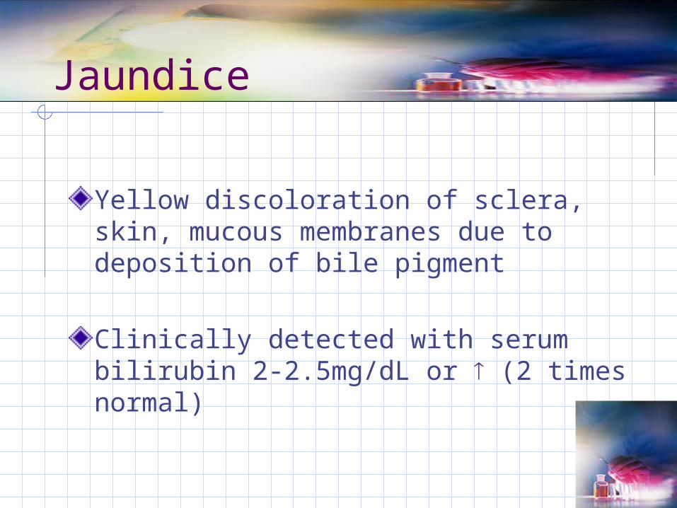
Jaundice
Yellow discoloration of sclera, skin, mucous membranes due to deposition of bile pigment
Clinically detected with serum bilirubin 2-2.5mg/dL or (2 times normal)
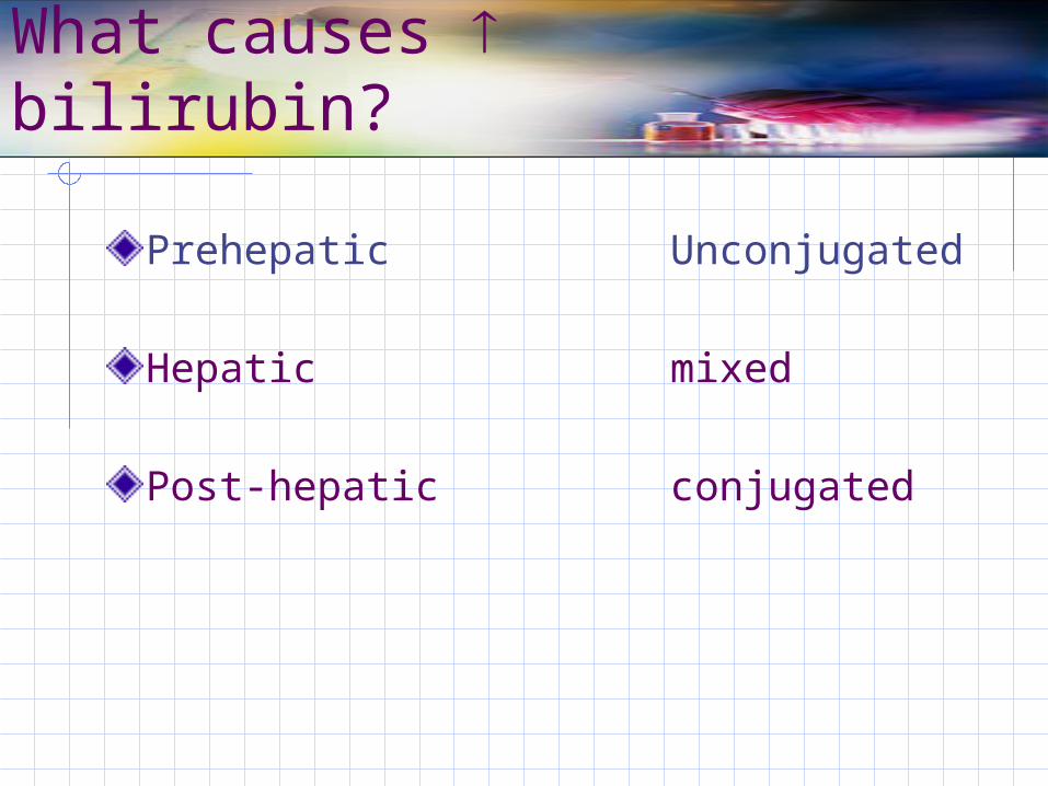
What causes bilirubin?
Prehepatic Unconjugated
Hepatic mixed
Post-hepatic conjugated
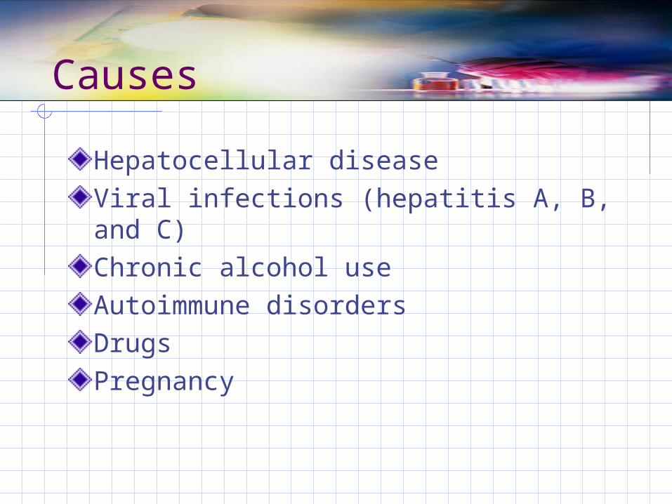
Causes
Hepatocellular diseaseViral infections (hepatitis A, B, and C)Chronic alcohol useAutoimmune disordersDrugsPregnancy
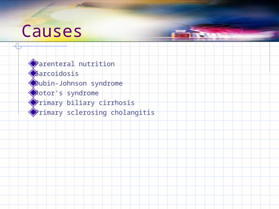
Causes
Parenteral nutritionSarcoidosis Dubin-Johnson syndromeRotor's syndromePrimary biliary cirrhosis Primary sclerosing cholangitis
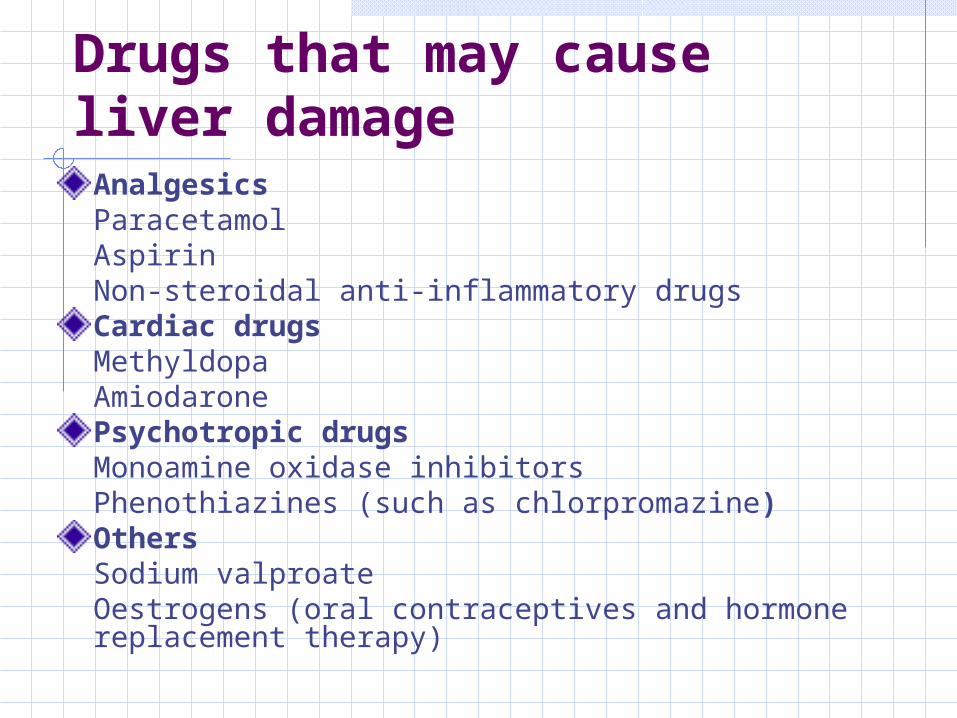
Drugs that may cause liver damageAnalgesicsParacetamolAspirinNon-steroidal anti-inflammatory drugsCardiac drugsMethyldopaAmiodaronePsychotropic drugsMonoamine oxidase inhibitorsPhenothiazines (such as chlorpromazine)OthersSodium valproateOestrogens (oral contraceptives and hormone replacement therapy)
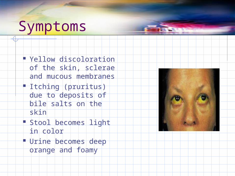
Symptoms
Yellow discoloration of the skin, sclerae and mucous membranes
Itching (pruritus) due to deposits of bile salts on the skin
Stool becomes light in color
Urine becomes deep orange and foamy
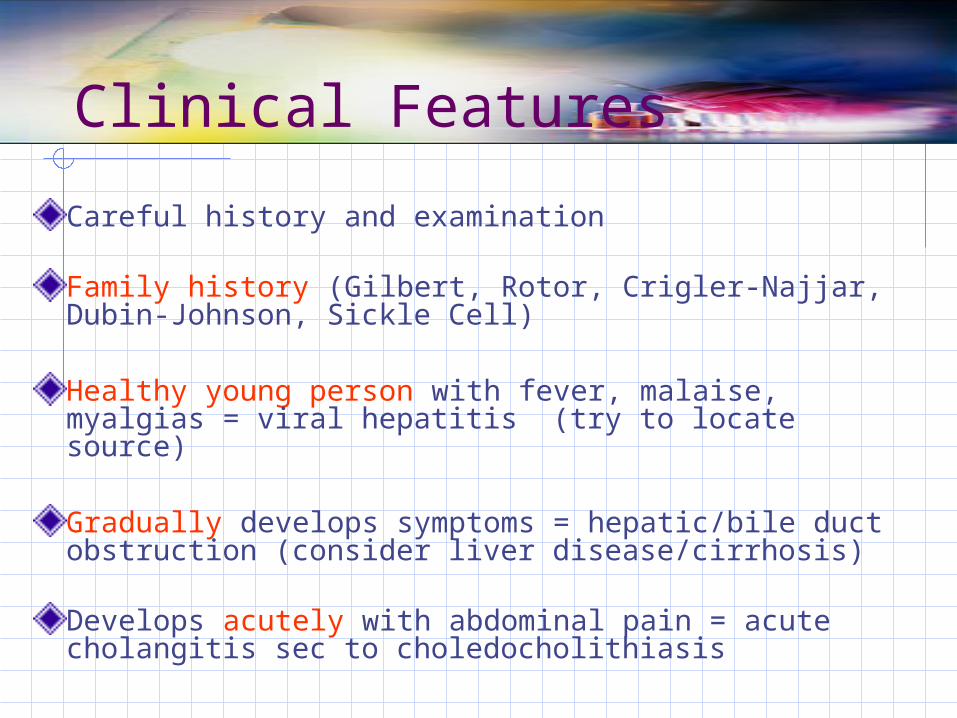
Clinical Features
Careful history and examination
Family history (Gilbert, Rotor, Crigler-Najjar, Dubin-Johnson, Sickle Cell)
Healthy young person with fever, malaise, myalgias = viral hepatitis (try to locate source)
Gradually develops symptoms = hepatic/bile duct obstruction (consider liver disease/cirrhosis)
Develops acutely with abdominal pain = acute cholangitis sec to choledocholithiasis
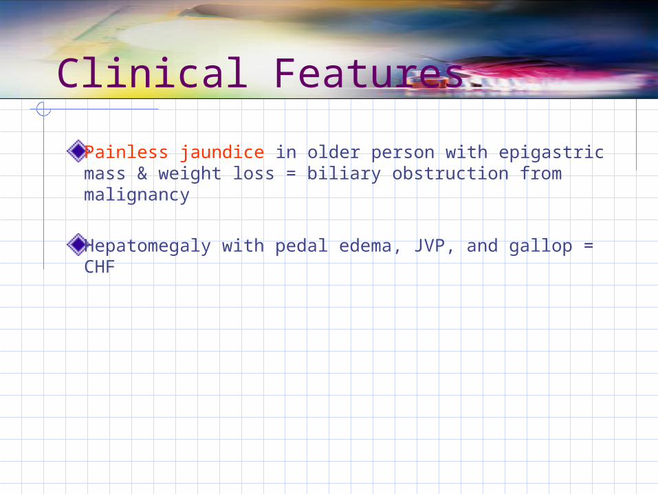
Clinical Features
Painless jaundice in older person with epigastric mass & weight loss = biliary obstruction from malignancy
Hepatomegaly with pedal edema, JVP, and gallop = CHF
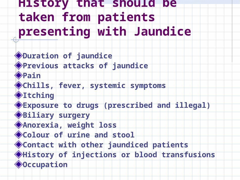
Duration of jaundicePrevious attacks of jaundicePainChills, fever, systemic symptomsItchingExposure to drugs (prescribed and illegal)Biliary surgeryAnorexia, weight lossColour of urine and stoolContact with other jaundiced patientsHistory of injections or blood transfusionsOccupation
History that should be taken from patients presenting with Jaundice
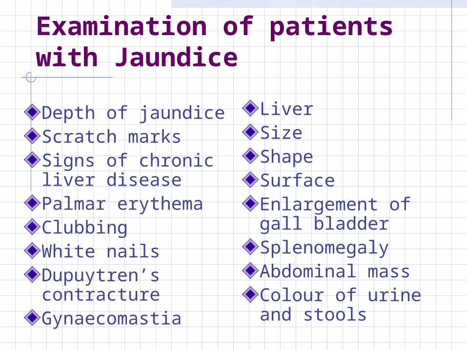
Examination of patients with Jaundice
Depth of jaundiceScratch marksSigns of chronic liver diseasePalmar erythemaClubbingWhite nailsDupuytren’s contractureGynaecomastia
LiverSizeShapeSurfaceEnlargement of gall bladderSplenomegalyAbdominal massColour of urine and stools
Laboratory Tests
Pigment studies Serum bilirubin, direct Serum bilirubin, total Urine R/E for bilirubin and urobilinogen
Alkaline PhosphataseLiver aminotransferrase levels
AST ALT
Elevated levels usually indicate cellular damage to the liver
> 70% of liver cells may be damaged before LFT’s become elevated
Blood Studies
Serum Ammonia Liver converts ammonia to urea. Ammonia
rises in liver failureProtein Studies Serum albumin
Low levels seen with liver disease Serum Globulin
Elevated levels with advanced cirrhosis and chronic active hepatitis
CBC
PT
Other labs pertinent to history
Coombs test
Hb electrophoresis
Viral hepatitis screen
ULTRASOUND
Tumor Marker Alpha-fetoprotein (AFP) Increased levels are seen with hepatic
carcinomaProthrombin Time (PT) Time required for a firm fibrin dot to form In liver dysfunction, increase clotting time
with increased risk of bleeding
Liver Biopsy
Used to obtain a specimen of liver tissueDone under local anesthesia
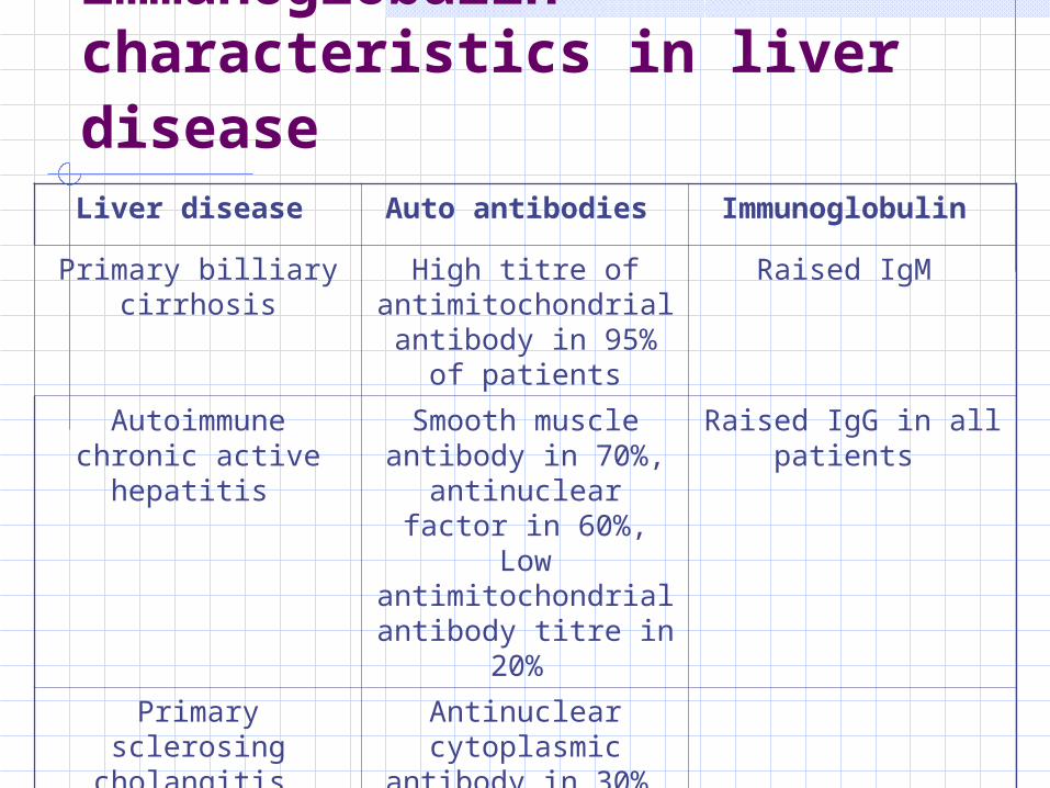
Autoantibody and immunoglobulin characteristics in liver disease Liver disease Auto antibodies Immunoglobulin
Primary billiary cirrhosis
High titre of antimitochondrial antibody in 95% of
patients
Raised IgM
Autoimmune chronic active hepatitis
Smooth muscle antibody in 70%,
antinuclear factor in 60%, Low
antimitochondrial antibody titre in
20%
Raised IgG in all patients
Primary sclerosing cholangitis
Antinuclear cytoplasmic
antibody in 30%
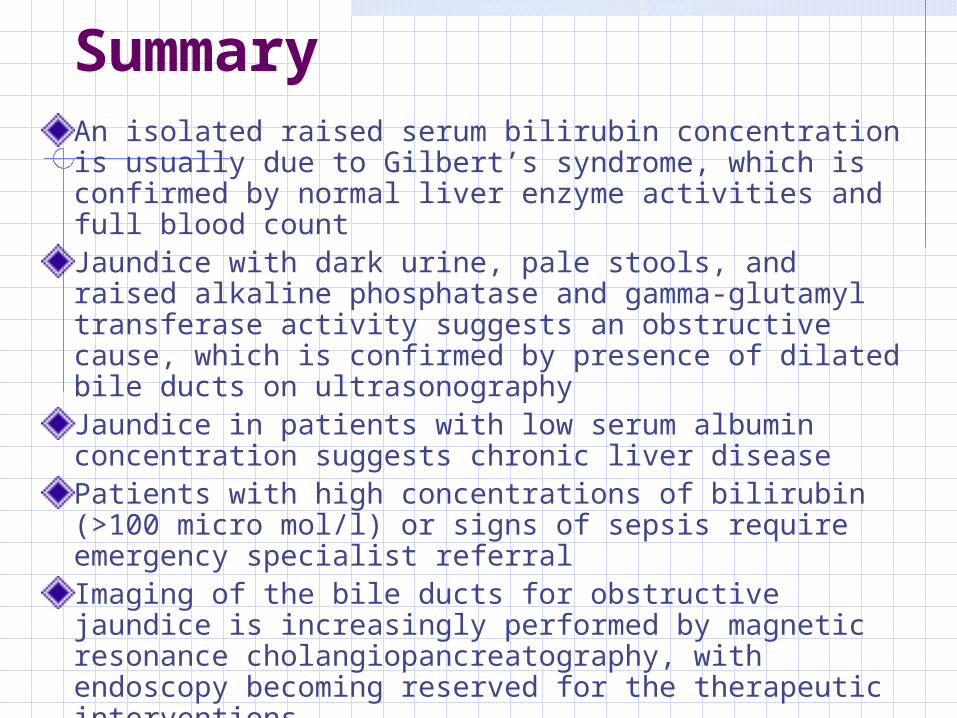
SummaryAn isolated raised serum bilirubin concentration is usually due to Gilbert’s syndrome, which is confirmed by normal liver enzyme activities and full blood countJaundice with dark urine, pale stools, and raised alkaline phosphatase and gamma-glutamyl transferase activity suggests an obstructive cause, which is confirmed by presence of dilated bile ducts on ultrasonographyJaundice in patients with low serum albumin concentration suggests chronic liver diseasePatients with high concentrations of bilirubin (>100 micro mol/l) or signs of sepsis require emergency specialist referralImaging of the bile ducts for obstructive jaundice is increasingly performed by magnetic resonance cholangiopancreatography, with endoscopy becoming reserved for the therapeutic interventions

Pre-Hepatic Disorders
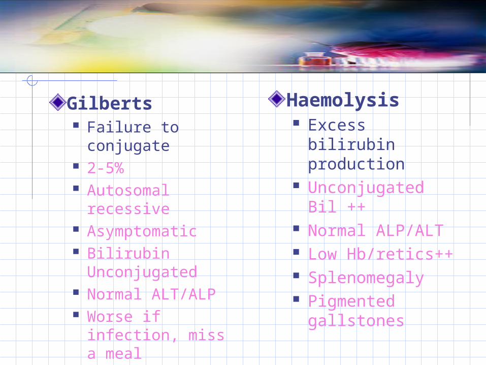
Gilberts Failure to
conjugate 2-5% Autosomal
recessive Asymptomatic Bilirubin
Unconjugated Normal ALT/ALP Worse if infection,
miss a meal
Haemolysis Excess bilirubin
production Unconjugated Bil
++ Normal ALP/ALT Low Hb/retics++ Splenomegaly Pigmented
gallstones

Hepatic Disorders & Hepatic Failure
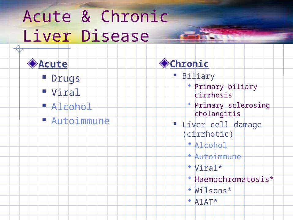
Acute & Chronic Liver Disease
Acute Drugs Viral Alcohol Autoimmune
Chronic Biliary
Primary biliary cirrhosis
Primary sclerosing cholangitis
Liver cell damage (cirrhotic) Alcohol Autoimmune Viral* Haemochromatosis* Wilsons* A1AT*
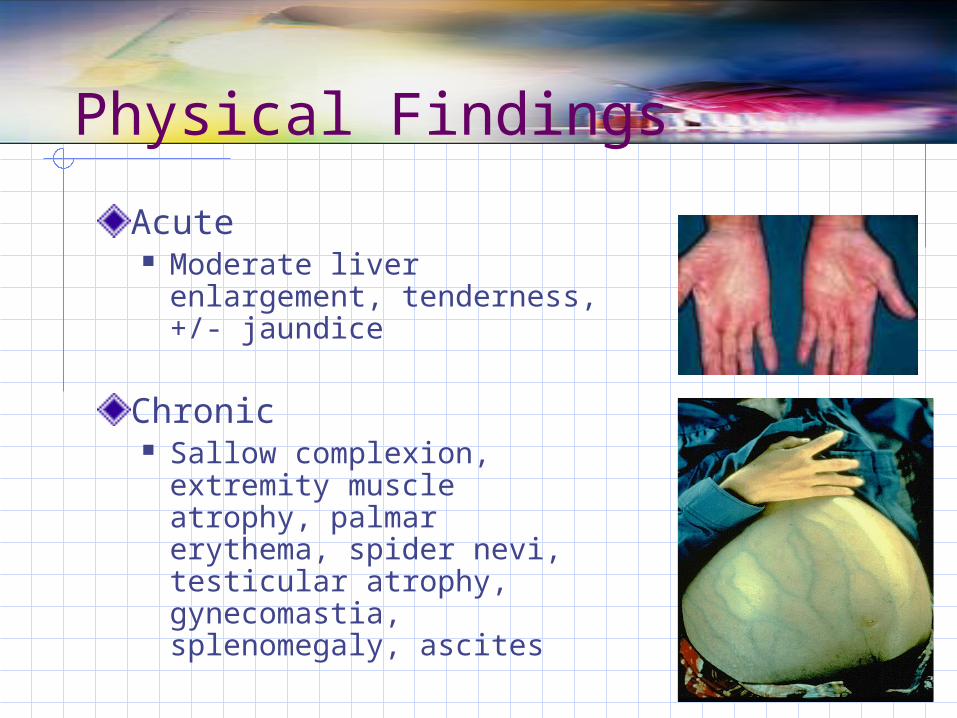
Physical Findings
Acute Moderate liver
enlargement, tenderness, +/- jaundice
Chronic Sallow complexion,
extremity muscle atrophy, palmar erythema, spider nevi, testicular atrophy, gynecomastia, splenomegaly, ascites
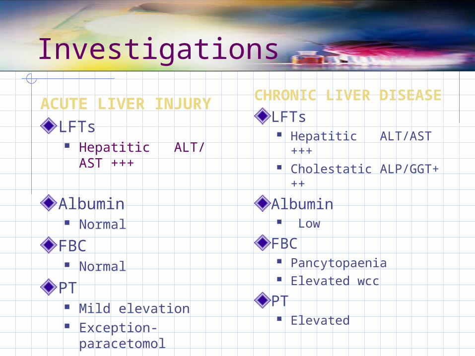
Investigations
ACUTE LIVER INJURYLFTs
Hepatitic ALT/AST +++
Albumin Normal
FBC Normal
PT Mild elevation Exception-
paracetomol
CHRONIC LIVER DISEASELFTs
Hepatitic ALT/AST +++
Cholestatic ALP/GGT+++
Albumin Low
FBC Pancytopaenia Elevated wcc
PT Elevated
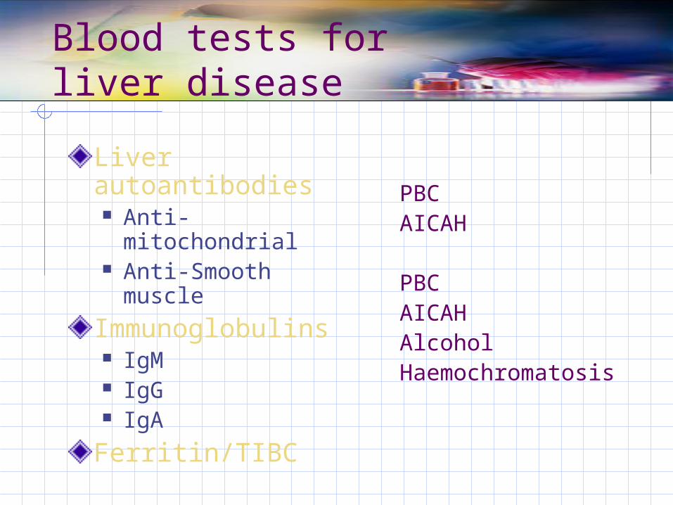
Blood tests for liver disease
Liver autoantibodies Anti-mitochondrial Anti-Smooth
muscle
Immunoglobulins IgM IgG IgA
Ferritin/TIBC
PBCAICAH
PBCAICAHAlcoholHaemochromatosis
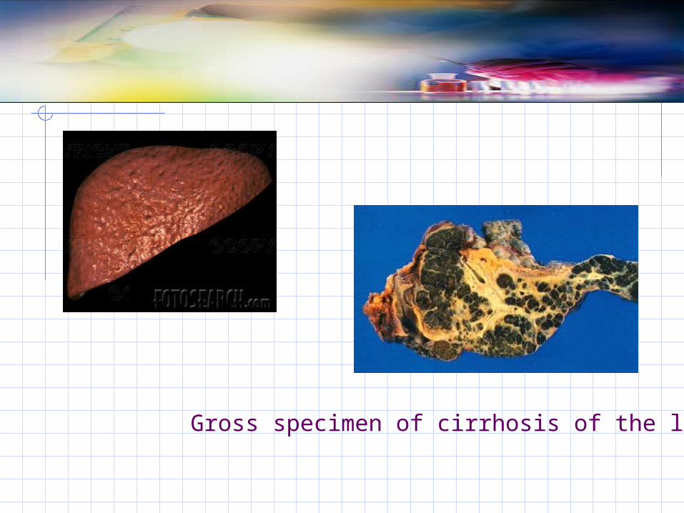
Gross specimen of cirrhosis of the liver
Post-Hepatic Disorders
Intrinsic to the ductal system Gallstones Surgical strictures Infection (cytomegalovirus,
Cryptosporidium infection in patients with acquired immunodeficiency syndrome)
Intrahepatic malignancy Cholangiocarcinoma
Extrinsic to the ductal system Extrahepatic malignancy (pancreas,
lymphoma) Pancreatitis
CASE SCENARIO
A 54 years old female is presented in emergency department with complaints of low grade fever, nausea and loss of appetite for last 10 days. now she is worried because of yellow discoloration of sclera and dark colored urine for one day.
What physical signs you can suspect in this case?

, CASE SCENARIO
On examination, she has fever of 100-F.she is jaundiced and having tender hepatomegaly.
How will you investigate this case ?
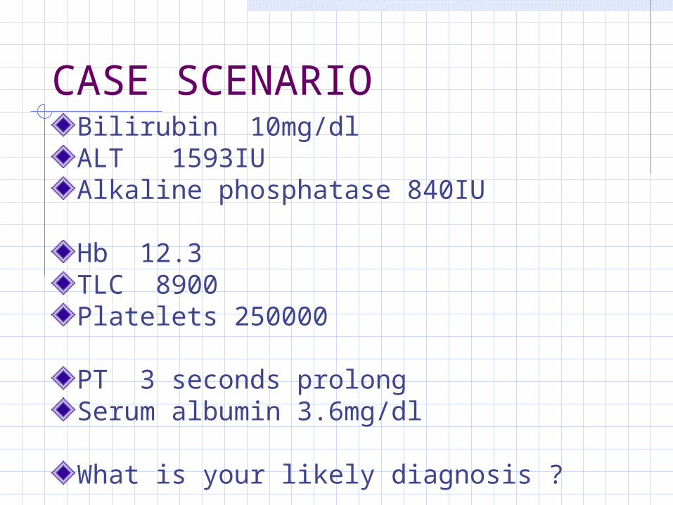
CASE SCENARIOBilirubin 10mg/dlALT 1593IUAlkaline phosphatase 840IU
Hb 12.3TLC 8900Platelets 250000
PT 3 seconds prolongSerum albumin 3.6mg/dl
What is your likely diagnosis ?

CASE SCENARIO
Hepatitis AHepatitis BHepatitis CHepatitis D

CASE SCENARIO
How will you differentiate hepatitis A and hepatitis E infection ?
How will you manage this case ?
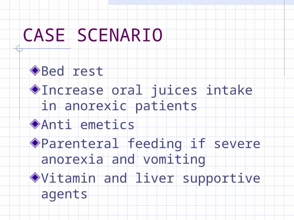
CASE SCENARIO
Bed restIncrease oral juices intake in anorexic patientsAnti emeticsParenteral feeding if severe anorexia and vomitingVitamin and liver supportive agents