Jason membrane collprotect membrane - Straumann · Jason® membrane collprotect ... 3 Frank...
24
1 Jason ® membrane collprotect ® membrane Natural collagen membranes for GBR/GTR Scientific and clinical evidence by PD Dr. Dr. Daniel Rothamel et al. soft tissue native reliable resorbable botiss biomaterials dental bone & tissue regeneration
Transcript of Jason membrane collprotect membrane - Straumann · Jason® membrane collprotect ... 3 Frank...
1
Jason® membranecollprotect® membraneNatural collagen membranes for GBR/GTR
Scientific and clinical evidence
by PD Dr. Dr. Daniel Rothamel et al.
soft tissue
native
reliable
resorbable
botissbiomaterials
dental
bone & tissue regeneration
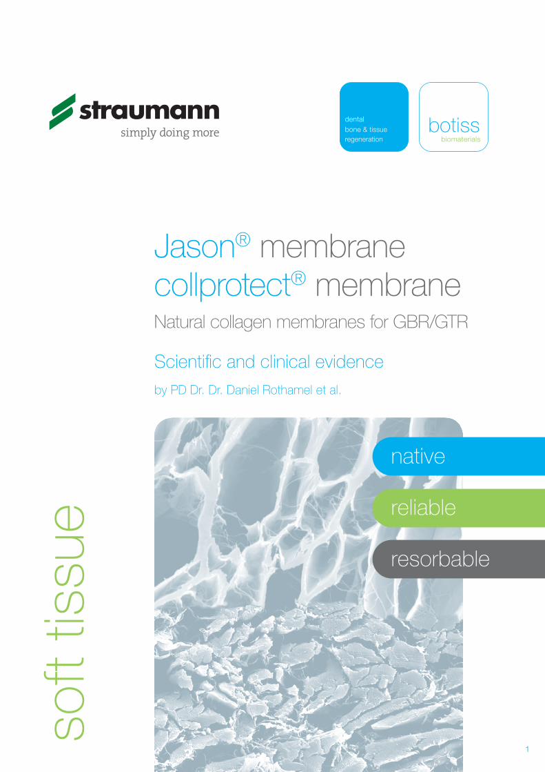
2
cerabone®
Natural bovine bone graft
maxresorb®
inject
maxgraft®
bonering
maxgraft®
bonebuilder
Patient matched allogenic bone implant
maxgraft®
Processed allogenicbone graft
Synthetic injectable bone paste
maxresorb®
Synthetic biphasiccalcium phosphate
Processed allogenic bone ring
maxresorb®
flexbone*
Flexible block (CaP / Collagen composite)
Straumann®
Emdogain®
Enamel matrix derivative
collacone®
max*
Cone(CaP / Collagen composite)
mucoderm®
3D-stable soft tissue (Collagen) graft
Jason®
membrane
Native pericardium GBR / GTR membrane
collprotect®
membrane
Native collagen membrane
Jason® fleece /
collacone®......
Collagenic haemostypt (Sponge / Cone)
* Coming soon
High Quality Learning
Activation
Flexib
ility
High Quality Learning
Activation
AcFlexibility
Flexibility
Controlled Degradation
Biological Potential
mucoderm®
collprotect® membrane
Jason® membrane
Jason® fleececollacone®......
hard tissue
cerabone®
Straumann® BoneCeramic™
maxresorb®
inject
maxgraft® boneringmaxgraft® bonebuilder
maxgraft®
EDUCATION
SCIENCE CLINIC
6 - 9
months6 - 9
months
6
months
4 - 6
months
4 - 6
months
3 - 4
months
6 - 9
months3 - 4
months
2 - 4
weeks
Regeneration
Augmentation
Preservation
Healing
IntegrationIntegration
Barrier
Resorption
2 - 3
months
3 - 6
months
bovi
ne
synth
etic
hum
an
nativ
e c
olla
gen
collacone®..max*
soft tissue
botiss regeneration system
synthetic + native collagen
botiss academy bone & tissue days
6 - 12
months
Regeneration
enamel matrix derivative
Straumann® Emdogain®maxresorb® flexbone*
Straumann®
BoneCeramic™
Synthetic biphasiccalcium phosphate
maxresorb®
3
PD Dr. Dr. Daniel Rothamel
PD Dr. med. Dr. med. dent.Daniel Rothamel
Department of Oral and Maxillofacial Plastic Surgery,
University of Cologne, Germany
.................................................
- Since 2010 Assistant Professor at the Department of Oral
and Maxillofacial Plastic Surgery (Prof. Dr. Dr. J. Zöller),
University of Cologne, Germany
- 2009, habilitation (post-doctoral lecturing qualification),
University of Cologne
Thesis: “Reconstruction of defects of the alveolar ridge using
artificial and autogenous bone blocks and growth factors”
- 2008, doctorate in human medicine (Dr. med.),
Heinrich-Heine University of Düsseldorf
Thesis: “Biocompatibility, biodegradation and angiogenetic
aspects of native and cross-linked collagen membranes”
- Since 2007 specialist in Oral Surgery
- 2004, doctorate in dental medicine (Dr. med. dent.),
Heinrich-Heine University of Düsseldorf, Germany
Thesis: „Establishing a new method for quantification of tooth
hypersensitivity“
Already during his medicine studies, PD Dr. Dr. Daniel Rothamel was
focused on scientific subjects in the field of bone regeneration and
implantology. He has published more than 80 articles, many of them
in renowned international scientific journals. He acts as a reviewer
for several journals and frequently participates as a lecturer on con-
gresses and training courses in Germany as well as other countries.
His research and lecture activities are focused on subjects such
as Guided Bone Regeneration (GBR), socket preservation, implant
surfaces, collagen membranes, bone substitute materials, growth
factors, face trauma, cancer rehabilitation and hemostyptics.
4
Collagen –a multifaceted protein
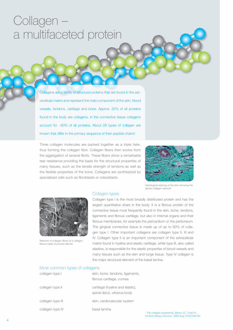
Collagen types
Collagen type I is the most broadly distributed protein and has the
largest quantitative share in the body. It is a fibrous protein of the
connective tissue most frequently found in the skin, bone, tendons,
ligaments and fibrous cartilage, but also in internal organs and their
fibrous membranes, for example the pericardium or the peritoneum.
The gingival connective tissue is made up of up to 60% of colla-
gen type I. Other important collagens are collagen type II, III and
IV. Collagen type II is an important component of the extracellular
matrix found in hyaline and elastic cartilage, while type III, also called
elastine, is responsible for the elastic properties of blood vessels and
many tissues such as the skin and lunge tissue. Type IV collagen is
the major structural element of the basal lamina.
Histological staining of the skin showing the dense collagen network
Network of collagen fibers of a collagen fleece made of porcine dermis
Collagens are a family of structural proteins that are found in the ext-
racellular matrix and represent the main component of the skin, blood
vessels, tendons, cartilage and bone. Approx. 25% of all proteins
found in the body are collagens, in the connective tissue collagens
account for ~80% of all proteins. About 28 types of collagen are
known that differ in the primary sequence of their peptide chains1.
Three collagen molecules are packed together as a triple helix,
thus forming the collagen fibril. Collagen fibers then evolve from
the aggregation of several fibrils. These fibers show a remarkable
tear resistance providing the basis for the structural properties of
many tissues, such as the tensile strength of tendons as well as
the flexible properties of the bone. Collagens are synthesized by
specialized cells such as fibroblasts or osteoblasts.
Most common types of collagens
collagen type I skin, bone, tendons, ligaments,
fibrous cartilage, cornea
collagen type II cartilage (hyaline and elastic),
spinal discs, vitreous body
collagen type III skin, cardiovascular system
collagen type IV basal lamina1 The collagen superfamily. Brown JC, Timpl R., Int Arch Allergy Immunol. 1995 Aug;107(4):484-90.
5
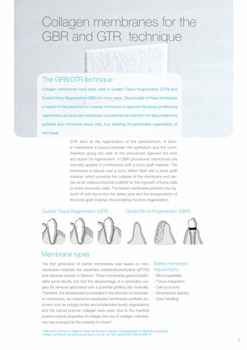
The GRB/GTR technique
Collagen membranes for the GBR and GTR technique
Collagen membranes have been used in Guided Tissue Regeneration (GTR) and
Guided Bone Regeneration (GBR) for many years. The principle of these techniques
is based on the placement of a barrier membrane to separate the slowly proliferating
regenerative cell types like osteoblasts and periodontal cells from the fast proliferating
epithelial and connective tissue cells, thus enabling the predictable regeneration of
lost tissue.
2 Rothamel D, Schwarz F, Sager M, Herten M, Sculean A, Becker J. Biodegradation of differently cross-linked collagen membranes: an experimental study in the rat. Clin Oral Implants Res 2005;16:369-78
GTR aims at the regeneration of the periodontium. A barri-
er membrane is placed between the epithelium and the tooth,
therefore giving the cells of the periodontal ligament the time
and space for regeneration. In GBR procedures membranes are
normally applied in combination with a bone graft material. The
membrane is placed over a bony defect filled with a bone graft
material, which prevents the collapse of the membrane and ser-
ves as an osteoconductive scaffold for the ingrowth of bone cells
(or bone precursor cells). The barrier membrane prevents the ing-
rowth of soft tissue into the defect area and the encapsulation of
the bone graft material, thus enabling the bony regeneration.
Guided Tissue Regeneration (GTR) Guided Bone Regeneration (GBR)
Membrane types
Barrier membrane
requirements
- Biocompatibility
- Tissue integration
- Cell occlusivity
- Dimensional stability
- Easy handling
The first generation of barrier membranes was based on non-
resorbable materials like expanded polytetrafluorethylene (ePTFE)
and cellulose acetate or titanium. These membranes gained predic-
table good results, but had the disadvantage of a secondary sur-
gery for removal associated with a potential grafting site morbidity.
Therefore, the development proceeded in the direction of resorbab-
le membranes. As material for resorbable membranes synthetic po-
lymers such as polyglycolides and polylactides (acidic degradation)
and the natural polymer collagen were used. Due to the manifold
positive natural properties of collagen the use of collagen membra-
nes has emerged as the material of choice2.
6
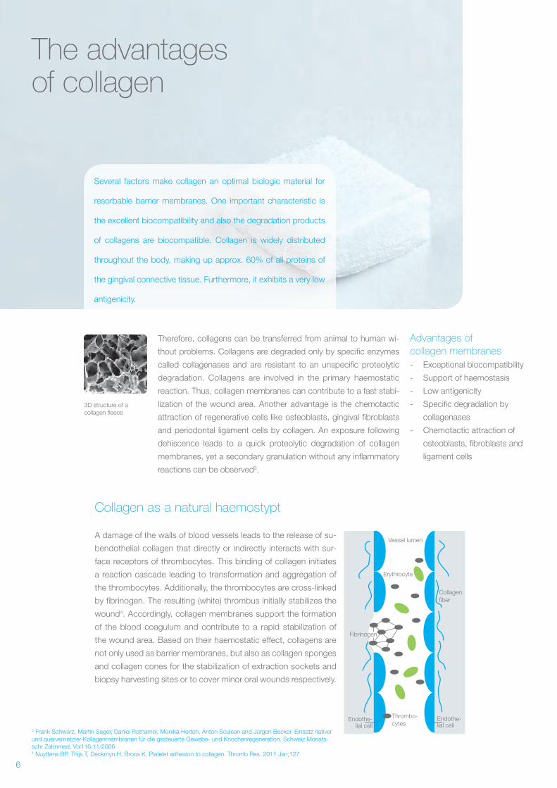
The advantages of collagen
Advantages of
collagen membranes
- Exceptional biocompatibility
- Support of haemostasis
- Low antigenicity
- Specific degradation by
collagenases
- Chemotactic attraction of
osteoblasts, fibroblasts and
ligament cells
Collagen as a natural haemostypt
A damage of the walls of blood vessels leads to the release of su-
bendothelial collagen that directly or indirectly interacts with sur-
face receptors of thrombocytes. This binding of collagen initiates
a reaction cascade leading to transformation and aggregation of
the thrombocytes. Additionally, the thrombocytes are cross- linked
by fibrinogen. The resulting (white) thrombus initially stabilizes the
wound4. Accordingly, collagen membranes support the formation
of the blood coagulum and contribute to a rapid stabilization of
the wound area. Based on their haemostatic effect, collagens are
not only used as barrier membranes, but also as collagen sponges
and collagen cones for the stabilization of extraction sockets and
biopsy harvesting sites or to cover minor oral wounds respectively.
Several factors make collagen an optimal biologic material for
resorbable barrier membranes. One important characteristic is
the excellent biocompatibility and also the degradation products
of collagens are biocompatible. Collagen is widely distributed
throughout the body, making up approx. 60% of all proteins of
the gingival connective tissue. Furthermore, it exhibits a very low
antigenicity.
Therefore, collagens can be transferred from animal to human wi-
thout problems. Collagens are degraded only by specific enzymes
called collagenases and are resistant to an unspecific proteolytic
degradation. Collagens are involved in the primary haemostatic
reaction. Thus, collagen membranes can contribute to a fast stabi-
lization of the wound area. Another advantage is the chemotactic
attraction of regenerative cells like osteoblasts, gingival fibroblasts
and periodontal ligament cells by collagen. An exposure following
dehiscence leads to a quick proteolytic degradation of collagen
membranes, yet a secondary granulation without any inflammatory
reactions can be observed3.
Vessel lumen
Endothe-lial cell
Endothe-lial cell
Collagen fiber
Fibrinogen
Thrombo-cytes
Erythrocyte
3 Frank Schwarz, Martin Sager, Daniel Rothamel, Monika Herten, Anton Sculean and Jürgen Becker Einsatz nativer und quervernetzter Kollagenmembranen für die gesteuerte Gewebe- und Knochenregeneration. Schweiz Monats-schr Zahnmed, Vol116:11/20064 Nuyttens BP, Thijs T, Deckmyn H, Broos K. Platelet adhesion to collagen. Thromb Res. 2011 Jan;127
3D structure of a collagen fleece
7
Origin of collagen membranes
The first collagen membranes available on the market were of
bovine origin (Achilles tendon and pericardium). Nowadays, por-
cine membranes are more widely used because their usage ex-
cludes the risk of a BSE transmission. Moreover, porcine collagen
exhibits a high homology to human collagen and therefore a very
good biocompatibility. Due to these reasons botiss membranes are
produced from porcine collagen.
Jason® membrane is very thin, but exhibits an excellent multidirectional tear resistance
Histology of a big blood vessel and some smaller ones
Collagen membranes can originate from various tissues ranging from
dermis, to peritoneum or pericardium. Accordingly, these membra-
nes differ in their handling and degradation properties and the resul-
ting barrier function.
Properties of barrier membranes – vascularization versus barrier function
The disadvantage of most collagen membranes, other than the bo-
tiss membranes, lays in their rapid enzymatic degradation by collage-
nases, resulting in a limited stability and correspondingly short barrier
function. A possibility to influence the barrier function is to choose a
specific original tissue to impart the membranes with a better stability.
In that way membranes made of pericardium, such as the Jason®
membrane, due to a structural speciality, exhibit a slowed degradation
and thus offer a prolonged barrier function. Furthermore, pericardium
membranes can be distinguished by an extraordinarily high tear
resistance and excellent handling properties (e.g. good adaptation to
surface contours, no sticking).
5 Daniel Rothamel, Roland Torök, Jörg Neugebauer, Tim Fienitz, Martin Scheer, Matthias Kreppel, Robert Mischkowski and Joachim E. Zöller. Clinical aspects of novel types of collagen membranes and matrices: Current issues in soft- and hard- tissue augmentation. EDI Journal 1rst Issue 2012
The barrier function can also be extended by the use of membranes
with a very dense collagen structure, but this dense structure might
oppose the early angiogenesis of the grafting site. The ingrowth of
blood vessels into the augmentation area is important not only for
the nutrition of the grafting site, but also because the surrounding
connective tissue of small capillaries contains undifferentiated proge-
nitor cells (pericytes). These cells can evolve into osteoblasts that are
responsible for new bone formation. Therefore, the selective perme-
ability of membranes for blood vessels is desirable5.
One example of such a semi- permeable membrane is the collpro-
tect® membrane. This membrane possesses loosely structured areas
(pores) punctuating the compact collagen matrix and supporting a
fast vascularization.
............................................................................
8
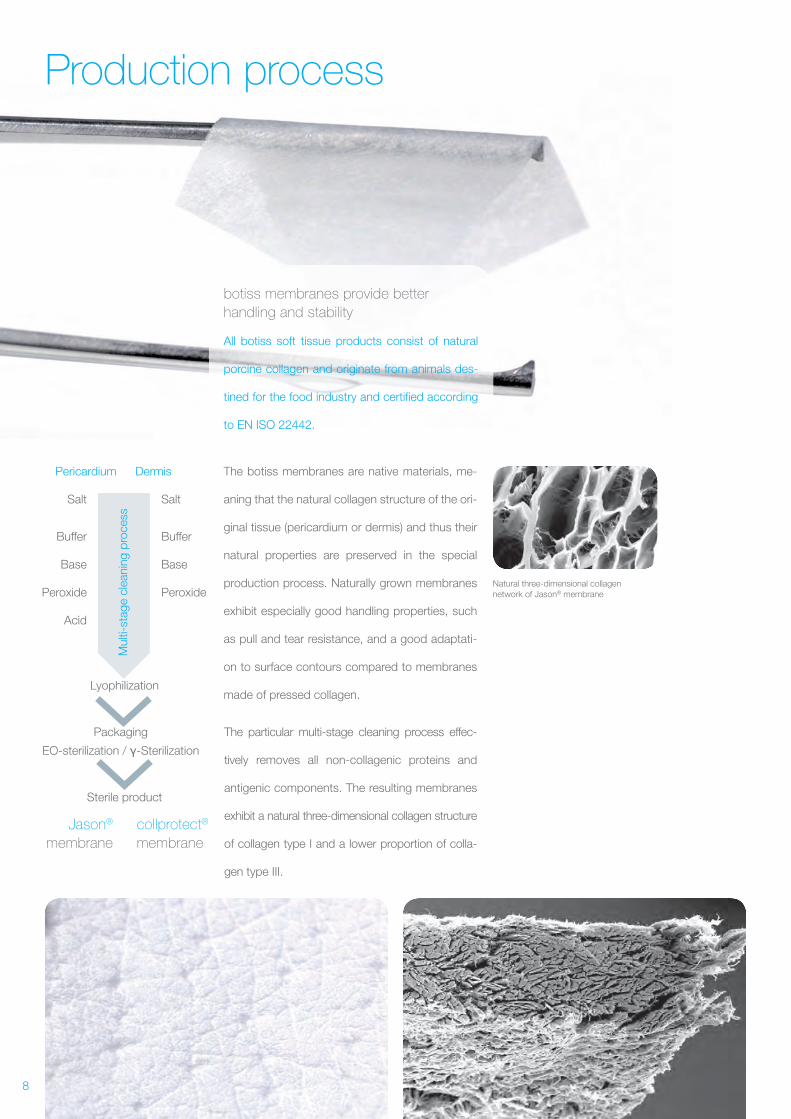
Production process
botiss membranes provide better
handling and stability
All botiss soft tissue products consist of natural
porcine collagen and originate from animals des-
tined for the food industry and certified according
to EN ISO 22442.
The botiss membranes are native materials, me-
aning that the natural collagen structure of the ori-
ginal tissue (pericardium or dermis) and thus their
natural properties are preserved in the special
production process. Naturally grown membranes
exhibit especially good handling properties, such
as pull and tear resistance, and a good adaptati-
on to surface contours compared to membranes
made of pressed collagen.
Pericardium
Salt Salt
Buffer Buffer
Base Base
Peroxide Peroxide
Acid
Lyophilization
Packaging
EO-sterilization / -Sterilization
Sterile product
Dermis
Jason®
membrane
collprotect®
membrane
Mul
ti-st
age
clea
ning
pro
cess
Natural three-dimensional collagen network of Jason® membrane
The particular multi-stage cleaning process effec-
tively removes all non- collagenic proteins and
antigenic components. The resulting membranes
exhibit a natural three-dimensional collagen structure
of collagen type I and a lower proportion of colla-
gen type III.
9
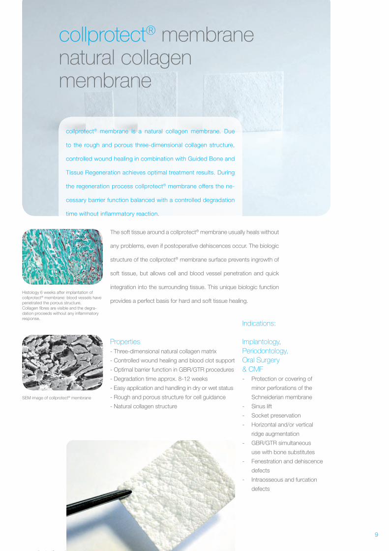
collprotect® membranenatural collagenmembrane
collprotect® membrane is a natural collagen membrane. Due
to the rough and porous three-dimensional collagen structure,
controlled wound healing in combination with Guided Bone and
Tissue Regeneration achieves optimal treatment results. During
the regeneration process collprotect® membrane offers the ne-
cessary barrier function balanced with a controlled degradation
time without inflammatory reaction.
Histology 6 weeks after implantation of collprotect® membrane: blood vessels havepenetrated the porous structure.Collagen fibres are visible and the degra-dation proceeds without any inflammatory response.
SEM image of collprotect® membrane
collprotect® membrane –
The soft tissue around a collprotect® membrane usually heals without
any problems, even if postoperative dehiscences occur. The biologic
structure of the collprotect® membrane surface prevents ingrowth of
soft tissue, but allows cell and blood vessel penetration and quick
integration into the surrounding tissue. This unique biologic function
provides a perfect basis for hard and soft tissue healing.
Properties
- Three-dimensional natural collagen matrix
- Controlled wound healing and blood clot support
- Optimal barrier function in GBR/GTR procedures
- Degradation time approx. 8- 12 weeks
- Easy application and handling in dry or wet status
- Rough and porous structure for cell guidance
- Natural collagen structure
Indications:
Implantology,
Periodontology,
Oral Surgery
& CMF
- Protection or covering of
minor perforations of the
Schneiderian membrane
- Sinus lift
- Socket preservation
- Horizontal and/or vertical
ridge augmentation
- GBR/GTR simultaneous
use with bone substitutes
- Fenestration and dehiscence
defects
- Intraosseous and furcation
defects
10
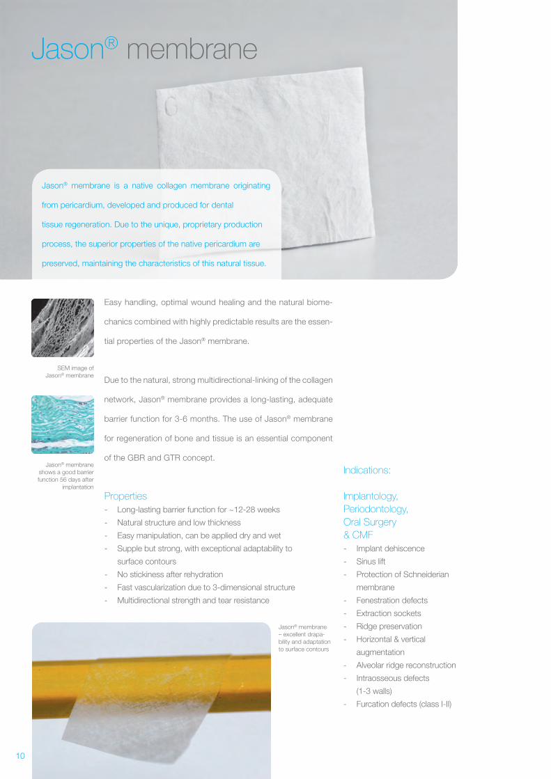
SEM image of Jason® membrane
Jason® membrane shows a good barrier function 56 days after
implantation
Jason® membrane
Jason® membrane is a native collagen membrane originating
from pericardium, developed and produced for dental
tissue regeneration. Due to the unique, proprietary production
process, the superior properties of the native pericardium are
preserved, maintaining the characteristics of this natural tissue.
Easy handling, optimal wound healing and the natural biome-
chanics combined with highly predictable results are the essen-
tial properties of the Jason® membrane.
Due to the natural, strong multidirectional-linking of the collagen
network, Jason® membrane provides a long- lasting, adequate
barrier function for 3- 6 months. The use of Jason® membrane
for regeneration of bone and tissue is an essential component
of the GBR and GTR concept.
Properties
- Long- lasting barrier function for ~12- 28 weeks
- Natural structure and low thickness
- Easy manipulation, can be applied dry and wet
- Supple but strong, with exceptional adaptability to
surface contours
- No stickiness after rehydration
- Fast vascularization due to 3- dimensional structure
- Multidirectional strength and tear resistance
Indications:
Implantology,
Periodontology,
Oral Surgery
& CMF
- Implant dehiscence
- Sinus lift
- Protection of Schneiderian
membrane
- Fenestration defects
- Extraction sockets
- Ridge preservation
- Horizontal & vertical
augmentation
- Alveolar ridge reconstruction
- Intraosseous defects
(1- 3 walls)
- Furcation defects (class I- II)
Jason® membrane– excellent drapa-bility and adaptation to surface contours
11
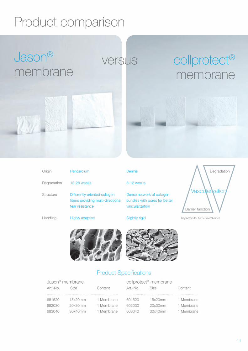
Product comparison
Jason® membrane
collprotect® membrane
versus
Origin Pericardium Dermis
Degradation 12- 28 weeks 8- 12 weeks
Structure Differently oriented collagen Dense network of collagen
fibers providing multi-directional bundles with pores for better
tear resistance vascularization
Handling Highly adaptive Slightly rigid
Product Specifications
Jason® membrane Art.-No. Size Content
....................................................................
681520 15x20mm 1 Membrane
682030 20x30mm 1 Membrane
683040 30x40mm 1 Membrane
collprotect® membrane Art.-No. Size Content
....................................................................
601520 15x20mm 1 Membrane
602030 20x30mm 1 Membrane
603040 30x40mm 1 Membrane
Barrier function
Degradation
Vascularization
Keyfactors for barrier membranes
12
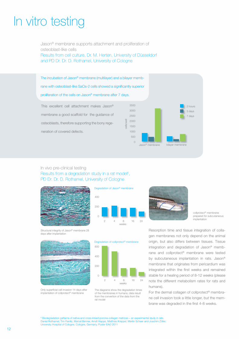
In vitro testing
The incubation of Jason® membrane (multilayer) and a bilayer memb-
rane with osteoblast- like SaOs-2 cells showed a significantly superior
proliferation of the cells on Jason® membrane after 7 days.
This excellent cell attachment makes Jason®
membrane a good scaffold for the guidance of
osteoblasts, therefore supporting the bony rege-
neration of covered defects.
Resorption time and tissue integration of colla-
gen membranes not only depend on the animal
origin, but also differs between tissues. Tissue
integration and degradation of Jason® memb-
rane and collprotect® membrane were tested
by subcutaneous implantation in rats. Jason®
membrane that originates from pericardium was
integrated within the first weeks and remained
stable for a healing period of 8- 12 weeks (please
note the different metabolism rates for rats and
humans).
For the dermal collagen of collprotect® membra-
ne cell invasion took a little longer, but the mem-
brane was degraded in the first 4- 8 weeks.
6 Biodegradation patterns of native and cross- linked porcine collagen matrices – an experimental study in rats. Daniel Rothamel, Tim Fienitz, Marcel Benner, Arndt Happe, Matthias Kreppel, Martin Scheer and Joachim Zöller, University Hospital of Cologne, Cologne, Germany, Poster EAO 2011
Jason® membrane supports attachment and proliferation of
osteoblast- like cells
Results from cell culture, Dr. M. Herten, University of Düsseldorf
and PD Dr. Dr. D. Rothamel, University of Cologne
In vivo pre- clinical testing
Results from a degradation study in a rat model6,
PD Dr. Dr. D. Rothamel, University of Cologne
Only superficial cell invasion 14 days after implantation of collprotect® membrane
collprotect® membrane prepared for subcutaneous implantation
Structural integrity of Jason® membrane 28 days after implantation
The diagrams show the degradation times of the membranes in humans, data result from the convertion of the data from the rat model
3500
3000
2500
2000
1500
1000
500
0Jason® membrane bilayer membrane®® bilayer mr membr membraneemb
2 hours
3 days
7 days
cells
/wel
l
400
200
0
2 4 8 2416weeks
600
400
200
0
2 4 8 2416weeks
Degradation of Jason® membrane
Degradation of collprotect® membrane
13
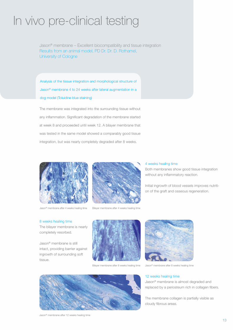
In vivo pre- clinical testing
Analysis of the tissue integration and morphological structure of
Jason® membrane 4 to 24 weeks after lateral augmentation in a
dog model (Toluidine blue staining)
The membrane was integrated into the surrounding tissue without
any inflammation. Significant degradation of the membrane started
at week 8 and proceeded until week 12. A bilayer membrane that
was tested in the same model showed a comparably good tissue
integration, but was nearly completely degraded after 8 weeks.
Jason® membrane – Excellent biocompatibility and tissue integration
Results from an animal model, PD Dr. Dr. D. Rothamel,
University of Cologne
Jason® membrane after 4 weeks healing time Bilayer membrane after 4 weeks healing time
Bilayer membrane after 8 weeks healing time Jason® membrane after 8 weeks healing time
Jason® membrane after 12 weeks healing time
4 weeks healing time
Both membranes show good tissue integration
without any inflammatory reaction.
Initial ingrowth of blood vessels improves nutriti-
on of the graft and osseous regeneration.
12 weeks healing time
Jason® membrane is almost degraded and
replaced by a periosteum rich in collagen fibers.
The membrane collagen is partially visible as
cloudy fibrous areas.
8 weeks healing time
The bilayer membrane is nearly
completely resorbed.
Jason® membrane is still
intact, providing barrier against
ingrowth of surrounding soft
tissue.
14
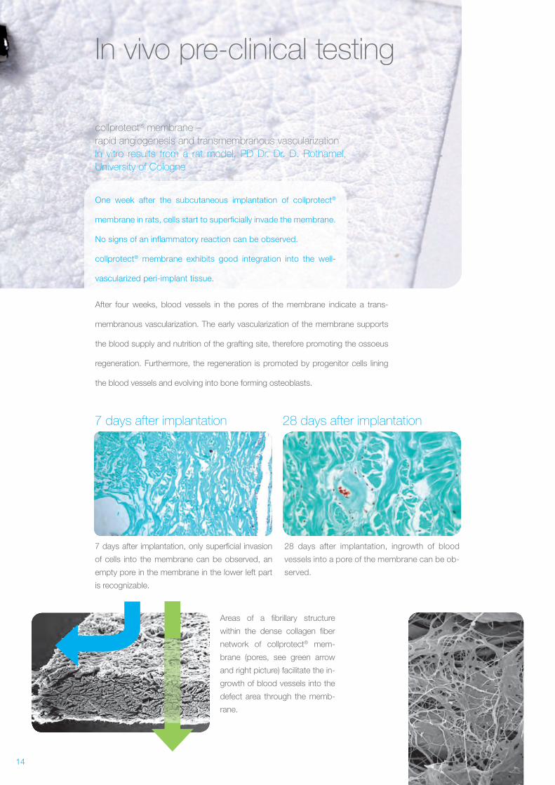
In vivo pre- clinical testing
One week after the subcutaneous implantation of collprotect®
membrane in rats, cells start to superficially invade the membrane.
No signs of an inflammatory reaction can be observed.
collprotect® membrane exhibits good integration into the well-
vascularized peri- implant tissue.
28 days after implantation7 days after implantation
After four weeks, blood vessels in the pores of the membrane indicate a trans-
membranous vascularization. The early vascularization of the membrane supports
the blood supply and nutrition of the grafting site, therefore promoting the ossoeus
regeneration. Furthermore, the regeneration is promoted by progenitor cells lining
the blood vessels and evolving into bone forming osteoblasts.
collprotect® membrane –
rapid angiogenesis and transmembranous vascularization
In vitro results from a rat model, PD Dr. Dr. D. Rothamel,
University of Cologne
7 days after implantation, only superficial invasion
of cells into the membrane can be observed, an
empty pore in the membrane in the lower left part
is recognizable.
Areas of a fibrillary structure
within the dense collagen fiber
network of collprotect® mem-
brane (pores, see green arrow
and right picture) facilitate the in-
growth of blood vessels into the
defect area through the memb-
rane.
28 days after implantation, ingrowth of blood
vessels into a pore of the membrane can be ob-
served.
15
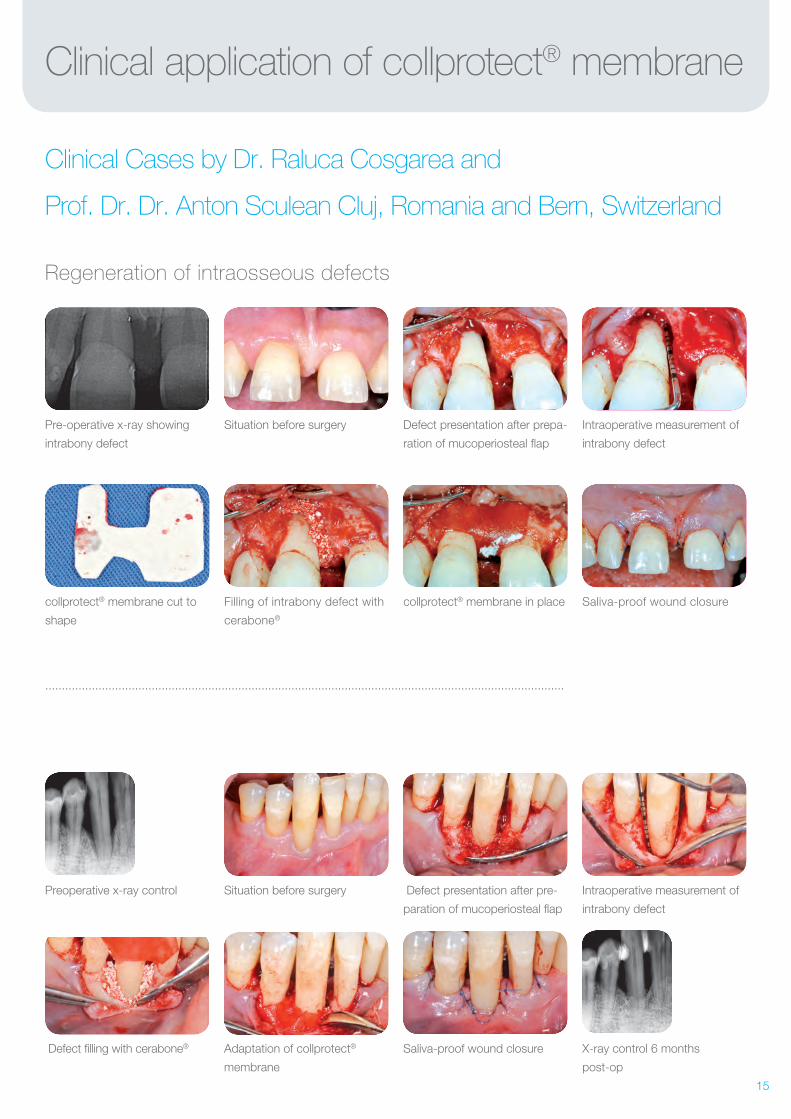
collprotect® membrane in placeFilling of intrabony defect with
cerabone®
collprotect® membrane cut to
shape
Saliva-proof wound closure
Situation before surgeryPre-operative x-ray showing
intrabony defect
Defect presentation after prepa-
ration of mucoperiosteal flap
Intraoperative measurement of
intrabony defect
Clinical application of collprotect® membrane
Clinical Cases by Dr. Raluca Cosgarea and
Prof. Dr. Dr. Anton Sculean Cluj, Romania and Bern, Switzerland
Regeneration of intraosseous defects
............................................................................................................................................................
Preoperative x-ray control
X-ray control 6 months
post-op
Saliva-proof wound closureAdaptation of collprotect®
membrane
Situation before surgery Defect presentation after pre-
paration of mucoperiosteal flap
Intraoperative measurement of
intrabony defect
Defect filling with cerabone®
16
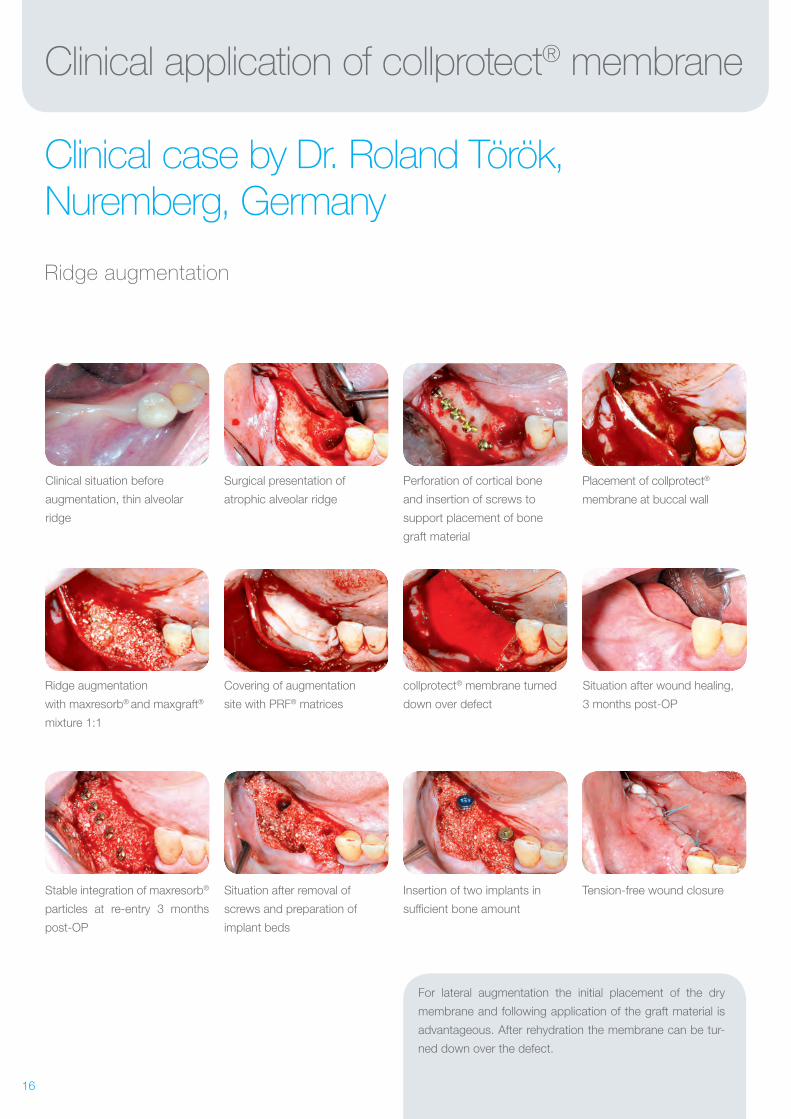
Clinical application of collprotect® membrane
Clinical case by Dr. Roland Török, Nuremberg, Germany
Ridge augmentation
Ridge augmentation
with maxresorb® and maxgraft®
mixture 1:1
Stable integration of maxresorb®
particles at re- entry 3 months
post-OP
Covering of augmentation
site with PRF® matrices
Situation after removal of
screws and preparation of
implant beds
Clinical situation before
augmentation, thin alveolar
ridge
Surgical presentation of
atrophic alveolar ridge
Perforation of cortical bone
and insertion of screws to
support placement of bone
graft material
Placement of collprotect®
membrane at buccal wall
collprotect® membrane turned
down over defect
Insertion of two implants in
sufficient bone amount
Tension- free wound closure
Situation after wound healing,
3 months post- OP
For lateral augmentation the initial placement of the dry
membrane and following application of the graft material is
advantageous. After rehydration the membrane can be tur-
ned down over the defect.
17
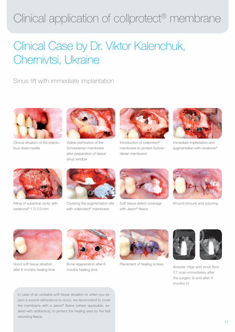
Soft tissue defect coverage
with Jason® fleece
Placement of healing screws
Covering the augmentation site
with collprotect® membrane
Filling of subantral cavity with
cerabone® 1.0- 2.0 mm
Bone regeneration after 6
months healing time
Good soft tissue situation
after 6 months healing time
Wound closure and suturing
Alveolar ridge and sinus floor
CT scan immediately after
the surgery (l) and after 6
months (r)
Visible perforation of the
Schneiderian membrane
after preparation of lateral
sinus window
Clinical situation of the edentu-
lous distal maxilla
Introduction of collprotect®
membrane to protect Schnei-
derian membrane
Immediate implantation and
augmentation with cerabone®
Clinical application of collprotect® membrane
Clinical Case by Dr. Viktor Kalenchuk, Chernivtsi, Ukraine
Sinus lift with immediate implantation
In case of an unstable soft tissue situation or when you ex-
pect a wound dehiscence to occur, we recommend to cover
the membrane with a Jason® fleece (where applicable, so-
aked with antibiotics), to protect the healing area by the fast
resorbing fleece.
18
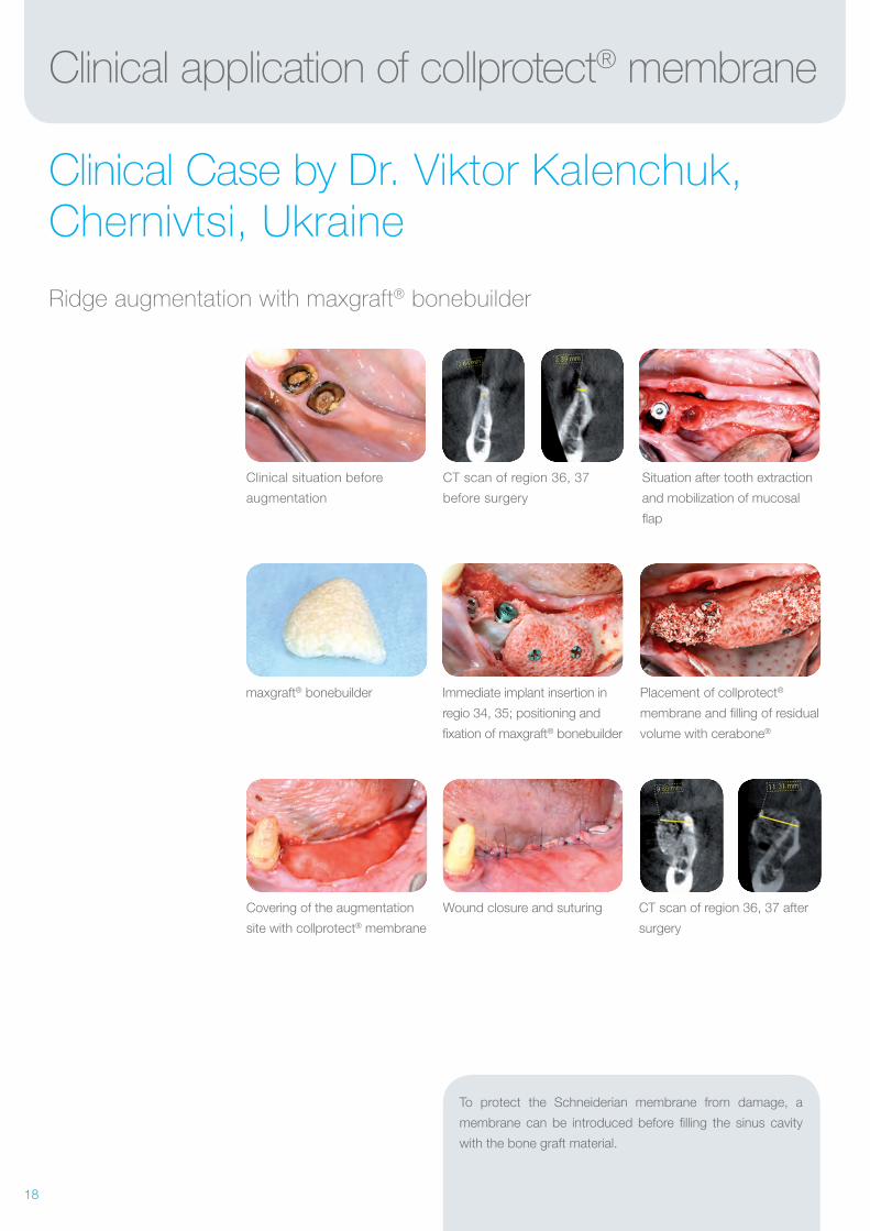
Clinical application of collprotect® membrane
Clinical Case by Dr. Viktor Kalenchuk, Chernivtsi, Ukraine
Ridge augmentation with maxgraft® bonebuilder
maxgraft® bonebuilder
Clinical situation before
augmentation
Covering of the augmentation
site with collprotect® membrane
CT scan of region 36, 37
before surgery
Wound closure and suturing
Immediate implant insertion in
regio 34, 35; positioning and
fixation of maxgraft® bonebuilder
Placement of collprotect®
membrane and filling of residual
volume with cerabone®
Situation after tooth extraction
and mobilization of mucosal
flap
CT scan of region 36, 37 after
surgery
To protect the Schneiderian membrane from damage, a
membrane can be introduced before filling the sinus cavity
with the bone graft material.
19
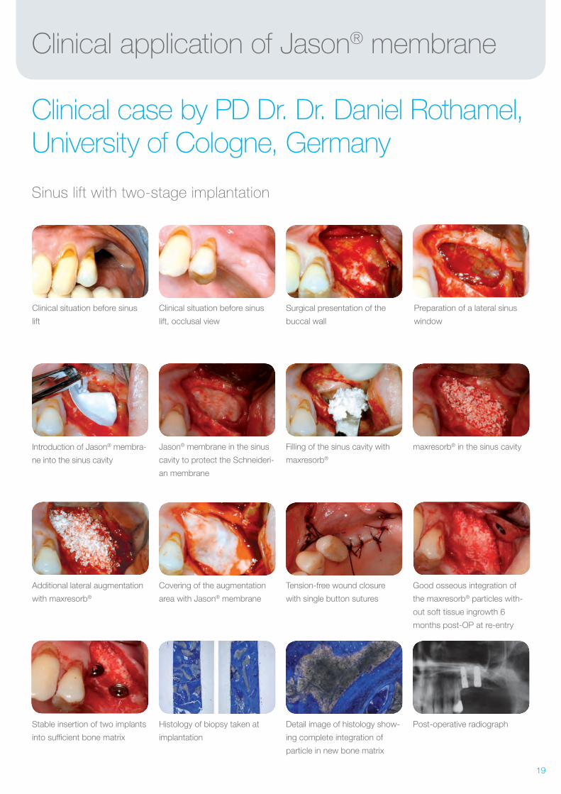
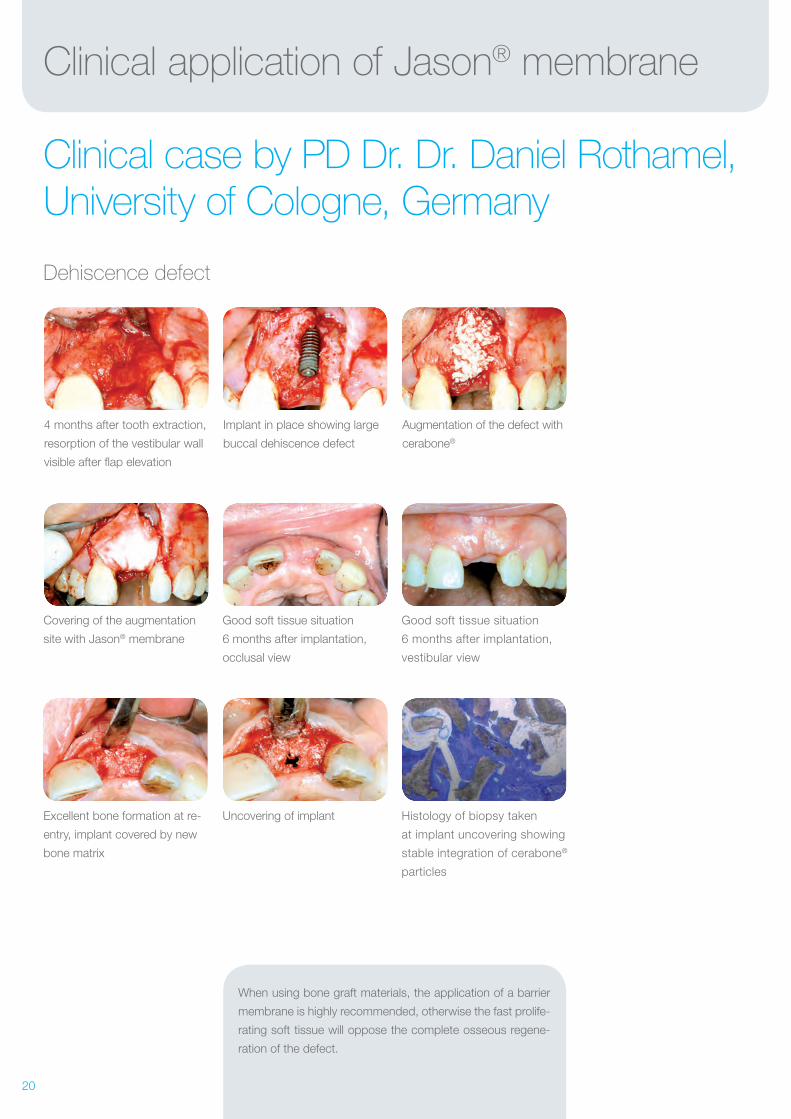
Clinical application of Jason® membrane
Clinical case by PD Dr. Dr. Daniel Rothamel, University of Cologne, Germany
Sinus lift with two-stage implantation
Jason® membrane in the sinus
cavity to protect the Schneideri-
an membrane
Introduction of Jason® membra-
ne into the sinus cavity
Covering of the augmentation
area with Jason® membrane
Stable insertion of two implants
into sufficient bone matrix
Filling of the sinus cavity with
maxresorb®
Tension-free wound closure
with single button sutures
Histology of biopsy taken at
implantation
Clinical situation before sinus
lift
Clinical situation before sinus
lift, occlusal view
Surgical presentation of the
buccal wall
Preparation of a lateral sinus
window
maxresorb® in the sinus cavity
Good osseous integration of
the maxresorb® particles with-
out soft tissue ingrowth 6
months post- OP at re- entry
Detail image of histology show-
ing complete integration of
particle in new bone matrix
Post-operative radiograph
Additional lateral augmentation
with maxresorb®
20
Good soft tissue situation
6 months after implantation,
occlusal view
Uncovering of implant
Covering of the augmentation
site with Jason® membrane
Excellent bone formation at re-
entry, implant covered by new
bone matrix
Good soft tissue situation
6 months after implantation,
vestibular view
Histology of biopsy taken
at implant uncovering showing
stable integration of cerabone®
particles
4 months after tooth extraction,
resorption of the vestibular wall
visible after flap elevation
Implant in place showing large
buccal dehiscence defect
Augmentation of the defect with
cerabone®
Clinical application of Jason® membrane
Clinical case by PD Dr. Dr. Daniel Rothamel, University of Cologne, Germany
Dehiscence defect
When using bone graft materials, the application of a barrier
membrane is highly recommended, otherwise the fast prolife-
rating soft tissue will oppose the complete osseous regene-
ration of the defect.
21
Clinical application of Jason® membrane
Clinical case by PD Dr. Dr. Daniel Rothamel, University of Cologne, Germany
Ridge augmentation
Bone spreading at 12 for lateral
widening of the crest
Covering of the augmentation
site with Jason® membrane
Perfect integration of the
cerabone® particles into newly
formed bone matrix
Internal sinus grafting to com-
pensate the vertical deficiency
at 15
Tension-free soft tissue closure
Implant uncovering and insertion
of gingiva formers
Instable bridge situation with
abscess formation at tooth 15
after apicectomia
OPG 6 months after tooth ext-
raction shows vertical deficiency
at 15
Clinical situation with scar
formation at former abscess
incision site
Mucoperiosteal flap elevation re-
veals a self-containing defect at
15 and a non-containing lateral
bone defect at 14 – 12
After implant installation, lateral
bone defects need further aug-
mentation
Post-operative x-ray showing
the position of implants and
internal sinus grafting
Prosthetic situation after one
year following professional
dental hygiene
Stable conditions after 6
months healing period
Radiological situation after one
year
Application of cerabone® and
autologous bone (mixture 1:2)
on the lateral aspect
22
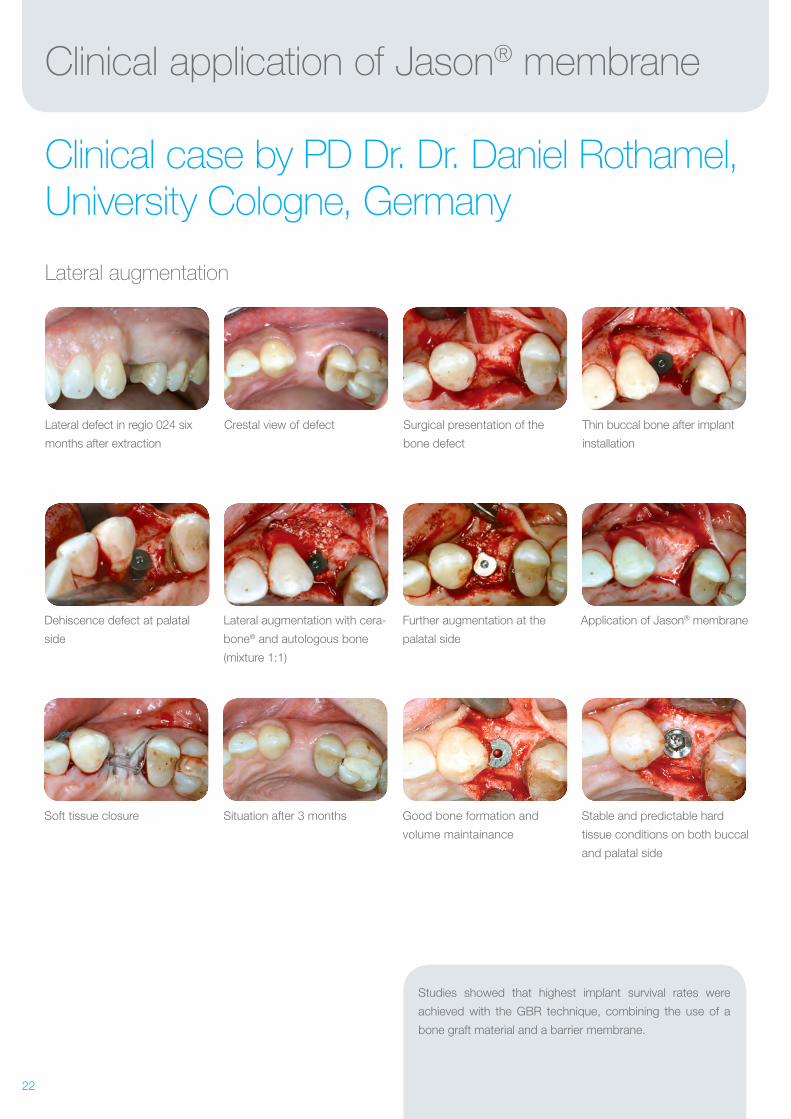
Further augmentation at the
palatal side
Good bone formation and
volume maintainance
Lateral augmentation with cera-
bone® and autologous bone
(mixture 1:1)
Dehiscence defect at palatal
side
Situation after 3 monthsSoft tissue closure
Application of Jason® membrane
Stable and predictable hard
tissue conditions on both buccal
and palatal side
Crestal view of defectLateral defect in regio 024 six
months after extraction
Surgical presentation of the
bone defect
Thin buccal bone after implant
installation
Clinical application of Jason® membrane
Clinical case by PD Dr. Dr. Daniel Rothamel, University Cologne, Germany
Lateral augmentation
Studies showed that highest implant survival rates were
achieved with the GBR technique, combining the use of a
bone graft material and a barrier membrane.
23
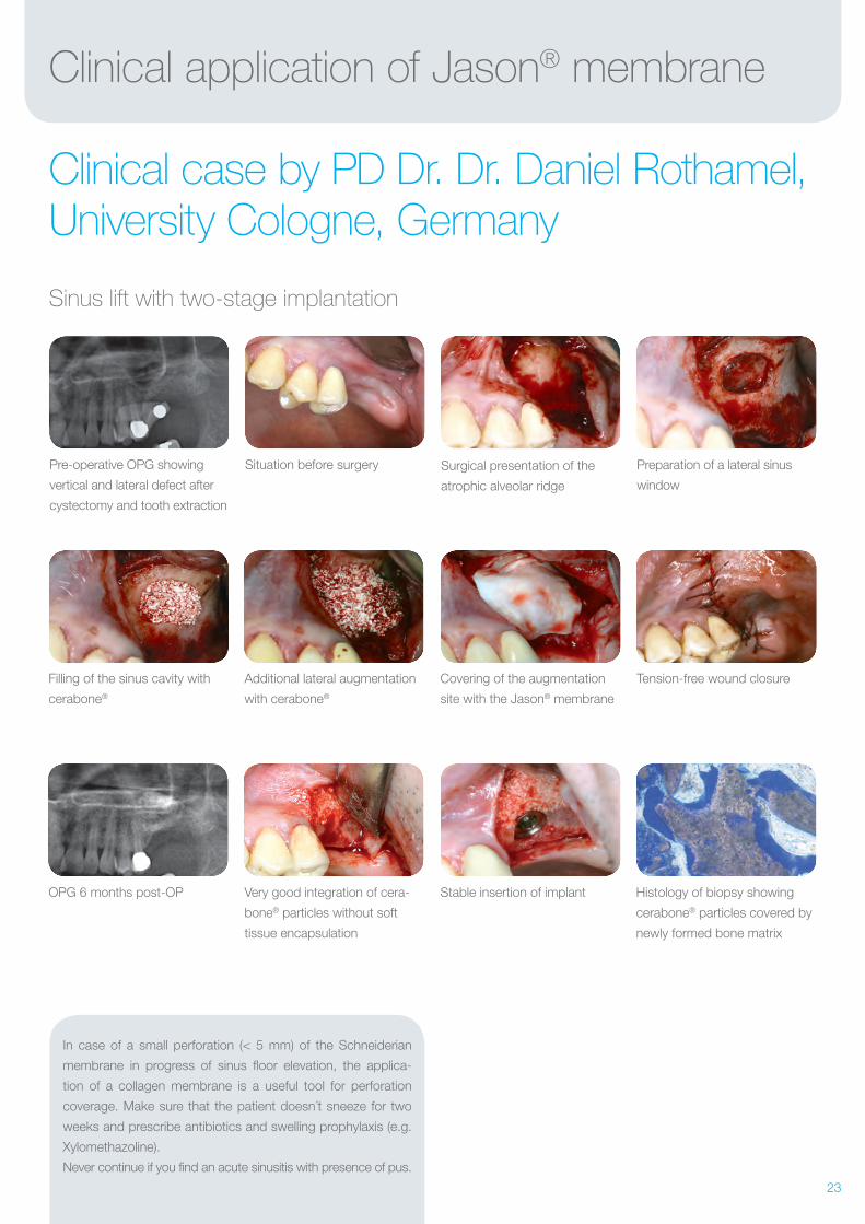
In case of a small perforation (< 5 mm) of the Schneiderian
membrane in progress of sinus floor elevation, the applica-
tion of a collagen membrane is a useful tool for perforation
coverage. Make sure that the patient doesn´t sneeze for two
weeks and prescribe antibiotics and swelling prophylaxis (e.g.
Xylomethazoline).
Never continue if you find an acute sinusitis with presence of pus.
Clinical application of Jason® membrane
Clinical case by PD Dr. Dr. Daniel Rothamel, University Cologne, Germany
Sinus lift with two-stage implantation
Filling of the sinus cavity with
cerabone®
OPG 6 months post-OP
Additional lateral augmentation
with cerabone®
Very good integration of cera-
bone® particles without soft
tissue encapsulation
Pre-operative OPG showing
vertical and lateral defect after
cystectomy and tooth extraction
Situation before surgery Surgical presentation of the
atrophic alveolar ridge
Preparation of a lateral sinus
window
Covering of the augmentation
site with the Jason® membrane
Stable insertion of implant Histology of biopsy showing
cerabone® particles covered by
newly formed bone matrix
Tension-free wound closure
24Rev.: CMSen-01/2014-08
soft tissue
education
hard tissue
botiss dental GmbH
Uhlandstraße 20-25
10623 Berlin / Germany
Fon +49 30 20 60 73 98 30
Fax +49 30 20 60 73 98 20
www.botiss.com
facebook: botiss biomaterials
Innovation.
Regeneration.
Aesthetics.
Straumann UK
3 Pegasus Place
Gatwick Road
West Sussex
Crawley RH10 9AY
Tel: +44 1293 65 12 30
Fax +44 1293 65 12 39
www.straumann.co.uk
Straumann SA/NV
Belgicastraat 3 Box 3
1930 Zaventem
Tel: +32 2 790 10 00
Fax +32 2 790 10 20
www.straumann.be
...................................
Straumann s.r.o.
Na Žertvách 2196
180 00 Prague 8
Czech Republic
Tel.: +420 284 094 650
Fax: +420 284 094 659
www.straumann.cz
Straumann B.V.
Einsteinweg 15
Postbus 338
3400 AH Ijsselstein
Tel: +31 30 604 66 11
Fax +31 30 604 67 28
www.straumann.nl
...................................
Straumann A/S
Post boks 1751, Vika
0122 Oslo
Tel: +47 23 35 44 88
Fax +47 23 35 44 80
www.straumann.no
...................................
Distributed by:
Straumann AB
Krokslätts Fabriker 45
431 37 Mölndal
Tel: +46 31 708 75 00
Fax +46 31 708 75 19
www.straumann.se
...................................
Some products may not be available in all countries. Please check with your local Straumann sales representative for more information.
botissbiomaterials
dental
bone & tissue regeneration


![Lecture 17 Membrane separations - CHERIC · Lecture 17. Membrane Separations [Ch. 14] •Membrane Separation •Membrane Materials •Membrane Modules •Transport in Membranes-Bulk](https://static.fdocuments.net/doc/165x107/5e688f368fbb145949438f76/lecture-17-membrane-separations-cheric-lecture-17-membrane-separations-ch-14.jpg)