Janice Santisi, RN 216 444 8836 santisj@ccf · - documentation guidelines - key nursing components...
116
Small Bowel Capsule Endoscopy Janice Santisi, RN 216 444 8836 [email protected] I have no stocks nor do I receive any compensation for this presentation. I do not represent or endorse any one manufacturer
Transcript of Janice Santisi, RN 216 444 8836 santisj@ccf · - documentation guidelines - key nursing components...
Small Bowel Capsule EndoscopyJanice Santisi, RN216 444 [email protected]
I have no stocks nor do I receive any compensation for this presentation. I do not represent or endorse any one manufacturer

Session GoalsAttendees will gain knowledge of Capsule Endoscopy of the small bowel including:
- preparation- pre-procedural considerations- safety factors- documentation guidelines- key nursing components
in current practice of capsule endoscopy for hospitalized and clinic-based patient groups.

Learning Objectives• Describe three pre-capsule endoscopy patient safety
considerations
• Actively discuss parameters of appropriate patient selection for capsule endoscopy
• Identify one clinical consideration difference between a hospitalized and non-hospitalized capsule endoscopy patient
• Wireless Endoscopy future in Digestive Disease
• Recognize RN scope of practice in interpretive processof capsule endoscopy video data.

Presentation Agenda
Small Bowel TestingCapsule Patients
• Non tethered• Photographic capsule• Collect 2 images per second• Moves by peristalsis• Images converted to RF and are collected and stored for later download• Capsule “working life” ranges 8-14hrs in current FDA approved devices• Capsule passes from body naturally in feces• Specific to examination of the Small bowel but other wireless device that exam remainder of GI tract

Traditional Upper Endoscope
-oral cavity>inches of SB
Traditional Lower Endoscope-Colonoscopy-rectal insertion> 5 feet> -through Ileal Cecal valve -From inches-1 foot examined
17-20 feet unexamined by direct visualization
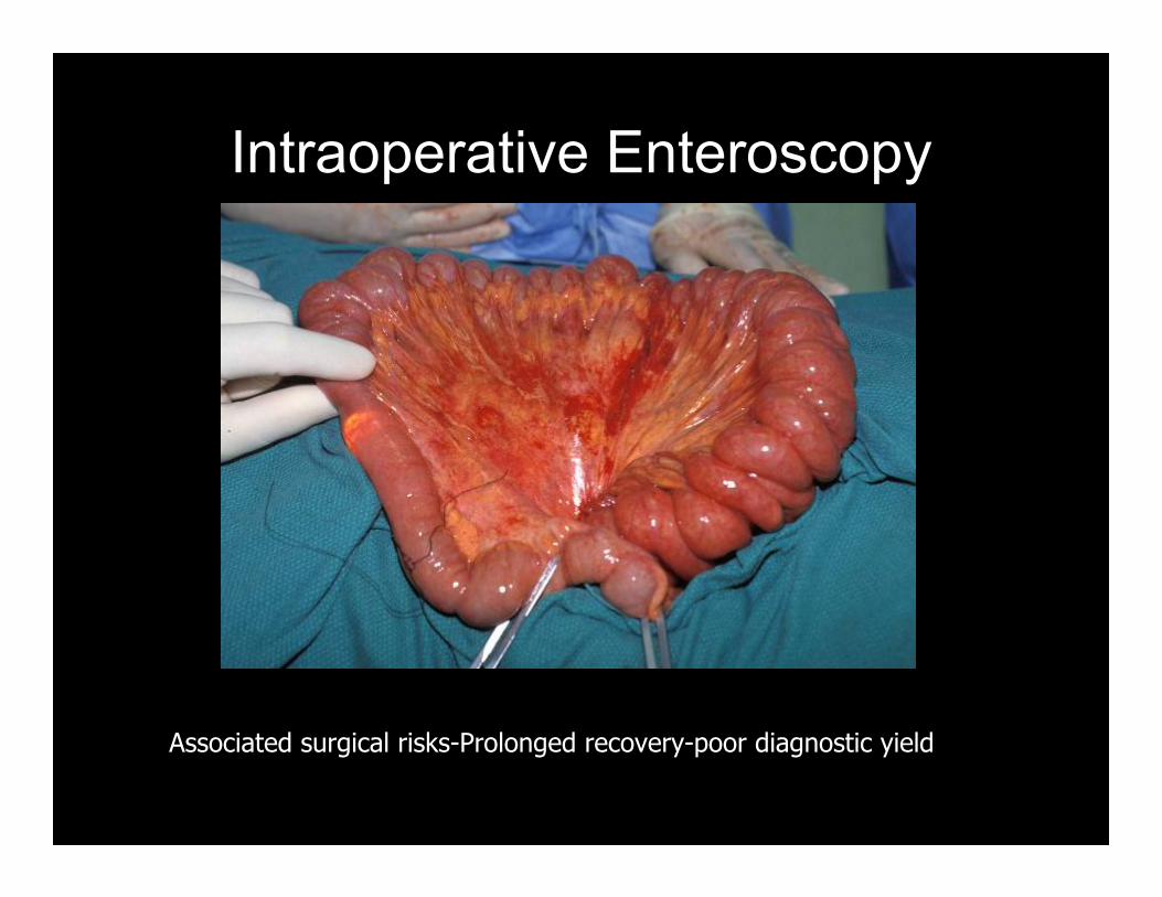
Intraoperative Enteroscopy
Associated surgical risks-Prolonged recovery-poor diagnostic yield

Balloon Enteroscopy
invasive-general anesthesia-risk of perforation - 2 hour procedure-long recovery -requires specifically trained team

Balloon Enteroscopy primarily used as treatment modality secondary to SB lesion diagnosis by capsule endoscopy
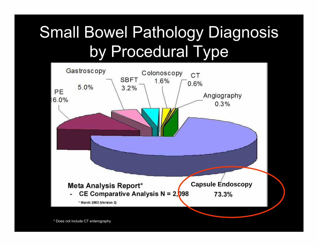
Small Bowel Pathology Diagnosis by Procedural Type
* Does not include CT enterography
Capsule Endoscopy
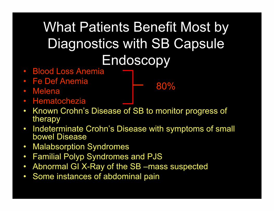
What Patients Benefit Most by Diagnostics with SB Capsule
Endoscopy• Blood Loss Anemia• Fe Def Anemia• Melena• Hematochezia• Known Crohn’s Disease of SB to monitor progress of
therapy• Indeterminate Crohn’s Disease with symptoms of small
bowel Disease• Malabsorption Syndromes• Familial Polyp Syndromes and PJS• Abnormal GI X-Ray of the SB –mass suspected• Some instances of abdominal pain
80%

Agenda
Small Bowel TestingSB Anatomy Review
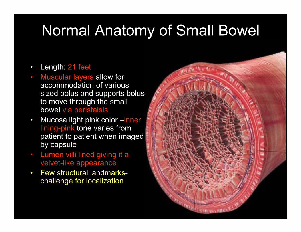
Normal Anatomy of Small Bowel
• Length: 21 feet• Muscular layers allow for
accommodation of various sized bolus and supports bolus to move through the small bowel via peristalsis
• Mucosa light pink color –inner lining-pink tone varies from patient to patient when imaged by capsule
• Lumen villi lined giving it a velvet-like appearance
• Few structural landmarks-challenge for localization
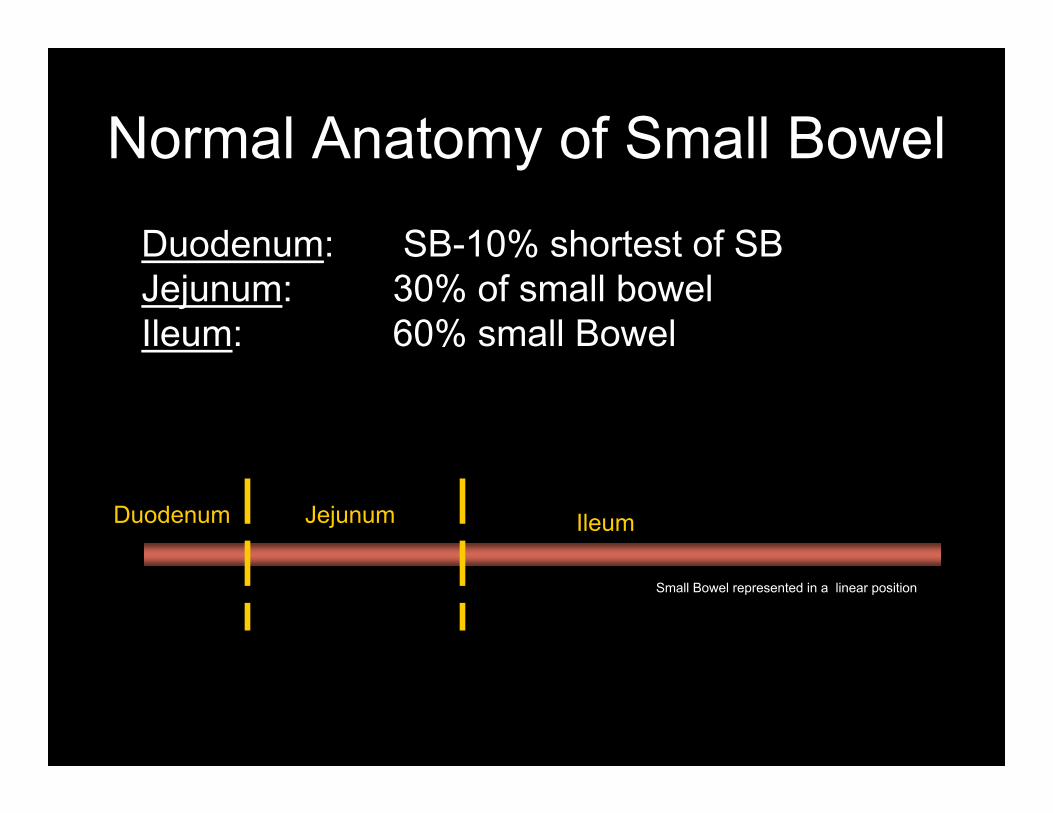
Normal Anatomy of Small Bowel
Duodenum: SB-10% shortest of SB Jejunum: 30% of small bowel Ileum: 60% small Bowel
IleumJejunumDuodenum
Small Bowel represented in a linear position

Normal Physiology of Small Bowel
• Localized Transit Pause: Capsule endoscope remains static recording same image for ½ hour but no structural obstruction
• Regional Transit Pause: Capsule endoscope moves forward and back but remains in same region for greater than ½ hour but no structural obstruction
• Failure to Traverse: Phrase used when capsule does not move beyond a point but no structural obstruction
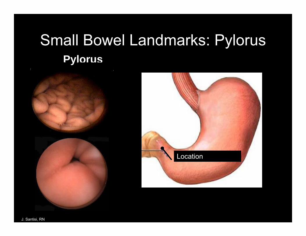
Small Bowel Landmarks: Pylorus
Location
Pylorus
J. Santisi, RN
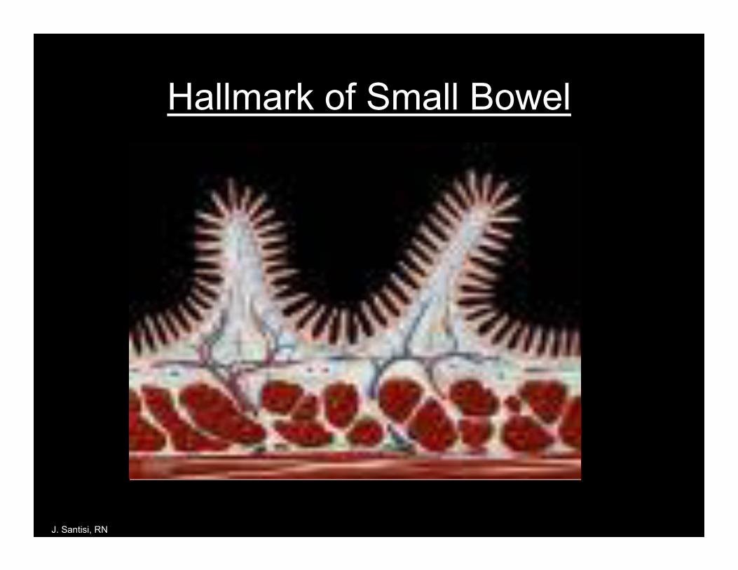
Hallmark of Small Bowel
J. Santisi, RN
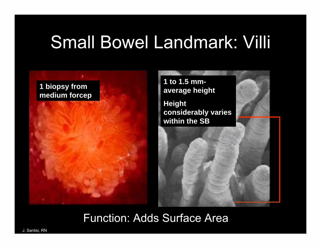
1 to 1.5 mm-average height
Height considerably varies within the SB
1 biopsy from medium forcep
J. Santisi, RN
Small Bowel Landmark: Villi
Function: Adds Surface Area

Small Bowel Landmarks: Villi
Ideal ImageDuodenum
Ideal ImageJejunum

Small Bowel Landmarks: Villi
Ideal ImageIleum

Small Bowel Landmarks
Capsule Endoscope Image 7 to 10 cm below the pylorus
Location
J. Santisi, RN
Landmark: Papilla
Ampulla's

Small Bowel LandmarksNon-specific-Non-structural landmark:BILECaution: not “hard evidence because bile can reflux into the stomach and actually be seen most anywhere along upper sb
J. Santisi, RN

Duodenojejunal flexureLigament of Treitz
“C curve”
Capsule often moves forward And back as it attempts toround the C-curve
Capsule often speeds in this area Passing past the flexure
Small Bowel Landmarks

Small Bowel Landmarks
Blood supply to the ileum is by way of the superior mesenteric artery.
Branched ARCADES of the superior mesenteric artery are prolific in the ileum leading to the luminal –capsule view of increased vascularity.
J. Santisi, RN
Increased vascularity good visual cue to passing from Jejunum to ileum
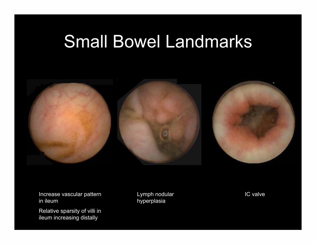
Increase vascular pattern in ileum
Relative sparsity of villi in ileum increasing distally
Lymph nodular hyperplasia
IC valve
Small Bowel Landmarks

Summary: Small Bowel Landmarks
• Pylorus• Villi length/population• Ampulla• Bile• Movement of capsule with the
lumen• Vascularity• Lymph nodular hyperplasia• IC valve
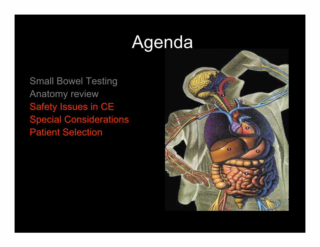
Agenda
Small Bowel TestingAnatomy reviewSafety Issues in CE Special ConsiderationsPatient Selection
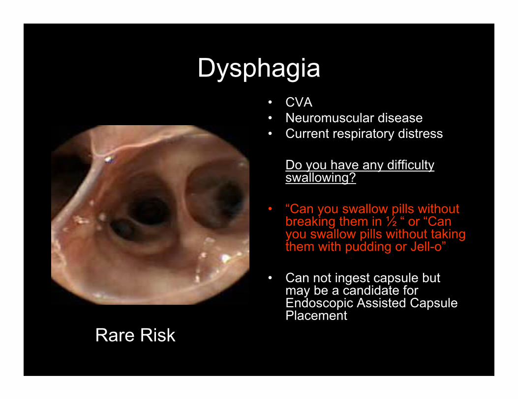
Dysphagia• CVA• Neuromuscular disease• Current respiratory distress
Do you have any difficulty swallowing?
• “Can you swallow pills without breaking them in ½ “ or “Can you swallow pills without taking them with pudding or Jell-o”
• Can not ingest capsule but may be a candidate for Endoscopic Assisted Capsule Placement
Rare Risk
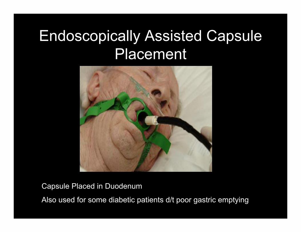
Endoscopically Assisted Capsule Placement
Capsule Placed in Duodenum
Also used for some diabetic patients d/t poor gastric emptying
Pregnancy
Not FDA approved for use in pregnancy

Not FDA approved for patient with Implanted Electromedical Device
Not FDA approved for patients with electromedical devices
J. Santisi, RN
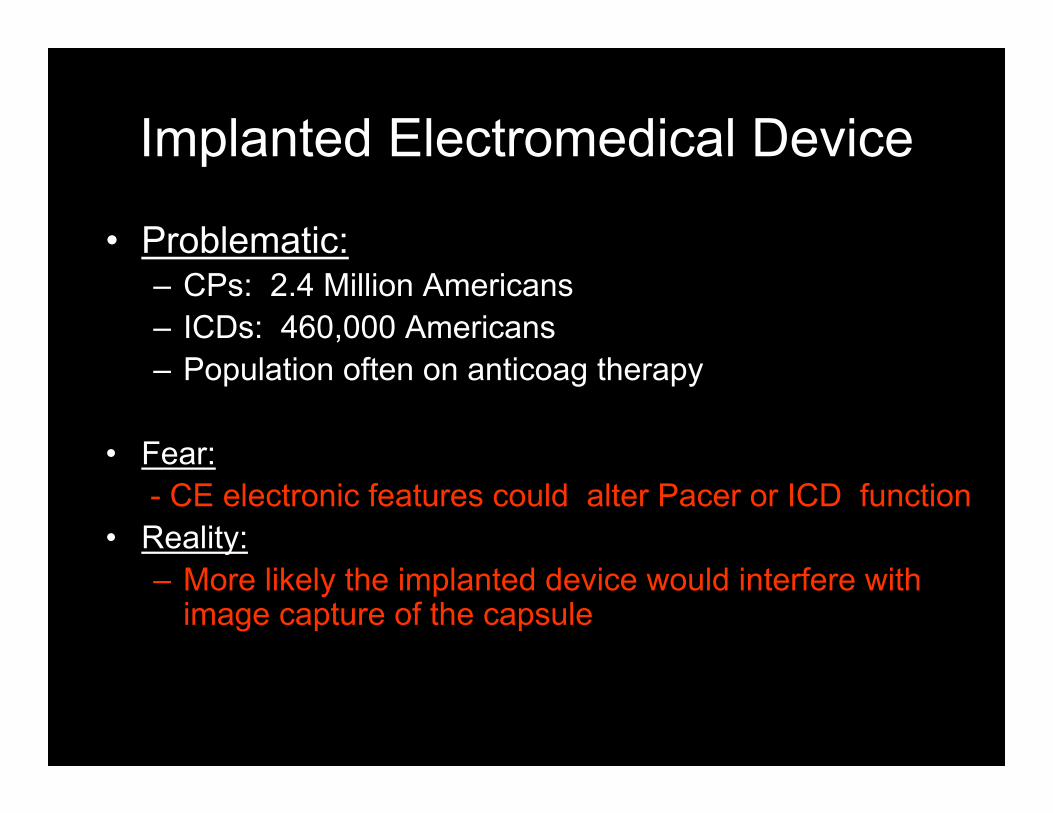
Implanted Electromedical Device
• Problematic:– CPs: 2.4 Million Americans– ICDs: 460,000 Americans– Population often on anticoag therapy
• Fear:- CE electronic features could alter Pacer or ICD function
• Reality:– More likely the implanted device would interfere with
image capture of the capsule
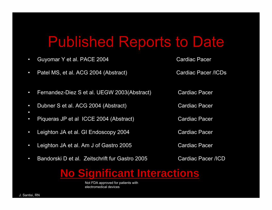
Published Reports to Date• Guyomar Y et al. PACE 2004 Cardiac Pacer
• Patel MS, et al. ACG 2004 (Abstract) Cardiac Pacer /ICDs
• Fernandez-Diez S et al. UEGW 2003(Abstract) Cardiac Pacer
• Dubner S et al. ACG 2004 (Abstract) Cardiac Pacer •• Piqueras JP et al ICCE 2004 (Abstract) Cardiac Pacer
• Leighton JA et al. GI Endoscopy 2004 Cardiac Pacer
• Leighton JA et al. Am J of Gastro 2005 Cardiac Pacer
• Bandorski D et al. Zeitschrift fur Gastro 2005 Cardiac Pacer /ICD
No Significant InteractionsNot FDA approved for patients with electromedical devices
J. Santisi, RN
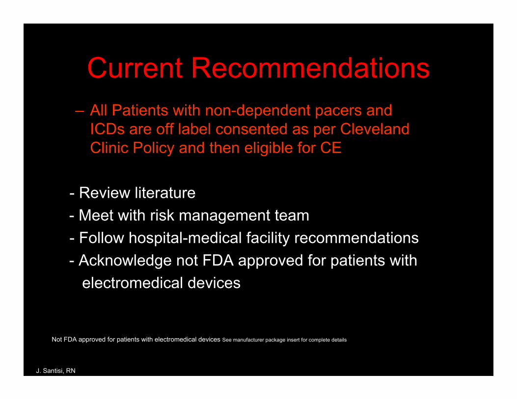
Current Recommendations– All Patients with non-dependent pacers and
ICDs are off label consented as per Cleveland Clinic Policy and then eligible for CE
- Review literature- Meet with risk management team- Follow hospital-medical facility recommendations- Acknowledge not FDA approved for patients with
electromedical devices
Not FDA approved for patients with electromedical devices See manufacturer package insert for complete details
J. Santisi, RN

Contraindicated: Known Small Bowel Obstruction

History of Adhesions Indeterminate Crohn’s History of Abdominal Surgery in past year Long term Use of NSAIDs
Capsule Endoscope Retention
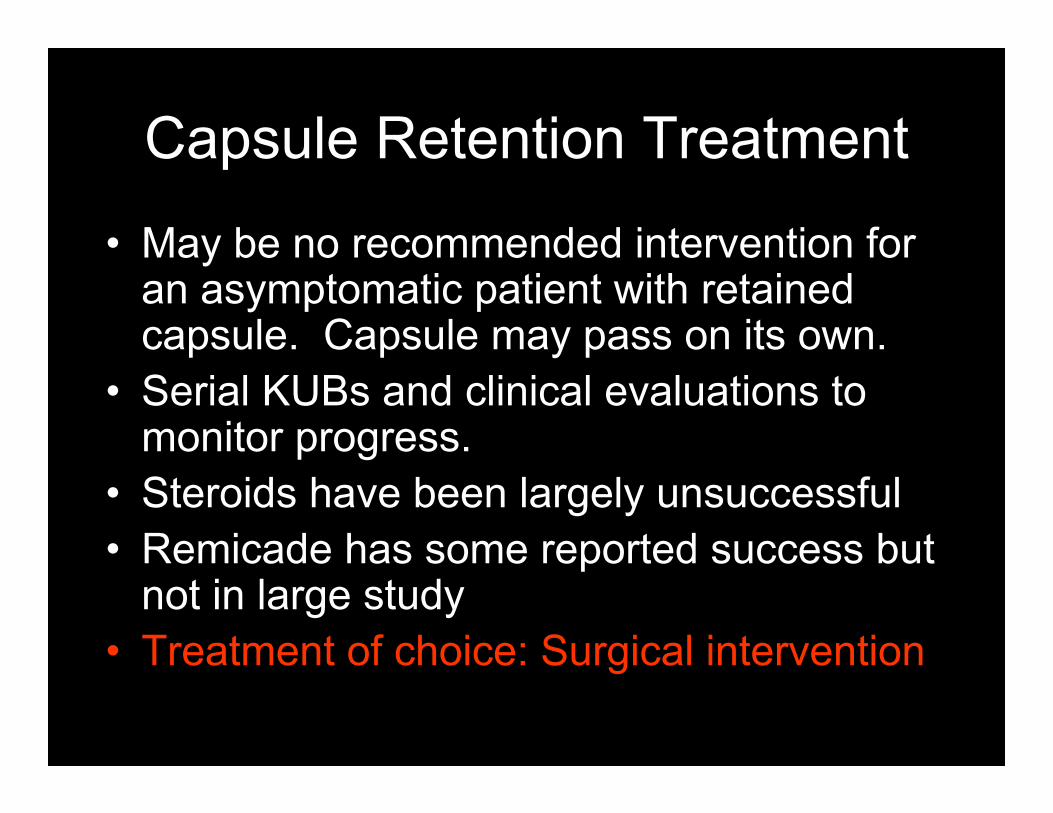
Capsule Retention Treatment
• May be no recommended intervention for an asymptomatic patient with retained capsule. Capsule may pass on its own.
• Serial KUBs and clinical evaluations to monitor progress.
• Steroids have been largely unsuccessful• Remicade has some reported success but
not in large study• Treatment of choice: Surgical intervention

Capsule Retention Treatment
• Capsule retention refers to “small bowel only”.
• Capsule may take 5-10 days to pass from the large bowel. Low risk of retention within the colon.
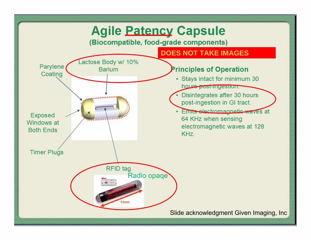
Radio opaqe
DOES NOT TAKE IMAGES
Slide acknowledgment Given Imaging, Inc

7:00 am Monday
7:00 am Tuesday
Ingest Patency Capsule with glass of water
KUB
NPO for 4 hours
24 hours later
No Agile Patency capsule in small bowel: CLEARED FOR PHOTOGRAPHIC CAPSULE

7:00 am Monday
7:00 am Tuesday
Ingest Patency Capsule with glass of water
KUB
NPO for 4 hours
24 hours later
Agile Patency Capsule noted in small bowel:
WAIT 4 HOURS AND REPEAT X-RAY
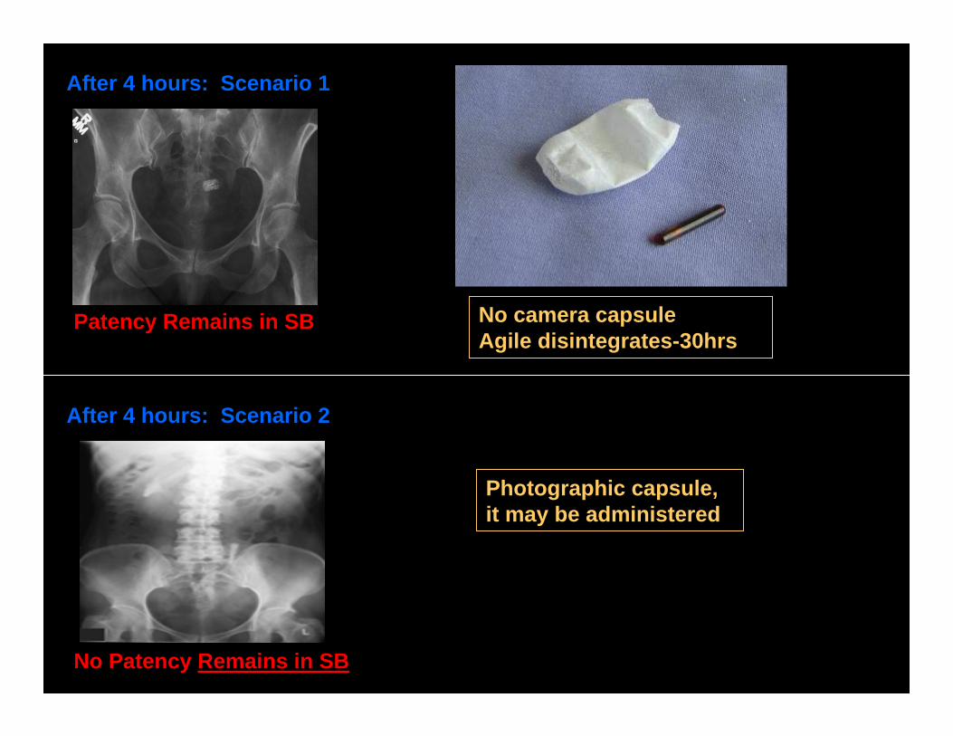
Patency Remains in SB
After 4 hours: Scenario 1
Photographic capsule, it may be administered
No camera capsule Agile disintegrates-30hrs
After 4 hours: Scenario 2
No Patency Remains in SB
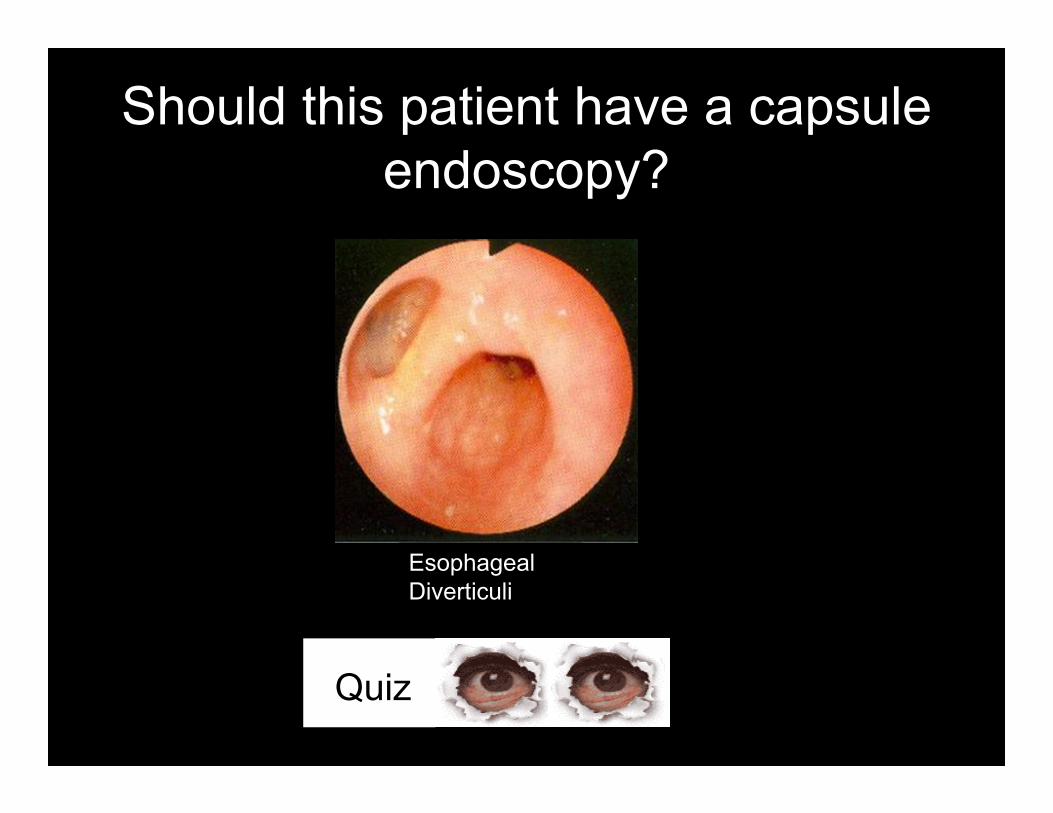
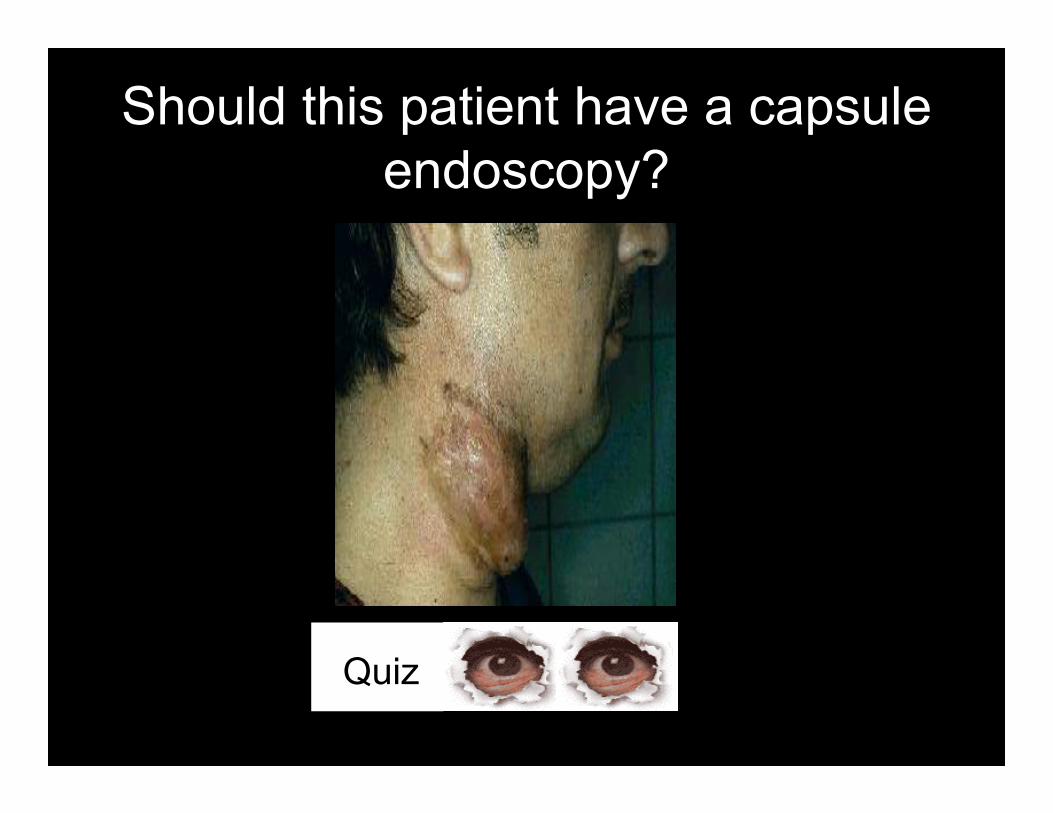
Should this patient have a capsule endoscopy?
Esophageal Diverticuli
Quiz
Should this patient have a capsule endoscopy?
Quiz

Should this patient have a capsule endoscopy?
J. Santisi, RN
Gastric bypass
Quiz
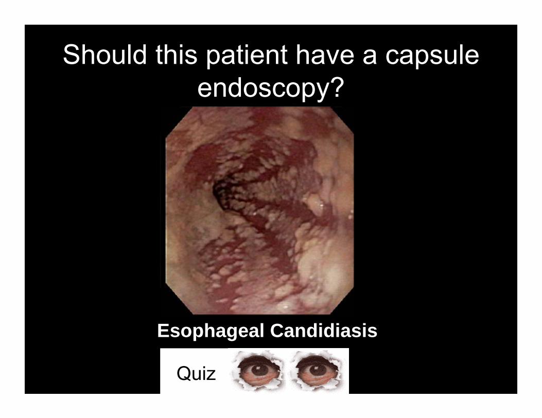
Should this patient have a capsule endoscopy?
Esophageal Candidiasis
Quiz

Should this patient have a capsule endoscopy?
Ileostomy

Should this patient have a capsule endoscopy?
J-Pouch

– BLOATING– FEVER– NAUSEA– VOMITING– ABD PAIN
• INTERMITTENT• CONTINUOUS
Should this patient have a capsule endoscopy?

• Aspirin• Celebrex• Ibuprofen, Motrin,
Tab-Profen• Vicoprofen
• CHRONIC= >10d/mo
Should this patient have a capsule endoscopy?

Should this patient have a capsule endoscopy?
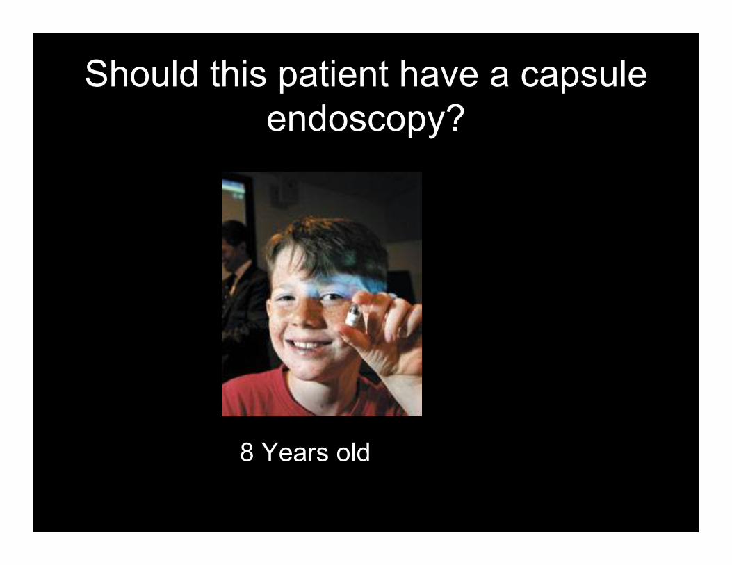
Should this patient have a capsule endoscopy?
8 Years old
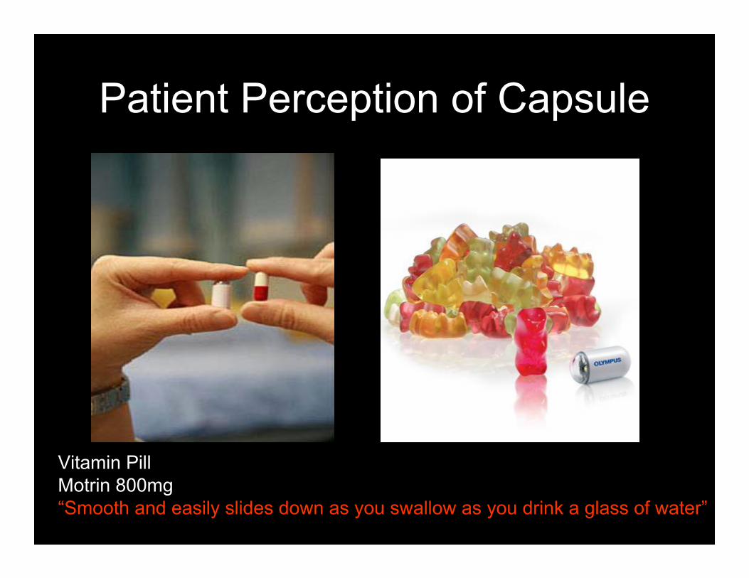
Patient Perception of Capsule
Vitamin Pill Motrin 800mg “Smooth and easily slides down as you swallow as you drink a glass of water”

Should this patient have a capsule endoscopy?
Morbid Obesity

Should this patient have a capsule endoscopy?
Chronic Constipation

Summary: Safety Issues• Dysphagia: May require endoscopically assisted capsule
placement• Poor Gastric Emptying/Gastroparesis: May require
endoscopically assisted capsule placement• Implanted Electromedical Device: Non-dependent
electromedical device with off label consent. Follow your medical facility policy.
• Known Bowel Obstruction: Contraindicated• Chronic NSAID use: Agile Patency Capsule prior to
photographic capsule• Crohn’s Disease: Possible Patency Capsule prior to
photographic capsule• Pregnancy: Contraindicated
•

Capsule Endoscopy and the Hospitalized Patient

Major Area of Recent Capsule Growth
• Economic: waiting longer to seek care• Statistical Influence:
– Outpatients Capsule Endoscopy yield is 30% significant pathology
– Inpatient Capsule Endoscopy yield in 60% significant pathology
Inpatient
OutPatient Main
OutpatientRegionOther

Hospitalized Patient• Must not share a room with patient also having a
capsule endoscopy• Patients taking narcotic pain medication may
have prolonged transit time of SB. Suggestion: Use product with longest battery life recording 14 hours
• Bed rest may slow SB transit time.• Dietary preparation: Clear liquids-nothing red-
day- before capsule administration-NPO after midnight: No po meds 2 hours before capsule ingestion

Hospitalized Patient
• NO MRI until capsule passes from body• Do not remove capsule equipment until
designated time. Consider re-scheduling routine x-ray.
• No barium 2-3 days pre- capsule• No po Fe products for 5 days pre- capsule.• May consider pre- capsule bowel
preparation
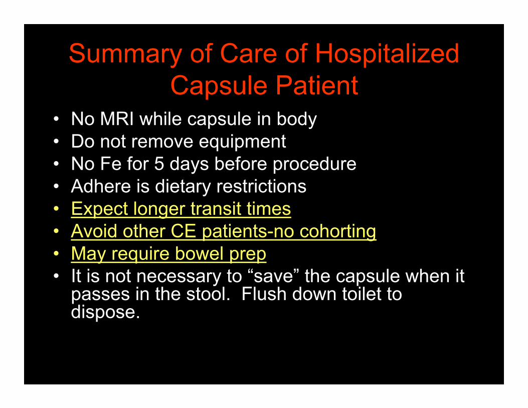
Summary of Care of Hospitalized Capsule Patient
• No MRI while capsule in body• Do not remove equipment• No Fe for 5 days before procedure• Adhere is dietary restrictions• Expect longer transit times• Avoid other CE patients-no cohorting• May require bowel prep• It is not necessary to “save” the capsule when it
passes in the stool. Flush down toilet to dispose.

Presentation Agenda
Small Bowel TestingAnatomy reviewSafety Issues in CE Special ConsiderationsPatient SelectionCapsule ProcedureDocumentation

Assessment Tool
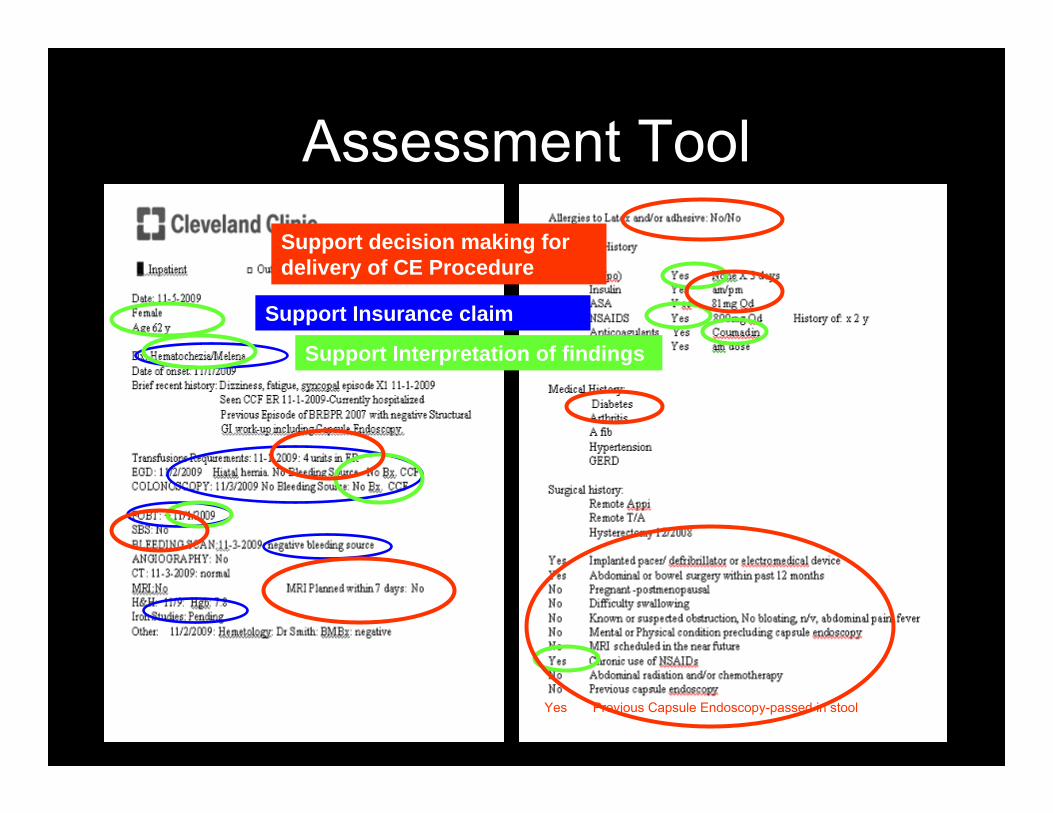
Yes Previous Capsule Endoscopy-passed in stool
Assessment Tool
Support Insurance claim
Support Interpretation of findings
Support decision making for delivery of CE Procedure
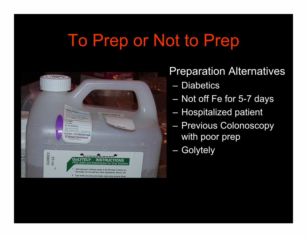
To Prep or Not to Prep
• Preparation Alternatives– Diabetics– Not off Fe for 5-7 days– Hospitalized patient– Previous Colonoscopy
with poor prep– Golytely

Pre- Capsule Instructions

Pre- Capsule Instruction Highlights
• One week before Capsule Endoscopy– Do not taking oral Iron medication for 5 days before your procedure– It is not necessary to stop taking anticoagulant medication like
Coumadin or Plavix before this test.– So that you may plan for your capsule endoscopy appointment, you
should know:
It is not necessary to have someone drive you to your procedure. This procedure will take approximately 30 minutes. You should not schedule other medical procedures on the same day
What about the day before?
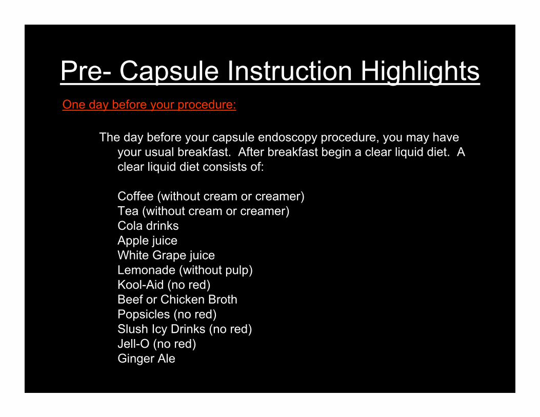
Pre- Capsule Instruction HighlightsOne day before your procedure:
The day before your capsule endoscopy procedure, you may have your usual breakfast. After breakfast begin a clear liquid diet. A clear liquid diet consists of:
Coffee (without cream or creamer)Tea (without cream or creamer)Cola drinksApple juice White Grape juiceLemonade (without pulp)Kool-Aid (no red)Beef or Chicken BrothPopsicles (no red)Slush Icy Drinks (no red)Jell-O (no red)Ginger Ale

Pre- Capsule Instruction HighlightsOne day before your procedure:
If you are a diabetic, your medication may have to be adjusted to accommodate this preparation. Check with the doctor who monitors your diabetes for further instructions.
You may continue your medication as usual the day before your procedure. Do not take any Pepto-Bismol, antacids, Carafate or medication that may coat your stomach and interfere with the capsule pictures.

Pre- Capsule Instruction HighlightsOne day before your procedure:
1. If you are a male with an abundance of body hair on your abdomen, please shave your abdomen 4 inches above your waist and 4 inches below your waist.Leave the body hair issue up to the patient
Generally, no other preparation is required.

Pre- Capsule Instruction HighlightsThe day of your procedure:
The morning of the procedure, you may take your medication with a sip of water before 7am.
Do not eat or drink anything ( other than sip of water with medication) before your procedure. NPO after midnight.
Wear loose fitting two- piece clothing to the procedure.
Do not wear lotions or powders on your abdomen.

Computer with Proprietary SoftwareEndocapsuleEndocapsule ActivatorData Recorder/Lithium Ion BatteryBattery Charging DeviceSensor Antenna/CoversBeltREAL Time ReaderUpload/Download
Patient information Patient discharge informationReturn bagUPS box/ labelDisposable cupGloves
1. Prepared Equipment
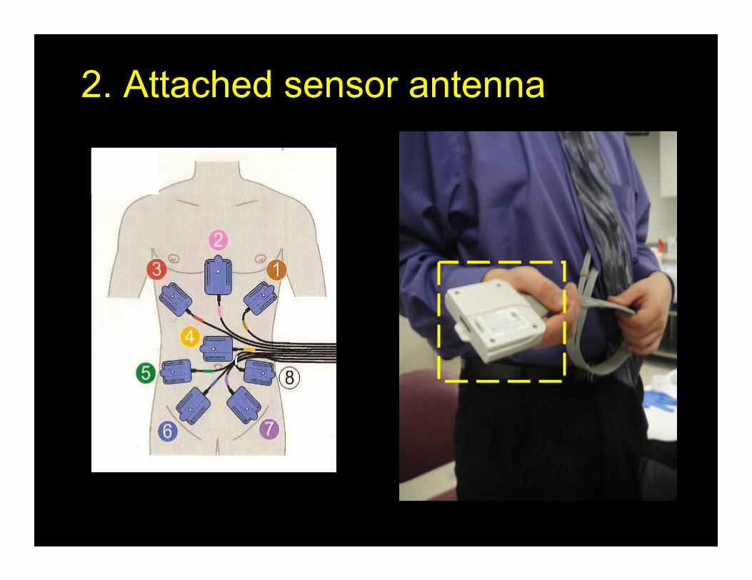
2. Attached sensor antenna
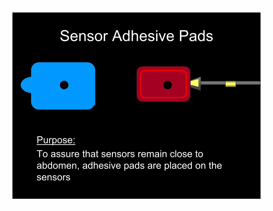
Sensor Adhesive Pads
Purpose:To assure that sensors remain close to abdomen, adhesive pads are placed on the sensors
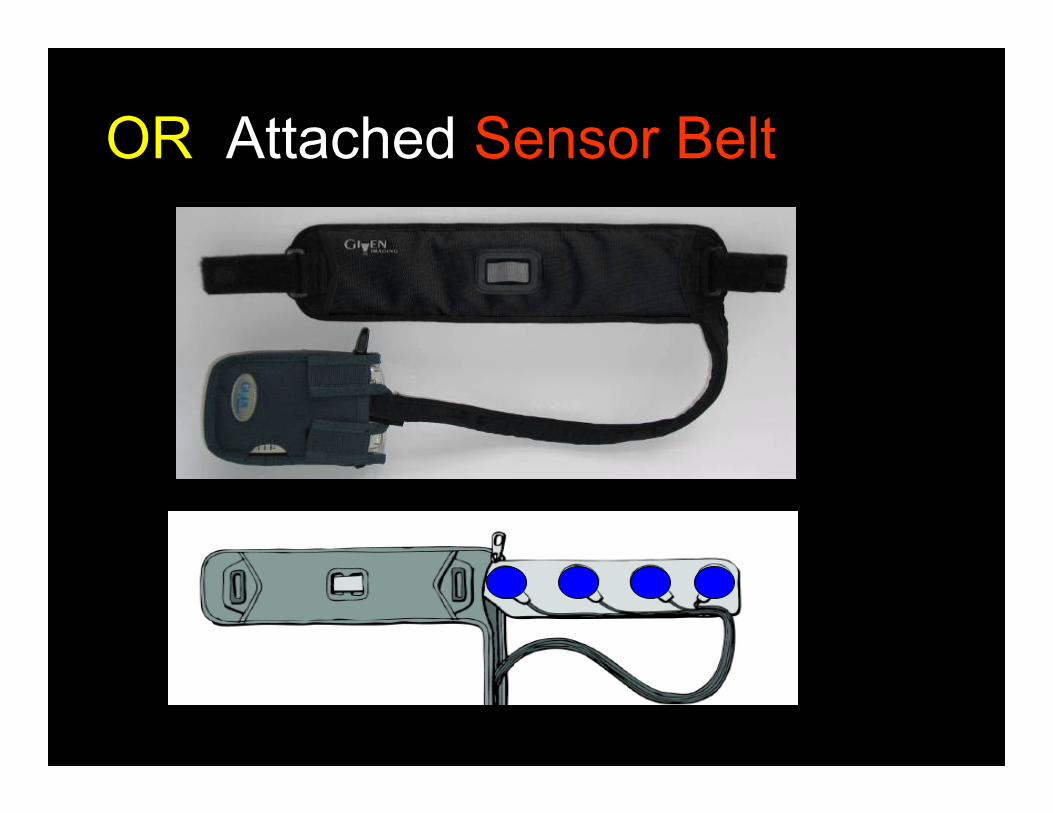
OR Attached Sensor Belt

• Sensor Antenna is attached using placement guideAttach Belt in Back
Attach Shoulder Strap
3. Apply belt
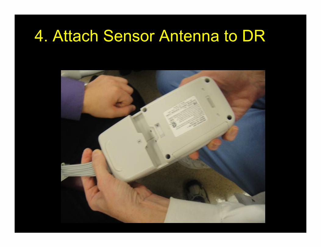
4. Attach Sensor Antenna to DR
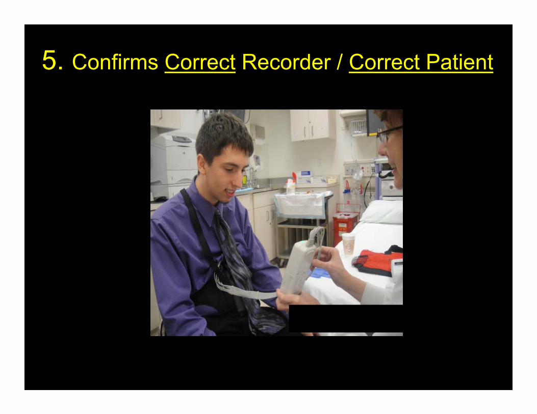
5. Confirms Correct Recorder / Correct Patient
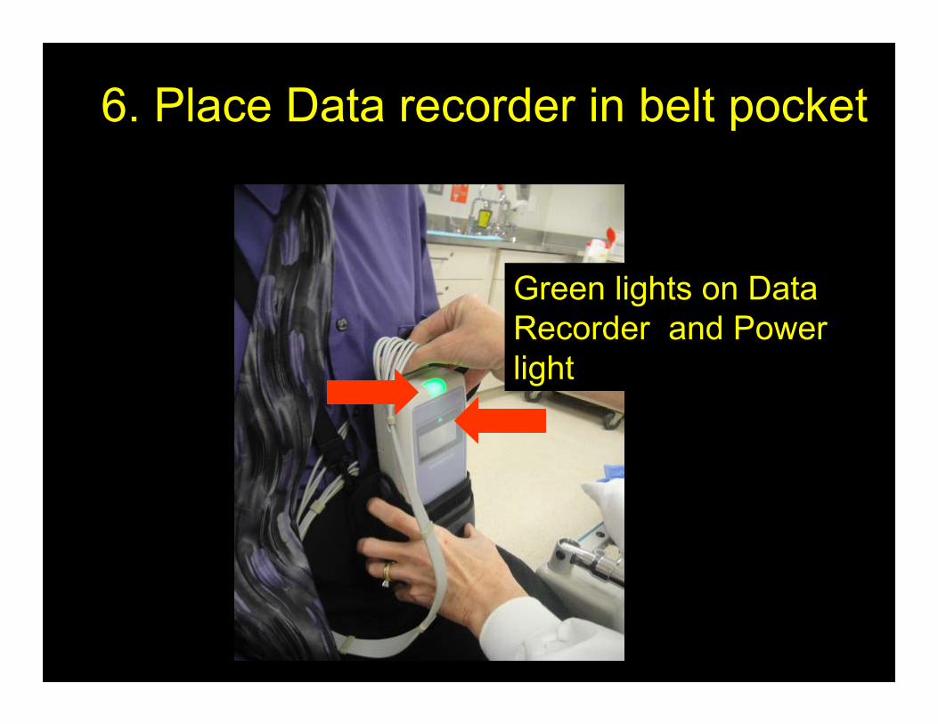
Green lights on Data Recorder and Power light
6. Place Data recorder in belt pocket

7. Activate the capsule

Activating the Endocapsule
De-activate-LEFTActivate-RIGHT

• Hold activated capsule next to data recorder
• Capsule and Data recorder light blink
8. Synchronizing
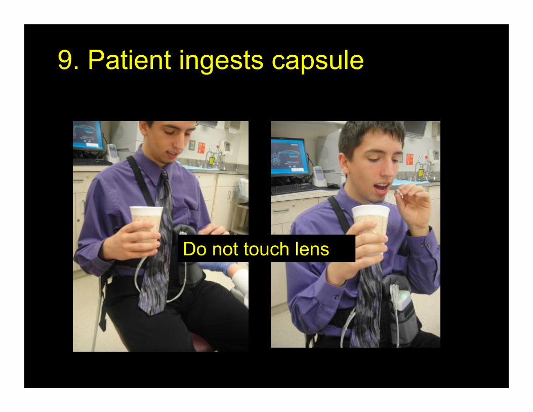
9. Patient ingests capsule
Do not touch lens
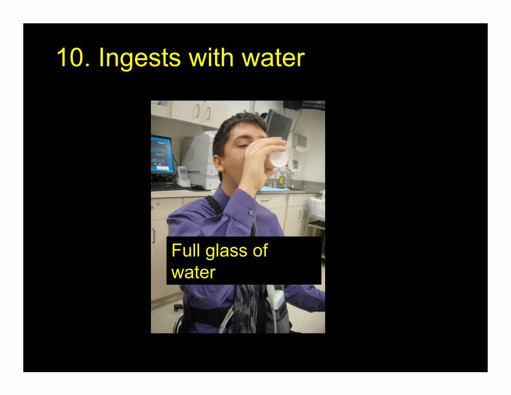
Full glass of water
10. Ingests with water

11. Teaching: return of equipment
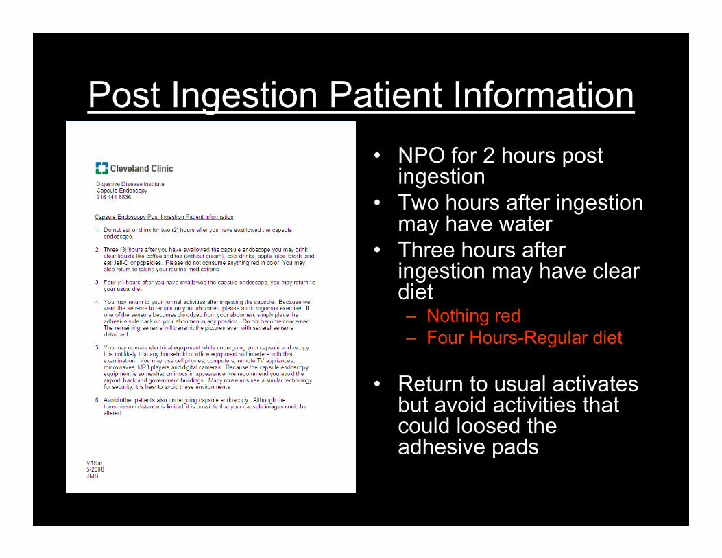
Post Ingestion Patient Information• NPO for 2 hours post
ingestion• Two hours after ingestion
may have water• Three hours after
ingestion may have clear diet – Nothing red– Four Hours-Regular diet
• Return to usual activates but avoid activities that could loosed the adhesive pads
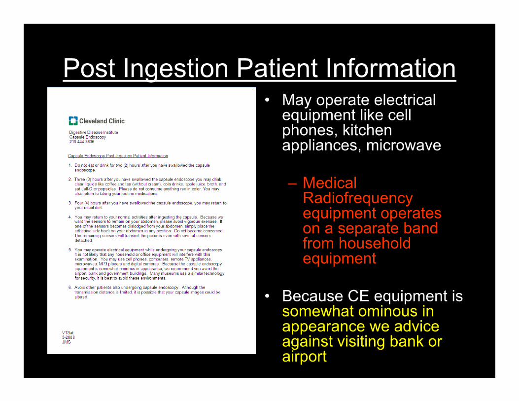
Post Ingestion Patient Information• May operate electrical
equipment like cell phones, kitchen appliances, microwave
– Medical Radiofrequency equipment operates on a separate band from household equipment
• Because CE equipment is somewhat ominous in appearance we advice against visiting bank or airport
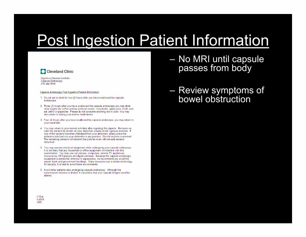
Post Ingestion Patient Information– No MRI until capsule
passes from body
– Review symptoms of bowel obstruction

Presentation Agenda
Small Bowel TestingAnatomy reviewSafety Issues in CE Special ConsiderationsPatient SelectionCapsule ProcedureDocumentationVideo TriageInterpretation Phase
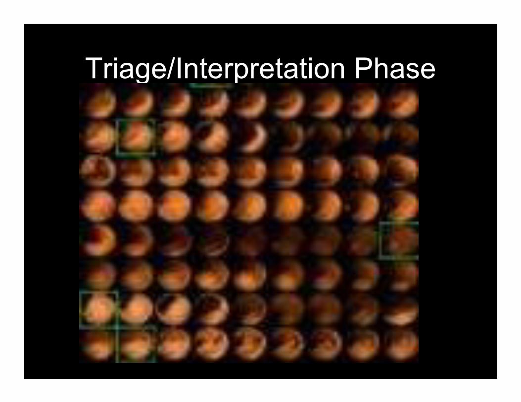
Triage/Interpretation Phase
Area of growth in nursing practice
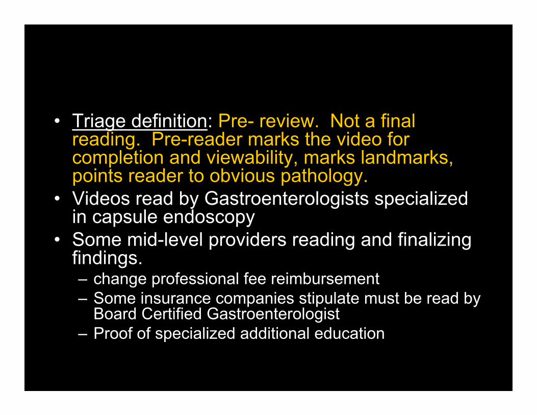
• Triage definition: Pre- review. Not a final reading. Pre-reader marks the video for completion and viewability, marks landmarks, points reader to obvious pathology.
• Videos read by Gastroenterologists specialized in capsule endoscopy
• Some mid-level providers reading and finalizing findings. – change professional fee reimbursement– Some insurance companies stipulate must be read by
Board Certified Gastroenterologist– Proof of specialized additional education
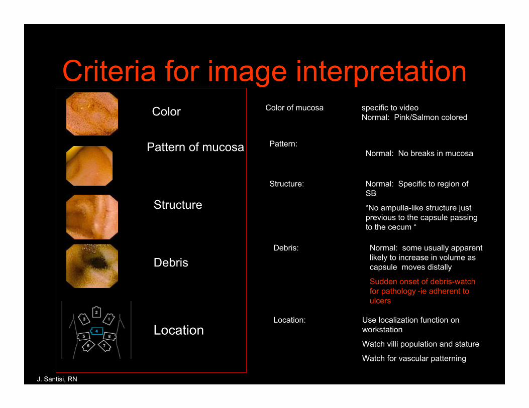
Criteria for image interpretation
J. Santisi, RN
Color
Pattern of mucosa
Structure
Debris
Location
Color of mucosa specific to videoNormal: Pink/Salmon colored
Pattern:Normal: No breaks in mucosa
Structure: Normal: Specific to region of SB
“No ampulla-like structure just previous to the capsule passing to the cecum “
Debris: Normal: some usually apparent likely to increase in volume as capsule moves distally
Sudden onset of debris-watch for pathology -ie adherent to ulcers
Location: Use localization function on workstation
Watch villi population and stature
Watch for vascular patterning

Peristaltic waveStromal tumor
• Color• Mucosal Integrity• Structure• Debris• Location MID SMALL BOWEL AS PER LOCALIZATION FEATURE
AND READERS ASSESSMENT
Quiz
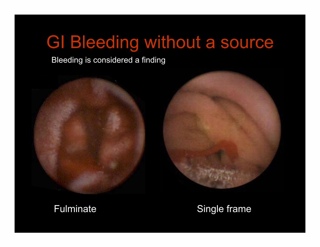
GI Bleeding without a source
Fulminate Single frame
Bleeding is considered a finding
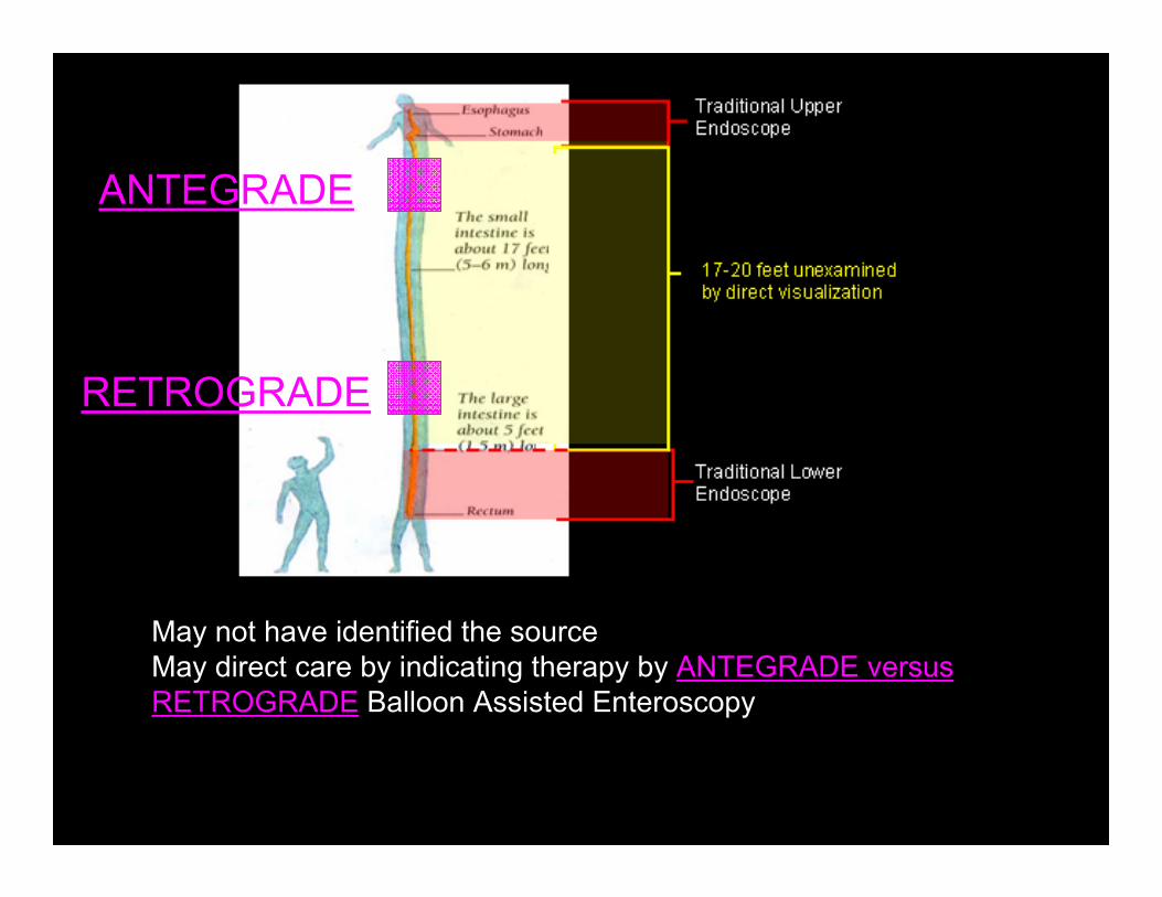
May not have identified the sourceMay direct care by indicating therapy by ANTEGRADE versus RETROGRADE Balloon Assisted Enteroscopy
ANTEGRADE
RETROGRADE

Capsule Findings: Angioectasia
● Feathery appearance
● Bright red
● Feeding vessel
● Singular or multiple
● Star shaped
● Easily identified
● flat- Missed on SBS
Often associated with renal disease, heart disease and anticoagulant therapy

Vascular Congestion
Distal venule fills with blood Muscle layer contracts
BLEED
Dilated and distorted microvascular complexes
Pressure rises within the venule
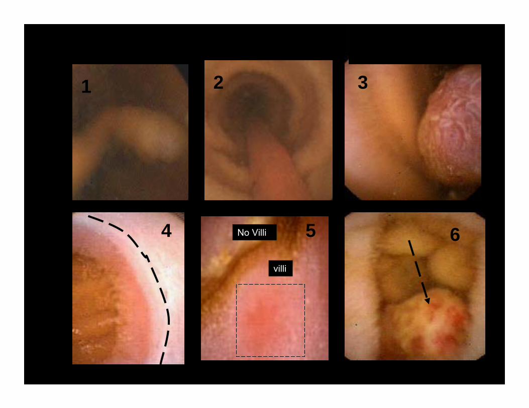
villi
No Villi
1 2 3
4 5 6
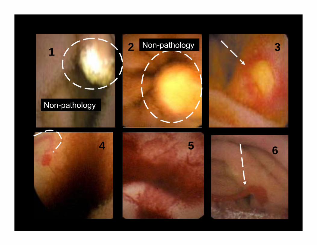
Non-pathology
Non-pathology1 2 3
4 5 6
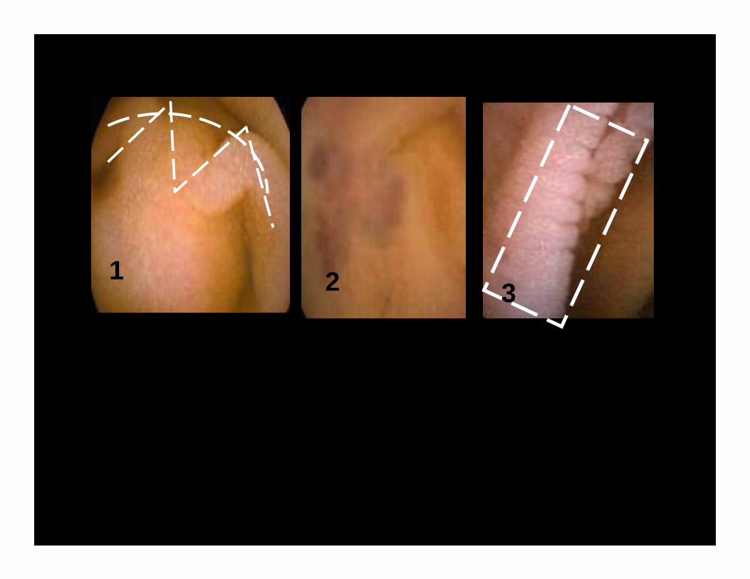
1 2 3
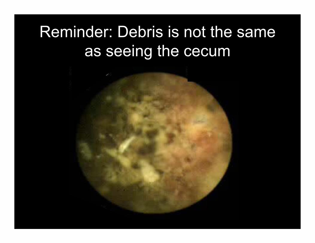
Reminder: Debris is not the same as seeing the cecum


Presentation Agenda
Small Bowel TestingAnatomy reviewSafety Issues in CE Special ConsiderationsPatient SelectionCapsule ProcedureDocumentationVideo TriageInterpretation PhaseFuture Technology
Goals of Capsule Research and Development
Colon Capsulecurrently in clinical trials for FDA approval – In use in Europe– Did not pass efficacy for FDA approval last
year– Will not doubt be used for screening
colonoscopies
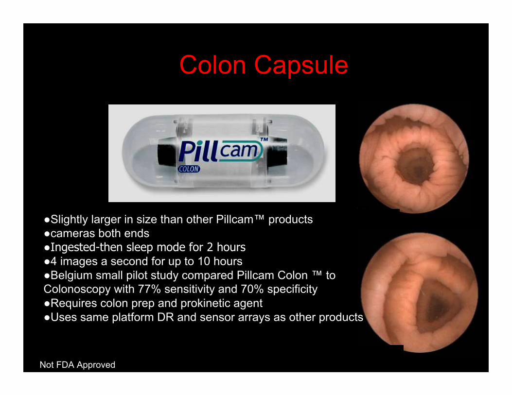
Colon Capsule
●Slightly larger in size than other Pillcam™ products ●cameras both ends ●Ingested-then sleep mode for 2 hours●4 images a second for up to 10 hours ●Belgium small pilot study compared Pillcam Colon ™ to Colonoscopy with 77% sensitivity and 70% specificity ●Requires colon prep and prokinetic agent ●Uses same platform DR and sensor arrays as other products
Not FDA Approved


Future Direction
Not FDA Approved

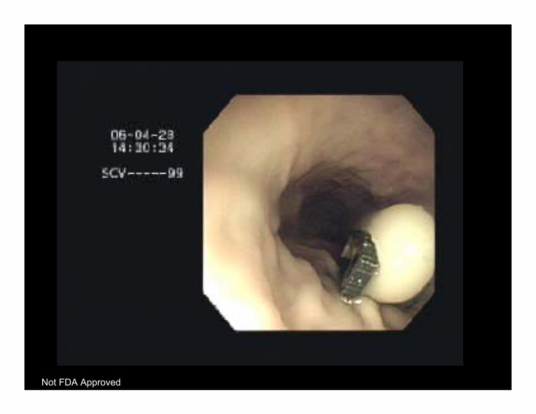
Not FDA Approved
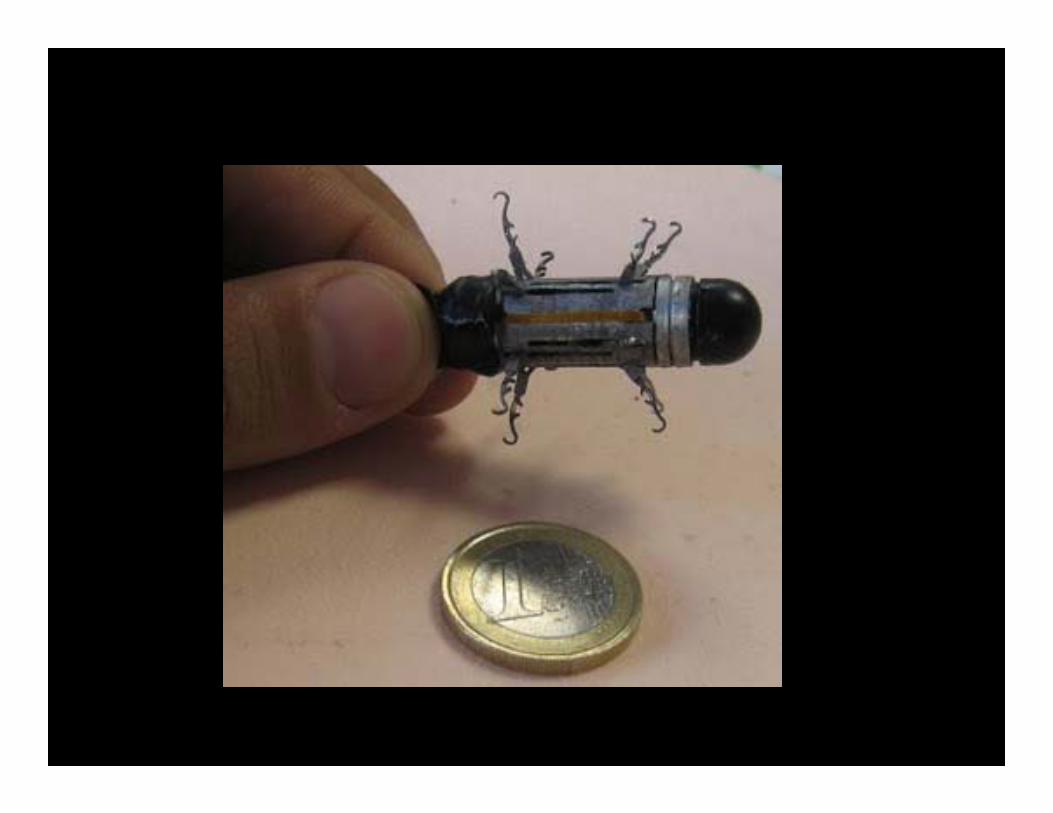

Increase Capsule Rate-Increase battery life
Not FDA Approved

• Non-Photographic Capsule
• GI motility disorders• Accurate biomedical
readings as it moves down GI tract– gastrointestinal
peristaltic pressure– pH levels– temperature and transit
time
SmartPill
Smart Pill™
FDA approved CCF Acquired Gastroparesis Future uses: IBS
Dyspepsia
Dual Sensors

Not FDA Approved

Not FDA Approved
Not FDA Approved

Questions, Thoughts, Concerns