
J-wave syndromes: Brugada and early repolarization syndromes.
36
Thomas Jefferson University Thomas Jefferson University Jefferson Digital Commons Jefferson Digital Commons Department of Medicine Faculty Papers Department of Medicine 8-1-2015 J-wave syndromes: Brugada and early repolarization syndromes. J-wave syndromes: Brugada and early repolarization syndromes. Charles Antzelevitch Gan-Xin Yan Thomas Jefferson University Follow this and additional works at: https://jdc.jefferson.edu/medfp Part of the Cardiology Commons Let us know how access to this document benefits you Recommended Citation Recommended Citation Antzelevitch, Charles and Yan, Gan-Xin, "J-wave syndromes: Brugada and early repolarization syndromes." (2015). Department of Medicine Faculty Papers. Paper 175. https://jdc.jefferson.edu/medfp/175 This Article is brought to you for free and open access by the Jefferson Digital Commons. The Jefferson Digital Commons is a service of Thomas Jefferson University's Center for Teaching and Learning (CTL). The Commons is a showcase for Jefferson books and journals, peer-reviewed scholarly publications, unique historical collections from the University archives, and teaching tools. The Jefferson Digital Commons allows researchers and interested readers anywhere in the world to learn about and keep up to date with Jefferson scholarship. This article has been accepted for inclusion in Department of Medicine Faculty Papers by an authorized administrator of the Jefferson Digital Commons. For more information, please contact: [email protected].
Transcript of J-wave syndromes: Brugada and early repolarization syndromes.
Thomas Jefferson University Thomas Jefferson University
Jefferson Digital Commons Jefferson Digital Commons
Department of Medicine Faculty Papers Department of Medicine
8-1-2015
J-wave syndromes: Brugada and early repolarization syndromes. J-wave syndromes: Brugada and early repolarization syndromes.
Charles Antzelevitch
Gan-Xin Yan Thomas Jefferson University
Follow this and additional works at: https://jdc.jefferson.edu/medfp
Part of the Cardiology Commons
Let us know how access to this document benefits you
Recommended Citation Recommended Citation
Antzelevitch, Charles and Yan, Gan-Xin, "J-wave syndromes: Brugada and early repolarization
syndromes." (2015). Department of Medicine Faculty Papers. Paper 175.
https://jdc.jefferson.edu/medfp/175
This Article is brought to you for free and open access by the Jefferson Digital Commons. The Jefferson Digital Commons is a service of Thomas Jefferson University's Center for Teaching and Learning (CTL). The Commons is a showcase for Jefferson books and journals, peer-reviewed scholarly publications, unique historical collections from the University archives, and teaching tools. The Jefferson Digital Commons allows researchers and interested readers anywhere in the world to learn about and keep up to date with Jefferson scholarship. This article has been accepted for inclusion in Department of Medicine Faculty Papers by an authorized administrator of the Jefferson Digital Commons. For more information, please contact: [email protected].
J Wave syndromes: Brugada and Early Repolarization Syndromes
Charles Antzelevitch, PhD, FHRS1 and Gan-Xin Yan, MD, PhD2,3,4
1 SUNY Upstate Medical University, Syracuse, NY, USA.
2Lankenau Institute for Medical Research and Lankenau Medical Center, Wynnewood, PA, USA.
3Jefferson Medical College, Philadelphia, PA, USA.
4The First Affiliated Hospital, Medical School of Xi'an Jiaotong University, Xi'an, China
Abstract
A prominent J wave is encountered in a number of life-threatening cardiac arrhythmia syndromes,
including the Brugada (BrS) and early repolarization (ERS) syndromes. BrS and ERS differ with
respect to the magnitude and lead location of abnormal J waves and are thought to represent a
continuous spectrum of phenotypic expression termed J wave syndromes. Despite two decades of
intensive research, risk stratification and the approach to therapy of these two inherited cardiac
arrhythmia syndromes are still undergoing rapid evolution. Our objective in this review is to
provide an integrated synopsis of the clinical characteristics, risk stratifiers, as well as the
molecular, ionic, cellular and genetic mechanisms underlying these two fascinating syndromes
that have captured the interest and attention of the cardiology community in recent years.
Keywords
Sudden cardiac death; J wave; Early repolarization; ST segment elevation; Cardiac arrhythmias; ventricular tachycardia; ventricular fibrillation; inherited cardiac arrhythmias syndromes
Introduction
A prominent J wave in humans has long been observed in the ECG in cases of
hypothermia1-3 and hypercalcemia.4, 5 More recently, the presence of prominent J waves has
been identified as a marker for a substrate capable of generating life-threatening ventricular
arrhythmias.6 In humans, the J wave more commonly appears as a J point elevation, with
part of the J wave buried inside the QRS. When greatly amplified, it can appear as an ST
segment elevation, as in cases of Brugada syndrome.
An early repolarization (ER) pattern in the electrocardiogram (ECG), consisting of a distinct
J wave or J point elevation, a notch or slur of the terminal part of the QRS and an ST
Address for correspondence: Dr. Charles Antzelevitch Department of Pharmacology SUNY Upstate Medical University Syracuse, NY 13210 [email protected].
Disclosures: Dr. Antzelevitch is a consultant for Gilead Sciences and received funding from Gilead Sciences and Buchang Pharmaceuticals.
HHS Public AccessAuthor manuscriptHeart Rhythm. Author manuscript; available in PMC 2016 August 01.
Published in final edited form as:Heart Rhythm. 2015 August ; 12(8): 1852–1866. doi:10.1016/j.hrthm.2015.04.014.
Author M
anuscriptA
uthor Manuscript
Author M
anuscriptA
uthor Manuscript
segment elevation (Figure 1), is generally found in healthy young males and has
traditionally been viewed as benign.7, 8 This view was challenged by us in the late 1990s and
early 2000s on the basis of experimental data showing that an ER pattern in the canine
coronary-perfused wedge preparation predisposes to the development of polymorphic
ventricular tachycardia and fibrillation (VT/VF).6, 9-11 Validation of this hypothesis was
provided eight years later by Haïssaguerre et al.12 and Nam et al.13 These reports together
with numerous additional case control and population-based studies provided clinical
evidence of an increased prevalence of ER pattern, particularly in inferior and infero-lateral
leads, among patients with a history of idiopathic ventricular fibrillation, thus confirming a
link between ER pattern in the ECG and life-threatening cardiac arrhythmias. Terminology
relative to ER has been a matter of confusion and contention. 14-16 A recent expert
consensus report recommends that peak of an end QRS notch and/or the onset of an end
QRS slur be designated as J(p) and that J(p) should exceed 0.1mV in ≥2 contiguous inferior
and/or lateral leads of a standard 12-lead ECG for early repolarization to be present.17
The two principal forms of J wave syndrome
Two distinct forms of inherited J wave syndromes are recognized: Early repolarization
syndrome (ERS) and Brugada syndrome (BrS). Both are associated with vulnerability to
polymorphic ventricular tachycardia (VT) and ventricular fibrillation (VF) leading to sudden
cardiac death6, 12, 13, 18 in young adults without apparent structural heart disease. J wave
syndromes are characterized by J-point and ST-elevation in distinct ECG-leads. The J wave
syndromes have also, although far less often, been associated with sudden infant death
syndrome (SIDS).19-21 BrS is the right ventricular variant of hereditary J wave syndromes.
The region most affected by the disease is the anterior right ventricular outflow tract. BrS
patients display J-point and ST-segment elevation in the right precordial leads.22, 23
An early repolarization ECG-pattern (ERP) is characterized by a distinct J wave or J-point-
elevation, notch or slur of the terminal part of the QRS and ST-elevation in the lateral (type
I), infero-lateral (type II) or in infero-lateral + anterior or right ventricular leads (type III).
ERP can be observed in acquired conditions such as hypothermia or ischemia.6, 24, 25 When
associated with VT/VF, it is referred to as early repolarization syndrome (ERS). Despite the
different regional localization within the heart, BrS and ERS display several clinical
similarities, suggesting similar pathophysiology (Table 1).10, 14, 26-28 Previous studies have
pointed to different pathophysiological basis based on the observation that sodium channel
blockers unmask or accentuate J wave manifestation in BrS, but reduces the amplitude in
ERS.29 The recent study by Nakagawa et al., however, showed that J waves recorded using
unipolar LV epicardial leads introduced into the left lateral coronary vein in ERS patients
were indeed augmented, even though J waves recorded in the lateral precordial leads were
diminished, due principally to engulfment of the surface J wave by the widened QRS.30
Interestingly, the study of Kawata et al. showing an effect of pilsicainide to augment J waves
and ST segment elevation in the right precordial leads, while reducing ER in the lateral leads
in the same patient, actually provides support for a similar mechanism of ERS and BrS in
light of the results advanced by Nakagawa and co-workers.29 Also in support of the notion
that these ECG patterns and syndromes are closely related are reports of cases of ERS that at
times transition to ERS plus BrS.31, 32
Antzelevitch and Yan Page 2
Heart Rhythm. Author manuscript; available in PMC 2016 August 01.
Author M
anuscriptA
uthor Manuscript
Author M
anuscriptA
uthor Manuscript
ERS and BrS share a number of common clinical features (Table 1): Males are dominant in
both syndromes: in the Brugada syndrome, the percentage of males involved ranges from 71
to 80 % in Caucasians but as high as 94%-96% in Japanese.33, 34 Similarly, in the setting of
ER pattern, males develop ventricular fibrillation in 60% of cases in the study of
Haïssaguerre et al 12 but in a much higher percentage reported by Japanese investigators.29
The patients with BrS or ERS may be totally asymptomatic until presenting with syncope
and sudden cardiac arrest often due to ventricular fibrillation. The risk for development of
VT/VF is dictated in large part by the ER subtype (Type I, II or III), as previously
discussed.6 In both syndromes, the highest incidence of ventricular fibrillation or sudden
cardiac death occurs in the third decade of life when testosterone levels peak in males.35
Another important clinical feature of both syndromes is that J wave and associated ST
segment elevation are accentuated during bradycardia or after pauses.36, 37 This may explain
why ventricular fibrillation in both syndromes often occurs during sleep or at a low level of
physical activities.29, 38
In addition to reports of SCD in otherwise healthy patients displaying an ER pattern, this
ECG pattern has been associated with an increased arrhythmogenic risk and SCD in patients
with acute myocardial infarction39, chronic coronary disease40, heart failure41 and
hypothermia 24, 42
Genetics
BrS has been associated with mutations in 19 different genes (Table 2). To date more than
300 BrS-related mutations in SCN5A have been described 14, 43, 44 Mutations in CACNA1C
(Cav1.2), CACNB2b (Cavß2b) and CACNA2D1 (Cavα2δ) are found in approximately 13%
of probands.45-48 Mutations in glycerol-3-phophate dehydrogenase 1-like enzyme gene
(GPD1L), SCN1B (β1-subunit of Na channel), KCNE3 (MiRP2), SCN3B (β3-subunit of Na
channel), KCNJ8 (Kir 6.1), KCND3 (Kv4.3), RANGRF (MOG1), SLMAP, ABCC9
(SUR2A), (Navß2), PKP2 (Plakophillin-2), FGF12 (FHAF1), HEY2, and SEMA3A
(Semaphorin) are more rare.49-5354-6153, 62-69 We recently reported an association of BrS
with SCN10A, a neuronal sodium channel that co-associates with SCN5A, with a yield of
16.7%.70 Mutations in these genes lead to loss of function in sodium channel current (INa)
and calcium channel current (ICa), as well as to a gain of function in transient outward
potassium current (Ito) or ATP-sensitive potassium current (IK-ATP). Mutations in KCNH2
and KCNE5, although not causative, have been identified as capable of modulating the
substrate for the development of BrS. Loss-of-function mutations in HCN4 causing a
reduction in pacemaker current, If, can unmask BrS by reducing heart rate.71
The familial nature of the ER pattern has been demonstrated in a number of studies.72-74 The
ER pattern and ERS have been associated with mutations in 7 genes. Consistent with the
findings that IK-ATP activation can generate an ER pattern in canine ventricular wedge
preparations, mutations in KCNJ8 and ABCC9, responsible for the pore forming and ATP-
sensing subunits of the IK-ATP channel, has been reported in a patients with ERS as
well.49, 51, 75 Loss of function mutations in the α1, α2 and α2δ subunits of the cardiac L-
type calcium channel (CACNA1C, CACNB2, and CACNA2D1) have been uncovered in
Antzelevitch and Yan Page 3
Heart Rhythm. Author manuscript; available in PMC 2016 August 01.
Author M
anuscriptA
uthor Manuscript
Author M
anuscriptA
uthor Manuscript
patients with ERS45 as have mutations in SCN5A.76 We have also recently reported an
association of SCN10A with ERS.70
A word of caution is appropriate here. It is important to recognize that a small fraction of
identified genetic variants in the numerous genes associated with BrS and ERS have been
examined using functional expression studies to establish causality or to establish a plausible
contribution to pathogenesis. Only a handful have been studied in genetically engineered
animal model and very few have been studied in native cardiac cells or in induced
pluripotent stem cell-derived cardiac myocytes isolated from ERS and BrS patients.
Computational strategies developed to predict the functional consequences of mutations are
helpful, but these methods have not been rigorously tested. The lack of functional or
biological validation of mutation effects remains the most severe limitation of genetic test
interpretation as recently highlighted by Schwartz et al.77 This limitation is still more
concerning in cases in which a susceptibility gene is identified on the basis of a single
proband and with the absence of familial segregation data. Recent studies have suggested a
more complex genetic background for BrS. Bezzina et al60 provided evidence that BrS is
associated with common genetic variants suggesting a multigenic origin of the syndrome.
Other authors, including Le Scouarnec et al 78 and Behr et al79, have questioned the impact
of rare gene-variants, with the exception of SCN5A, in the pathogenesis of the synmdrome.
These studies once again call for caution in the interpretation of genetic resutls as well as the
need for genotype-phenotype correlation data and functional expression data before
designating a rare variants as causative of the disease.
Ionic and Cellular Mechanisms
The J wave is inscribed as a consequence of a transmural voltage gradient caused by the
presence of an action potential notch in epicardium but not endocardium, secondary to a
transmural distribution of transient outward current (Ito).27
The cellular mechanisms underlying J wave syndromes have been mired in
controversy.80, 81 Two principal hypotheses have been advanced in the case of BrS (Figure 2): 1) The repolarization hypothesis maintains that an outward shift in the balance of
currents in right ventricular epicardium leads to repolarization abnormalities resulting in the
development of phase 2 reentry, which generates closely-coupled premature beats capable of
precipitating VT/VF; 2) The depolarization hypothesis maintains that slow conduction in
the right ventricular outflow tract plays a primary role in the development of the
electrocardiographic and arrhythmic manifestations of the syndrome. Although these
theories are not mutually exclusive and may indeed be synergistic, from the standpoint of
appropriate therapy, correct assessment of the cellular pathophysiology is important.
The most compelling evidence in support of the depolarization hypothesis comes from a
study by Nademanee et al.82 showing that radiofrequency (RF) ablation of epicardial sites
displaying late potentials and fractionated bipolar electrograms (EGs) in the right ventricular
outflow tract (RVOT) of BrS patients significantly reduced the arrhythmia-vulnerability and
ECG-manifestation of the disease. These authors concluded that the late potential (LP) and
fractionated electrogram activity are due to conduction delays within the RVOT and
elimination of the sites of slow conduction is the basis for the ameliorative effect of ablation
Antzelevitch and Yan Page 4
Heart Rhythm. Author manuscript; available in PMC 2016 August 01.
Author M
anuscriptA
uthor Manuscript
Author M
anuscriptA
uthor Manuscript
therapy.82 In a direct test of this hypothesis, our group recently suggested an alternative
cellular electrophysiological mechanism as the basis for late potentials and fractionated
electrogram activity in the setting of BrS.83 As illustrated in Figure 3, when the genetic
defects associated with BrS are pharmacologically mimicked in the coronary-perfused
canine right ventricular wedge model of BrS, high frequency late potentials develop in the
right ventricular (RV) epicardium secondary to concealed phase 2 reentry. At other sites,
low-voltage fractionated electrogram activity develops due to regional desynchronization in
the appearance of the second action potential upstroke, secondary to accentuation of the
epicardial action potential notch. In no case was delayed conduction of the primary beat
observed.
If late potentials and fractionated electrogram activity recorded from the RVOT do not
reflect depolarization and conduction abnormalities, what is the basis for the ameliorative
effect of RVOT ablation? Figure 4 illustrates that ablation of sites of phase 2 reentry in the
canine ventricular wedge model of BrS markedly diminishes the manifestation of J waves
and ST segment elevation and abolishes all arrhythmic activity. The data provide support for
the hypothesis that ablation destroys the cells with the most prominent action potential
notch, thus eliminating the cells responsible for the repolarization abnormalities that give
rise to phase 2 reentry and VT/VF. These findings collectively lend strong support for the
repolarization hypothesis. Also in support of this thesis is the congruence between BrS and
ERS, which by virtue of its name is considered to be due to a repolarization defect.
Conduction delays are known to give rise to notching of the QRS complex. Although such
notching often occurs on the rising phase of the QRS or during the middle of the descending
phase, it can occur at the terminal portion of the QRS, thus masquerading as a J wave. 14, 84
How then can we differentiate an end of QRS notch from a J wave? One method that we and
others have previously suggested is to gauge the response to prematurity or to an increase in
rate. Delayed conduction invariably becomes worse at faster rates or during premature beats
thus leading to an accentuation of the notch, whereas repolarization defects are usually
mitigated resulting in a diminution of the J wave at faster rates. Although typical J waves are
usually accentuated with bradycardia or long pauses, the opposite has been described.85, 86 J
waves are often seen in young males with no apparent structural heart diseases, whereas
intra-ventricular conduction delay is often observed in older individuals or those with a
history of myocardial infarction or cardiomyopathy. Figure 5 illustrates examples of both.
The tachycardia-augmented notches (Figure 5D) can result in apparent “J” waves that are as
tall or taller than 50% of the R wave85 and as such may be more reasonably characterized as
an R’ due to intra-ventricular conduction delay.84 The prognostic value of a fragmented
QRS has been demonstrated in BrS, 87, 88 although fragmentation of the QRS is not
associated with increased risk in the absence of cardiac disease. 89 Factors that may aid in
the differential diagnosis of J wave vs IVCD-mediated syndromes are summarized in Table 4.
The ionic and cellular mechanisms underlying ERS were recently advanced in a report by
Koncz and co-workers.90 The authors provided evidence in support of the hypothesis that,
similar to the mechanism operative in BrS, an accentuation of transmural gradients, in this
case across the LV wall, underlies the repolarization abnormalities responsible for ERS,
Antzelevitch and Yan Page 5
Heart Rhythm. Author manuscript; available in PMC 2016 August 01.
Author M
anuscriptA
uthor Manuscript
Author M
anuscriptA
uthor Manuscript
giving rise to J point elevation, distinct J waves, or slurring of the terminal part of the QRS
(Figure 1). The repolarization defect is accentuated by cholinergic agonists and reduced by
quinidine, isoproterenol, cilostazol and milrinone, accounting for the ability of these agents
to reverse the repolarization abnormalities responsible for ERS. 90, 91 These authors also
showed significantly higher intrinsic levels of Ito in the inferior LV, providing an
explanation for the greater vulnerability of the inferior LV wall to VT/VF.90 The clinical
translation of these experimental findings has been facilitated by the advent and
implementation of electrocardiographic imaging (ECGI) by Rudy and co-workers. Using
ECGI mapping, Ghosh et al. identified abnormally short activation-recovery intervals (ARI)
in the inferior and lateral regions of LV and a marked dispersion of repolarization in support
of regional accelerated repolarization.92 More recent studies involving ECGI mapping in an
ERS patient during VF have demonstrated VF rotors anchored in the inferior-lateral left
ventricular wall.15
Risk stratification
A great deal of effort has been devoted to assessment of risk for the development of life-
threatening arrhythmias. The incidental discovery of a J wave on routine screening should
not be interpreted as a marker of “high risk” for SCD since the odds for this fatal disease is
approximately 1:10,000.93 Rosso et al. indicated that the presence of a J wave on the ECG
increases the probability of VF from 3.4:100,000 to 11:100,000.94, 95 However, careful
attention should be paid to subjects with “high risk” ER or J waves. Table 3 presents the
available data of studies designed to identify patients at high risk. Among these risk
stratifiers, some are highly predictive, including: 1) history of cardiac events or syncope
likely due to VT/VF, 2) pause-dependent augmentation of J waves, especially when
accompanied by T wave inversion, and 3) prominent J waves in global leads including Type
1 ST segment elevation in the right precordial leads. Fragmentation of the QRS, although
predictive of cardiac events in the J wave syndromes, is non-specific in that it is associated
with a high risk of sudden death in other cardiac arrhythmia syndromes.
In the case of ERS, the morphology of the ST segment is reported to be associated with
increased arrhythmic risk. Tikkanen et al were the first to suggest that a horizontal or
descending ST segments portends a higher risk for arrhythmic events and SCD.96 In
contrast, an ER pattern with a rapidly ascending pattern, typically observed in healthy
athletes, was associated with a relatively benign prognosis. Rosso et al. and Rollin et al.
reported additional evidence linking a horizontal ST segment to an increased risk for SCD
soon thereafter. 97, 98
In a recent review, Mahida and coworkers concluded that the presence of inferior or
widespread J point elevation, a markedly elevated J point, a horizontal or descending ST
segment, pause-dependent augmentation of the J wave amplitude, short coupled
extrasystoles, and QRS notching predict progressively greater risk for SCD in patients
presenting with syncope. 15
Antzelevitch and Yan Page 6
Heart Rhythm. Author manuscript; available in PMC 2016 August 01.
Author M
anuscriptA
uthor Manuscript
Author M
anuscriptA
uthor Manuscript
Approaches to therapy
Implantation of an ICD is the mainstay of therapy for J wave syndrome patients presenting
with aborted SCD or documented VT/VF with or without syncope (Class I
recommendation). 99, 100 According to the 2013 HRS/EHRA/APHRS consensus statement,
ICDs can be useful (Class IIa) in symptomatic BrS patients with Type I pattern, in whom
syncope was likely caused by VT/VF and may be considered (Class IIb) in asymptomatic
patients inducible by programmed electrical stimulation (PES).101 ICDs are not indicated in
asymptomatic patients. There is no clear role for PES in patients with ERS.
Pacemaker therapy—Although arrhythmias and sudden cardiac death generally occur
during sleep or at rest and have been associated with slow heart rates, a potential therapeutic
role for cardiac pacing remains largely unexplored.102
Only a few case reports are available in the literature.103, 104
Radiofrequency Ablation Therapy—Nademanee et al.82 showed that radiofrequency
(RF) ablation of epicardial sites displaying late potentials and fractionated bipolar
electrograms (EGs) in the right ventricular outflow tract (RVOT) of BrS patients can
significantly reduce arrhythmia-vulnerability and the ECG-manifestation of the disease.
Ablation at these sites was reported to render VT/VF non-inducible and to normalize the
Brugada ECG pattern in the majority of patients over a period of weeks or months. Long-
term follow-up (20±6months) showed no recurrent VT/VF with only 1 patient on medical
therapy with amiodarone. Since then, case reports have been published in support of these
effects.105 Ablation therapy can be life-saving in otherwise uncontrollable cases or cases in
which ICD therapy is contraindicated. In the HRS/EHRA/APHRS expert consensus
guideline, radiofrequency ablation is a Class IIb recommendation in BrS-patient with
frequent appropriate ICD-shocks due to recurrent electrical storms.101 There are no clinical
reports of ablation of the LV substrate in patients with ERS.
Pharmacologic approach to therapy
Approach to therapy of BrS—ICD implantation may not be a practical solution for
infants and young children due to a high complication rate or for patients residing in regions
of the world where an ICD is out of reach because of economic factors. A pharmacologic
approach to therapy, based on a rebalancing of currents active during the early phases of the
epicardial action potential in the right ventricle so as to reduce the magnitude of the action
potential notch and/or restore the action potential dome, has been a focus of basic and
clinical research in recent years. Table 5 lists the various pharmacologic agents thus far
investigated. Antiarrhythmic agents such as amiodarone and β blockers have been shown to
be ineffective. 106 Class IC antiarrhythmic drugs (such as flecainide and propafenone) and
class IA agents, such as procainamide, are contraindicated because of their effects to unmask
the Brugada syndrome and induce arrhythmogenesis. Disopyramide is a class IA
antiarrhythmic that has been demonstrated to normalize ST segment elevation in some
Brugada patients but to unmask the syndrome in others. 107
Antzelevitch and Yan Page 7
Heart Rhythm. Author manuscript; available in PMC 2016 August 01.
Author M
anuscriptA
uthor Manuscript
Author M
anuscriptA
uthor Manuscript
Because the presence of a prominent transient outward current, Ito, is central to the
mechanism underlying BrS and ERS, the most rationale approach to therapy, regardless of
the ionic or genetic basis for the disease, is to partially inhibit Ito. Cardio-selective and Ito-
specific blockers are not currently available. 4-aminopyridine (4-AP) is an agent that is ion-
channel specific at low concentrations, but is not cardio-selective in that it inhibits Ito in the
nervous system. Although it is effective in suppressing arrhythmogenesis in wedge models
of the Brugada syndrome, 10 it is unlikely to be of clinical benefit because of neurally-
mediated and other side effects.
The only agent on the market in the United States and around the world with significant Ito
blocking properties is quinidine. It is for this reason that we suggested in 1999 that this agent
may be of therapeutic value in BrS.10, 108 Experimental studies have since shown quinidine
to be effective in restoring the epicardial action potential dome, thus normalizing the ST
segment and preventing phase 2 reentry and polymorphic VT in different experimental
models of the Brugada syndrome, regardless of which pharmacologic agents were used to
mimic BrS-phenotype. 10, 83, 109-111 Additionally, a recent experimental study suggests that
quinidine, owing to its effect to block Ito, can exert a protective effect against hypothermia-
induced VT/VF in a J wave syndrome model.42 It is noteworthy that historically, quinidine
was used to prevent ventricular fibrillation in patients who required hypothermia for surgical
procedures. 111
Clinical evidence of the effectiveness of quinidine in normalizing ST segment elevation and
or preventing arrhythmic events in patients with the BrS has been reported in numerous
studies and case reports 112-126 The first prospective study describing the effects of
quinidine to prevent inducible and spontaneous ventricular fibrillation (VF) was reported by
Belhassen and coworkers.115 The results were consistent with those reported the same group
in prior years 113, 127 and later by other investigators 128, 129. The data highlight the need for
randomized clinical trials to assess the effectiveness of quinidine, preferably in patients with
frequent events who have already received an ICD. Hermida et al. reported 76 % efficacy in
prevention of VF induced by PES.128
Because of the GI side effects of high dose quinidine, low-dose quinidine (<600mg) has
been suggested to be as a therapeutic option. Marquez et al. evaluated the clinical history of
symptomatic patients with recurrent arrhythmias and frequent ICD discharges and reported
that relatively low dose quinidine, as adjunctive therapy, completely prevented arrhythmias
in 85 % of the patients (median follow-up of 4years).118.
In a more recent trial conducted at two French centers, 44 asymptomatic BrS patients with
inducible VT/VF were enrolled (47 ± 10 years, 95% male).130 Of these, 34 (77%) were no
longer inducible while treated with 600 mg/day hydroquinidine (HQ) for 6.2 ± 3 years.
Among the 10 other patients (22%), who remained inducible and received ICD (Group PVS
+), none received appropriate therapy during a mean follow-up of 7.7 ± 2 years. The overall
annual rate of arrhythmic events was 1.04%, without significant difference between
inducibility under HQ. One-third of patients experienced device-related complications.
Antzelevitch and Yan Page 8
Heart Rhythm. Author manuscript; available in PMC 2016 August 01.
Author M
anuscriptA
uthor Manuscript
Author M
anuscriptA
uthor Manuscript

A prospective registry of empiric quinidine for asymptomatic Brugada syndrome has been
established. The study appears at the National Institutes of Health website
(ClinicalTrials.gov) and can be accessed at http://clinicaltrials.gov/ct2/show/NCT00789165?
term_brugada&rank_2. Doses between 600 and 900 mg were recommended, if tolerated. 116
In the latest_HRS/EHRA/APHRS expert consensus statement, quinidine was given a class
IIa recommendation in BrS patients who are qualified for an ICD but in whom hindering
factors are present, IIa in ICD-patients with electrical storms, and IIb recommendation in
asymptomatic BrS-patients displaying a spontaneous Type I ECG. 101
The development of a more cardio-selective and Ito-specific blocker would be a most
welcome addition to the limited therapeutic armamentarium currently available to combat
this disease. Agents that augment the L-type calcium channel current, such as β adrenergic
agents like isoproterenol, denopamine or orciprenaline, are useful as well. 10, 121, 125, 131-133
Isoproterenol, sometimes in combination with quinidine, has been utilized successfully to
control VF storms and normalizing ST elevation particularly in
children 113, 114, 125, 129, 134-141. 119, 142-146. The occurrence of spontaneous VF in patients
with Brugada syndrome is often related to increases in vagal tone and correspondingly
electrical storm is sometimes treatable by the increase of sympathetic tone via isoproterenol
administration. In the latest HRS/EHRA/APHRS guideline, Isoprotereonol has a Class IIa
recommendation for BrS patients presenting with electrical storms.101
Another promising pharmacologic approach is the administration of phosphodiesterase III
inhibitor cilostazol 125, 132, 147, which normalizes the ST segment, most likely by
augmenting calcium current (ICa) as well as by reducing Ito secondary to an increase in
cAMP and heart rate.148 Other diverse effects of cilostazol in playing a role in its beneficial
impact cannot be excluded. (e.g.: adenosine, NO, mitochondrial IKATP 149) Its efficacy in
combination with bepridil in preventing VF-episodes was recently reported by Shinoharaet
al.150 A case report describing the failure of cilostazol in the treatment of a BrS-patient is
also available in the literature.151
Milrinone is another phosphodiesterase III inhibitor recently identified as a more potent
alternative to cilostazol in suppressing ST elevation and arrhythmogenesis in an
experimental model of BrS.83, 152 No clinical reports have appeared as yet.
Wenxin Keli, a traditional Chinese medicine (TCM), in addition to its actions to suppress
atrial fibrillation by atrial-selective inhibition of INa-dependent parameters153, has recently
been shown to inhibit Ito and thus to suppress polymorphic VT in experimental models of
BrS when combined with low concentrations of quinidine (5 μM). 110 A recent study has
also reported the effect of Wenxin Keli to suppress ischemia-induced ventricular
arrhythmias.154
Agents that augment sodium channel current active during the early phases of the action
potential, including bepridil and dimethyl lithospermate B (dmLSB), have been suggested to
be of value in BrS. Bepridil has been reported to suppress VT/VF in several studies of
patients with BrS.125, 155-157 The drug's action are thought to be mediated by: 1) inhibition
of Ito; 2) augmentation of INa via upregulation of the channels158; and 3) prolongation of QT
Antzelevitch and Yan Page 9
Heart Rhythm. Author manuscript; available in PMC 2016 August 01.
Author M
anuscriptA
uthor Manuscript
Author M
anuscriptA
uthor Manuscript
interval at slow rates thus increasing the QT/RR slope.155, 157 Dimethyl lithospermate B, an
extract of Danshen, a traditional Chinese herbal remedy, has been reported to slow
inactivation of INa thus increasing INa during the early phases of the action potential (AP)
and thus to suppress arrhythmogenesis in experimental models of BrS.159
Approach to therapy of ERS—It is not surprising that the approach to therapy of ERS is
similar to that of BrS, since the mechanisms underlying the two syndromes are similar.
Quinidine, phosphodiesterase III inhibitors and isoproterenol have all been shown to exert
an ameliorative effect in preventing or quieting arrhythmias associated with ERS.
Isoproterenol has been shown to be effective in quieting electrical storms developing in
patients with either BrS125, 134 or ERS75. Isoproterenol acts by increasing ICa, thus leading
to a reversal of repolarization abnormalities secondary to restoration of the epicardial action
potential dome in experimental models of BrS 10, 109 and ERS 90.
The phosphodiesterase (PDE) III inhibitor cilostazol has been reported to reduce the ECG
and arrhythmic manifestations of ERS.160 PDE inhibitors are known to activate ICa
secondary to an increase in cAMP.132, 148, 161-165 The augmentation of ICa is thought to
prevent arrhythmias associated with the J wave syndromes by reversing the repolarization
defects and restoring electrical homogeneity across the ventricular wall secondary to
restoration of the epicardial action potential dome in both BrS152 and ERS.42 Cilostazol has
been hypothesized to also block Ito. Augmentation of ICa together with inhibition of Ito are
expected to produce an inward shift in the balance of currents active during the early phases
of the epicardial action potential that should be especially effective in suppressing J wave
activity.
The effectiveness of bepridil in ERS has been reported in a single patient thus far.166
No clinical data are available regarding the effectiveness of radiofrequency (RF) ablation in
the setting of ERS, despite the fact that low voltage fractionated electrogram activity and
high frequency late potentials are observed in the LV in patients with ERS30 and in
experimental models of ERS (Yoon and Antzelevitch, unpublished observation). Nakagawa
et al.30 reported the results of a study in which they recorded epicardial electrograms
directly from the left ventricle of patients diagnosed with ERS by introducing a multipolar
catheter into the left lateral (marginal) coronary vein, anterior interventricular vein (AIV),
and middle cardiac vein (MCV) via the coronary sinus. The authors reported late potentials
in the bipolar electrograms recorded from the left ventricular (LV) epicardium of the ERS
patients.30
Acknowledgments
Funding: This study was supported by grant HL47678 from the NHLBI (CA), the Sharpe-Strumia Research Foundation (GXY), and grant NSFC-81370289 from National Natural Science Foundation of China (GXY)
References
1. Clements SD, Hurst JW. Diagnostic value of ECG abnormalities observed in subjects accidentally exposed to cold. AmJCardiol. 1972; 29:729–734.
Antzelevitch and Yan Page 10
Heart Rhythm. Author manuscript; available in PMC 2016 August 01.
Author M
anuscriptA
uthor Manuscript
Author M
anuscriptA
uthor Manuscript

2. Thompson R, Rich J, Chmelik F, Nelson WL. Evolutionary changes in the electrocardiogram of severe progressive hypothermia. JElectrocardiol. 1977; 10:67–70. [PubMed: 833527]
3. Eagle K. Images in clinical medicine. Osborn waves of hypothermia. NEnglJMed. 1994; 10:680.
4. Kraus F. Ueber die wirkung des kalziums auf den kreislauf 1 ). DtschMedWochenschr. 1920; 46:201–203.
5. Sridharan MR, Horan LG. Electrocardiographic J wave of hypercalcemia. AmJCardiol. 1984; 54:672–673.
6. Antzelevitch C, Yan GX. J wave syndromes. Heart Rhythm. 2010; 7:549–558. [PubMed: 20153265]
7. Wasserburger RH, Alt WJ. The normal RS-T segment elevation variant. Am J Cardiol. 1961; 8:184–192. [PubMed: 13783301]
8. Mehta MC, Jain AC. Early repolarization on scalar electrocardiogram. Am J MedSci. 1995; 309:305–311.
9. Gussak I, Antzelevitch C. Early repolarization syndrome: clinical characteristics and possible cellular and ionic mechanisms. JElectrocardiol. 2000; 33:299–309. [PubMed: 11099355]
10. Yan GX, Antzelevitch C. Cellular basis for the Brugada syndrome and other mechanisms of arrhythmogenesis associated with ST segment elevation. Circulation. 1999; 100:1660–1666. [PubMed: 10517739]
11. Shu J, Zhu T, Yang L, Cui C, Yan GX. ST-segment elevation in the early repolarization syndrome, idiopathic ventricular fibrillation, and the Brugada syndrome: cellular and clinical linkage. J Electrocardiol. 2005; 38:26–32. [PubMed: 16226071]
12. Haissaguerre M, Derval N, Sacher F, Jesel L, Deisenhofer I, De Roy L, Pasquie JL, Nogami A, Babuty D, Yli-Mayry S, De Chillou C, Scanu P, Mabo P, Matsuo S, Probst V, Le Scouarnec S, Defaye P, Schlaepfer J, Rostock T, Lacroix D, Lamaison D, Lavergne T, Aizawa Y, Englund A, Anselme F, O'Neill M, Hocini M, Lim KT, Knecht S, Veenhuyzen GD, Bordachar P, Chauvin M, Jais P, Coureau G, Chene G, Klein GJ, Clementy J. Sudden cardiac arrest associated with early repolarization. NEnglJ Med. 2008; 358:2016–2023.
13. Nam GB, Kim YH, Antzelevitch C. Augmentation of J waves and electrical storms in patients with early repolarization. NEnglJ Med. 2008; 358:2078–2079.
14. Antzelevitch C. J wave syndromes: molecular and cellular mechanisms. J Electrocardiol. 2013; 46:510–518. [PubMed: 24011992]
15. Mahida S, Derval N, Sacher F, Berte B, Yamashita S, Hooks DA, Denis A, Lim H, Amraoui S, Aljefairi N, Hocini M, Jais P, Haissaguerre M. History and clinical significance of early repolarization syndrome. Heart Rhythm. 2015; 12:242–9. [PubMed: 25257090]
16. Wellens HJ, Schwartz PJ, Lindemans FW, Buxton AE, Goldberger JJ, Hohnloser SH, Huikuri HV, Kaab S, La Rovere MT, Malik M, Myerburg RJ, Simoons ML, Swedberg K, Tijssen J, Voors AA, Wilde AA. Risk stratification for sudden cardiac death: current status and challenges for the futuredagger. EurHeart J. 2014; 35:1642–1651.
17. Macfarlane P, Antzelevitch C, Haissaguerre M, Huikuri HPMRR, Sacher F, Tikkanen J, Wellens H, Yan G-X. CONSENSUS PAPER - EARLY REPOLARIZATION PATTERN. J Amer Coll Cardiol. 2015 In Press.
18. Brugada P, Brugada J. Right bundle branch block, persistent ST segment elevation and sudden cardiac death: a distinct clinical and electrocardiographic syndrome: a multicenter report. JAmCollCardiol. 1992; 20:1391–1396.
19. Kanter RJ, Pfeiffer R, Hu D, Barajas-Martinez H, Carboni MP, Antzelevitch C. Brugada-like syndrome in infancy presenting with rapid ventricular tachycardia and intraventricular conduction delay. Circulation. 2012; 125:14–22. [PubMed: 22090166]
20. Antzelevitch C. Molecular biology and cellular mechanisms of brugada and long QT syndromes in infants and young children. JElectrocardiol. 2001; 34:177–181. [PubMed: 11781953]
21. Wedekind H, Smits JP, Schulze-Bahr E, Arnold R, Veldkamp MW, Bajanowski T, Borggrefe M, Brinkmann B, Warnecke I, Funke H, Bhuiyan ZA, Wilde AA, Breithardt G, Haverkamp W. De novo mmutation in the SCN5A gene associated with early onset of sudden infant death. Circulation. 2001; 104:1158–1164. [PubMed: 11535573]
22. Antzelevitch C. Brugada syndrome. Pacing ClinElectrophysiol. 2006; 29:1130–1159.
Antzelevitch and Yan Page 11
Heart Rhythm. Author manuscript; available in PMC 2016 August 01.
Author M
anuscriptA
uthor Manuscript
Author M
anuscriptA
uthor Manuscript

23. Coronel R, Casini S, Koopmann TT, Wilms-Schopman FJ, Verkerk AO, de Groot JR, Bhuiyan Z, Bezzina CR, Veldkamp MW, Linnenbank AC, van der Wal AC, Tan HL, Brugada P, Wilde AA, de Bakker JM. Right ventricular fibrosis and conduction delay in a patient with clinical signs of Brugada syndrome: a combined electrophysiological, genetic, histopathologic, and computational study. Circulation. 2005; 112:2769–2777. [PubMed: 16267250]
24. Bastiaenen R, Hedley PL, Christiansen M, Behr ER. Therapeutic hypothermia and ventricular fibrillation storm in early repolarization syndrome. Heart Rhythm. 2010; 7:832–834. [PubMed: 20206297]
25. Federman NJ, Mechulan A, Klein GJ, Krahn AD. Ventricular fibrillation induced by spontaneous hypothermia in a patient with early repolarization syndrome. J Cardiovasc Electrophysiol. 2013; 24:586–588. [PubMed: 23140469]
26. Nam GB. Idiopathic ventricular fibrillation, early repolarization and other J wave-related ventricular fibrillation syndromes. Circ J. 2012; 76:2723–2731. [PubMed: 23131759]
27. Yan GX, Antzelevitch C. Cellular basis for the electrocardiographic J wave. Circulation. 1996; 93:372–379. [PubMed: 8548912]
28. McIntyre WF, Perez-Riera AR, Femenia F, Baranchuk A. Coexisting early repolarization pattern and Brugada syndrome: recognition of potentially overlapping entities. J Electrocardiol. 2012; 45:195–198. [PubMed: 22178622]
29. Kawata H, Noda T, Yamada Y, Okamura H, Satomi K, Aiba T, Takaki H, Aihara N, Isobe M, Kamakura S, Shimizu W. Effect of sodium-channel blockade on early repolarization in inferior/lateral leads in patients with idiopathic ventricular fibrillation and Brugada syndrome. Heart Rhythm. 2012; 9:77–83. [PubMed: 21855521]
30. Nakagawa K, Nagase S, Morita H, Ito H. Left ventricular epicardial electrogram recordings in idiopathic ventricular fibrillation with inferior and lateral early repolarization. Heart Rhythm. 2013; 11:314–317. [PubMed: 24184784]
31. McIntyre WF, Perez-Riera AR, Femenia F, Baranchuk A. Coexisting early repolarization pattern and Brugada syndrome: recognition of potentially overlapping entities. J Electrocardiol. 2012; 45:195–8. [PubMed: 22178622]
32. Nam GB, Ko KH, Kim J, Park KM, Rhee KS, Choi KJ, Kim YH, Antzelevitch C. Mode of onset of ventricular fibrillation in patients with early repolarization pattern vs. Brugada syndrome. Eur Heart J. 2010; 31:330–339. [PubMed: 19880418]
33. Benito B, Sarkozy A, Mont L, Henkens S, Berruezo A, Tamborero D, Arzamendi D, Berne P, Brugada R, Brugada P, Brugada J. Gender differences in clinical manifestations of Brugada syndrome. J Am Coll Cardiol. 2008; 52:1567–1573. [PubMed: 19007594]
34. Kamakura T, Kawata H, Nakajima I, Yamada Y, Miyamoto K, Okamura H, Noda T, Satomi K, Aiba T, Takaki H, Aihara N, Kamakura S, Kimura T, Shimizu W. Significance of non-type 1 anterior early repolarization in patients with inferolateral early repolarization syndrome. J Am Coll Cardiol. 2013; 62:1610–1618. [PubMed: 23850930]
35. Matsumoto AM. Fundamental aspects of hypogonadism in the aging male. Reviews in urology. 2003; 5(Suppl 1):S3–s10. [PubMed: 16985941]
36. Kalla H, Yan GX, Marinchak R. Ventricular fibrillation in a patient with prominent J (Osborn) waves and ST segment elevation in the inferior electrocardiographic leads: a Brugada syndrome variant? J Cardiovasc Electrophysiol. 2000; 11:95–98. [PubMed: 10695469]
37. Aizawa Y, Sato A, Watanabe H, Chinushi M, Furushima H, Horie M, Kaneko Y, Imaizumi T, Okubo K, Watanabe I, Shinozaki T, Aizawa Y, Fukuda K, Joo K, Haissaguerre M. Dynamicity of the J-wave in idiopathic ventricular fibrillation with a special reference to pause-dependent augmentation of the J-wave. J Am Coll Cardiol. 2012; 59:1948–1953. [PubMed: 22624834]
38. Nademanee K. Sudden unexplained death syndrome in southeast Asia. American Journal of Cardiology. 1997; 79(6A):10–11. [PubMed: 9080856]
39. Patel RB, Ng J, Reddy V, Chokshi M, Parikh K, Subacius H, sheikh-Ali AA, Nguyen T, Link MS, Goldberger JJ, Ilkhanoff L, Kadish AH. Early repolarization associated with ventricular arrhythmias in patients with chronic coronary artery disease. Circ ArrhythmElectrophysiol. 2010; 3:489–495.
Antzelevitch and Yan Page 12
Heart Rhythm. Author manuscript; available in PMC 2016 August 01.
Author M
anuscriptA
uthor Manuscript
Author M
anuscriptA
uthor Manuscript
40. Naruse Y, Tada H, Harimura Y, Hayashi M, Noguchi Y, Sato A, Yoshida K, Sekiguchi Y, Aonuma K. Early repolarization is an independent predictor of occurrences of ventricular fibrillation in the very early phase of acute myocardial infarctions. Circ Arrhythm Electrophysiol. 2012; 5:506–513. [PubMed: 22534250]
41. Furukawa Y, Yamada T, Morita T, Iwasaki Y, Kawasaki M, Kikuchi A, Naito T, Fujimoto T, Ozu K, Kondo T, Sengoku K, Yamamoto H, Masuyama T, Fukunami M. Early repolarization pattern associated with sudden cardiac death: long-term follow-up in patients with chronic heart failure. J Cardiovasc Electrophysiol. 2013; 24:632–9. [PubMed: 23397903]
42. Gurabi Z, Koncz I, Patocskai B, Nesterenko VV, Antzelevitch C. Cellular mechanism underlying hypothermia-induced VT/VF in the setting of early repolarization and the protective effect of quinidine, cilostazol and milrinone. Circ Arrhythm Electrophysiol. 2014; 7:134–142. [PubMed: 24429494]
43. Antzelevitch C. Genetic, molecular and cellular mechanisms underlying the J wave syndromes. Circ J. 2012; 76:1054–1065. [PubMed: 22498570]
44. Kapplinger JD, Tester DJ, Alders M, et al. An international compendium of mutations in the SCN5A encoded cardiac sodium channel in patients referred for Brugada syndrome genetic testing. Heart Rhythm. 2010; 7:33–46. [PubMed: 20129283]
45. Burashnikov E, Pfeiffer R, Barajas-Martinez H, et al. Mutations in the cardiac L-type calcium channel associated J wave sydnrome and sudden cardiac death. Heart Rhythm. 2010; 7:1872–1882. [PubMed: 20817017]
46. Cordeiro JM, Marieb M, Pfeiffer R, Calloe K, Burashnikov E, Antzelevitch C. Accelerated inactivation of the L-type calcium due to a mutation in CACNB2b due to a mutation in CACNB2b underlies Brugada syndrome. J MolCell Cardiol. 2009; 46:695–703.
47. Antzelevitch C, Pollevick GD, Cordeiro JM, et al. Loss-of-function mutations in the cardiac calcium channel underline a new clinical entity characterized by ST segment elevation, short QT intervals, and sudden cardiac death. CircRes. 2006; 99:1279.
48. Gurnett CA, De WM, Campbell KP. Dual function of the voltage-dependent Ca2+ channel alpha 2 delta subunit in current stimulation and subunit interaction. Neuron. 1996; 16:431–440. [PubMed: 8789958]
49. Barajas-Martinez H, Hu D, Ferrer T, Onetti CG, Wu Y, Burashnikov E, Boyle M, Surman T, Urrutia J, Veltmann C, Schimpf R, Borggrefe M, Wolpert C, Ibrahim BB, Sanchez-Chapula JA, Winters S, Haissaguerre M, Antzelevitch C. Molecular genetic and functional association of Bugada and early repolarization syndromes with S422L missense mutation in KCNJ8. Heart Rhythm. 2012; 9:548–555. [PubMed: 22056721]
50. Delaney JT, Muhammad R, Blair MA, Kor K, Fish FA, Roden DM, Darbar D. A KCNJ8 mutation associated with early repolarization and atrial fibrillation. Europace. 2012; 14:1428–1432. [PubMed: 22562657]
51. Medeiros-Domingo A, Tan BH, Crotti L, Tester DJ, Eckhardt L, Cuoretti A, Kroboth SL, Song C, Zhou Q, Kopp D, Schwartz PJ, Makielski JC, Ackerman MJ. Gain-of-function mutation S422L in the KCNJ8-encoded cardiac K(ATP) channel Kir6.1 as a pathogenic substrate for J-wave syndromes. Heart Rhythm. 2010; 7:1466–1471. [PubMed: 20558321]
52. Hu D, Barajas-Martinez H, Medeiros-Domingo A, Crotti L, Tester DJ, Veltmann C, Schimpf R, Pfeiffer R, Dezi F, Liu Y, Burashnikov E, Giudicessi JR, Ye D, Wolpert C, Borggrefe M, Schwartz P, Ackerman MJ, Antzelevitch C. Novel mutations in the sodium channel 2 subunit gene (SCN2B) associated with Brugada syndrome and atrial fibrillation. Circulation. 2012; 126:A16521.
53. Riuro H, Beltran-Alvarez P, Tarradas A, Selga E, Campuzano O, Verges M, Pagans S, Iglesias A, Brugada J, Brugada P, Vazquez FM, Perez GJ, Scornik FS, Brugada R. A missense mutation in the sodium channel ß2 subunit reveals SCN2B as a new candidate gene for Brugada syndrome. Hum Mutat. 2013; 34:961–966. [PubMed: 23559163]
54. Giudicessi JR, Ye D, Tester DJ, Crotti L, Mugione A, Nesterenko VV, Albertson RM, Antzelevitch C, Schwartz PJ, Ackerman MJ. Transient outward current (Ito) gain-of-function mutations in the KCND3-encoded Kv4.3 potassium channel and Brugada syndrome. Heart Rhythm. 2011; 8:1024–1032. [PubMed: 21349352]
Antzelevitch and Yan Page 13
Heart Rhythm. Author manuscript; available in PMC 2016 August 01.
Author M
anuscriptA
uthor Manuscript
Author M
anuscriptA
uthor Manuscript
55. Delpón E, Cordeiro JM, Núñez L, Thomsen PEB, Guerchicoff A, Pollevick GD, Wu Y, Kanters JK, Larsen CT, Burashnikov A, Christiansen M, Antzelevitch C. Functional effects of KCNE3 mutation and its role in the development of Brugada syndrome. Circ Arrhythm Electrophysiol. 2008; 1:209–218. [PubMed: 19122847]
56. Olesen MS, Jensen NF, Holst AG, Nielsen JB, Tfelt-Hansen J, Jespersen T, Sajadieh A, Haunso S, Lund JT, Calloe K, Schmitt N, Svendsen JH. A novel nonsense variant in Nav1.5 cofactor MOG1 eliminates its sodium current increasing effect and may increase the risk of arrhythmias. CanJ Cardiol. 2011; 27:523–523. [PubMed: 21621375]
57. Kattygnarath D, Maugenre S, Neyroud N, Balse E, Ichai C, Denjoy I, Dilanian G, Martins RP, Fressart V, Berthet M, Schott JJ, Leenhardt A, Probst V, Le MH, Hainque B, Coulombe A, Hatem SN, Guicheney P. MOG1: a new susceptibility gene for Brugada syndrome. Circ Cardiovasc Genet. 2011; 4:261–268. [PubMed: 21447824]
58. Cerrone M, Lin X, Zhang M, Agullo-Pascual E, Pfenniger A, Chkourko GH, Novelli V, Kim C, Tirasawadischai T, Judge DP, Rothenberg E, Chen HV, Napolitano C, Priori S, Delmar M. Missense mutations in plakophilin-2 cause sodium current deficit and associate with a Brugada syndrome phenotype. Circulation. 2013; 129:1092–1103. [PubMed: 24352520]
59. Hennessey JA, Marcou CA, Wang C, Wei EQ, Wang C, Tester DJ, Torchio M, Dagradi F, Crotti L, Schwartz PJ, Ackerman MJ, Pitt GS. FGF12 is a candidate Brugada syndrome locus. Heart Rhythm. 2013; 10:1886–1894. [PubMed: 24096171]
60. Bezzina CR, Barc J, Mizusawa Y, Remme CA, et al. Common variants at SCN5A-SCN10A and HEY2 are associated with Brugada syndrome, a rare disease with high risk of sudden cardiac death. Nat Genet. 2013; 45:1044–1049. [PubMed: 23872634]
61. Boukens BJ, Sylva M, de Gier-de VC, Remme CA, Bezzina C, Christoffels VM, Coronel R. Reduced sodium channel function unmasks residual embryonic slow conduction in the adult right ventricular outflow tract. Circ Res. 2013; 113:137–141. [PubMed: 23661717]
62. Hartman ME, Liu Y, Zhu WZ, Chien WM, Weldy CS, Fishman GI, Laflamme MA, Chin MT. Myocardial deletion of transcription factor CHF1/Hey2 results in altered myocyte action potential and mild conduction system expansion but does not alter conduction system function or promote spontaneous arrhythmias. FASEB J. 2014; 28:3007–3015. [PubMed: 24687990]
63. Ishikawa T, Takahashi N, Ohno S, Sakurada H, Nakamura K, On YK, Park JE, Makiyama T, Horie M, Arimura T, Makita N, Kimura A. Novel SCN3B mutation associated with brugada syndrome affects intracellular trafficking and function of Nav1.5. Circ J. 2013; 77:959–967. [PubMed: 23257389]
64. Hu D, Barajas-Martinez H, Burashnikov E, Springer M, Wu Y, Varro A, Pfeiffer R, Koopmann TT, Cordeiro JM, Guerchicoff A, Pollevick GD, Antzelevitch C. A mutation in the beta 3 subunit of the cardiac sodium channel associated with Brugada ECG phenotype. Circ Cardiovasc Genet. 2009; 2:270–278. [PubMed: 20031595]
65. Watanabe H, Koopmann TT, LS S, Yang T, Ingram CR, Schott JJ, Demolombe S, Probst V, Anselme F, Escande D, Wiesfeld AC, Pfeufer A, Kaab S, Wichmann HE, Hasdemir C, Aizawa Y, Wilde AA, Roden DM, Bezzina CR. Sodium channel b1 subunit mutations associated with Brugada syndrome and cardiac conduction disease in humans. J ClinInvest. 2008; 118:2260–2268.
66. Valdivia CR, Ueda K, Ackerman MJ, Makielski JC. GPD1L links redox state to cardiac excitability by PKC-dependent phosphorylation of the sodium channel SCN5A. Am J Physiol Heart Circ Physiol. 2009; 297:H1446–H1452. [PubMed: 19666841]
67. Shy D, Gillet L, Abriel H. Cardiac sodium channel NaV1.5 distribution in myocytes via interacting proteins: the multiple pool model. BiochimBiophys Acta. 2013; 1833:886–894.
68. Weiss R, Barmada MM, Nguyen T, Seibel JS, Cavlovich D, Kornblit CA, Angelilli A, Villanueva F, McNamara DM, London B. Clinical and molecular heterogeneity in the Brugada syndrome: a novel gene locus on chromosome 3. Circulation. 2002; 105:707–713. [PubMed: 11839626]
69. Hu D, Barajas-Martinez H, Medeiros-Domingo A, Crotti L, Veltmann C, Schimpf R, Urrutia J, Alday A, Casis O, Pfeiffer R, Burashnikov E, Caceres G, Tester DJ, Wolpert C, Borggrefe M, Schwartz P, Ackerman MJ, Antzelevitch C. A novel rare variant in SCN1Bb linked to Brugada syndrome and SIDS by combined modulation of Na(v)1.5 and K(v)4.3 channel currents. Heart Rhythm. 2012; 9:760–769. [PubMed: 22155597]
Antzelevitch and Yan Page 14
Heart Rhythm. Author manuscript; available in PMC 2016 August 01.
Author M
anuscriptA
uthor Manuscript
Author M
anuscriptA
uthor Manuscript
70. Hu D, Barajas-Martinez H, Pfeiffer R, Dezi F, Pfeiffer J, Buch T, Betzenhauser MJ, Belardinelli L, Kahlig KM, Rajamani S, DeAntonio HJ, Myerburg RJ, Ito H, Deshmukh P, Marieb M, Nam GB, Bhatia A, Hasdemir C, Haissaguerre M, Veltmann C, Schimpf R, Borggrefe M, Viskin S, Antzelevitch C. Mutations in SCN10A are responsible for a large fraction of cases of Brugada syndrome. J Am Coll Cardiol. 2014; 64:66–79. [PubMed: 24998131]
71. Ueda K, Nakamura K, Hayashi T, Inagaki N, Takahashi M, Arimura T, Morita H, Higashiuesato Y, Hirano Y, Yasunami M, Takishita S, Yamashina A, Ohe T, Sunamori M, Hiraoka M, Kimura A. Functional characterization of a trafficking-defective HCN4 mutation, D553N, associated with cardiac arrhythmia. J Biol Chem. 2004; 279:27194–27198. [PubMed: 15123648]
72. Noseworthy PA, Tikkanen JT, Porthan K, Oikarinen L, Pietila A, Harald K, Peloso GM, Merchant FM, Jula A, Vaananen H, Hwang SJ, O'Donnell CJ, Salomaa V, Newton-Cheh C, Huikuri HV. The early repolarization pattern in the general population clinical correlates and heritability. J Am Coll Cardiol. 2011; 57:2284–2289. [PubMed: 21600720]
73. Reinhard W, Kaess BM, Debiec R, Nelson CP, Stark K, Tobin MD, Macfarlane PW, Tomaszewski M, Samani NJ, Hengstenberg C. Heritability of early repolarization: a population-based study. Circ Cardiovasc Genet. 2011; 4:134–138. [PubMed: 21282333]
74. Nunn LM, Bhar-Amato J, Lowe MD, Macfarlane PW, Rogers P, McKenna WJ, Elliott PM, Lambiase PD. Prevalence of J-point elevation in sudden arrhythmic death syndrome families. J Am Coll Cardiol. 2011; 58:286–290. [PubMed: 21737021]
75. Haissaguerre M, Chatel S, Sacher F, Weerasooriya R, Probst V, Loussouarn G, Horlitz M, Liersch R, Schulze-Bahr E, Wilde A, Kaab S, Koster J, Rudy Y, Le MH, Schott JJ. Ventricular fibrillation with prominent early repolarization associated with a rare variant of KCNJ8/KATP channel. J CardiovascElectrophysiol. 2009; 20:93–98.
76. Watanabe H, Nogami A, Ohkubo K, Kawata H, Hayashi Y, Ishikawa T, Makiyama T, Nagao S, Yagihara N, Takehara N, Kawamura Y, Sato A, Okamura K, Hosaka Y, Sato M, Fukae S, Chinushi M, Oda H, Okabe M, Kimura A, Maemura K, Watanabe I, Kamakura S, Horie M, Aizawa Y, Shimizu W, Makita N. Electrocardiographic characteristics and SCN5A mutations in idiopathic ventricular fibrillation associated with early repolarization. Circ ArrhythmElectrophysiol. 2011; 4:874–881.
77. Schwartz PJ, Ackerman MJ, George AL Jr. Wilde AA. Impact of genetics on the clinical management of channelopathies. J Am Coll Cardiol. 2013; 62:169–180. [PubMed: 23684683]
78. Le Scouarnec S, Karakachoff M, Gourraud JB, Lindenbaum P, Bonnaud S, Portero V, Duboscq-Bidot L, Daumy X, Simonet F, Teusan R, Baron E, Violleau J, Persyn E, Bellanger L, Barc J, Chatel S, Martins R, Mabo P, Sacher F, Haissaguerre M, Kyndt F, Schmitt S, Bezieau S, Le Marec H, Dina C, Schott JJ, Probst V, Redon R. Testing the burden of rare variation in arrhythmia-susceptibility genes provides new insights into molecular diagnosis for Brugada syndrome. Hum Mol Genet. 2015
79. Behr ER, Savio-Galimberti E, Barc J, Holst AG, Petropoulou E, Prins BP, Jabbari J, Torchio M, Berthet M, Mizusawa Y, Yang T, Nannenberg EA, Dagradi F, Weeke P, Bastiaenan R, Ackerman MJ, Haunso S, Leenhardt A, Kaab S, Probst V, Redon R, Sharma S, Wilde A, Tfelt-Hansen J, Schwartz P, Roden DM, Bezzina CR, Olesen M, Darbar D, Guicheney P, Crotti L, Jamshidi Y. Role of common and rare variants in SCN10A: Results from the Brugada syndrome QRS locus gene discovery collaborative study. Cardiovasc Res. 2015
80. Wilde AA, Postema PG, Di Diego JM, Viskin S, Morita H, Fish JM, Antzelevitch C. The pathophysiological mechanism underlying Brugada syndrome: depolarization versus repolarization. J Mol Cell Cardiol. 2010; 49:543–553. [PubMed: 20659475]
81. Morita H, Zipes DP, Wu J. Brugada syndrome: insights of ST elevation, arrhythmogenicity, and risk stratification from experimental observations. Heart Rhythm. 2009; 6:S34–S43. [PubMed: 19880072]
82. Nademanee K, Veerakul G, Chandanamattha P, Chaothawee L, Ariyachaipanich A, Jirasirirojanakorn K, Likittanasombat K, Bhuripanyo K, Ngarmukos T. Prevention of ventricular fibrillation episodes in Brugada syndrome by catheter ablation over the anterior right ventricular outflow tract epicardium. Circulation. 2011; 123:1270–1279. [PubMed: 21403098]
Antzelevitch and Yan Page 15
Heart Rhythm. Author manuscript; available in PMC 2016 August 01.
Author M
anuscriptA
uthor Manuscript
Author M
anuscriptA
uthor Manuscript
83. Szel T, Antzelevitch C. Abnormal repolarization as the basis for late potentials and fractionated electrograms recorded from epicardium in experimental models of brugada syndrome. J Am Coll Cardiol. 2014; 63:2037–2045. [PubMed: 24657694]
84. Huikuri HV. Separation of Benign from Malignant J waves. Heart Rhythm. 2015; 12:384–5. [PubMed: 25460858]
85. Aizawa Y, Sato M, Kitazawa H, Aizawa Y, Takatsuki S, Oda E, Okabe M, Fukuda K. Tachycardia-dependent augmentation of “notched J waves” in a general patient population without ventricular fibrillation or cardiac arrest: Not a repolarization but a depolarization abnormality? Heart Rhythm. 2015; 12:376–83. [PubMed: 25460863]
86. Badri M, Patel A, Yan G. Cellular and ionic basis of J-wave syndromes. Trends Cardiovasc Med. 2015; 25:12–21. [PubMed: 25446046]
87. Morita H, Kusano KF, Miura D, Nagase S, Nakamura K, Morita ST, Ohe T, Zipes DP, Wu J. Fragmented QRS as a marker of conduction abnormality and a predictor of prognosis of Brugada syndrome. Circulation. 2008; 118:1697–1704. [PubMed: 18838563]
88. Priori SG, Gasparini M, Napolitano C, Della BP, Ottonelli AG, Sassone B, Giordano U, Pappone C, Mascioli G, Rossetti G, De NR, Colombo M. Risk stratification in Brugada syndrome: results of the PRELUDE (PRogrammed ELectrical stimUlation preDictive valuE) registry. J Am Coll Cardiol. 2012; 59:37–45. [PubMed: 22192666]
89. Terho HK, Tikkanen JT, Junttila JM, Anttonen O, Kentta TV, Aro AL, Kerola T, Rissanen HA, Reunanen A, Huikuri HV. Prevalence and prognostic significance of fragmented QRS complex in middle-aged subjects with and without clinical or electrocardiographic evidence of cardiac disease. Am J Cardiol. 2014; 114:141–7. [PubMed: 24819902]
90. Koncz I, Gurabi Z, Patocskai B, Panama BK, Szel T, Hu D, Barajas-Martinez H, Antzelevitch C. Mechanisms underlying the development of the electrocardiographic and arrhythmic manifestations of early repolarization syndrome. J Mol Cell Cardiol. 2014; 68C:20–28. [PubMed: 24378566]
91. Gurabi Z, Koncz I, Patocskai B, Nesterenko VV, Antzelevitch C. Cellular mechanism underlying hypothermia-induced ventricular tachycardia/ventricular fibrillation in the setting of early repolarization and the protective effect of quinidine, cilostazol, and milrinone. Circ Arrhythm Electrophysiol. 2014; 7:134–42. [PubMed: 24429494]
92. Ghosh S, Cooper DH, Vijayakumar R, Zhang J, Pollak S, Haissaguerre M, Rudy Y. Early repolarization associated with sudden death: insights from noninvasive electrocardiographic imaging. Heart Rhythm. 2010; 7:534–7. [PubMed: 20153422]
93. Viskin S, Adler A, Halkin A, Rosso R. Reply: is the J wave or the ST slope malignant...or neither? J Am Coll Cardiol. 2014; 63:1812–1813. [PubMed: 24315912]
94. Rosso R, Adler A, Halkin A, Viskin S. Risk of sudden death among young individuals with J waves and early repolarization: putting the evidence into perspective. Heart Rhythm. 2011; 8:923–929. [PubMed: 21295159]
95. Rosso R, Kogan E, Belhassen B, Rozovski U, Scheinman MM, Zeltser D, Halkin A, Steinvil A, Heller K, Glikson M, Katz A, Viskin S. J-point elevation in survivors of primary ventricular fibrillation and matched control subjects: incidence and clinical significance. J Am Coll Cardiol. 2008; 52:1231–1238. [PubMed: 18926326]
96. Tikkanen JT, Junttila MJ, Anttonen O, Aro AL, Luttinen S, Kerola T, Sager SJ, Rissanen HA, Myerburg RJ, Reunanen A, Huikuri HV. Early repolarization: electrocardiographic phenotypes associated with favorable long-term outcome. Circulation. 2011; 123:2666–2673. [PubMed: 21632493]
97. Rosso R, Glikson E, Belhassen B, Katz A, Halkin A, Steinvil A, Viskin S. Distinguishing “benign” from “malignant early repolarization”: The value of the ST-segment morphology. Heart Rhythm. 2012; 9:225–229. [PubMed: 21914497]
98. Rollin A, Maury P, Bongard V, Sacher F, Delay M, Duparc A, Mondoly P, Carrie D, Ferrieres J, Ruidavets JB. Prevalence, prognosis, and identification of the malignant form of early repolarization pattern in a population-based study. Am J Cardiol. 2012; 110:1302–1308. [PubMed: 22819431]
Antzelevitch and Yan Page 16
Heart Rhythm. Author manuscript; available in PMC 2016 August 01.
Author M
anuscriptA
uthor Manuscript
Author M
anuscriptA
uthor Manuscript
99. Brugada J, Brugada R, Brugada P. Pharmacological and device approach to therapy of inherited cardiac diseases associated with cardiac arrhythmias and sudden death. JElectrocardiol. 2000; 33(Suppl):41–47. [PubMed: 11265735]
100. Brugada P, Brugada R, Brugada J, Geelen P. Use of the prophylactic implantable cardioverter defibrillator for patients with normal hearts. AmJCardiol. 1999; 83:98D–100D.
101. Priori SG, Wilde AA, Horie M, et al. Executive Summary: HRS/EHRA/APHRS Expert Consensus Statement on the Diagnosis and Management of Patients with Inherited Primary Arrhythmia Syndromes. Heart Rhythm. 2013; 15:1389–1406.
102. van Den Berg MP, Wilde AA, Viersma TJW, Brouwer J, Haaksma J, van der Hout AH, Stolte-Dijkstra I, Bezzina TCR, Van Langen IM, Beaufort-Krol GC, Cornel JH, Crijns HJ. Possible bradycardic mode of death and successful pacemaker treatment in a large family with features of long QT syndrome type 3 and Brugada syndrome. JCardiovascElectrophysiol. 2001; 12:630–636.
103. Bertomeu-Gonzalez V, Ruiz-Granell R, Garcia-Civera R, Morell-Cabedo S, Ferrero A. Syncopal monomorphic ventricular tachycardia with pleomorphism, sensitive to antitachycardia pacing in a patient with Brugada syndrome. Europace. 2006; 8:1048–1050. [PubMed: 17098780]
104. Lee KL, Lau C, Tse H, Wan S, Fan K. Prevention of ventricular fibrillation by pacing in a man with Brugada syndrome. J Cardiovasc Electrophysiol. 2000; 11:935–937. [PubMed: 10969759]
105. Cortez-Dias N, Placido R, Marta L, Bernardes A, Sobral S, Carpinteiro L, de SJ. Epicardial ablation for prevention of ventricular fibrillation in a patient with Brugada syndrome. RevPortCardiol. 2014; 33:305–305.
106. Brugada J, Brugada R, Brugada P. Right bundle-branch block and ST-segment elevation in leads V1 through V3. A marker for sudden death in patients without demonstrable structural heart disease. Circulation. 1998; 97:457–460. [PubMed: 9490240]
107. Chinushi M, Aizawa Y, Ogawa Y, Shiba M, Takahashi K. Discrepant drug action of disopyramide on ECG abnormalities and induction of ventricular arrhythmias in a patient with Brugada syndrome. JElectrocardiol. 1997; 30:133–136. [PubMed: 9141608]
108. Antzelevitch, C.; Brugada, P.; Brugada, J.; Brugada, R.; Nademanee, K.; Towbin, JA. The Brugada syndrome. Futura Publishing Company, Inc.; Armonk, NY: 1999. Clinical approaches to tachyarrhythmias..
109. Minoura Y, Di Diego JM, Barajas-Martinez H, Zygmunt AC, Hu D, Sicouri S, Antzelevitch C. Ionic and cellular mechanisms underlying the development of acquired Brugada syndrome in patients treated with antidepressants. J Cardiovasc Electrophysiol. 2012; 23:423–432. [PubMed: 22034916]
110. Minoura Y, Panama BK, Nesterenko VV, Betzenhauser M, Barajas-Martinez H, Hu D, Di Diego JM, Antzelevitch C. Effect of Wenxin Keli and quinidine to suppress arrhythmogenesis in an experimental model of Brugada syndrome. Heart Rhythm. 2013; 10:1054–1062. [PubMed: 23499631]
111. Johnson P, Lesage A, Floyd WL, Young WG Jr. Sealy WC. Prevention of ventricular fibrillation during profound hypothermia by quinidine. Ann Surg. 1960; 151:490–495. [PubMed: 14407499]
112. Belhassen B, Shapira I, Shoshani D, Paredes A, Miller H, Laniado S. Idiopathic ventricular fibrillation: inducibility and beneficial effects of class I antiarrhythmic agents. Circulation. 1987; 75:809–16. [PubMed: 3829343]
113. Belhassen B, Viskin S, Antzelevitch C. The Brugada syndrome: is an implantable cardioverter defibrillator the only therapeutic option? Pacing Clin Electrophysiol. 2002; 25:1634–1640. [PubMed: 12494624]
114. Alings M, Dekker L, Sadee A, Wilde A. Quinidine induced electrocardiographic normalization in two patients with Brugada syndrome. Pacing ClinElectrophysiol. 2001; 24:1420–1422.
115. Belhassen, B.; Viskin, S. Pharmacologic approach to therapy of Brugada syndrome: quinidine as an alternative to ICD therapy?. In: Antzelevitch, C.; Brugada, P.; Brugada, J.; Brugada, R., editors. The Brugada Syndrome: From Bench to Bedside. Blackwell Futura; Oxford: 2004. p. 202-211.
116. Viskin S, Wilde AA, Tan HL, Antzelevitch C, Shimizu W, Belhassen B. Empiric quinidine therapy for asymptomatic Brugada syndrome: time for a prospective registry. Heart Rhythm. 2009; 6:401–404. [PubMed: 19251219]
Antzelevitch and Yan Page 17
Heart Rhythm. Author manuscript; available in PMC 2016 August 01.
Author M
anuscriptA
uthor Manuscript
Author M
anuscriptA
uthor Manuscript
117. Belhassen B, Glick A, Viskin S. Excellent long-term reproducibility of the electrophysiologic efficacy of quinidine in patients with idiopathic ventricular fibrillation and Brugada syndrome. Pacing Clin Electrophysiol. 2009; 32:294–301. [PubMed: 19272057]
118. Marquez MF, Bonny A, Hernandez-Castillo E, De SA, Gomez-Flores J, Nava S, Hidden-Lucet F, Iturralde P, Cardenas M, Tonet J. Long-term efficacy of low doses of quinidine on malignant arrhythmias in Brugada syndrome with an implantable cardioverter-defibrillator: A case series and literature review. Heart Rhythm. 2013; 9:1995–2000. [PubMed: 23059185]
119. Pellegrino PL, Di BM, Brunetti ND. Quinidine for the management of electrical storm in an old patient with Brugada syndrome and syncope. Acta Cardiol. 2013; 68:201–203. [PubMed: 23705565]
120. Probst V, Evain S, Gournay V, Marie A, Schott JJ, Boisseau P, Le MH. Monomorphic ventricular tachycardia due to Brugada syndrome successfully treated by hydroquinidine therapy in a 3-year-old child. J Cardiovasc Electrophysiol. 2006; 17:97–100. [PubMed: 16426410]
121. Schweizer PA, Becker R, Katus HA, Thomas D. Successful acute and long-term management of electrical storm in Brugada syndrome using orciprenaline and quinine/quinidine. Clin Res Cardiol. 2010; 99:467–470. [PubMed: 20221832]
122. Hasegawa K, Ashihara T, Kimura H, Jo H, Itoh H, Yamamoto T, Aizawa Y, Horie M. Long-term pharmacological therapy of Brugada syndrome: is J-wave attenuation a marker of drug efficacy? InternMed. 2014; 53:1523–1526.
123. Marquez MF, Rivera J, Hermosillo AG, Iturralde P, Colin L, Moragrega JL, Cardenas M. Arrhythmic storm responsive to quinidine in a patient with Brugada syndrome and vasovagal syncope. Pacing ClinElectrophysiol. 2005; 28:870–873.
124. Viskin S, Antzelevitch C, Marquez MF, Belhassen B. Quinidine: a valuable medication joins the list of 'endangered species'. Europace. 2007; 12:1105–1106. [PubMed: 17761793]
125. Ohgo T, Okamura H, Noda T, Satomi K, Suyama K, Kurita T, Aihara N, Kamakura S, Ohe T, Shimizu W. Acute and chronic management in patients with Brugada syndrome associated with electrical storm of ventricular fibrillation. Heart Rhythm. 2007; 4:695–700. [PubMed: 17556186]
126. Rosso R, Glick A, Glikson M, Wagshal A, Swissa M, Rosenhek S, Shetboun I, Khalamizer V, Fuchs T, Boulos M, Geist M, Strasberg B, Ilan M, Belhassen B. Outcome after implantation of cardioverter defribrillator in patients with Brugada syndrome: a multicenter Israeli study (ISRABRU). IsrMed AssocJ. 2008; 10:435–439.
127. Belhassen B, Viskin S, Fish R, Glick A, Setbon I, Eldar M. Effects of electrophysiologic-guided therapy with Class IA antiarrhythmic drugs on the long-term outcome of patients with idiopathic ventricular fibrillation with or without the Brugada syndrome. JCardiovasc Electrophysiol. 1999; 10:1301–1312. [PubMed: 10515552]
128. Hermida JS, Denjoy I, Clerc J, Extramiana F, Jarry G, Milliez P, Guicheney P, Di Fusco S, Rey JL, Cauchemez B, Leenhardt A. Hydroquinidine therapy in Brugada syndrome. J Am Coll Cardiol. 2004; 43:1853–1860. [PubMed: 15145111]
129. Mok NS, Chan NY, Chi-Suen CA. Successful use of quinidine in treatment of electrical storm in Brugada syndrome. Pacing ClinElectrophysiol. 2004; 27:821–823.
130. Bouzeman A, Traulle S, Messali A, Extramiana F, Denjoy I, Narayanan K, Marijon E, Hermida JS, Leenhardt A. Long-term follow-up of asymptomatic Brugada patients with inducible ventricular fibrillation under hydroquinidine. Europace. 2014; 16:572–7. [PubMed: 24068450]
131. Antzelevitch C. The Brugada syndrome: ionic basis and arrhythmia mechanisms. JCardiovasc Electrophysiol. 2001; 12:268–272. [PubMed: 11232628]
132. Tsuchiya T, Ashikaga K, Honda T, Arita M. Prevention of ventricular fibrillation by cilostazol, an oral phosphodiesterase inhibitor, in a patient with Brugada syndrome. JCardiovascElectrophysiol. 2002; 13:698–701.
133. Kyriazis K, Bahlmann E, van der SH KH. Electrical storm in Brugada syndrome successfully treated with orciprenaline; effect of low-dose quinidine on the electrocardiogram. Europace. 2009; 11:665–666. [PubMed: 19346290]
134. Maury P, Couderc P, Delay M, Boveda S, Brugada J. Electrical storm in Brugada syndrome successfully treated using isoprenaline. Europace. 2004; 6:130–133. [PubMed: 15018871]
Antzelevitch and Yan Page 18
Heart Rhythm. Author manuscript; available in PMC 2016 August 01.
Author M
anuscriptA
uthor Manuscript
Author M
anuscriptA
uthor Manuscript

135. Maury P, Hocini M, Haissaguerre M. Electrical storms in Brugada syndrome: review of pharmacologic and ablative therapeutic options. Indian Pacing Electrophysiol J. 2005; 5:25–34. [PubMed: 16943940]
136. Jongman JK, Jepkes-Bruin N, Ramdat Misier AR, Beukema WP, Delnoy PP, Oude LH, Dambrink JH, Hoorntje JC, Elvan A. Electrical storms in Brugada syndrome successfully treated with isoproterenol infusion and quinidine orally. NethHeart J. 2007; 15:151–155.
137. Watanabe A, Fukushima KK, Morita H, Miura D, Sumida W, Hiramatsu S, Banba K, Nishii N, Nagase S, Nakamura K, Sakuragi S, Ohe T. Low-dose isoproterenol for repetitive ventricular arrhythmia in patients with Brugada syndrome. EurHeart J. 2006; 27:1579–1583.
138. Furniss G. Isoprenaline and quinidine to calm Brugada VF storm. BMJ CaseRep. 2012
139. Shimizu W, Matsuo K, Takagi M, Tanabe Y, Aiba T, Taguchi A, Suyama K, Kurita T, Aihara N, Kamakura S. Body surface distribution and response to drugs of ST segment elevation in Brugada syndrome: clinical implication of eighty-seven-lead body surface potential mapping and its application to twelve-lead electrocardiograms. J Cardiovasc Electrophysiol. 2000; 11:396–404. [PubMed: 10809492]
140. Suzuki H, Torigoe K, Numata O, Yazaki S. Infant case with a malignant form of Brugada syndrome. J Cardiovasc Electrophysiol. 2000; 11:1277–1280. [PubMed: 11083249]
141. Tanaka H, Kinoshita O, Uchikawa S, Kasai H, Nakamura M, Izawa A, Yokoseki O, Kitabayashi H, Takahashi W, Yazaki Y, Watanabe N, Imamura H, Kubo K. Successful prevention of recurrent ventricular fibrillation by intravenous isoproterenol in a patient with Brugada syndrome. Pacing ClinElectrophysiol. 2001; 24:1293–1294.
142. Miyazaki T, Mitamura H, Miyoshi S, Soejima K, Aizawa Y, Ogawa S. Autonomic and antiarrhythmic drug modulation of ST segment elevation in patients with Brugada syndrome. JAmCollCardiol. 1996; 27:1061–1070.
143. Shimizu W, Kamakura S. Catecholamines in children with congenital long QT syndrome and Brugada syndrome. J Electrocardiol. 2001; 34(Suppl):173–175. [PubMed: 11781952]
144. Sharif-Kazemi MB, Emkanjoo Z, Tavoosi A, Kafi M, Kheirkhah J, Alizadeh A, Sadr-Ameli MA. Electrical storm in Brugada syndrome during pregnancy. Pacing Clin Electrophysiol. 2011; 34:e18–e21. [PubMed: 20353417]
145. Roten L, Derval N, Sacher F, Pascale P, Scherr D, Komatsu Y, Ramoul K, Daly M, Denis A, Shah AJ, Hocini M, Jais P, Haissaguerre M. Heterogeneous response of J wave syndromes to beta-adrenergic stimulation. Heart Rhythm. 2012; 9:1970–1976. [PubMed: 22864265]
146. Kaneko Y, Horie M, Niwano S, Kusano K, Takatsuki S, Kurita T, Mitsuhashi T, Nakajima T, Irie T, Hasegawa K, Noda T, Kamakura S, Aizawa Y, Yasuoka R, Torigoe K, Suzuki H, Ohe T, Shimizu A, Fukuda K, Kurabayashi M, Aizawa Y. Electrical storm in patients with Brugada syndrome is associated with early repolarization. Circ ArrhythmElectrophysiol. 2014
147. Agac MT, Erkan H, Korkmaz L. Conversion of Brugada type I to type III and successful control of recurrent ventricular arrhythmia with cilostazol. ArchCardiovasc Dis. 2013
148. Kanlop N, Chattipakorn S, Chattipakorn N. Effects of cilostazol in the heart. J Cardiovasc Med (Hagerstown). 2011; 12:88–95. [PubMed: 21200326]
149. Bai Y, Muqier, Murakami H, Iwasa M, Sumi S, Yamada Y, Ushikoshi H, Aoyama T, Nishigaki K, Takemura G, Uno B, Minatoguchi S. Cilostazol protects the heart against ischaemia reperfusion injury in a rabbit model of myocardial infarction: focus on adenosine, nitric oxide and mitochondrial ATP-sensitive potassium channels. Clin ExpPharmacolPhysiol. 2011; 38:658–665.
150. Shinohara T, Ebata Y, Ayabe R, Fukui A, Okada N, Yufu K, Nakagawa M, Takahashi N. Combination therapy of cilostazol and bepridil suppresses recurrent ventricular fibrillation related to J-wave syndromes. Heart Rhythm. 2014; 11:1441–1445. [PubMed: 24813378]
151. Abud A, Bagattin D, Goyeneche R, Becker C. Failure of cilostazol in the prevention of ventricular fibrillation in a patient with Brugada syndrome. J CardiovascElectrophysiol. 2006; 17:210–212.
152. Szel T, Koncz I, Antzelevitch C. Cellular mechanisms underlying the effects of milrinone and cilostazol to supress arrhythmogenesis associated with Brugada syndrome. Heart Rhythm. 2013; 10:1720–1727. [PubMed: 23911896]
Antzelevitch and Yan Page 19
Heart Rhythm. Author manuscript; available in PMC 2016 August 01.
Author M
anuscriptA
uthor Manuscript
Author M
anuscriptA
uthor Manuscript
153. Burashnikov A, Petroski A, Hu D, Barajas-Martinez H, Antzelevitch C. Atrial-selective inhibition of sodium channel current by Wenxin Keli is effective in suppressing atrial fibrillation. Heart Rhythm. 2012; 9:125–131. [PubMed: 21884675]
154. Wang X, Wang X, Gu Y, Wang T, Huang C. Wenxin Keli attenuates ischemia-induced ventricular arrhythmias in rats: Involvement of Ltype calcium and transient outward potassium currents. Mol Med Report. 2013; 7:519–524.
155. Sugao M, Fujiki A, Nishida K, Sakabe M, Tsuneda T, Iwamoto J, Mizumaki K, Inoue H. Repolarization dynamics in patients with idiopathic ventricular fibrillation: pharmacological therapy with bepridil and disopyramide. Journal of Cardiovascular Pharmacology. 2005; 45:545–549. [PubMed: 15897781]
156. Murakami M, Nakamura K, Kusano KF, Morita H, Nakagawa K, Tanaka M, Tada T, Toh N, Nishii N, Nagase S, Hata Y, Kohno K, Miura D, Ohe T, Ito H. Efficacy of low-dose bepridil for prevention of ventricular fibrillation in patients with Brugada syndrome with and without SCN5A mutation. J Cardiovasc Pharmacol. 2010; 56:389–395. [PubMed: 20625312]
157. Aizawa Y, Yamakawa H, Takatsuki S, Katsumata Y, Nishiyama T, Kimura T, Nishiyama N, Fukumoto K, Tanimoto Y, Tanimoto K, Mitamura H, Ogawa S, Fukuda K. Efficacy and safety of bepridil for prevention of ICD shocks in patients with Brugada syndrome and idiopathic ventricular fibrillation. IntJ Cardiol. 2013; 168:5083–5085. [PubMed: 23954005]
158. Kang L, Zheng MQ, Morishima M, Wang Y, Kaku T, Ono K. Bepridil up-regulates cardiac Na+ channels as a long-term effect by blunting proteasome signals through inhibition of calmodulin activity. BrJ Pharmacol. 2009; 157:404–414. [PubMed: 19371335]
159. Fish JM, Welchons DR, Kim YS, Lee SH, Ho WK, Antzelevitch C. Dimethyl lithospermate B, an extract of danshen, suppresses arrhythmogenesis associated with the Brugada syndrome. Circulation. 2006; 113:1393–1400. [PubMed: 16534004]
160. Iguchi K, Noda T, Kamakura S, Shimizu W. Beneficial effects of cilostazol in a patient with recurrent ventricular fibrillation associated with early repolarization syndrome. Heart Rhythm. 2013; 10:604–606. [PubMed: 23142636]
161. Kanlop N, Shinlapawittayatorn K, Sungnoon R, Weerateerangkul P, Chattipakorn S, Chattipakorn N. Cilostazol attenuates ventricular arrhythmia induction and improves defibrillation efficacy in swine. CanJ Physiol Pharmacol. 2010; 88:422–428. [PubMed: 20555410]
162. Endoh M, Yanagisawa T, Taira N, Blinks JR. Effects of new inotropic agents on cyclic nucleotide metabolism and calcium transients in canine ventricular muscle. Circulation. 1986; 73:III117–III133. [PubMed: 2417745]
163. Rapundalo ST, Grupp I, Grupp G, Abdul MM, Solaro RJ, Schwartz A. Myocardial actions of milrinone: characterization of its mechanism of action. Circulation. 1986; 73:III134–III144. [PubMed: 2417746]
164. Atarashi H, Endoh Y, Saitoh H, Kishida H, Hayakawa H. Chronotropic effects of cilostazol, a new antithrombotic agent, in patients with bradyarrhythmias. J Cardiovasc Pharmacol. 1998; 31:534–539. [PubMed: 9554801]
165. Matsui K, Kiyosue T, Wang JC, Dohi K, Arita M. Effects of pimobendan on the L-type Ca2+ current and developed tension in guinea-pig ventricular myocytes and papillary muscle: comparison with IBMX, milrinone, and cilostazol. Cardiovasc Drugs Ther. 1999; 13:105–113. [PubMed: 10372225]
166. Aizawa Y, Chinushi M, Hasegawa K, Naiki N, Horie M, Kaneko Y, Kurabayashi M, Ito S, Imaizumi T, Aizawa Y, Takatsuki S, Joo K, Sato M, Ebe K, Hosaka Y, Haissaguerre M, Fukuda K. Electrical storm in idiopathic ventricular fibrillation is associated with early repolarization. J Am Coll Cardiol. 2013; 62:1015–1019. [PubMed: 23747791]
167. Antzelevitch C, Yan GX, Viskin S. Rationale for the use of the terms J-wave syndromes and early repolarization. J Am Coll Cardiol. 2011; 57:1587–1590. [PubMed: 21474038]
168. Patocskai B, Antzelevitch C. Expert Opinion on Orphan Drugs. Expert opinion on orphan drugs. 2015
169. Yan GX, Antzelevitch C. Cellular basis for the electrocardiographic J wave. Circulation. 1996; 93:372–9. [PubMed: 8548912]
Antzelevitch and Yan Page 20
Heart Rhythm. Author manuscript; available in PMC 2016 August 01.
Author M
anuscriptA
uthor Manuscript
Author M
anuscriptA
uthor Manuscript
170. Priori SG, Napolitano C, Gasparini M, Pappone C, Della BP, Giordano U, Bloise R, Giustetto C, De Nardis R, Grillo M, Ronchetti E, Faggiano G, Nastoli J. Natural history of Brugada syndrome: insights for risk stratification and management. Circulation. 2002; 105:1342–1347. [PubMed: 11901046]
171. Probst V, Veltmann C, Eckardt L, Meregalli PG, Gaita F, Tan HL, Babuty D, Sacher F, Giustetto C, Schulze-Bahr E, Borggrefe M, Haissaguerre M, Mabo P, Le Marec H, Wolpert C, Wilde AAM. Long-term prognosis of aatients diagnosed with Brugada syndrome: results from the FINGER Brugada Syndrome Registry. Circulation. 2010; 121:635–643. [PubMed: 20100972]
172. Delise P, Allocca G, Marras E, Giustetto C, Gaita F, Sciarra L, Calo L, Proclemer A, Marziali M, Rebellato L, Berton G, Coro L, Sitta N. Risk stratification in individuals with the Brugada type 1 ECG pattern without previous cardiac arrest: usefulness of a combined clinical and electrophysiologic approach. EurHeart J. 2011; 32:169–176.
173. Antzelevitch C, Brugada P, Borggrefe M, Brugada J, Brugada R, Corrado D, Gussak I, LeMarec H, Nademanee K, Perez Riera AR, Shimizu W, Schulze-Bahr E, Tan H, Wilde A. Brugada syndrome: report of the second consensus conference. Heart Rhythm. 2005; 2:429–440. [PubMed: 15898165]
174. Takagi M, Aonuma K, Sekiguchi Y, Yokoyama Y, Aihara N, Hiraoka M. The prognostic value of early repolarization (J wave) and ST-segment morphology after J wave in Brugada syndrome: Multicenter study in Japan. Heart Rhythm. 2013; 10:533–539. [PubMed: 23274366]
175. Kawata H, Morita H, Yamada Y, Noda T, Satomi K, Aiba T, Isobe M, Nagase S, Nakamura K, Fukushima KK, Ito H, Kamakura S, Shimizu W. Prognostic significance of early repolarization in inferolateral leads in Brugada patients with documented ventricular fibrillation: a novel risk factor for Brugada syndrome with ventricular fibrillation. Heart Rhythm. 2013; 10:1161–1168. [PubMed: 23587501]
176. Adler A, Rosso R, Viskin D, Halkin A, Viskin S. What do we know about the “malignant form” of early repolarization? J Am Coll Cardiol. 2013; 62:863–869. [PubMed: 23770177]
177. Watanabe H, Makiyama T, Koyama T, Kannankeril PJ, Seto S, Okamura K, Oda H, Itoh H, Okada M, Tanabe N, Yagihara N, Kamakura S, Horie M, Aizawa Y, Shimizu W. High prevalence of early repolarization in short QT syndrome. Heart Rhythm. 2010; 7:647–652. [PubMed: 20206319]
178. Gang ES, Priori SS, Chen PS. Short Coupled Premature Ventricular Contraction Initiating Ventricular Fibrillation in a Patient with Brugada Syndrome. J Cardiovasc Electrophysiol. 2004; 15:837–837. [PubMed: 15250875]
179. Viskin S, Rosso R, Rogowski O, Belhassen B. The “short-coupled” variant of right ventricular outflow ventricular tachycardia: a not-so-benign form of benign ventricular tachycardia? J Cardiovasc Electrophysiol. 2005; 16:912–916. [PubMed: 16101636]
180. Qi X, Sun F, An X, Yang J. A case of Brugada syndrome with ST segment elevation through entire precordial leads. ChinJCardiol. 2004; 32:272–273.
181. Ikeda T, Sakurada H, Sakabe K, Sakata T, Takami M, Tezuka N, Nakae T, Noro M, Enjoji Y, Tejima T, Sugi K, Yamaguchi T. Assessment of noninvasive markers in identifying patients at risk in the Brugada syndrome: insight into risk stratification. JAmCollCardiol. 2001; 37:1628–1634.
182. Yoshioka K, Amino M, Zareba W, Shima M, Matsuzaki A, Fujii T, Kanda S, Deguchi Y, Kobayashi Y, Ikari Y, Kodama I, Tanabe T. Identification of high-risk brugada syndrome patients by combined analysis of late potential and T-wave amplitude variability on ambulatory electrocardiograms. Circ J. 2013; 77:610–618. [PubMed: 23439592]
183. Aizawa Y, Chinushi M, Tagawa M, Furushima H, Okada S, Iijima K, Izumi D, Watanabe H, Komura S. A post-QRS potential in Brugada syndrome: its relation to electrocardiographic pattern and possible genesis. J Am Coll Cardiol. 2008; 51:1720–1721. [PubMed: 18436126]
184. Furushima H, Chinushi M, Okamura K, Iijima K, Komura S, Tanabe Y, Okada S, Izumi D, Aizawa Y. Comparison of conduction delay in the right ventricular outflow tract between Brugada syndrome and right ventricular cardiomyopathy: investigation of signal average ECG in the precordial leads. Europace. 2007; 9:951–956. [PubMed: 17636305]
185. Nagase S, Kusano KF, Morita H, Fujimoto Y, Kakishita M, Nakamura K, Emori T, Matsubara H, Ohe T. Epicardial electrogram of the right ventricular outflow tract in patients with the Brugada
Antzelevitch and Yan Page 21
Heart Rhythm. Author manuscript; available in PMC 2016 August 01.
Author M
anuscriptA
uthor Manuscript
Author M
anuscriptA
uthor Manuscript
syndrome: using the epicardial lead. J Am Coll Cardiol. 2002; 39:1992–1995. [PubMed: 12084598]
186. Huang Z, Patel C, Li W, Xie Q, Wu R, Zhang L, Tang R, Wan X, Ma Y, Zhen W, Gao L, Yan GX. Role of signal-averaged electrocardiograms in arrhythmic risk stratification of patients with Brugada syndrome: a prospective study. Heart Rhythm. 2009; 6:1156–1162. [PubMed: 19632627]
187. Merchant FM, Noseworthy PA, Weiner RB, Singh SM, Ruskin JN, Reddy VY. Ability of terminal QRS notching to distinguish benign from malignant electrocardiographic forms of early repolarization. Am J Cardiol. 2009; 104:1402–1406. [PubMed: 19892058]
188. Junttila MJ, Brugada P, Hong K, Lizotte E, de Zutter M, Sarkozy A, Brugada J, Benito B, Perkiomaki JS, Makikallio TH, Huikuri HV, Brugada R. Differences in 12-lead electrocardiogram between symptomatic and asymptomatic Brugada syndrome patients. J CardiovascElectrophysiol. 2008; 19:380–383.
189. Castro Hevia J, Antzelevitch C, Tornés Bárzaga F, Dorantes Sánchez M, Dorticós Balea F, Zayas Molina R, Quiñones Pérez MA, Fayad RY. Tpeak-Tend and Tpeak-Tend dispersion as risk factors for ventricular tachycardia/ventricular fibrillation in patients with the Brugada syndrome. J Am Coll Cardiol. 2006; 47:1828–1834. [PubMed: 16682308]
190. Letsas KP, Weber R, Astheimer K, Kalusche D, Arentz T. Tpeak-Tend interval and Tpeak-Tend/QT ratio as markers of ventricular tachycardia inducibility in subjects with Brugada ECG phenotype. Europace. 2010; 12:271–274. [PubMed: 19897501]
191. Karim Talib A, Sato N, Sakamoto N, Tanabe Y, Takeuchi T, Saijo Y, Kawamura Y, Hasebe N. Enhanced transmural dispersion of repolarization in patients with J wave syndromes. J Cardiovasc Electrophysiol. 2012; 23:1109–14. [PubMed: 22612896]
192. Lambiase PD. Tpeak-Tend interval and Tpeak-Tend/QT ratio as markers of ventricular tachycardia inducibility in subjects with Brugada ECG phenotype. Europace. 2010; 12:158–9. [PubMed: 20045864]
193. Uzieblo-Zyczkowska B, Gielerak G, Michalkiewicz D. Usefulness of patient's history and noninvasive electrocardiographic parameters in prediction of ajmaline test results in patients with suspected Brugada syndrome. Archives of medical science : AMS. 2014; 10:899–912. [PubMed: 25395941]
194. Wang JF, Shan QJ, Yang B, Chen ML, Zou JG, Chen C, Xu DJ, Cao KJ. [Tpeak-Tend interval and risk of cardiac events in patients with Brugada syndrome]. Zhonghua xin xue guan bing za zhi. 2007; 35:629–32. [PubMed: 17961428]
195. Talib AK, Sato N, Kawabata N, Sugiyama E, Sakamoto N, Tanabe Y, Fujino T, Takeuchi T, Saijo Y, Akasaka K, Kawamura Y, Hasebe N. Repolarization characteristics in early repolarization and brugada syndromes: insight into an overlapping mechanism of lethal arrhythmias. J Cardiovasc Electrophysiol. 2014; 25:1376–84. [PubMed: 25329037]
196. Makimoto H, Nakagawa E, Takaki H, Yamada Y, Okamura H, Noda T, Satomi K, Suyama K, Aihara N, Kurita T, Kamakura S, Shimizu W. Augmented ST-segment elevation during recovery from exercise predicts cardiac events in patients with Brugada syndrome. J Am Coll Cardiol. 2010; 56:1576–1584. [PubMed: 21029874]
197. Tatsumi H, Takagi M, Nakagawa E, Yamashita H, Yoshiyama M. Risk stratification in patients with Brugada syndrome: analysis of daily fluctuations in 12-lead electrocardiogram (ECG) and signal-averaged electrocardiogram (SAECG). J CardiovascElectrophysiol. 2006; 17:705–711.
198. Haissaguerre M, Extramiana F, Hocini M, Cauchemez B, Jais P, Cabrera JA, Farre G, Leenhardt A, Sanders P, Scavee C, Hsu LF, Weerasooriya R, Shah DC, Frank R, Maury P, Delay M, Garrigue S, Clementy J. Mapping and ablation of ventricular fibrillation associated with long-QT and Brugada syndromes. Circulation. 2003; 108:925–928. [PubMed: 12925452]
199. Nakagawa E, Takagi M, Tatsumi H, Yoshiyama M. Successful radiofrequency catheter ablation for electrical storm of ventricular fibrillation in a patient with Brugada syndrome. Circ J. 2008; 72:1025–1029. [PubMed: 18503235]
200. Shah AJ, Hocini M, Lamaison D, Sacher F, Derval N, Haissaguerre M. Regional substrate ablation abolishes Brugada syndrome. J Cardiovasc Electrophysiol. 2011; 22:1290–1291. [PubMed: 21457386]
Antzelevitch and Yan Page 22
Heart Rhythm. Author manuscript; available in PMC 2016 August 01.
Author M
anuscriptA
uthor Manuscript
Author M
anuscriptA
uthor Manuscript
201. Sunsaneewitayakul B, Yao Y, Thamaree S, Zhang S. Endocardial mapping and catheter ablation for ventricular fibrillation prevention in Brugada syndrome. J Cardiovasc Electrophysiol. 2012; 23(Suppl 1):S10–S16. [PubMed: 22988965]
202. Shimizu W, Antzelevitch C, Suyama K, Kurita T, Taguchi A, Aihara N, Takaki H, Sunagawa K, Kamakura S. Effect of sodium channel blockers on ST segment, QRS duration, and corrected QT interval in patients with Brugada syndrome. JCardiovasc Electrophysiol. 2000; 11:1320–1329. [PubMed: 11196553]
203. Matana A, Goldner V, Stanic K, Mavric Z, Zaputovic L, Matana Z. Unmasking effect of propafenone on the concealed form of the Brugada phenomenon. Pacing ClinElectrophysiol. 2000; 23:416–418.
204. Brugada R, Brugada J, Antzelevitch C, Kirsch GE, Potenza D, Towbin JA, Brugada P. Sodium channel blockers identify risk for sudden death in patients with ST-segment elevation and right bundle branch block but structurally normal hearts. Circulation. 2000; 101:510–515. [PubMed: 10662748]
205. Fish JM, Extramiana F, Antzelevitch C. Tedisamil abolishes the arrhythmogenic substrate responsible for VT/VF in an experimental model of the Brugada syndrome. Heart Rhythm. 2004; 1(1S):S158.
206. Fish JM, Extramiana F, Antzelevitch C. AVE0118, an Ito and IKur blocker, suppresses VT/VF in an experimental model of the Brugada syndrome. Circulation. 2004; 110(17):III–193.
Antzelevitch and Yan Page 23
Heart Rhythm. Author manuscript; available in PMC 2016 August 01.
Author M
anuscriptA
uthor Manuscript
Author M
anuscriptA
uthor Manuscript
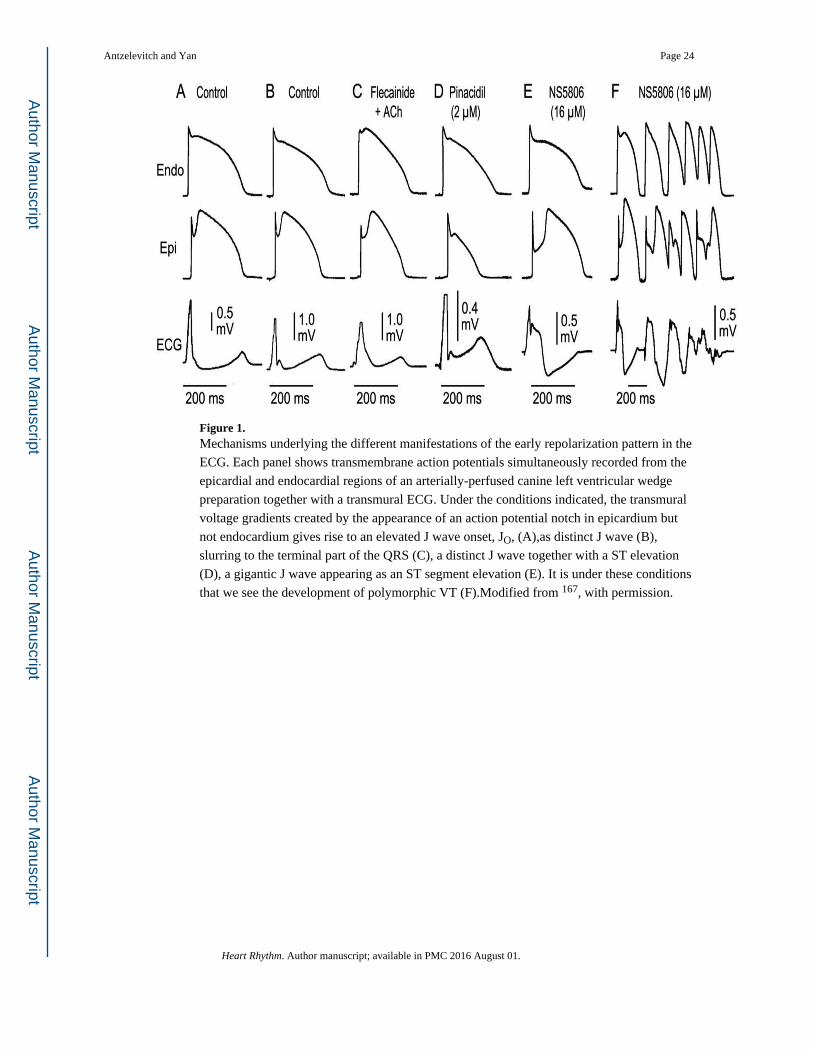
Figure 1. Mechanisms underlying the different manifestations of the early repolarization pattern in the
ECG. Each panel shows transmembrane action potentials simultaneously recorded from the
epicardial and endocardial regions of an arterially-perfused canine left ventricular wedge
preparation together with a transmural ECG. Under the conditions indicated, the transmural
voltage gradients created by the appearance of an action potential notch in epicardium but
not endocardium gives rise to an elevated J wave onset, JO, (A),as distinct J wave (B),
slurring to the terminal part of the QRS (C), a distinct J wave together with a ST elevation
(D), a gigantic J wave appearing as an ST segment elevation (E). It is under these conditions
that we see the development of polymorphic VT (F).Modified from 167, with permission.
Antzelevitch and Yan Page 24
Heart Rhythm. Author manuscript; available in PMC 2016 August 01.
Author M
anuscriptA
uthor Manuscript
Author M
anuscriptA
uthor Manuscript
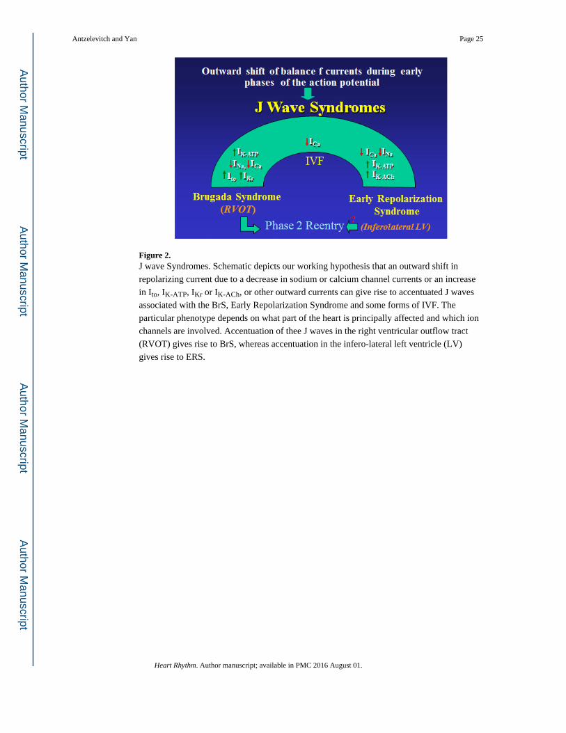
Figure 2. J wave Syndromes. Schematic depicts our working hypothesis that an outward shift in
repolarizing current due to a decrease in sodium or calcium channel currents or an increase
in Ito, IK-ATP, IKr or IK-ACh, or other outward currents can give rise to accentuated J waves
associated with the BrS, Early Repolarization Syndrome and some forms of IVF. The
particular phenotype depends on what part of the heart is principally affected and which ion
channels are involved. Accentuation of thee J waves in the right ventricular outflow tract
(RVOT) gives rise to BrS, whereas accentuation in the infero-lateral left ventricle (LV)
gives rise to ERS.
Antzelevitch and Yan Page 25
Heart Rhythm. Author manuscript; available in PMC 2016 August 01.
Author M
anuscriptA
uthor Manuscript
Author M
anuscriptA
uthor Manuscript
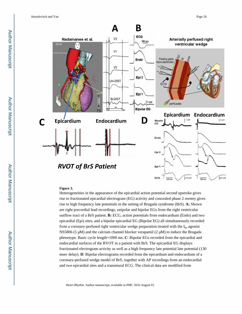
Figure 3. Heterogeneities in the appearance of the epicardial action potential second upstroke gives
rise to fractionated epicardial electrogram (EG) activity and concealed phase 2 reentry gives
rise to high frequency late potentials in the setting of Brugada syndrome (BrS). A: Shown
are right precordial lead recordings, unipolar and bipolar EGs from the right ventricular
outflow tract of a BrS patient. B: ECG, action potentials from endocardium (Endo) and two
epicardial (Epi) sites, and a bipolar epicardial EG (Bipolar EG) all simultaneously recorded
from a coronary-perfused right ventricular wedge preparation treated with the Ito agonist
NS5806 (5 μM) and the calcium channel blocker verapamil (2 μM) to induce the Brugada
phenotype. Basic cycle length=1000 ms. C: Bipolar EGs recorded from the epicardial and
endocardial surfaces of the RVOT in a patient with BrS. The epicardial EG displays
fractionated electrogram activity as well as a high frequency late potential late potential (130
msec delay). D: Bipolar electrograms recorded from the epicardium and endocardium of a
coronary-perfused wedge model of BrS, together with AP recordings from an endocardial
and two epicardial sites and a transmural ECG. The clinical data are modified from
Antzelevitch and Yan Page 26
Heart Rhythm. Author manuscript; available in PMC 2016 August 01.
Author M
anuscriptA
uthor Manuscript
Author M
anuscriptA
uthor Manuscript

Nademanee et al.82 and the experimental data are from Szel and co-workers 83, with
permission).
Antzelevitch and Yan Page 27
Heart Rhythm. Author manuscript; available in PMC 2016 August 01.
Author M
anuscriptA
uthor Manuscript
Author M
anuscriptA
uthor Manuscript
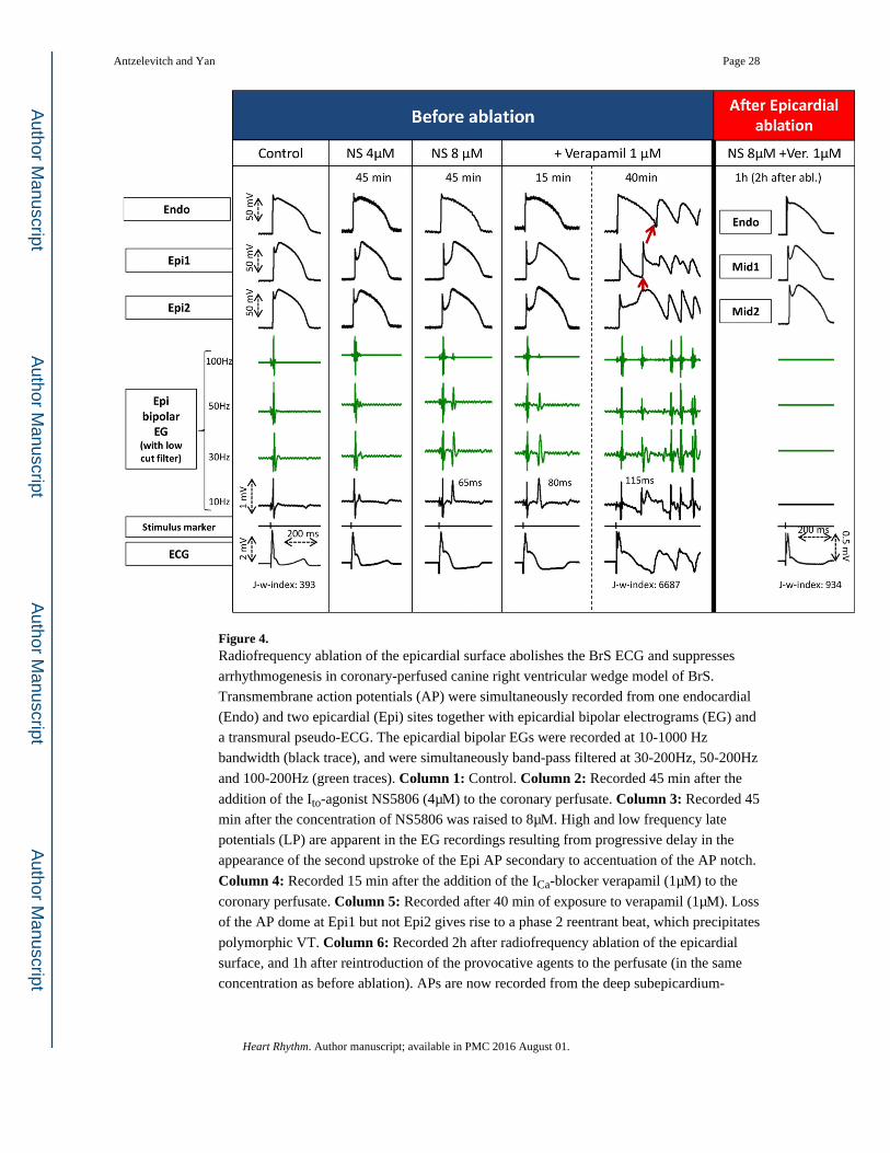
Figure 4. Radiofrequency ablation of the epicardial surface abolishes the BrS ECG and suppresses
arrhythmogenesis in coronary-perfused canine right ventricular wedge model of BrS.
Transmembrane action potentials (AP) were simultaneously recorded from one endocardial
(Endo) and two epicardial (Epi) sites together with epicardial bipolar electrograms (EG) and
a transmural pseudo-ECG. The epicardial bipolar EGs were recorded at 10-1000 Hz
bandwidth (black trace), and were simultaneously band-pass filtered at 30-200Hz, 50-200Hz
and 100-200Hz (green traces). Column 1: Control. Column 2: Recorded 45 min after the
addition of the Ito-agonist NS5806 (4μM) to the coronary perfusate. Column 3: Recorded 45
min after the concentration of NS5806 was raised to 8μM. High and low frequency late
potentials (LP) are apparent in the EG recordings resulting from progressive delay in the
appearance of the second upstroke of the Epi AP secondary to accentuation of the AP notch.
Column 4: Recorded 15 min after the addition of the ICa-blocker verapamil (1μM) to the
coronary perfusate. Column 5: Recorded after 40 min of exposure to verapamil (1μM). Loss
of the AP dome at Epi1 but not Epi2 gives rise to a phase 2 reentrant beat, which precipitates
polymorphic VT. Column 6: Recorded 2h after radiofrequency ablation of the epicardial
surface, and 1h after reintroduction of the provocative agents to the perfusate (in the same
concentration as before ablation). APs are now recorded from the deep subepicardium-
Antzelevitch and Yan Page 28
Heart Rhythm. Author manuscript; available in PMC 2016 August 01.
Author M
anuscriptA
uthor Manuscript
Author M
anuscriptA
uthor Manuscript
midmyocardium (Mid1, Mid2) instead of the epicardial surface. Ablation markedly
suppressed the BrS phenotype and abolished all arrhythmic activity. Modified from 168, with
permission.
Antzelevitch and Yan Page 29
Heart Rhythm. Author manuscript; available in PMC 2016 August 01.
Author M
anuscriptA
uthor Manuscript
Author M
anuscriptA
uthor Manuscript
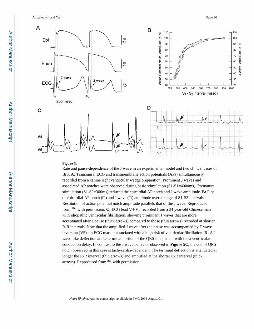
Figure 5. Rate and pause-dependence of the J wave in an experimental model and two clinical cases of
BrS. A: Transmural ECG and transmembrane action potentials (APs) simultaneously
recorded from a canine right ventricular wedge preparation: Prominent J waves and
associated AP notches were observed during basic stimulation (S1-S1=4000ms). Premature
stimulation (S1-S2=300ms) reduced the epicardial AP notch and J wave amplitude. B: Plot
of epicardial AP notch (□) and J wave (○) amplitude over a range of S1-S2 intervals.
Restitution of action potential notch amplitude parallels that of the J wave. Reproduced
from 169 with permission. C: ECG lead V4-V5 recorded from a 34 year-old Chinese man
with idiopathic ventricular fibrillation, showing prominent J waves that are more
accentuated after a pause (thick arrows) compared to those (thin arrows) recorded at shorter
R-R intervals. Note that the amplified J wave after the pause was accompanied by T wave
inversion (V5), an ECG marker associated with a high risk of ventricular fibrillation. D: A J-
wave-like deflection at the terminal portion of the QRS in a patient with intra-ventricular
conduction delay. In contrast to the J wave behavior observed in Figure 5C, the end of QRS
notch observed in this case is tachycardia-dependent. The terminal deflection is attenuated at
longer the R-R interval (thin arrows) and amplified at the shorter R-R interval (thick
arrows). Reproduced from 86, with permission.
Antzelevitch and Yan Page 30
Heart Rhythm. Author manuscript; available in PMC 2016 August 01.
Author M
anuscriptA
uthor Manuscript
Author M
anuscriptA
uthor Manuscript
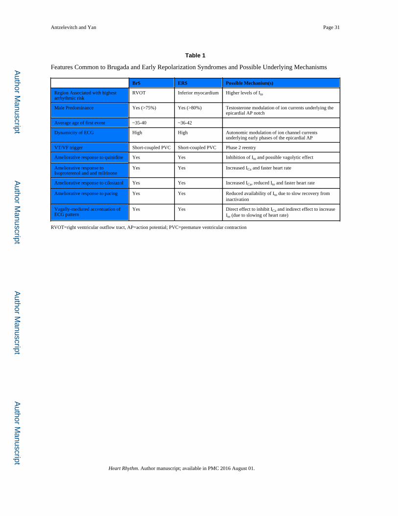
Author M
anuscriptA
uthor Manuscript
Author M
anuscriptA
uthor Manuscript
Antzelevitch and Yan Page 31
Table 1
Features Common to Brugada and Early Repolarization Syndromes and Possible Underlying Mechanisms
BrS ERS Possible Mechanism(s)
Region Associated with highest arrhythmic risk
RVOT Inferior myocardium Higher levels of Ito
Male Predominance Yes (>75%) Yes (>80%) Testosterone modulation of ion currents underlying the epicardial AP notch
Average age of first event ~35-40 ~36-42
Dynamicity of ECG High High Autonomic modulation of ion channel currents underlying early phases of the epicardial AP
VT/VF trigger Short-coupled PVC Short-coupled PVC Phase 2 reentry
Ameliorative response to quinidine Yes Yes Inhibition of Ito and possible vagolytic effect
Ameliorative response to Isoproterenol and and milrinone
Yes Yes Increased ICa and faster heart rate
Ameliorative response to cilostazol Yes Yes Increased ICa, reduced Ito and faster heart rate
Ameliorative response to pacing Yes Yes Reduced availability of Ito due to slow recovery from inactivation
Vagally-mediated accentuation of ECG pattern
Yes Yes Direct effect to inhibit ICa and indirect effect to increase Ito (due to slowing of heart rate)
RVOT=right ventricular outflow tract, AP=action potential; PVC=premature ventricular contraction
Heart Rhythm. Author manuscript; available in PMC 2016 August 01.
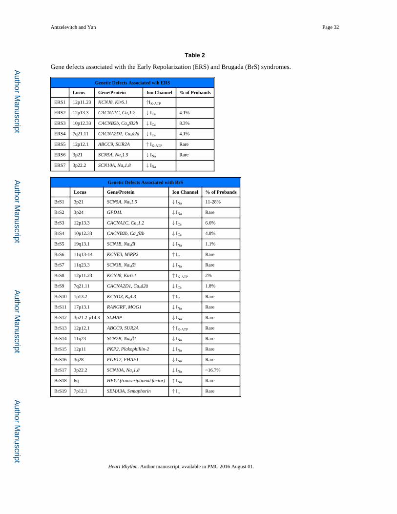
Author M
anuscriptA
uthor Manuscript
Author M
anuscriptA
uthor Manuscript
Antzelevitch and Yan Page 32
Table 2
Gene defects associated with the Early Repolarization (ERS) and Brugada (BrS) syndromes.
Genetic Defects Associated wih ERS
Locus Gene/Protein Ion Channel % of Probands
ERS1 12p11.23 KCNJ8, Kir6.1 ↑IK-ATP
ERS2 12p13.3 CACNA1C, Cav1.2 ↓ ICa 4.1%
ERS3 10p12.33 CACNB2b, Cavβ32b ↓ ICa 8.3%
ERS4 7q21.11 CACNA2D1, Cavá2ä ↓ ICa 4.1%
ERS5 12p12.1 ABCC9, SUR2A ↑ IK-ATP Rare
ERS6 3p21 SCN5A, Nav1.5 ↓ INa Rare
ERS7 3p22.2 SCN10A, Nav1.8 ↓ INa
Genetic Defects Associated with BrS
Locus Gene/Protein Ion Channel % of Probands
BrS1 3p21 SCN5A, Nav1.5 ↓ INa 11-28%
BrS2 3p24 GPD1L ↓ INa Rare
BrS3 12p13.3 CACNA1C, Cav1.2 ↓ ICa 6.6%
BrS4 10p12.33 CACNB2b, Cavβ2b ↓ ICa 4.8%
BrS5 19q13.1 SCN1B, Navβ1 ↓ INa 1.1%
BrS6 11q13-14 KCNE3, MiRP2 ↑ Ito Rare
BrS7 11q23.3 SCN3B, Navβ3 ↓ INa Rare
BrS8 12p11.23 KCNJ8, Kir6.1 ↑ IK-ATP 2%
BrS9 7q21.11 CACNA2D1, Cavá2ä ↓ ICa 1.8%
BrS10 1p13.2 KCND3, Kv4.3 ↑ Ito Rare
BrS11 17p13.1 RANGRF, MOG1 ↓ INa Rare
BrS12 3p21.2-p14.3 SLMAP ↓ INa Rare
BrS13 12p12.1 ABCC9, SUR2A ↑ IK-ATP Rare
BrS14 11q23 SCN2B, Navβ2 ↓ INa Rare
BrS15 12p11 PKP2, Plakophillin-2 ↓ INa Rare
BrS16 3q28 FGF12, FHAF1 ↓ INa Rare
BrS17 3p22.2 SCN10A, Nav1.8 ↓ INa ~16.7%
BrS18 6q HEY2 (transcriptional factor) ↑ INa Rare
BrS19 7p12.1 SEMA3A, Semaphorin ↑ Ito Rare
Heart Rhythm. Author manuscript; available in PMC 2016 August 01.
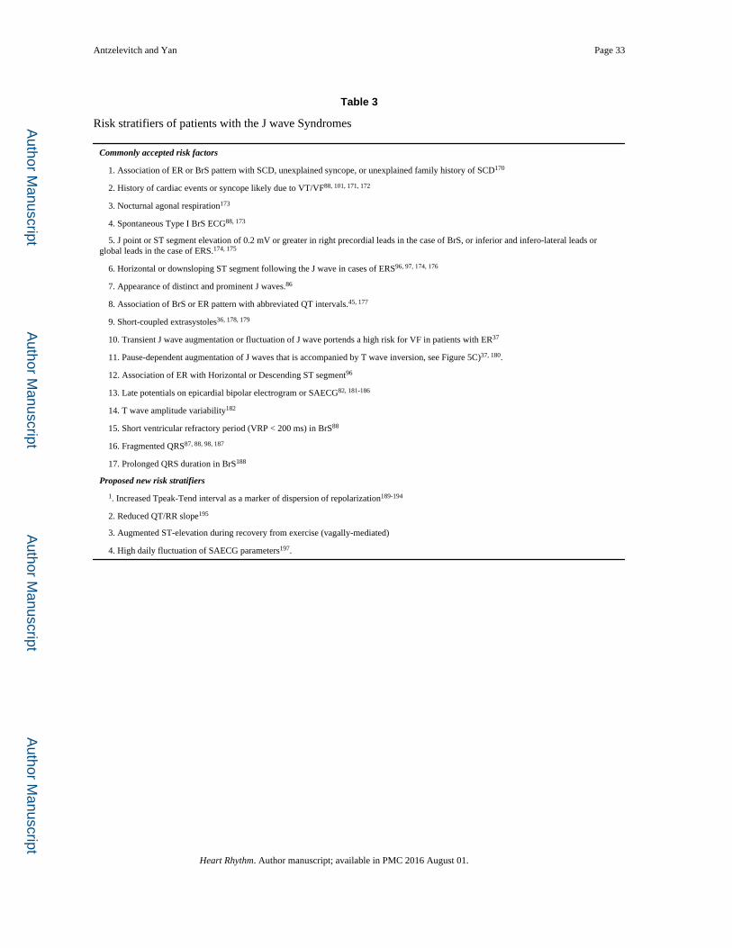
Author M
anuscriptA
uthor Manuscript
Author M
anuscriptA
uthor Manuscript
Antzelevitch and Yan Page 33
Table 3
Risk stratifiers of patients with the J wave Syndromes
Commonly accepted risk factors
1. Association of ER or BrS pattern with SCD, unexplained syncope, or unexplained family history of SCD170
2. History of cardiac events or syncope likely due to VT/VF88, 101, 171, 172
3. Nocturnal agonal respiration173
4. Spontaneous Type I BrS ECG88, 173
5. J point or ST segment elevation of 0.2 mV or greater in right precordial leads in the case of BrS, or inferior and infero-lateral leads or global leads in the case of ERS.174, 175
6. Horizontal or downsloping ST segment following the J wave in cases of ERS96, 97, 174, 176
7. Appearance of distinct and prominent J waves.86
8. Association of BrS or ER pattern with abbreviated QT intervals.45, 177
9. Short-coupled extrasystoles36, 178, 179
10. Transient J wave augmentation or fluctuation of J wave portends a high risk for VF in patients with ER37
11. Pause-dependent augmentation of J waves that is accompanied by T wave inversion, see Figure 5C)37, 180.
12. Association of ER with Horizontal or Descending ST segment96
13. Late potentials on epicardial bipolar electrogram or SAECG82, 181-186
14. T wave amplitude variability182
15. Short ventricular refractory period (VRP < 200 ms) in BrS88
16. Fragmented QRS87, 88, 98, 187
17. Prolonged QRS duration in BrS188
Proposed new risk stratifiers
1. Increased Tpeak-Tend interval as a marker of dispersion of repolarization189-194
2. Reduced QT/RR slope195
3. Augmented ST-elevation during recovery from exercise (vagally-mediated)
4. High daily fluctuation of SAECG parameters197.
Heart Rhythm. Author manuscript; available in PMC 2016 August 01.

Author M
anuscriptA
uthor Manuscript
Author M
anuscriptA
uthor Manuscript
Antzelevitch and Yan Page 34
Table 4
Differential diagnosis of J wave vs. Intra-ventricular conduction defect-mediated notch syndromes. (IVCD)
J wave IVCD-induced end QRS notch
Male Predominance Yes No
Average Age at Initial Presentation Young adults Older adults
Most common Morphology dome-like smooth appearance Relatively sharp appearance
Response to Change in Heart Rate Bradycardia- and pause-dependent augmentation of J wave which may be accompanied by T
wave inversion.
Tachycardia and prematurity-dependent augmentation of the notch
Structural Heart Diseases Rare Common History of myocardial infarction and/or cardiomyopathy
Heart Rhythm. Author manuscript; available in PMC 2016 August 01.
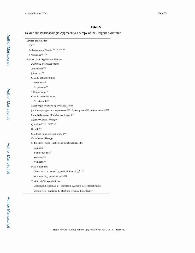
Author M
anuscriptA
uthor Manuscript
Author M
anuscriptA
uthor Manuscript
Antzelevitch and Yan Page 35
Table 5
Device and Pharmacologic Approach to Therapy of the Brugada Syndrome
Devices and Ablation
ICD99
Radiofrequency Ablation82, 105, 198-201
? Pacemaker102-104
Pharmacologic Approach to Therapy
Ineffective or Proarrhythmic
Amiodarone106
β Blockers106
Class IC antiarrhythmics
Flecainide202
Propafenone203
? Disopyramide107
Class IA antiarrhythmics
Procainamide204
Effective for Treatment of Electrical Storms
β Adrenergic agonists – isoproterenol139, 142, denopamine125, orciprenaline121, 133
Phosphodiesterase III Inhibitors-cilostazol132
Effective General Therapy
Quinidine10, 113-115, 127-129
Bepridil157
Cilostazol combined with bepridil150
Experimental Therapy
Ito Blockers - cardioselective and ion channel specific
Quinidine10
4-aminopyridine10
Tedisamil205
AVE0118206
PDE-3-inhibitors
Cilostazol – Increase in ICa and inhibition of Ito91, 152
Milrinone - ICa augmentation91, 152
Traditional Chinese Medicine
Dimethyl lithospermate B – Increase in INa due to slowed inactivation
Wenxin Keli - combined Ito-block and tyramine-like effect110
Heart Rhythm. Author manuscript; available in PMC 2016 August 01.


