IVUS image interpretation...Pocket guide Eagle Eye Platinum RX digital IVUS catheter Normal vessel...
17
IVUS image interpretation Pocket guide Eagle Eye Platinum RX digital IVUS catheter
Transcript of IVUS image interpretation...Pocket guide Eagle Eye Platinum RX digital IVUS catheter Normal vessel...

IVUS image interpretationPocket guide
Eagle Eye Platinum
RX digitalIVUS catheter

Normal vessel
• In a normal vessel, the lumen border is almost indistinguishable from the vessel border. While IVUS allows visualization of vessel and lumen, angiograms only provide a shadow of the lumen. In patients with diffuse disease, relying on the angiogram can potentially lead to underestimation of stenosis.
• Lumen border is drawn inside the intima or plaque.
• Intimal layer is normally not seen unless it has begun to thicken.
• Vessel border is drawn just inside the adventitia, which appears as a bright white ring due to highly reflective collagen sheets.
• The catheter mask (blue area) indicates the location of the ultrasound transducer.
Lumen border Intimal thickening Vessel border
1 2

Concentric mixed plaque
• Concentric plaques are distributed circumferentially in the vessel.
• Concentric plaques tend to occur in areas of negative remodeling; use of angiography alone could result in too large a stent diameter.
• Mixed plaque is a combination of tissues of varying echogenecity. The distribution of light and dark may be distinct, or light and dark variations may be intermingled as shown here.
• MLA < 4 mm2 in LAD, LCX, and RCA vessels > 3 mm in diameter correlates with physiological significance.1
• MLA < 6 mm2 in left main correlates with FFR < 0.75 indicating physiological significance.1, 2, 3
• Nearby vessels on the periphery can be seen moving in and out of the field of view and can be used as landmarks.
Nearby vessel
1 Jasti, et al. Correlations between Fractional Flow Reserve and Intravascular Ultrasound in Patients with an Ambiguous Left Main Coronary Artery Stenosis, Circulation, 2004; 110: 2831-2836
2 Jose M. de la Torre Hernandez et al. Prospective Application of Pre-Defined Intravascular Ultrasound Criteria for Assessment of Intermediate Left Main Coronary Artery Lesions: Results From the Multicenter LITRO Study, J Am Coll Cardiol, 2011 58: 351-358.
3 Levine et al. 2011 ACCF/AHA/SCAI Guideline for Percutaneous Coronary Intervention. J Am Coll Cardiol, 2011; 58:44-122
3 4

Eccentric mixed plaque
• Eccentric plaques are distributed non-circumferentially in the vessel; this makes the assessment of disease by angiography especially prone to underestimation or overestimation depending on the angle of view.
• Calcium is indicated by very bright areas with acoustic shadowing that blocks out the image behind. This shadowing occurs because the high density of calcium dampens the ultrasound echo.
• VH IVUS* imaging can provide a more detailed and objective view of plaque composition (see pages 23-24).
Echolucent plaque Echogenic plaque Calcium
4 Acoustic shadowing
4*Safety and effectiveness of VH IVUS for use in the characterization of vascular lesions and tissue types has not been established
5 6

Plaque with calcium
• The calcium in this plaque produces two arcs of acoustic shadowing.
• Calcium is indicated by very bright areas with acoustic shadowing that blocks out the image behind. This shadowing occurs because the high density of calcium prevents the ultrasound from passing through.
• Detection of calcium is a critical factor in determining the optimal PCI strategy.
• Calcium is not always seen on the angiogram. In carotid cases angiography can fail to identify calcium 46% of the time.1
• Nearby vessels on the periphery can be seen moving in and out of the field of view and can be used as landmarks.
Calcium Acoustic shadowing
1 Diethrich et al. Journal of Endovascular Therapy, 2007; 14:676-686
7 8
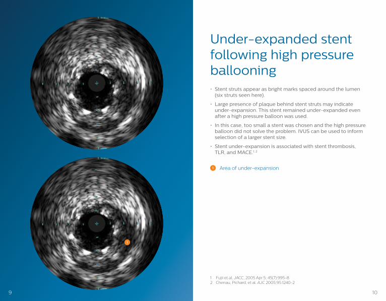
Under-expanded stent following high pressure ballooning• Stent struts appear as bright marks spaced around the lumen
(six struts seen here).
• Large presence of plaque behind stent struts may indicate under-expansion. This stent remained under-expanded even after a high pressure balloon was used.
• In this case, too small a stent was chosen and the high pressure balloon did not solve the problem. IVUS can be used to inform selection of a larger stent size.
• Stent under-expansion is associated with stent thrombosis, TLR, and MACE.1, 2
Area of under-expansion
1 Fujii et al, JACC, 2005 Apr 5; 45(7):995-82 Chenau, Pichard, et al. AJC 2005;95:1240-2
9 10
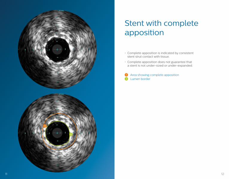
Stent with complete apposition
• Complete apposition is indicated by consistent stent strut contact with tissue.
• Complete apposition does not guarantee that a stent is not under-sized or under-expanded.
Area showing complete apposition Lumen border
11 12

Stent with malapposition
• Malapposition is indicated by blood visible behind stent struts.
• Blood may appear as a very faint speckle or black on grayscale IVUS.
• Blood flow will appear red with ChromaFlo imaging.
• Stents may be completely or only partially malapposed depending on how much of the stent is in contact with the lumen wall.
Blood behind struts indicates malapposition
Grayscale ChromaFlo
13 14

Dissection
• A dissection, or tear in the vessel wall, can be seen as a flap with blood flow behind it.
• ChromaFlo imaging, shown here, enhances identification of dissection flaps by verifying presence of blood flow (in red) behind the flap.
Dissection flap
15 16
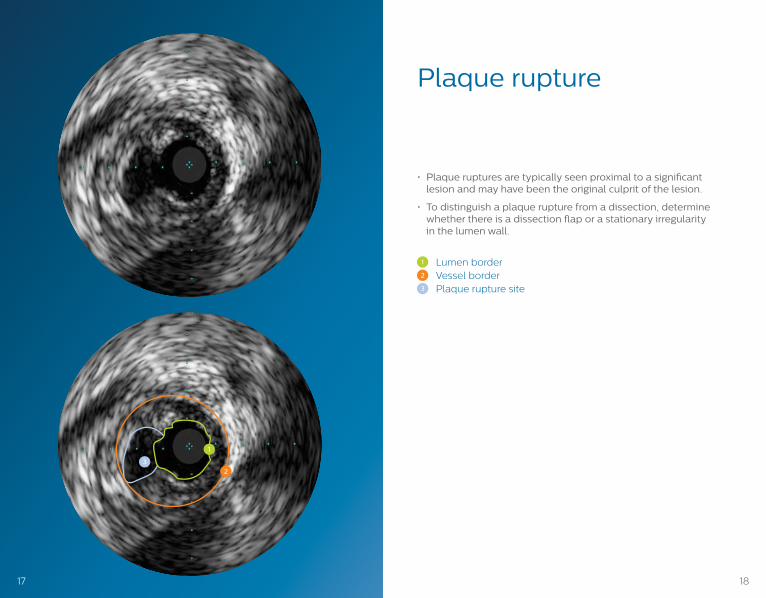
Plaque rupture
• Plaque ruptures are typically seen proximal to a significant lesion and may have been the original culprit of the lesion.
• To distinguish a plaque rupture from a dissection, determine whether there is a dissection flap or a stationary irregularity in the lumen wall.
Lumen border Vessel border Plaque rupture site
17 18

Proximal cap
• When a proximal cap is located near a side branch, the IVUS catheter can be positioned in the side branch to view and confirm the location of the proximal cap.
• As a wire penetrates the cap, IVUS can confirm wire position.
The border of the proximal cap The border of the side branch
19 20

True and false lumen
• IVUS can help confirm wire position in the true or false lumen.
• The true lumen is often compressed by the false lumen.
Compressed true lumen (separate echolucent area) False lumen
21 22

VH IVUS imaging
• VH IVUS imaging classifies plaque into four tissue types.1
• Based on 15 years of research with over 200 scientific publications.
• Used in multiple clinical studies including the landmark PROSPECT study of 700 ACS patients published in the New England Journal of Medicine.
• PROSPECT results demonstrated that VH IVUS adds valuable information for physicians to determine lesion risk profile.2
Complete lesion assessment Pathological intimal thickening (PIT) Calcified fibroatheroma
4 Thin cap fibroatheroma (TCFA)
Fibrous Fibrofatty
Dense calciumNecrotic core
4
1. Nair A et al. Automated coronary plaque characterisation with intravascular ultrasound backscatter: ex vivo validation. EuroInterv 2007; 3:113-120.
2. Stone et al. A prospective natural history study of atherosclerosis. N Engl J Med 2011;364:226-35.
Safety and effectiveness of VH IVUS for use in the characterization of vascular lesions and tissue types has not been established.
23 24

Bioresorbable scaffold, well apposed
• Because of the thickness and material of the struts, it is common for the struts to present on ultrasound as a double line representing the leading and trailing edges of the strut.
• IVUS may be used to optimize BRS implantation by allowing you to observe: scaffold expansion, apposition, edge complications.
BRS, well apposed
25 26

Bioresorbable scaffold, in small vessel
BRS in small vessel
27 28
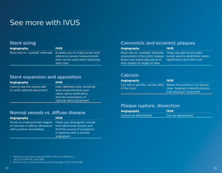
See more with IVUS
Stent sizingAngiography IVUSMust rely on “eyeball” estimate Enables you to make lumen and
reference vessel measurements that can be used when selecting stent size
Stent expansion and appositionAngiography IVUSCannot see the vessel wall to verify optimal placement
User-defined cross-sectional area measurements and views, allow verification and documen tation of optimal stent place ment
Normal vessels vs. diffuse diseaseAngiography IVUSTends to underestimate degree of stenosis in diffuse disease or with positive remodeling1
Helps you distinguish normal from abnormal vessels and find the source of symptoms in patients with a normal angiogram
Concentric and eccentric plaquesAngiography IVUSMust rely on “eyeball” estimate; assessment of eccentric plaque lesion size especially prone to error based on angle of view
Helps you get an accurate lumen area to determine lesion significance and stent size
CalciumAngiography IVUSCan fail to identify calcium 46% of the time2
Makes the presence of calcium clear, helping to identify lesions that need pre-treatment
Plaque rupture, dissectionAngiography IVUSCannot be determined Can be determined
1 Reference vessels are diseased 93% of the time. Mintz et al. JACC 25;7:1479-85, June 1995.
2 Diethrich et al. Journal of Endovascular Therapy, 2007; 14:676-686.
29 30

© 2017 Koninklijke Philips N.V. All rights reserved. Trademarks are the property of Koninklijke Philips N.V. or their respective owners.
Philips Volcano3721 Valley Centre Drive, Suite 500San Diego, CA 92130 USAwww.volcanocorp.com
600.0500.03/LB