Italian Journal of Zoology, March 2007; 74(1): 83–99 · Interstitial harpacticoids from...
17
Interstitial harpacticoids from groundwater in Tuscany (Central Italy): Parastenocaris reidae sp. nov., Nitocrella ensifera sp. nov., and notes on the morphology of Parastenocaris cf. glacialis Noodt (Crustacea: Copepoda) V. COTTARELLI, M. C. BRUNO & R. BERERA Department of Environmental Sciences, ‘‘della Tuscia’’ University, Italy (Received 13 April 2006; accepted 25 July 2006) Abstract Two new interstitial harpacticoids were recently collected from groundwater in Tuscany (Central Italy). The Parastenocarididae Parastenocaris reidae sp. nov. was collected from both phreatic and hyporheic habitats of River Serchio, Lucca province, together with specimens of Parastenocaris cf. glacialis Noodt. We describe and discuss the affinities of the former species, and give a preliminary description of the latter. Nitocrella ensifera sp. nov. was collected from the phreatic habitat of Fiora River, Grosseto, and from a well in the same area; this new species is described and its affinities are discussed. Keywords: Parastenocaris, Nitocrella, interstitial habitat, Harpacticoida, hyporheos, groundwater Introduction During the last decade, we have been investigating groundwater copepods from phreatic and hyporheic habitats of several rivers in Central Italy (Cottarelli et al. 2002; Cottarelli & Berera 2003; Berera et al. 2003). Harpacticoid and cyclopoid copepods represent a dominant component of groundwater communities in shallow aquifers (Galassi 2001; Cottarelli & Berera 2003). The first results regarding the study of Fiora, Orcia, and Serchio rivers were published elsewhere (Cottarelli et al. 2002; Berera et al. 2003; Cottarelli & Berera 2003), and they provided the faunistic and ecological description of interstitial copepod assemblages. In the present work we describe and discuss two taxa of Parastenocarididae, which belong to the genus Parastenocaris Kessler, collected from Serchio River: Parastenocaris reidae sp. nov. (previously listed as Parastenocaris sp. A in Cottarelli et al. 2002), and Parastenocaris cf. glacialis (previously listed as Parastenocaris cfr. glacialis in Cottarelli et al. 2002 and in Berera et al. 2005); these taxa are new to science or rarely collected in Italy. We present the most important morphological characters of P. cf. glacialis and underline the differences with the description of P. glacialis by Pesce et al. (1995), which is the most recent description available in literature. We also describe and discuss the Ameridae Nito- crella ensifera sp. nov. (previously listed as Nitocrella sp. 2 in Cottarelli & Berera 2003), collected from the Fiora River. The genera Parastenocaris and Nitocrella Petkovski include species almost exclusive to groundwater habitats; Nitocrella ensifera sp. nov. shows unusual morphological peculiarities never recorded before for the genus. Materials and methods Specimens were collected using the Karaman– Chappuis method (Delamare Deboutteville 1960) along the river banks, and with a Cvetkov phreato- biological net (Cvetkov 1968) in wells. Specimens were fixed in 5% buffered formalin solution, sorted and mounted on slides in Faure’s medium. Drawings were made at 12506, with an oil *Correspondence: M. C. Bruno, Universita ´ della Tuscia, Largo dell’Universita ` snc, I-01100 Viterbo, Italy. Email: [email protected] Italian Journal of Zoology, March 2007; 74(1): 83–99 ISSN 1125-0003 print/ISSN 1748-5851 online # 2007 Unione Zoologica Italiana DOI: 10.1080/11250000601022605
-
Upload
nguyenquynh -
Category
Documents
-
view
214 -
download
0
Transcript of Italian Journal of Zoology, March 2007; 74(1): 83–99 · Interstitial harpacticoids from...
Interstitial harpacticoids from groundwater in Tuscany (Central Italy):Parastenocaris reidae sp. nov., Nitocrella ensifera sp. nov., and noteson the morphology of Parastenocaris cf. glacialis Noodt (Crustacea:Copepoda)
V. COTTARELLI, M. C. BRUNO & R. BERERA
Department of Environmental Sciences, ‘‘della Tuscia’’ University, Italy
(Received 13 April 2006; accepted 25 July 2006)
AbstractTwo new interstitial harpacticoids were recently collected from groundwater in Tuscany (Central Italy). TheParastenocarididae Parastenocaris reidae sp. nov. was collected from both phreatic and hyporheic habitats of RiverSerchio, Lucca province, together with specimens of Parastenocaris cf. glacialis Noodt. We describe and discuss the affinitiesof the former species, and give a preliminary description of the latter. Nitocrella ensifera sp. nov. was collected from thephreatic habitat of Fiora River, Grosseto, and from a well in the same area; this new species is described and its affinities arediscussed.
Keywords: Parastenocaris, Nitocrella, interstitial habitat, Harpacticoida, hyporheos, groundwater
Introduction
During the last decade, we have been investigating
groundwater copepods from phreatic and hyporheic
habitats of several rivers in Central Italy (Cottarelli
et al. 2002; Cottarelli & Berera 2003; Berera et al.
2003). Harpacticoid and cyclopoid copepods
represent a dominant component of groundwater
communities in shallow aquifers (Galassi 2001;
Cottarelli & Berera 2003). The first results regarding
the study of Fiora, Orcia, and Serchio rivers were
published elsewhere (Cottarelli et al. 2002; Berera
et al. 2003; Cottarelli & Berera 2003), and they
provided the faunistic and ecological description of
interstitial copepod assemblages.
In the present work we describe and discuss
two taxa of Parastenocarididae, which belong to the
genus Parastenocaris Kessler, collected from Serchio
River: Parastenocaris reidae sp. nov. (previously
listed as Parastenocaris sp. A in Cottarelli et al.
2002), and Parastenocaris cf. glacialis (previously
listed as Parastenocaris cfr. glacialis in Cottarelli et al.
2002 and in Berera et al. 2005); these taxa are new
to science or rarely collected in Italy. We present the
most important morphological characters of P. cf.
glacialis and underline the differences with the
description of P. glacialis by Pesce et al. (1995),
which is the most recent description available in
literature.
We also describe and discuss the Ameridae Nito-
crella ensifera sp. nov. (previously listed as Nitocrella
sp. 2 in Cottarelli & Berera 2003), collected from
the Fiora River. The genera Parastenocaris and
Nitocrella Petkovski include species almost exclusive
to groundwater habitats; Nitocrella ensifera sp. nov.
shows unusual morphological peculiarities never
recorded before for the genus.
Materials and methods
Specimens were collected using the Karaman–
Chappuis method (Delamare Deboutteville 1960)
along the river banks, and with a Cvetkov phreato-
biological net (Cvetkov 1968) in wells.
Specimens were fixed in 5% buffered formalin
solution, sorted and mounted on slides in Faure’s
medium. Drawings were made at 12506, with an oil
*Correspondence: M. C. Bruno, Universita della Tuscia, Largo dell’Universita snc, I-01100 Viterbo, Italy. Email: [email protected]
Italian Journal of Zoology, March 2007; 74(1): 83–99
ISSN 1125-0003 print/ISSN 1748-5851 online # 2007 Unione Zoologica Italiana
DOI: 10.1080/11250000601022605
immersion lens, using a drawing tube mounted on a
Zeiss AxioskopH phase-contrast microscope.
Eight females and one male of Parastenocaris reidae
sp. nov., and one female and one male of Nitocrella
ensifera sp. nov. were prepared for scanning elec-
tron microscopy. They were fixed for 24 h in 10%
formalin solution, washed twice in cacodylate buffer
(pH 7.2), post-fixed in 1% osmium tetraoxide in the
same buffer, dehydrated in a graded ethanol series,
critical-point-dried in a Balzers UnionH CPD 020
apparatus, and coated with gold in a Balzers UnionHMED 010 sputter coater. Observations were per-
formed with a 1,200 JEOL JEMH EX II scanning
electron microscope.
The following abbreviations are used, when
required, throughout the text and figures: Enp5
endopod; Exp5exopod; P1–P55thoracic appen-
dages. The nomenclature and descriptive terminol-
ogy follow Huys and Boxshall (1991).
Specimens are deposited at the Museo Civico
di Storia Naturale di Genova (MCSNG); at the
Museo Civico di Storia Naturale di Verona
(MCSNV); at the Natural History Museum,
London (NHM); at the National Museum of
Natural History, Smithsonian Institution (USNM).
The remaining material is located at the senior
author’s collection at the Department of Environ-
mental Sciences, ‘‘della Tuscia’’ University, Viterbo
(DSAUT). The stubs prepared for SEM are
deposited at the Interdepartmental Center for
Electron Microscopy, Tuscia University (CIME).
Taxonomic account
Family Parastenocarididae Chappuis 1940
Genus Parastenocaris Kessler 1913
Parastenocaris reidae sp. nov.
Material examined
Turrite Secca stream (Garfagnana, Lucca province,
Tuscany, Italy), right tributary joining the Serchio
river at about 30 km from the spring and 68 km from
the rivermouth, location ‘‘Isola Santa’’, 850 m asl,
about 13 km upstream, coordinates 44 03.125 N
010 14.171 E, from parafluvial phreatic habitat in
mixed gravel and sand bank. Holotype: male, dis-
sected and mounted on slide labelled: ‘‘Parasteno-
caris reidae holotype’’ (MCSNG 53219a). Paratypes:
female, dissected and mounted on slide labelled:
‘‘Parastenocaris reidae paratype, female no. 1’’
(MCSNG 53219b); four females, mounted on
different slides labelled: ‘‘Parastenocaris reidae para-
type, female no. 2–5’’ (DSAUT). Material collected
by V. Cottarelli, 20 May 1998.
Serchio river (Garfagnana, Lucca province,
Tuscany, Italy), location ‘‘Piaggione’’, 160 m asl,
about 54 km from the spring and 44 km from the
rivermouth, coordinates 43 57.665 N 010 31.053 E,
from hyporheic habitat in a mid-river sand bar on
the left side of the river. Paratypes: three males,
each dissected and mounted on different slides
labelled: ‘‘Parastenocaris reidae paratype, male no.
1–3’’ (MCSNV 633, NHM 2006.1271, USNM
1091070, respectively; three males, each dissected
and mounted on different slides labelled:
‘‘Parastenocaris reidae paratype, male no. 4–6’’
(DSAUT); one female mounted on slide labelled:
‘‘Parastenocaris reidae paratype, female no. 7’’
(MCSNV 632). Two females, each dissected and
mounted on different slides labelled: ‘‘Parastenocaris
reidae paratype, female no. 8, 9’’ (NHM 2006.1272,
and USNM 1091072, respectively). Material col-
lected by V. Cottarelli, 27 June 1998.
Serchio river (Garfagnana, Lucca province,
Tuscany, Italy), location ‘‘Sillano’’, 730 m asl, about
6 km from the spring and 92 km from the river-
mouth, coordinates 44 12.489 N 010 18.204 E,
from hyporheic habitat in a mid-river sand bar on
the left side of the river. Three dissected and one
whole females, mounted on different slides labelled:
‘‘Parastenocaris reidae paratype, female no. 10–13’’
(DSAUT). Material collected by V. Cottarelli, 31
May 1998. Ten females and one male, prepared for
scanning electron microscopy, on one stub labelled:
‘‘Parastenocaris reidae sp. nov. Fiume Serchio’’
(CIME). Material collected by M. C. Bruno, 22
October 2004.
Description of male
Length averaged among holotype and six paratypes,
from rostrum to distal apex of caudal rami: 0.34
mm. Body vermiform, slender, unpigmented, eye-
less. Hyaline frills of cephalotorax, thoracic
and abdominal somites and urosome smooth.
Cephalotorax with round dorsal hyaline integumen-
tal window; four abdominal somites with oval dorsal
integumental windows. Anal somite (Figure 1A):
with paired sensilla on dorsal side, two lateral rows
of small proctodeal spinules, and ventral spinules
near caudal rami. Anal operculum (Figure 1A): with
straight, smooth distal margin, protruding slightly
beyond insertion line of caudal rami. Caudal rami
(Figures 1A, 3F): longer than last abdominal somite;
length to width ratio: 3.6. Anterolateral accessory
seta (I), anterolateral seta (II) and posterolateral seta
(III) short, subequal. Outer terminal seta (IV) short
(length seta/length caudal ramus: 0.8), unipinnate.
Inner terminal seta (V) without breaking planes,
84 V. Cottarelli et al.
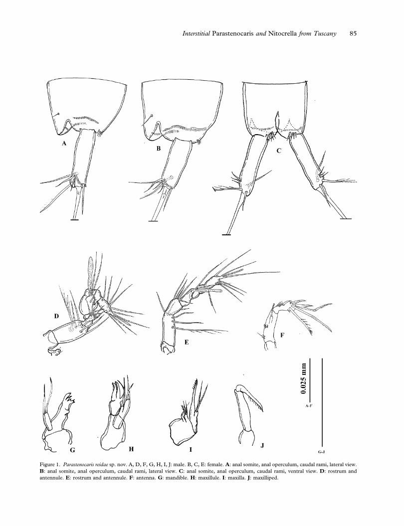
Figure 1. Parastenocaris reidae sp. nov. A, D, F, G, H, I, J: male. B, C, E: female. A: anal somite, anal operculum, caudal rami, lateral view.
B: anal somite, anal operculum, caudal rami, lateral view. C: anal somite, anal operculum, caudal rami, ventral view. D: rostrum and
antennule. E: rostrum and antennule. F: antenna. G: mandible. H: maxillule. I: maxilla. J: maxilliped.
Interstitial Parastenocaris and Nitocrella from Tuscany 85
unipinnate. Terminal accessory seta (VI) smooth
(length seta/length caudal ramus 0.5). Dorsal seta
(VII) articulate (length seta/length caudal ramus:
0.6). All setae inserted on the distal third of the
caudal ramus.
Rostrum (Figure 1D): small, reaching the end of
first segment of antennule, with two apical sensilla.
Antennule(Figure 1D):geniculate,eight-segmented.
First segment bare, second segment with six setae,
one plumose. Third segment with four distal setae;
fourth segment represented by a U-shaped sclerite,
with two setae. Fifth segment enlarged, with a dis-
tal tubercle with two setae and one long aesthetasc
on the apex, reaching the end of the antennule, and
one subapical short seta. Sixth segment partially
merged with the fifth one near the tubercle, with no
armature, small and cylindrical. Seventh segment
bare, with a tooth-like expansion that matches with
the enlarged fifth segment. Eighth segment with
seven setae and apical acrothek consisting of two
setae of different lengths and one slender, short
aesthetasc.
Antenna (Figure 1F): coxa unarmed; allobasis
with two transversal rows of spinules on medial
margin. Exopod one-segmented, not defined at base,
with one short pinnate apical spine. Endopod
bearing two geniculate and one transformed setae
(Figure 3D), and two distal spines. The transformed
seta is smooth in the first half, curved and one-side
pinnate in the second half (Figure 3D). Some
spinules along the distal margin, near the insertion
of the apical setae. On the lateral margin one smooth
and slightly curved spine with four short transversal
spinules near its insertion.
Mandible (Figures 1G, 3A, 3B): cutting edge of
coxal gnathobase with two strong teeth and a row of
smaller teeth; one-segmented palp, with two distal
setae.
Maxillule (Figures 1H, 3A, 3B): praecoxal arthrite
with two transformed spines (Figure 3B) and one
subapical curved seta. Coxa with one distal seta,
basis with two apical setae.
Maxilla (Figures 1I, 3A, 3B): syncoxa with two
endites, the proximal one with one seta, the distal
one with two normal ad one transformed setae.
Allobasis prolonged in an apical claw, on the medial
side a proximal transversal row of spinules and a
large pore (Figure 3B). Endopod with two setae.
Maxilliped (Figures 1J, 3A 3B): prehensile. Syn-
coxa small and unarmed; basis slim and elongate,
unarmed; endopod represented by distally unipin-
nate claw.
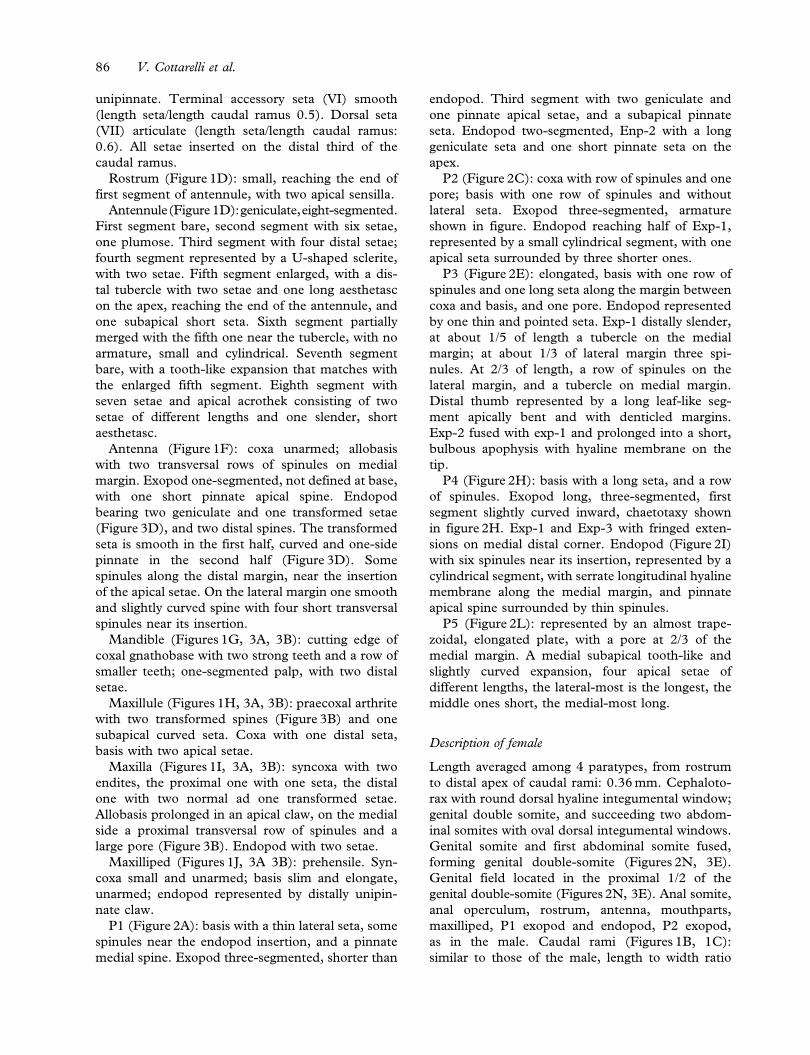
P1 (Figure 2A): basis with a thin lateral seta, some
spinules near the endopod insertion, and a pinnate
medial spine. Exopod three-segmented, shorter than
endopod. Third segment with two geniculate and
one pinnate apical setae, and a subapical pinnate
seta. Endopod two-segmented, Enp-2 with a long
geniculate seta and one short pinnate seta on the
apex.
P2 (Figure 2C): coxa with row of spinules and one
pore; basis with one row of spinules and without
lateral seta. Exopod three-segmented, armature
shown in figure. Endopod reaching half of Exp-1,
represented by a small cylindrical segment, with one
apical seta surrounded by three shorter ones.
P3 (Figure 2E): elongated, basis with one row of
spinules and one long seta along the margin between
coxa and basis, and one pore. Endopod represented
by one thin and pointed seta. Exp-1 distally slender,
at about 1/5 of length a tubercle on the medial
margin; at about 1/3 of lateral margin three spi-
nules. At 2/3 of length, a row of spinules on the
lateral margin, and a tubercle on medial margin.
Distal thumb represented by a long leaf-like seg-
ment apically bent and with denticled margins.
Exp-2 fused with exp-1 and prolonged into a short,
bulbous apophysis with hyaline membrane on the
tip.
P4 (Figure 2H): basis with a long seta, and a row
of spinules. Exopod long, three-segmented, first
segment slightly curved inward, chaetotaxy shown
in figure 2H. Exp-1 and Exp-3 with fringed exten-
sions on medial distal corner. Endopod (Figure 2I)
with six spinules near its insertion, represented by a
cylindrical segment, with serrate longitudinal hyaline
membrane along the medial margin, and pinnate
apical spine surrounded by thin spinules.
P5 (Figure 2L): represented by an almost trape-
zoidal, elongated plate, with a pore at 2/3 of the
medial margin. A medial subapical tooth-like and
slightly curved expansion, four apical setae of
different lengths, the lateral-most is the longest, the
middle ones short, the medial-most long.
Description of female
Length averaged among 4 paratypes, from rostrum
to distal apex of caudal rami: 0.36 mm. Cephaloto-
rax with round dorsal hyaline integumental window;
genital double somite, and succeeding two abdom-
inal somites with oval dorsal integumental windows.
Genital somite and first abdominal somite fused,
forming genital double-somite (Figures 2N, 3E).
Genital field located in the proximal 1/2 of the
genital double-somite (Figures 2N, 3E). Anal somite,
anal operculum, rostrum, antenna, mouthparts,
maxilliped, P1 exopod and endopod, P2 exopod,
as in the male. Caudal rami (Figures 1B, 1C):
similar to those of the male, length to width ratio
86 V. Cottarelli et al.
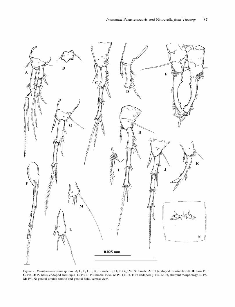
Figure 2. Parastenocaris reidae sp. nov. A, C, E, H, I, K, L: male. B, D, F, G, J,M, N: female. A: P1 (endopod disarticulated). B: basis P1.
C: P2. D: P2 basis, endopod and Exp-1. E: P3. F: P3, medial view. G: P3. H: P3. I: P3 endopod. J: P4. K: P3, aberrant morphology. L: P5.
M: P5. N: genital double somite and genital field, ventral view.
Interstitial Parastenocaris and Nitocrella from Tuscany 87
3.5. Lateral terminal seta proportionally shorter
than in male.
Antennule (Figure 1E): seven-segmented, with
aesthetasc on fourth segment almost reaching the
end of segment seven. First segment with row
of short spinules. Number of setae beginning at
proximal segment: 0, 6, 5, 2+aesthetasc, 1, 0,
5+acrothek. Apical acrothek represented by two
setae of different lengths and one slender, short
aesthetasc.
Figure 3. Parastenocaris reidae sp. nov. A, B, D, F: male. C, E: female. A: mandible, maxillule, maxilla, maxilliped, P1, ventral view. B:
mandible, maxillule, maxilla, maxilliped, ventral view. C: seventh segment of antennule, apical acrothek. D: antennal endopod, apical setae.
E: P5, genital double somite and genital field, ventral view. F: anal somite, anal operculum, caudal rami, lateral view.
88 V. Cottarelli et al.

P1 basis (Figure 2B): ornamentation as in the
male, but the medial spine is thin and bare.
P2 endopod (Figure 2D): similar to that of the
male, but longer.
P3 (Figures 2F, 2G): exopod two-segmented,
Exp-1 with one apical spine, Exp-2 with fringed
extensions on medial distal corner, one spine and
one pinnate seta on the apex. Endopod reduced to a
thin, curved and pointed segment, reaching 2/3 of
Exp-1, with a row of spinules near its insertion.
P4 (Figures 2J): basis with lateral seta and row of
spinules, and one pore. Exopod similar to that of the
male. Exp-3 with fringed extension on medial distal
corner. Endopod represented by a small cylindrical
segment reaching 1/2 of Exp-1, with one apical,
short spine, and some spinules at its insertion.
P5 (Figure 2M): similar to the male’s, but more
markedly trapezoidal in shape; the medial tooth-like
expansion is apical, pointed, curved, and stronger than
in male; the medial-most apical seta is the shortest.
Variability
No significant variability has been observed on
morphological characters, except one male paratype
(Figure 2K) with aberrant P3 endopod, represented
by a cylindrical segment with one apical short spine.
Etymology
The new species is named after our dear friend Dr
Janet W. Reid, Virginia Museum of Natural History,
in appreciation of her outstanding contribution to
the study of copepods, and Parastenocarididae in
particular, and for her continuous and friendly
support to M.C.B. research in the USA. Specific
epithet in singular feminine genitive.
Family Parastenocarididae Chappuis 1940
Genus Parastenocaris Kessler 1913
Parastenocaris cf. glacialis Noodt 1952
Material examined
Turrite Secca stream (Garfagnana, Lucca province,
Tuscany, Italy), same location as Parastenocaris
reidae sp. nov. One male, mounted on slide labelled:
‘‘Parastenocaris cf. glacialis Noodt, 1952 male’’. One
male and one female, mounted on one slide labelled:
‘‘Parastenocaris cf. glacialis Noodt, 1952 female and
male’’. Material collected by V. Cottarelli, 20 May
1998.
All material is deposited at the Department of
Environmental Sciences, ‘‘della Tuscia’’ University,
Viterbo (senior author’s collection).
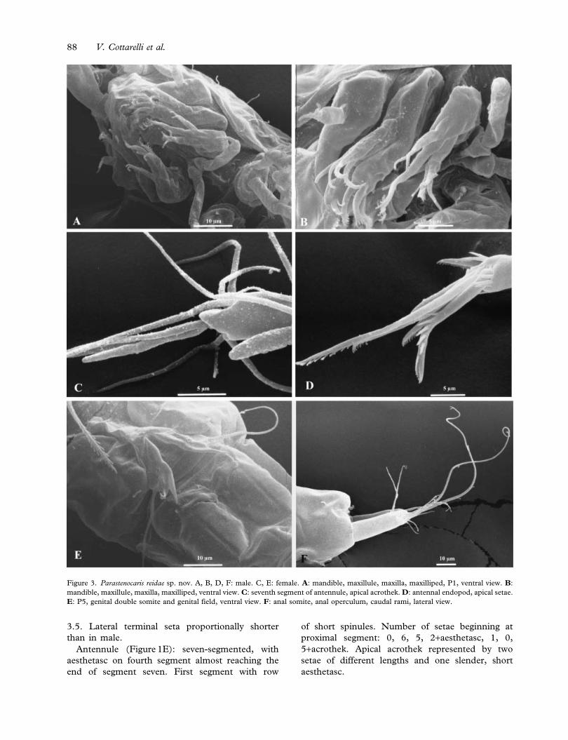
Description
Cephalotorax of both sexes with round dorsal
hyaline integumental window; genital double-somite
and succeeding abdominal somites of female, first,
second third and fourth abdominal somites of male
with oval dorsal integumental windows. Genital
field of female (Figure 4P) located in the pro-
ximal 1/2 of genital double-somite; opercula visible
in Figure 3P. Furcal rami (Figure 4A, B): length/
width ratio about 4.1 in male, and 3.8 in female.
Lateral terminal seta 1.7 and 1.5 times longer than
terminal accessory seta, respectively, in males and
females.
Male antennule (Figure 4C): eight-segmented,
first segment bare; second segment with five normal
and one plumose seta; third segment small, with four
setae; fourth segment reduced to a U-shaped
sclerite, with two setae. Fifth segment enlarged,
with a subapical seta and one apical tubercle with
two setae and one short aesthetasc. Sixth segment
bare; seventh segment short, with lateral tooth-like
expansion. Eighth segment with seven setae and
acrothek represented by two subequal setae and one
slender, short aesthetasc.
Mandible (Figure 4D): coxal gnathobase with
subapical thin seta; one-segmented palp, with two
distal setae.
Maxillule (Figure 4E, 4F): praecoxal arthrite with
three claw-like elements and one subapical curved
seta. Coxa with one distal seta, basis with two apical
setae.
Maxilla (Figure 4G): syncoxa with two endites;
one small seta on the proximal endite; distal endite
with one normal and one leaf-like pinnate setae.
Allobasis prolonged into an apical pinnate claw.
Endopod reduced to a small tubercle with one seta.
P1 basis (Figure 4H) with two short setae near the
endopod insertion in the male (Figure 4I), only one
longer and stronger seta in the female (Figure 4J).
Male P3 (Figure 4K): basis with a lateral long-
itudinal row of spinules, and one pore. Endopod
represented by one thin and pointed seta. Exp-1
distally slender, at about 1/5 of length a tubercle on
medial margin and a row of spinules on lateral
margin. At 2/3 of length, a row of short spinules on
lateral margin, and a tubercle on medial margin.
Distal thumb represented by a leaf-like segment with
blunt tip, shorter than Exp-3. Exp-2 fused with exp-
1 and prolonged into a short, bulbous and inwardly
curved apophysis.
Male P4 (Figure 4L): with row of cuticular
spinules of variable number from four to six even
in the same individual. Female endopod (Figure 4M)
with short apical seta surrounded by few spinules.
Interstitial Parastenocaris and Nitocrella from Tuscany 89
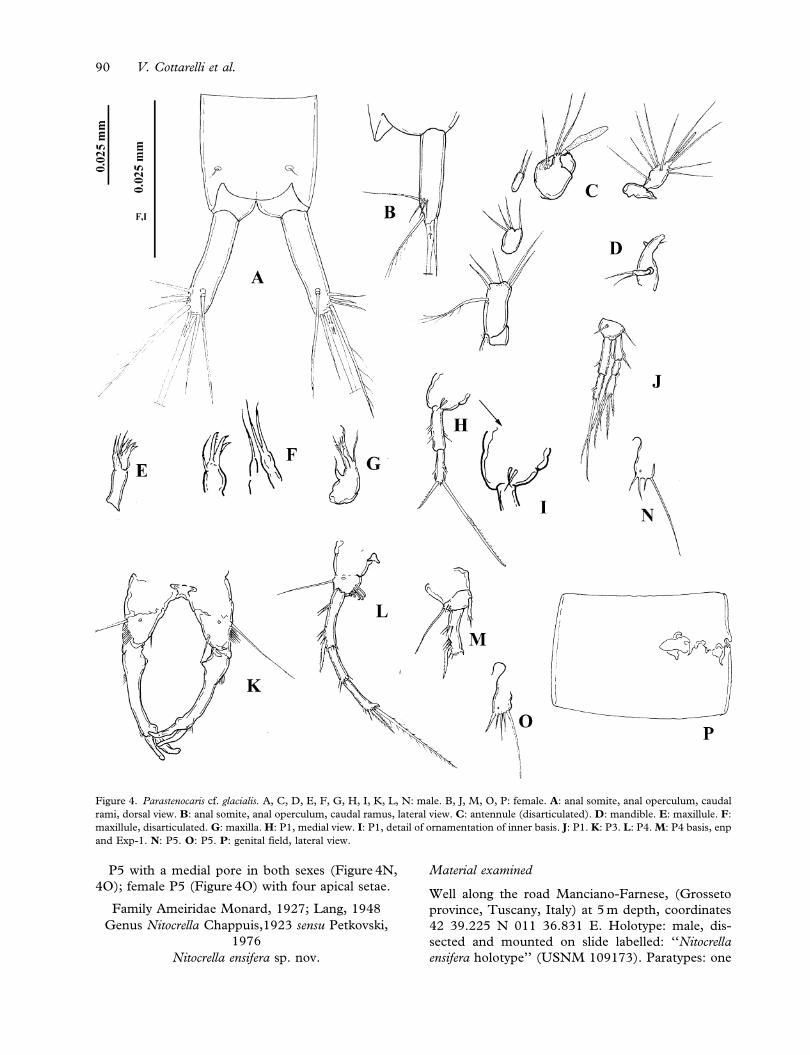
P5 with a medial pore in both sexes (Figure 4N,
4O); female P5 (Figure 4O) with four apical setae.
Family Ameiridae Monard, 1927; Lang, 1948
Genus Nitocrella Chappuis,1923 sensu Petkovski,
1976
Nitocrella ensifera sp. nov.
Material examined
Well along the road Manciano-Farnese, (Grosseto
province, Tuscany, Italy) at 5 m depth, coordinates
42 39.225 N 011 36.831 E. Holotype: male, dis-
sected and mounted on slide labelled: ‘‘Nitocrella
ensifera holotype’’ (USNM 109173). Paratypes: one
Figure 4. Parastenocaris cf. glacialis. A, C, D, E, F, G, H, I, K, L, N: male. B, J, M, O, P: female. A: anal somite, anal operculum, caudal
rami, dorsal view. B: anal somite, anal operculum, caudal ramus, lateral view. C: antennule (disarticulated). D: mandible. E: maxillule. F:
maxillule, disarticulated. G: maxilla. H: P1, medial view. I: P1, detail of ornamentation of inner basis. J: P1. K: P3. L: P4. M: P4 basis, enp
and Exp-1. N: P5. O: P5. P: genital field, lateral view.
90 V. Cottarelli et al.

female, dissected and mounted on slide labelled:
‘‘Nitocrella ensifera paratype, female no. 1’’ (USNM
109175); five females, three of which dissected,
mounted on different slides labelled: ‘‘Nitocrella
ensifera paratype, female no. 2–6’’ (DSAUT); seven
males, 3 of which dissected and mounted on
different slides labelled: ‘‘Nitocrella ensifera paratype,
male no. 1–7’’ (DSAUT). One female, one male,
prepared for scanning electron microscopy, on one
stub labelled: ‘‘Nitocrella ensifera sp. nov. Manciano-
Farnese’’ (CIME). Material collected by V.
Cottarelli and A. Santacroce, 17 April 1996.
Fiora river (Grosseto province, Tuscany, Italy),
locality ‘‘Sovana’’, 340 m asl, about about 28.2 km
from the spring and 29.2 km from the rivermouth,
coordinates 42 39 688N, 011 37 259 E, from
parafluvial phreatic habitat. Paratypes: two females,
each mounted on different slides labelled: ‘‘Nitocrella
ensifera paratype, female no. 7, 8’’ (DSAUT); one
male mounted on slide labelled: ‘‘Nitocrella ensifera
paratype, male no. 8’’ (DSAUT). Material collected
by V. Cottarelli, 12 March 2003.
Description of male
Length averaged among 5 paratypes, from rostrum
to distal apex of caudal rami: 0.42 mm. Body
slender, cephalosome smooth with no dorsal hyaline
window, hyaline frills of all abdominal somites
smooth, body surface variolated. Each abdominal
somite with one row of ventral spinules extending
laterally. Second, third, and fourth abdominal
somite ornamented with one row of spinules anter-
ior to hyaline frill (Figure 7C). Anal somite
(Figure 5A) with two dorsal sensilla, and two dorsal
pores (Figure 5A); four short rows of ventral spi-
nules and four ventral pores (Figure 7C); one row
of spinules around insertion of each ramus, and
one row of thin hair-like setules on each side of
the anal operculum. Rows of proctodeal spinules
(Figure 7C). Anal operculum (Figures 5A, 7C)
slightly convex, with seven spinules on the distal
margin. Caudal ramus (Figures 5A, 7C) subconical,
short; length/width: 1. Posterolateral seta (III) longer
than terminal accessory seta (VI), and as long as
anterolateral seta (II). Anterolateral accessory seta
(I) thin. Two small spinules near insertion of ter-
minal accessory seta, two spinules close to ante-
rolateral seta, and one spinule near insertion of
posterolateral seta (III). Dorsal seta (VII) 2.2 times
as long as caudal ramus, inserted near distomedial
corner. Inner terminal seta (V) 2.4 as long as outer
terminal seta (IV), both terminal setae with break-
ing planes (Figure 7C).
Rostrum (Figure 5B) small and triangular, with
two sensilla.
Antennule (Figures 5B, 7B) 10-segmented; first
segment with one seta and a transversal row of
spinules; second and third segments with eighth
setae each. Fourth segment small, with one seta.
Fifth segment with four normal ventral setae and
distal tubercle with two setae and one aesthetasc of
same lengths, reaching past end of ninth segment;
dorsally three pinnate spiniform (arrowed in figure)
and one normal setae. Sixth segment bare, partially
fused with the fifth one. Seventh segment with one
normal and one transformed seta. Eighth segment
with one distal seta; ninth segment with three setae;
last segment with seven setae.
Antenna (Figure 5C) basis unarmed, exopod one-
segmented, with two transversal rows of spinules and
two transformed and one pinnate apical setae.
Endopod two-segmented, second segment bearing
on the apex four geniculate setae, and two setae
fused together in the proximal part (Figure 5C).
Two smooth spines and a longitudinal spinule row
on lateral margin.
Labrum (Figure 5D): with lateral row of spinules.
Mandible (Figures 5E, 7E): coxal gnathobase
elongate, cutting edge with two strong teeth and
row of small teeth; one plumose seta near dorsal
corner. Palp two-segmented: basis naked, endopod
with five apical setae.
Maxillule (Figure 5F): arthrite of precoxa with
three curved unipinnate apical spines and six naked
subapical spines. Coxa with three setae, one is
geniculate; basis with four setae. Endopod reduced
to a tubercle with one slender seta.
Maxilla (Figures 5G, 7D): syncoxa with one end-
ite with one apical normal and one transformed seta.
Allobasis with terminal pinnate claw and one lateral
seta. Endopod reduced to a tubercle with two apical
setae of same length.
Maxilliped (Figure 5H): prehensile; syncoxa with
one plumose seta, one pore and one transversal row
of spinules on the medial margin. Basis with row of
spinules; endopod with strong unipinnate claw.
Major ornamentation of P1–P4 as follows:
P1 basis 1–1 exp 0–1; 0–1; 1,2,1 enp 1–0; 0–0; 1,2,0
P2 basis 0–1 exp 0–1; 1–1; 0,2,2 enp 0–0; 0,2,0
P3 basis 0–1 exp 0–1; 1–1; 0,2,2 enp 0–0; 0,2,0
P4 basis 0–1 exp 0–1; 1–1; 1,2,2 enp 0–0; 0,2,0
Distal spines on Exp-1 and Exp-2 of P2 and P3
strong.
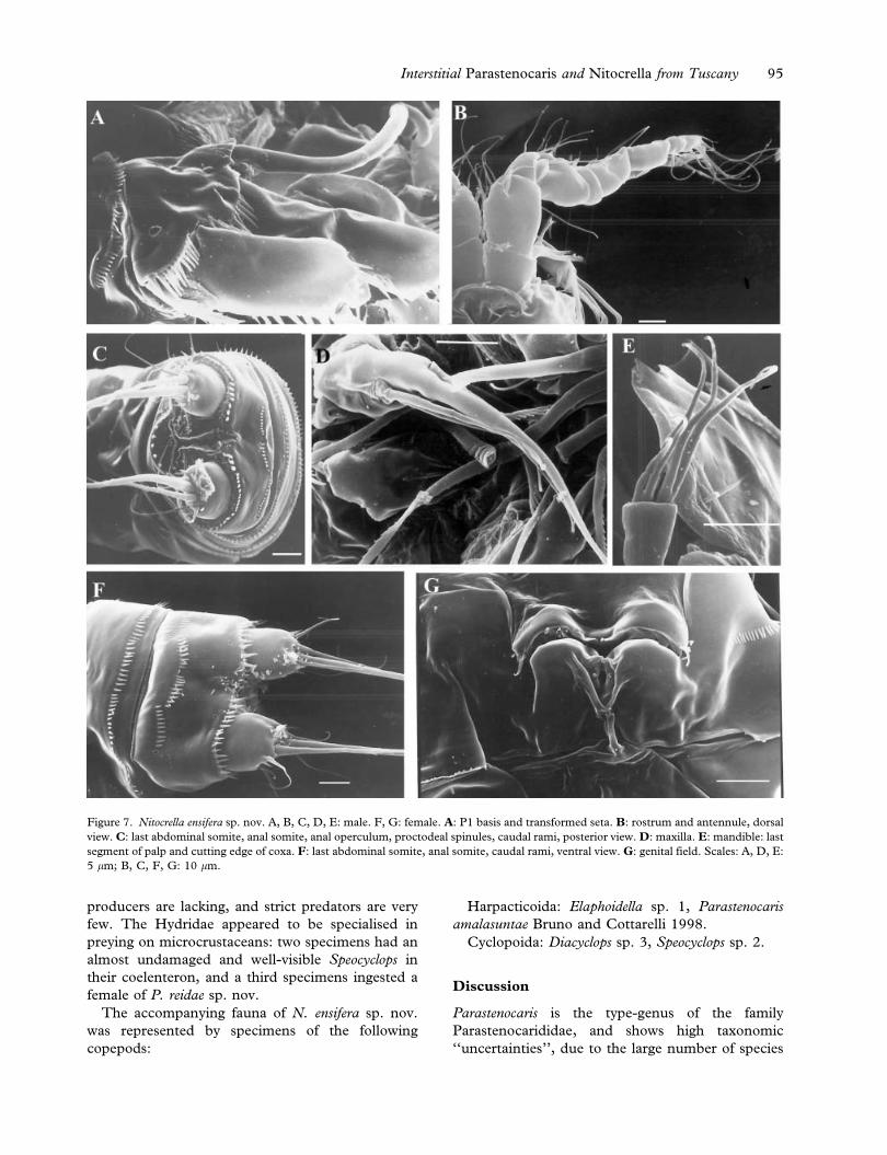
P1 (Figure 5I): basis with two pores visible with
SEM (Figure 7A), one strong lateral spine. Medial
corner with transformed spine which is extremely
long, curved, ending in an enlarged and bilobed
Interstitial Parastenocaris and Nitocrella from Tuscany 91

Figure 5. Nitocrella ensifera sp. nov., male. A: anal somite, anal operculum, caudal rami, dorsal view. B: antennule (disarticulated), ventral
view. C: antenna. D: labrum. E: mandible. F: maxillule. G: maxilla. H: maxilliped. I: P1. J: transformed basal medial seta and Enp-1. K:
P2. L: P3. M: P4. N: P5. O: P6.
92 V. Cottarelli et al.
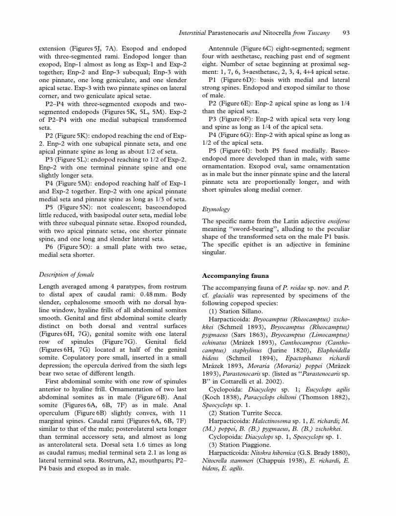
extension (Figures 5J, 7A). Exopod and endopod
with three-segmented rami. Endopod longer than
exopod, Enp-1 almost as long as Exp-1 and Exp-2
together; Enp-2 and Enp-3 subequal; Enp-3 with
one pinnate, one long geniculate, and one slender
apical setae. Exp-3 with two pinnate spines on lateral
corner, and two geniculate apical setae.
P2–P4 with three-segmented exopods and two-
segmented endopods (Figures 5K, 5L, 5M). Exp-2
of P2–P4 with one medial subapical transformed
seta.
P2 (Figure 5K): endopod reaching the end of Exp-
2. Enp-2 with one subapical pinnate seta, and one
apical pinnate spine as long as about 1/2 of seta.
P3 (Figure 5L): endopod reaching to 1/2 of Exp-2.
Enp-2 with one terminal pinnate spine and one
slightly longer seta.
P4 (Figure 5M): endopod reaching half of Exp-1
and Exp-2 together. Enp-2 with one apical pinnate
medial seta and pinnate spine as long as 1/3 of seta.
P5 (Figure 5N): not coalescent; baseoendopod
little reduced, with basipodal outer seta, medial lobe
with three subequal pinnate setae. Exopod rounded,
with two apical pinnate setae, one shorter pinnate
spine, and one long and slender lateral seta.
P6 (Figure 5O): a small plate with two setae,
medial seta shorter.
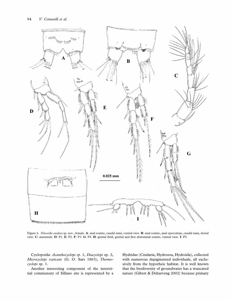
Description of female
Length averaged among 4 paratypes, from rostrum
to distal apex of caudal rami: 0.48 mm. Body
slender, cephalosome smooth with no dorsal hya-
line window, hyaline frills of all abdominal somites
smooth. Genital and first abdominal somite clearly
distinct on both dorsal and ventral surfaces
(Figures 6H, 7G), genital somite with one lateral
row of spinules (Figure 7G). Genital field
(Figures 6H, 7G) located at half of the genital
somite. Copulatory pore small, inserted in a small
depression; the opercula derived from the sixth legs
bear two setae of different length.
First abdominal somite with one row of spinules
anterior to hyaline frill. Ornamentation of two last
abdominal somites as in male (Figure 6B). Anal
somite (Figures 6A, 6B, 7F) as in male. Anal
operculum (Figure 6B) slightly convex, with 11
marginal spines. Caudal rami (Figures 6A, 6B, 7F)
similar to that of the male; posterolateral seta longer
than terminal accessory seta, and almost as long
as anterolateral seta. Dorsal seta 1.6 times as long
as caudal ramus; medial terminal seta 2.1 as long as
lateral terminal seta. Rostrum, A2, mouthparts; P2–
P4 basis and exopod as in male.
Antennule (Figure 6C) eight-segmented; segment
four with aesthetasc, reaching past end of segment
eight. Number of setae beginning at proximal seg-
ment: 1, 7, 6, 3+aesthetasc, 2, 3, 4, 4+4 apical setae.
P1 (Figure 6D): basis with medial and lateral
strong spines. Endopod and exopod similar to those
of male.
P2 (Figure 6E): Enp-2 apical spine as long as 1/4
than the apical seta.
P3 (Figure 6F): Enp-2 with apical seta very long
and spine as long as 1/4 of the apical seta.
P4 (Figure 6G): Enp-2 with apical spine as long as
1/2 of the apical seta.
P5 (Figure 6I): both P5 fused medially. Baseo-
endopod more developed than in male, with same
ornamentation. Exopod oval, same ornamentation
as in male but the inner pinnate spine and the lateral
pinnate seta are proportionally longer, and with
short spinules along medial corner.
Etymology
The specific name from the Latin adjective ensiferus
meaning ‘‘sword-bearing’’, alluding to the peculiar
shape of the transformed seta on the male P1 basis.
The specific epithet is an adjective in feminine
singular.
Accompanying fauna
The accompanying fauna of P. reidae sp. nov. and P.
cf. glacialis was represented by specimens of the
following copepod species:
(1) Station Sillano.
Harpacticoida: Bryocamptus (Rheocamptus) zscho-
kkei (Schmeil 1893), Bryocamptus (Rheocamptus)
pygmaeus (Sars 1863), Bryocamptus (Limocamptus)
echinatus (Mrazek 1893), Canthocamptus (Cantho-
camptus) staphylinus (Jurine 1820), Elaphoidella
bidens (Schmeil 1894), Epactophanes richardi
Mrazek 1893, Moraria (Moraria) poppei (Mrazek
1893), Parastenocaris sp. (listed as ‘‘Parastenocaris sp.
B’’ in Cottarelli et al. 2002).
Cyclopoida: Diacyclops sp. 1; Eucyclops agilis
(Koch 1838), Paracyclops chiltoni (Thomson 1882),
Speocyclops sp. 1.
(2) Station Turrite Secca.
Harpacticoida: Halectinosoma sp. 1, E. richardi; M.
(M.) poppei, B. (B.) pygmaeus, B. (B.) zschokkei.
Cyclopoida: Diacyclops sp. 1, Speocyclops sp. 1.
(3) Station Piaggione.
Harpacticoida: Nitokra hibernica (G.S. Brady 1880),
Nitocrella stammeri (Chappuis 1938), E. richardi, E.
bidens, E. agilis.
Interstitial Parastenocaris and Nitocrella from Tuscany 93
Cyclopoida: Acanthocyclops sp. 1, Diacyclops sp. 2,
Microcyclops varicans (G. O. Sars 1863), Thermo-
cyclops sp. 1.
Another interesting component of the intersti-
tial communuty of Sillano site is represented by a
Hydridae (Cnidaria, Hydrozoa, Hydroida), collected
with numerous depigmented individuals, all exclu-
sively from the hyporheic habitat. It is well known
that the biodiversity of groundwater has a truncated
nature (Gibert & Deharveng 2002) because primary
Figure 6. Nitocrella ensifera sp. nov., female. A: anal somite, caudal rami, ventral view. B: anal somite, anal operculum, caudal rami, dorsal
view. C: antennule. D: P1. E: P2. F: P3. G: P4. H: genital field, genital and first abdominal somite, ventral view. I: P5.
94 V. Cottarelli et al.
producers are lacking, and strict predators are very
few. The Hydridae appeared to be specialised in
preying on microcrustaceans: two specimens had an
almost undamaged and well-visible Speocyclops in
their coelenteron, and a third specimens ingested a
female of P. reidae sp. nov.
The accompanying fauna of N. ensifera sp. nov.
was represented by specimens of the following
copepods:
Harpacticoida: Elaphoidella sp. 1, Parastenocaris
amalasuntae Bruno and Cottarelli 1998.
Cyclopoida: Diacyclops sp. 3, Speocyclops sp. 2.
Discussion
Parastenocaris is the type-genus of the family
Parastenocarididae, and shows high taxonomic
‘‘uncertainties’’, due to the large number of species
Figure 7. Nitocrella ensifera sp. nov. A, B, C, D, E: male. F, G: female. A: P1 basis and transformed seta. B: rostrum and antennule, dorsal
view. C: last abdominal somite, anal somite, anal operculum, proctodeal spinules, caudal rami, posterior view. D: maxilla. E: mandible: last
segment of palp and cutting edge of coxa. F: last abdominal somite, anal somite, caudal rami, ventral view. G: genital field. Scales: A, D, E:
5 mm; B, C, F, G: 10 mm.
Interstitial Parastenocaris and Nitocrella from Tuscany 95
assigned to it, and to the often incomplete older
descriptions, with drawings of poor quality. Several
authors recently underlined these problems
(Schminke & Notenboom 1990; Reid 1995,
Cottarelli & Bruno 1997; Martınez-Arbizu 1997;
Berera & Cottarelli 2003; Galassi & De Laurentiis
2004) and discussed how the characters traditionally
used in the taxonomy of the genus are no longer
adequate to define true phylogenetic affinities.
Among the species-groups instituted by Lang
(1948) for the genus, the minuta species-group
includes 51 species in continental and insular
Europe (Galassi & De Laurentiis 2004). According
to Rouch (1990) and Galassi (1997) these species
share a similar morphology of the male P3 and
Enp-P4 and, but they actually differ in several
phylogenetic informative characters; the minuta
species-group was recognised as polyphyletic by
Galassi and De Laurentiis (2004). Berera and
Cottarelli (2003) defined the hera group on the
basis of morphological, biogeographical and ecolo-
gical characters; this new group included eight
species previously assigned to the minuta-group.
Even if the existing species-groups are question-
able, we think they should be still used pending
the revision of the genus Parastenocaris, and the
formal assessment of natural species-groups.
Within the minuta-group, Parastenocaris reidae sp.
nov. is distinguishable from the remaining species by
the unique combination of structure and ornamen-
tation of male P3 and Enp-P4, and of P5 and caudal
rami in both sexes. The medial seta on P1 basis of
the male is not modified and it is not accompanied
by a chitinous ‘‘protrusion’’ (see Galassi & De
Laurentiis 2004), it is longer and stronger than the
corresponding seta of the female, as it occurs, for
instance, in P. silvana Cottarelli, Bruno and Berera
2000. The remaining affinities are represented by
the simple male P4 endopod, the caudal rami of
both sexes cylindrical and longer than the last
abdominal somite, with setae inserted towards the
apex, and the P5 of both sexes with a medial tip.
These features resemble to those of P. silvana and
partly to those of P. tryphida Cottarelli and Bruno
1993 and P. nertensis Rouch 1990. The male P3 is
not well characterized in several species of the
minuta-group; in P. reidae sp. nov. it is slim and
elongated, partially resembling the male P3 of P.
nertensis.
Both the morphology and position of the genital
field were recently listed among the characters that
should be taken into account when assessing
affinities within the family (Reid 1995; Galassi &
De Laurentiis 2004). The genital field can be best
examined from SEM images; comparison of images
of P. reidae sp. nov. with the few images or drawings
available for other species (Glatzel 1991; Reid 1995;
Cottarelli & Bruno 1997; Galassi 1997; Martınez-
Arbizu 1997; Bruno & Cottarelli 1998, 1999;
Galassi & De Laurentiis 2004) showed marked
differences between genera (i.e. Parastenocaris and
Simplicaris), among species-groups, and also within a
species-group (such as P. reidae sp. nov., P. crenobia
Galassi 1997, P. pasquinii Cottarelli 1972 within the
minuta-group).
Parastenocaris cf. glacialis is the second species
collected in Garfagnana, Tuscany; these specimens
correspond only in part to the original description
and the subsequent ones given by Kiefer (1960,
1961), Kulhavy & Noodt (1968), and Pesce et al.
(1995). The final definition of the taxonomic status
of P. cf. glacialis requires observation of new and
more abundant material of P. cf. glacialis and of P.
glacialis collected from the type locality. The most
important morphological characters of P. cf. glacialis
and differences from the descriptions of P. glacialis
available in literature are as follows:
(1) Integumental windows on cephalotorax and
first three abdominal somites are present in P.
cf. glacialis; versus absent in P. glacialis
according to Noodt (1955); no longer detect-
able in diaphanized specimens (Galassi, perso-
nal communication), and not recorded (neither
as present nor as absent) in Pesce et al. (1995).
(2) Caudal rami: more elongated than in the
specimens described by Pesce et al. (1995),
where the length/width ratio was about 3 in
males and 2.6–2.75 in females. Lateral term-
inal seta is 1.7 and 1.5 times longer than
terminal accessory seta, respectively, in males
and females, whereas it was reported as twice
as long for both sexes in Pesce et al. (1995).
(3) Mouthparts not previously described; they fit
the basic structure known for the genus, with
the exception of the proximal maxillar endite,
which bears only one apical seta, a condition
rarely found in species of Parastenocaris.
(4) Basis of male P4 with 4–6 cuticular spinules,
the number varies even in the same individual;
5 spinules are reported in the original descrip-
tion and in Kiefer (1960), 4 in Kulhavy and
Noodt (1968), and in Pesce et al. (1995); the
exopod of the male P4 is longer and more
curved than in Pesce et al. (1995).
(5) Female P5 with 4 apical setae, whereas it was
reported with 3 in the original description and
in Kulhavy and Noodt (1968), and 4 in Pesce
et al. (1995); the lateral-most seta is longer
than previously reported. The pore is present
96 V. Cottarelli et al.
in other species of Parastenocaris (e.g. P.
pasquinii, P. amalasuntae Bruno & Cottarelli
1998, P. silvana).
(6) Basis of P1 with two medial setae in the male
and only one longer seta in the female. Pesce
et al. (1995) report only one medial seta for
both male and female, confirmed by Galassi
(personal communication). In all the other
species of Parastenocaris the male has a longer
and stronger seta. The male of P. cf. glacialis,
moreover, has a second true seta on the medial
side, almost identical to the first one. This
peculiar feature could represent a new orna-
mentation pattern never recorded before in
Parastenocaris (see Galassi & De Laurentiis
2004 for a more detailed discussion). However,
as two males only were collected, a deeper
interpretation of this feature will be given,
pending the availability of additional material.
As for Parastenocarididae, the systematics of
Ameridae is currently in revision. After the older
revisions by Lang (1965), and Petkovski (1976),
more recently several other authors (Cottarelli &
Forniz 1993; Galassi & De Laurentiis 1997; Conroy
Dalton & Huys 1998; Galassi et al. 1999; Fiers &
Iliffe 2000; Lee & Huys 2002; Karanovic 2004)
underlined how the systematics traditionally based
on swimming legs segmentation can lead to results
sometimes misleading and not informative, if char-
acters with greater phylogenetic significance (such
as setation patterns, and morphology and orna-
mentation of mouthparts and of other cephalic
appendages) are not taken into account (Cottarelli
& Forniz 1993; Galassi et al. 1999). Within this
framework, ‘‘central to this confusion stands the
genus Nitocrella which has served as a taxonomic
repository for freshwater Ameiridae’’ (Lee & Huys
2002). As a consequence, the phylogenetic value
of the hirta, chappuisi and vasconica groups of
Nitocrella proposed by Petkovski (1976) on the
basis of the number of setae/spines present on
the P4 Exp-3, requires confirmation. However,
Petkovski’s species-groups are, according to us
and to Karanovic (2004), still useful for a first
taxonomic screening of the species, which must be
followed by discussion and comparison of other
characters with higher phylogenetic relevance.
The new species can be easily distinguished from
all the other Nitocrella and also from all the Ameridae
by the remarkable autapomorphy represented by the
uniquely transformed medial seta of the male P1
basis. Nitocrella ensifera sp. nov. has 5 spines/setae on
the P4 Exp-3, and therefore can be included in the
chappuisi species-group. Within this group, the new
species shares the highest number of characters with
Nitocrella stammeri, reported for several localities
in Italy (Cottarelli & Fasano 1979; Pesce 1985;
Cottarelli et al. 1996). The two species have a
similar structure and chaetotaxy of legs P1–P4 in
both sexes (with the exception of the above-
mentioned modified seta on male P1 basis); the
same number of segments of male A1 [the male A1
of N. stammeri is 10-segmented and not 8-segmented
as previously reported, for instance, by Cottarelli
& Fasano (1979)]; similar A2, with distinct basis;
female genital somite not fused with the first abdo-
minal somite in both species; similar ornamentation
of anal operculum and abdominal somites; similar
shape and size of caudal rami which nonetheless
differ in length between the two species. The genital
field is similar for the two species but the two
setae near the operculum are of same length in
N. stammeri, whereas the lateral one is longer in
N. ensifera sp. nov. The Italian populations of N.
stammeri and N. ensifera sp. nov. differ in the
following morphological features:
(1) Mandibular palp with one distal seta on the
basis in N. stammeri versus naked in N. ensifera
sp. nov.; endopod with 5 apical setae in the
new species, and 4 in N. stammeri.
(2) Maxillule differing in the ornamentation of
coxa, basis, and endopod, which bear, respec-
tively, 3, 4, and 2 setae in N. ensifera sp. nov.,
and 3, 2, and 1 setae in N. stammeri.
(3) Maxilla with two endites with 2 setae each in
N. stammeri, and one endite with 2 setae in N.
ensifera sp. nov.
(4) P1–P4 very similar in the two species, but the
P2–P3 endopods are proportionally shorter in
the new species, and the apical setae of the
P2–P4 endopods differ in length between the
two species.
(5) P5 with the same number of setae/spines in
the two species, but the setae differ in length
between the two species; the male P5 exopod
of N. ensifera sp. nov. is oval, whereas it is
smaller and almost rectangular in N. stammeri.
Rouch (1985) redescribed of N. stammeri from
specimens collected in Andalusia (Spain), but the
ornamentation and morphology of mandible and
maxillule he described do not correspond to those
of the Italian populations of N. stammeri, and of
N. ensifera sp. nov. It is likely that N. stammeri is in
fact a group of cryptic species, as suggested also by
the wide distribution of N. stammeri.
Nitocrella ensifera sp. nov. also has some affinities
with N. achaiae Pesce 1981, in the morphology and
Interstitial Parastenocaris and Nitocrella from Tuscany 97
ornamentation of P1–P4, and the morphology of A2
and the unfused genital and first abdominal somites.
It is likely that N. achaiae has a 10-segmented male
A1, because this seems to be the common feature
in Nitocrella and in other Ameiridae genera (Galassi
et al. 1999; Lee & Huys 2002; Karanovic 2004).
The shape and ornamentation of P5 are similar
in the two species as well. N. ensifera sp. nov. differs
from N. achaiae in the size of P2–P4 endopods, the
ornamentation of the anal operculum, chaetotaxy
and morphology of caudal rami. The mouthparts
can not be compared because they were not
described for N. achaiae.
Notes on distribution and ecology
The already-mentioned state of flux of the systema-
tic of Parastenocaris complicates the interpretation
of the biogeography of this genus. Nonetheless, P.
reidae sp. nov. is closely related to species from
Sardinia, Corsica, and the Pyrenean Massif, suggest-
ing that their origin dates back to the fragmentation
of the Sardinian–Corsica microplate, during the
Early Miocene, 13 MYA (Bernini & Avanzati
1980; Cottarelli et al. 2000, 2002). The origin of
this taxon is related to the fragmentation of this land
unit, which may have led to vicariance events, and
consequently to speciation.
Parastenocaris cf. glacialis and P. glacialis appear to
have similar ecology: they can be considered cold-
stenotherm relicts (Husmann 1975; Pesce et al.
1995).
Nitocrella is a stygobitic ameirid genus with 11
species recorded mostly from peninsular Italy, and
with few species from Sicily, Sardinia and small
Tyrrhenian islands (Berera et al. 2005). Some Italian
Nitocrella have wide distributions: for instance N.
stammeri is circum-Mediterranean, N. psammophila
Chappuis 1954 is alpine, N. achaiae is reported for
Greece and Italy. Other species have very narrow
distributions and are recorded for only one site
(Berera et al. 2005), such as N. fedelitae Pesce 1985,
N. juturnae Cottarelli 1975, N. stochi Pesce & Galassi
1986. These ‘‘spot’’ distributions can be due to
narrow ecological requirements of the species that
are associated to peculiar microhabitats (see Galassi
& De Laurentiis 1997), but can also be the con-
sequence of missing records from undersampled
regions, as it occurs for several other stygobitic taxa.
Until further data are available, N. ensifera sp. nov.
can be considered endemic to Tuscany. Its origin
can be related to vicariance phenomena which
affected the ancestral population from which both
N. ensifera sp. nov. and its sister-species N. stammeri
may have derived.
N. ensifera sp. nov. lives in several groundwater
habitats such as the phreatic waters in the parafluvial
of the Fiora River, and the deeper phreatic aquifer in
the same area.
Acknowledgements
We want to thank Dr Annalisa Santacroce for
collecting part of the material. Dr Annarita Taddei
(Interdepartmental Center for Electron Microscopy,
‘‘della Tuscia’’ University) helped us to prepare
specimens for SEM, and took the pictures. Dr
Diana M. P. Galassi (University of L’Aquila, Italy)
provided useful information on Parastenocaris cf.
glacialis.
References
Berera R, Bianchini C, Pariciani L, Raschioni R, Cottarelli V.
2003. Studio della biocenosi interstiziale come possibile
strumento per la valutazione della qualita ambientale del
Fiume Orcia. Studi Trentini di Scienze Naturali, Acta
Biologica 80:101–105.
Berera R, Cottarelli V. 2003. Two new species of interstitial
harpacticoids from southern Italy and proposal of a new
Parastenocaris species-group. Italian Journal of Zoology 70:
261–268.
Berera R, Cottarelli V, De Laurentiis P, Galassi DMP, Stoch F.
2005. Crustacea Copepoda Harpacticoida. In: Ruffo S,
Stoch F, editors. Checklist e distribuzione della fauna italiana.
Memorie del Museo Civico di Storia Naturale di Verona
(2u ser.), Sezione Scienza Vita 16:93–95.
Bernini F, Avanzati A. 1980. Notulae chernetologicae XII—Le
Oribatelidae (Acaridae, Oribatia) viventi sul massiccio sardo-
corso. Il popolamento animale e vegetale della Sardegna.
Lavori della Societa Italiana di Biogeografia (n. ser.) 8:
347–399.
Bruno MC, Cottarelli V. 1998. Description of Parastenocaris
amalasuntae n. sp. and new data on Parastenocaris proserpina
and Parastenocaris pasquinii from subterranean waters of
central Italy (Copepoda, Harpacticoida). Italian Journal of
Zoology 65:121–136.
Bruno MC, Cottarelli V. 1999. Harpacticoids from groundwaters
in the Philippines: Parastenocaris mangyans new species,
Epactophanes philippinus new species and redescription of
Phyllognathopus bassoti (Crustacea, Copepoda). Journal of
Crustacean Biology 19:510–529.
Conroy-Dalton S, Huys R. 1998. Towards a revision of Ameira
Boeck, 1865 (Harpacticoida, Ameiridae): Reinstatement of
Psammameira Noodt, 1952. Zoologica Scripta 27:247–261.
Cottarelli V, Berera R. 2003. Il fiume sotto il fiume: ricerche sul
popolamento iporreico a crostacei del Fiume Fiora (Italia
centrale). Studi Trentini di Scienze Naturali, Acta Biologica
80:27–30.
Cottarelli V, Berera R, Maiolini B. 2002. Annotazioni faunistiche
ed ecologiche su Copepodi di alta e media quota di sorgenti e
corsi d’acqua alpini, appenninici e sardi. Studi Trentini di
Scienze Naturali, Acta Biologica 78:25–30.
Cottarelli V, Bruno MC. 1997. First record of Parastenocarididae
(Crustacea, Copepoda, Harpacticoida) from subterranean
freshwater of insular Greece and description of two new
species. International Journal of Speleology 5:43–57.
98 V. Cottarelli et al.
Cottarelli V, Bruno MC, Berera R. 2000. Parastenocaris corsica sp.
nov. and Parastenocaris silvana sp. nov., first Parastenocarididae
from groundwater of Corsica (Copepoda, Harpacticoida).
Crustaceana 7:345–364.
Cottarelli V, Bruno MC, Forniz C. 1996. Copepodi Arpacticoidi
e Sincaridi (Crustacea) di acque sotterranee delle isole
circumsarde. Biogeographia 18:261–272.
Cottarelli V, Fasano L. 1979. Nitocrella stammeri Chappuis
(Crustacea, Copepoda, Harpacticoida): nuovi reperti italiani
e descrizione del maschio. Animalia 5:187–196.
Cottarelli V, Forniz C. 1993. Due nuove specie di Nitocrellopsis
Petkovski di acque freatiche delle isole di Kos e Tilos
(Sporadi Meridionali) (Crustacea, Copepoda, Harpacticoida).
Fragmenta Entomologica 24:131–145.
Cvetkov L. 1968. Un filet phreatobiologique. Bulletin de
l’Institut de Zoologie et Musee, Academie Bulgare des
Sciences 27:215–218.
Delamare Deboutteville C. 1960. Biologie des eaux soutterraines
littorales et continentales. Paris: Hermann.
Fiers F, Iliffe TM. 2000. Nitocrellopsis texana n. sp. from central
Texas (U.S.A.) and N. ahaggarensis n. sp. from the central
Algerian Sahara (Copepoda, Harpacticoida). Hydrobiologia
418:81–97.
Galassi DMP. 1997. Little known harpacticoid copepods from
Italy, and description of Parastenocaris crenobia n. sp.
(Copepoda, Harpacticoida). Crustaceana 70:694–709.
Galassi DMP. 2001. Groundwater copepods: Diversity patterns
over ecological and evolutionary scales. Hydrobiologia 453/
454:227–253.
Galassi DMP, De Laurentiis P. 1997. Two new species of
Nitocrella from groundwaters of Italy (Crustacea, Copepoda,
Harpacticoida). Italian Journal of Zoology 64:367–376.
Galassi DMP, De Laurentiis P. 2004. Towards a revision of
the genus Parastenocaris Kessler, 1913: establishment of
Simplicaris gen. nov. grom groundwaters in central Italy and
review of the P. brevipes-group (Copepoda, Harpacticoida,
Parastenocarididae). Zoological Journal of the Linnean
Society, London 140:417–436.
Galassi DMP, De Laurentiis P, Dole-Oliver MJ. 1999. Nito-
crellopsis rouchi sp. n., a new ameirid harpacticoid from phreatic
waters in France (Copepoda: Harpacticoida: Ameiridae).
Hydrobiologia 412:77–189.
Gibert J, Deharveng L. 2002. Subterranean ecosystems: A
truncated functional biodiversity. BioScience 52:473–481.
Glatzel T. 1991. Neue morphologische Aspekte und die
Copepodid-Stadien von Parastenocaris phyllura Kiefer
(Copepoda, Harpacticoida). Zoologica Scripta 20:375–393.
Husmann S. 1975. The boreoalpine distribution of groundwater
organisms in Europe. Verhandlungen der Internationale
Vereinigung fur Theoretische und Angewandte Limnologie
19:2983–2988.
Huys R, Boxshall GA. 1991. Copepod evolution. London: The
Ray Society.
Karanovic T. 2004. Subterranean copepods (Crustacea,
Copepoda) from arid Western Australia. Crustaceana 3
(suppl.):1–366.
Kiefer F. 1960. Subterrane Ruderfusskrebse (Crust. Cop) aus
dem Ruhrtal. Zoologischer Anzeiger 165:323–329.
Kiefer F. 1961. Uber einige Parastenocariden (Copepoda Har-
pacticoida) aus den Niederlanden. Crustaceana 3:115–119.
Kulhavy V, Noodt W. 1968. Uber Copepoden (Crustacea) aus
dem limnischen Mesopsammal Islands. Gewasser und
Abwasser 46:50–61.
Lang K. 1948. Monographie der Harpacticiden. Vols. 1, 2.
Stockholm: Nordiska Bokhandeln.
Lang K. 1965. Copepoda Harpacticoida from the Californian
Pacific coast. Stockholm: Almqvist & Wiksell.
Lee W, Huys R. 2002. A new genus of groundwater Ameiridae
(Copepoda, Harpacticoida) from boreholes in Western
Australia and the artificial status of Stygonitocrella Petkovski,
1976. Bulletin of the Natural History Museum, Zoology Series
68:39–50.
Martınez-Arbizu P. 1997. Parastenocaris hispanica n.sp.
(Copepoda: Harpacticoida: Parastenocarididae) from hypor-
heic groundwaters in Spain and its phylogenetic position
within the fontinalis-group of species. Contributions to
Zoology 66:215–226.
Noodt W. 1955. Die Verbreitung des Genus Parastenocaris, ein
Beispiel einer subterranen Crustaceen-Gruppe. Zoologischer
Anzeiger 18 (suppl.):429–435.
Pesce GL. 1985. Un nuovo arpacticoide di acque freatiche del
Molise e considerazioni sullo ‘status’ tassonomico e distribu-
zione del genere Nitocrella Chappuis in Italia (Crustacea
Copepoda: Ameiridae). Rivista di Idrobiologia 24:65–72.
Pesce GL, Galassi DMP, Cottarelli V. 1995. Parastenocaris
lorenzae n. sp., and first record of Parastenocaris glacialis
Noodt (Copepoda, Harpacticoida) from Italy. Hydrobiologia
302:97–101.
Petkovski TK. 1976. Drei neue Nitocrella-Arten von Kuba,
zugleich eine Revision des Genus Nitocrella Chappuis (s.
restr.) (Crustacea, Copepoda, Ameiridae Acta Musei
Macedonici Scientiarum Naturalium 15:1–26.
Reid JW. 1995. Redescription of Parastenocaris brevipes Kessler
and description of a new species of Parastenocaris (Copepoda:
Harpacticoida: Parastenocarididae) from the USA. Canadian
Journal of Zoology 73:173–187.
Rouch R. 1985. Une nouvelle Stygonitocrella (Copepoda,
Harpacticoida) des eaux souterraines d’Andalousie, Espagne.
Stygologia 1:118–127.
Rouch R. 1990. Deux nouvelles Parastenocaris (Copepodes,
Harpacticoıdes) des Pyrenees. Annales de Limnologie
26:19–28.
Schminke HK, Notenboom J. 1990. Parastenocarididae
(Copepoda, Harpacticoida) from the Netherlands. Bijdragen
tot de Dierkunde 60:299–304.
Interstitial Parastenocaris and Nitocrella from Tuscany 99