ISUOG Practice Guidelines (updated): sonographic ...
9
Ultrasound Obstet Gynecol 2020 Published online in Wiley Online Library (wileyonlinelibrary.com). DOI: 10.1002/uog.22145 isuog.org GUIDELINES ISUOG Practice Guidelines (updated): sonographic examination of the fetal central nervous system. Part 1: performance of screening examination and indications for targeted neurosonography Clinical Standards Committee The International Society of Ultrasound in Obstetrics and Gynecology (ISUOG) is a scientific organization that encourages sound clinical practice, and high-quality teaching and research related to diagnostic imaging in women’s healthcare. The ISUOG Clinical Standards Committee (CSC) has a remit to develop Practice Guidelines and Consensus Statements as educational recommendations that provide healthcare practitioners with a consensus-based approach, from experts, for diagnostic imaging. They are intended to reflect what is considered by ISUOG to be the best practice at the time at which they are issued. Although ISUOG has made every effort to ensure that Guidelines are accurate when issued, neither the Society nor any of its employees or members accepts any liability for the consequences of any inaccurate or misleading data, opinions or statements issued by the CSC. The ISUOG CSC documents are not intended to establish a legal standard of care, because interpretation of the evidence that underpins the Guidelines may be influenced by individual circumstances, local protocol and available resources. Approved Guidelines can be distributed freely with the permission of ISUOG ([email protected]). INTRODUCTION Central nervous system (CNS) malformations are some of the most common congenital abnormalities. Neural tube defects are the most frequent CNS malformations and amount to about one to two cases per 1000 births. The incidence of intracranial abnormalities with an intact neural tube is uncertain, as most of these abnormalities probably go undetected at birth and manifest only in later life. However, long-term follow-up studies suggest that the incidence may be as high as one in 100 births 1 . Ultrasound has been used for nearly 30 years as the main modality to help diagnose fetal CNS anomalies. The aim of these Guidelines is to review, describe and update the technical aspects of the screening evaluation of the fetal brain to be performed as part of the midtrimester anomaly scan, which is referred to in this document as a ‘screening examination’. This Guideline also presents the indications for the detailed evaluation of the fetal CNS, which constitutes ‘targeted fetal neurosonography’, a dedicated examination of the fetal brain and spine that requires specific expertise and sophisticated ultrasound equipment. This examination is described in Part 2 of this Guideline, in which we also discuss the indications for fetal brain magnetic resonance imaging (MRI). Details of the grades of recommendation and levels of evidence used in this Guideline are given in Appendix 1. GENERAL CONSIDERATIONS Gestational age Recommendation • Examiners involved in screening for CNS abnormalities should be familiar with normal CNS appearance at different gestational ages (GOOD PRACTICE POINT). The appearance of the brain and the spine changes throughout gestation. To avoid diagnostic errors, it is important to be familiar with normal CNS appearance at different gestational ages (Figure 1), although most efforts to diagnose CNS anomalies are focused around midgestation 2 . Hence, it is recommended that this Guide- line is applied during the midtrimester anomaly scan. However, during the last decade, it has become evident that an increasing number of CNS and neural tube abnormalities, mainly dorsal and rhombencephalic induction defects, may be visible from the end of the first trimester 3–9 . Although these are in the minority, they are usually severe and therefore deserve special consideration. While early examination of the CNS requires certain skills, it is always worthwhile paying particular attention to the fetal head and brain at early gestational ages. The advantage of early fetal neurosonography at 12 – 15 weeks is that the bones are thin and the brain may be evaluated © 2020 International Society of Ultrasound in Obstetrics and Gynecology ISUOG GUIDELINES
Transcript of ISUOG Practice Guidelines (updated): sonographic ...

Ultrasound Obstet Gynecol 2020Published online in Wiley Online Library (wileyonlinelibrary.com). DOI: 10.1002/uog.22145
isuog.org GUIDELINES
ISUOG Practice Guidelines (updated): sonographicexamination of the fetal central nervous system. Part 1:performance of screening examination and indications fortargeted neurosonography
Clinical Standards Committee
The International Society of Ultrasound in Obstetricsand Gynecology (ISUOG) is a scientific organizationthat encourages sound clinical practice, and high-qualityteaching and research related to diagnostic imagingin women’s healthcare. The ISUOG Clinical StandardsCommittee (CSC) has a remit to develop PracticeGuidelines and Consensus Statements as educationalrecommendations that provide healthcare practitionerswith a consensus-based approach, from experts, fordiagnostic imaging. They are intended to reflect whatis considered by ISUOG to be the best practice atthe time at which they are issued. Although ISUOGhas made every effort to ensure that Guidelines areaccurate when issued, neither the Society nor any ofits employees or members accepts any liability for theconsequences of any inaccurate or misleading data,opinions or statements issued by the CSC. The ISUOGCSC documents are not intended to establish a legalstandard of care, because interpretation of the evidencethat underpins the Guidelines may be influenced byindividual circumstances, local protocol and availableresources. Approved Guidelines can be distributed freelywith the permission of ISUOG ([email protected]).
INTRODUCTION
Central nervous system (CNS) malformations are someof the most common congenital abnormalities. Neuraltube defects are the most frequent CNS malformationsand amount to about one to two cases per 1000 births.The incidence of intracranial abnormalities with an intactneural tube is uncertain, as most of these abnormalitiesprobably go undetected at birth and manifest only in laterlife. However, long-term follow-up studies suggest thatthe incidence may be as high as one in 100 births1.
Ultrasound has been used for nearly 30 years as themain modality to help diagnose fetal CNS anomalies. Theaim of these Guidelines is to review, describe and updatethe technical aspects of the screening evaluation of the
fetal brain to be performed as part of the midtrimesteranomaly scan, which is referred to in this document asa ‘screening examination’. This Guideline also presentsthe indications for the detailed evaluation of the fetalCNS, which constitutes ‘targeted fetal neurosonography’,a dedicated examination of the fetal brain and spine thatrequires specific expertise and sophisticated ultrasoundequipment. This examination is described in Part 2 of thisGuideline, in which we also discuss the indications forfetal brain magnetic resonance imaging (MRI). Details ofthe grades of recommendation and levels of evidence usedin this Guideline are given in Appendix 1.
GENERAL CONSIDERATIONS
Gestational age
Recommendation
• Examiners involved in screening for CNS abnormalitiesshould be familiar with normal CNS appearance atdifferent gestational ages (GOOD PRACTICE POINT).
The appearance of the brain and the spine changesthroughout gestation. To avoid diagnostic errors, it isimportant to be familiar with normal CNS appearanceat different gestational ages (Figure 1), although mostefforts to diagnose CNS anomalies are focused aroundmidgestation2. Hence, it is recommended that this Guide-line is applied during the midtrimester anomaly scan.
However, during the last decade, it has becomeevident that an increasing number of CNS and neuraltube abnormalities, mainly dorsal and rhombencephalicinduction defects, may be visible from the end of the firsttrimester3–9. Although these are in the minority, they areusually severe and therefore deserve special consideration.While early examination of the CNS requires certainskills, it is always worthwhile paying particular attentionto the fetal head and brain at early gestational ages. Theadvantage of early fetal neurosonography at 12–15 weeksis that the bones are thin and the brain may be evaluated
© 2020 International Society of Ultrasound in Obstetrics and Gynecology ISUOG GUIDELINES

2 ISUOG Guidelines
12 weeks
Tra
nsve
ntri
cula
rT
rans
thal
amic
Tra
nsce
rebe
llar
21 weeks 32 weeks
Figure 1 Normal morphological changes of fetal brain throughout gestation, as visualized on sonographic examination in axial planes:views in transventricular, transthalamic and transcerebellar planes at 12, 21 and 32 gestational weeks. Note significant structural change oflateral ventricles and choroid plexus from late first trimester to midgestation, along with appearance of cavum septi pellucidi only from earlysecond trimester onwards. Nevertheless, ventricular atrial width remains relatively stable during second and third trimesters.
from almost all angles, especially with a high-frequencytransvaginal transducer.
Generally, a satisfactory evaluation of the fetal CNScan be performed from the end of the first trimester.As pregnancy advances, visualization of the intracranialstructures becomes more difficult due to advancedossification of the calvarium.
Technical factors
Ultrasound transducers
High-frequency ultrasound transducers increase spatialresolution but decrease the penetration of the soundbeam. The choice of optimal transducer and operatingfrequency is influenced by a number of factors, includingmaternal habitus, fetal position, gestational age andthe approach used. Most screening examinations areperformed satisfactorily with a 3–5-MHz transabdominaltransducer, although recent wideband transducers canalso be employed advantageously.
Imaging parameters
The examination is performed with grayscale two-dimensional ultrasound. Harmonic and crossbeam imag-ing, as well as speckle-reduction filters, may enhance
visualization of subtle anatomic details and in patientswho scan poorly, for example those with increased bodymass index or abdominal scarring.
SCREENING EXAMINATION OF FETALBRAIN AFTER 18 WEEKS
Qualitative evaluation
Recommendation
• Transabdominal sonography is the technique of choicefor the screening examination of the fetal CNS duringthe midtrimester scan in low-risk pregnancies. Thisexamination should include evaluation of the fetal headand spine (GOOD PRACTICE POINT).
The fetal CNS screening examination during themidtrimester scan in low-risk pregnancies should includeevaluation of the fetal head and spine, using transabdom-inal sonography. Evaluation of two axial planes allowsvisualization of the relevant cerebral structures to assessthe anatomic integrity of the fetal brain10. These planes arecommonly referred to as the transventricular (Figure 2a)and transcerebellar (Figure 2b) planes. A third plane, the
© 2020 International Society of Ultrasound in Obstetrics and Gynecology Ultrasound Obstet Gynecol 2020.

ISUOG Guidelines 3
Th
Th
CM
Cerebellarvermis
Cerebellum
Th
Th
b
ac
FalxAnterior
horns
CSP
Atrium
CP
Figure 2 Fetal central nervous system screening examination (normal 21-week fetus) in three axial planes. (a) Transventricular plane,showing anterior and posterior portions of lateral ventricles. Comma-shaped anterior horns are separated centrally by cavum septi pellucidi(CSP). Atrium and posterior horn of ventricle distal to transducer are also demonstrated, along with choroid plexus (CP), as anatomicreference for measurement of atrial width, and parieto-occipital fissure ( ). (b) For transcerebellar plane, transducer is tilted posteriorly inorder to depict middle and posterior fossa structures: thalami (Th), cerebellar hemispheres and cerebellar vermis, demon-strated as butterfly shape, and retrocerebellar anechoic space corresponding to cisterna magna (CM). (c) Transthalamic plane is frequentlyused for biometry of fetal head (biparietal diameter, occipitofrontal distance and head circumference) and is inferior and parallel totransventricular plane. In this plane, falx, anterior horns of lateral ventricles and CSP are also observed, as well as thalami (Th) andhippocampal gyri bilaterally. Line diagram (top left) illustrates positions of axial planes.
Table 1 Structures usually noted on screening ultrasoundexamination of fetal central nervous system
• Head shape• Lateral ventricles• Cavum septi pellucidi• Thalami• Cerebellum• Cisterna magna• Spine
so-called transthalamic plane (Figure 2c), is frequentlyadded, mostly for the purpose of biometry. Structuresthat should be noted in the routine examination includethe lateral ventricles, the cerebellum and cisterna magna,
and the cavum septi pellucidi (CSP). Head shape and braintexture should also be noted on these views (Table 1).
Transventricular plane (Figure 2a)
Recommendation
• In the transventricular plane, the aspect of the atriumdistal to the transducer and the presence of theCSP should be assessed and documented (GOODPRACTICE POINT).
The transventricular plane demonstrates the anteriorand posterior portions of the lateral ventricles. Theanterior portion (frontal or anterior horns) appears as
© 2020 International Society of Ultrasound in Obstetrics and Gynecology Ultrasound Obstet Gynecol 2020.

4 ISUOG Guidelines
two comma-shaped, fluid-filled structures. They have awell-defined lateral wall and are separated medially bythe CSP. The CSP is a fluid-filled cavity between two thinmembranes. In late gestation or the early neonatal period,these membranes usually fuse to become the septumpellucidum. The CSP becomes visible between 17 and20 weeks and disappears near term. Using transabdominalultrasound, it should always be demonstrable between17–20 and 37 weeks, or at a biparietal diameter (BPD)of 44–88 mm11. Failure to demonstrate the CSP priorto 16 weeks or later than 37 weeks is a normal finding;rarely, absence of fluid in the CSP is seen in completelynormal fetuses12. The importance of visualizing the CSPbetween 17 and 37 gestational weeks is due to the factthat its non-visualization or an abnormal appearance isassociated with commissural anomalies, which may be anindirect sign of corpus callosal agenesis on screening views(usually in conjunction with a tear-shaped appearanceof the lateral ventricles, known as colpocephaly13).Failure to visualize the membranes of the septumpellucidum is highly suspicious for the presence ofa number of severe cerebral malformations, such asholoprosencephaly, severe hydrocephaly and septo-opticdysplasia14. Recently, an abnormal shape of the CSP hasbeen described as a relatively reliable marker of partialagenesis of the corpus callosum15,16.
From about 16 weeks, the posterior portion of thelateral ventricles (also referred to as occipital horns) is,in reality, a complex formed by the atrium that continuesposteriorly into the occipital horn. The atrium is char-acterized by the presence of the glomus of the choroidplexus, which is highly echogenic, while the occipital hornis filled with cerebrospinal fluid. Particularly in the secondtrimester of gestation, both medial and lateral walls ofthe ventricle are parallel to the midline and are there-fore well-depicted sonographically as well-demarcatedechogenic lines. Under normal conditions, the glomusof the choroid plexus completely fills the cavity of theventricle at the level of the atrium, being in close contactwith the medial and lateral walls, although in somenormal cases a small amount of fluid may be presentbetween the medial wall and the choroid plexus17–20.
It should be noted that, due to artifacts in the near fieldof the image, caused by shadowing from the proximalparietal bone, in the standard transventricular plane, onlythe hemisphere and the lateral ventricle on the far sideof the transducer are usually visualized clearly. However,most severe cerebral lesions are bilateral or associated witha significant deviation or distortion of the midline echo,and it has been suggested that, in screening examinations,symmetry of the brain can be assumed.
Transcerebellar plane (Figure 2b)
Recommendation
• In the transcerebellar plane, the presence and shape ofthe cerebellum, as well as the presence of cerebrospinalfluid in the cisterna magna, should be assessed anddocumented (GOOD PRACTICE POINT).
The transcerebellar plane is slightly caudal to the transven-tricular one, and it is usually obtained with slight posteriortilting of the transducer. It is used to visualize the thalami,cerebellum and cisterna magna. The cerebellum appears asa butterfly-shaped structure formed by the round cerebel-lar hemispheres joined in the middle by the slightly moreechogenic cerebellar vermis. The cisterna magna, or cis-terna cerebellomedullaris, is a fluid-filled space posteriorto the cerebellum. It normally contains thin septations,which are not usually demonstrated in the presence ofpathology21. In the second half of gestation, the antero-posterior diameter of the cisterna magna remains stableand should not exceed 10 mm10. Before 19–20 gestationalweeks, the cerebellar vermis has not yet completely cov-ered the fourth ventricle, and this unusual appearancemay give the false impression of a defect of the vermis. Asa rule of thumb, by 19 gestational weeks, there should beno midline fluid-filled space between the two cerebellarhemispheres; should this finding, referred to as ‘keyholesign’, be detected, it may be associated with an anomaly ofthe cerebellar vermis and the fetus should be referred forneurosonography22. Care should be taken to avoid ‘over-tilting’ of the probe, since this will increase the likelihoodof false-positive diagnosis of a vermian anomaly.
Transthalamic plane (Figure 2c)
Commonly referred to as the transthalamic or BPD plane,a third scanning plane, obtained parallel but caudal tothe transventricular plane, is also frequently used in thesonographic assessment of the fetal head. The anatomiclandmarks include, from anterior to posterior, the frontalhorns of the lateral ventricles, the CSP, the thalami andthe hippocampal gyri23. This plane is used for biometryof the fetal head. It is easier to identify in late gestationand allows more reproducible measurements than doesthe transventricular plane24.
Fetal spine
Recommendation
• When technically feasible, a longitudinal section of thefetal spine should be obtained, in order to screen foropen and closed spinal dysraphism (GOOD PRACTICEPOINT).
Technical advice
• Up to 97% of cases of open spina bifida presentwith the so-called ‘banana sign’, which is due toChiari-II malformation25 (GRADE OF RECOMMEN-DATION: C).
Detailed examination of the fetal spine requires expertiseand meticulous scanning, and the results are heavilydependent on the fetal position. Therefore, a full anddetailed evaluation of the fetal spine in every planeis not part of the screening examination. One of themost frequent severe spinal abnormalities, open spina
© 2020 International Society of Ultrasound in Obstetrics and Gynecology Ultrasound Obstet Gynecol 2020.

ISUOG Guidelines 5
bifida, is usually associated with abnormal intracranialanatomy: up to 97% of cases present with the so-called‘banana sign’, which is due to Chiari-II malformation25.However, a longitudinal section of the fetal spine shouldbe sought4 if technically feasible, because it may reveal, atleast in some cases, other spinal malformations, includingvertebral abnormalities and sacral agenesis, although thelatter diagnosis may be challenging even for experts,due to the physiological non-ossification of the caudalspine in the mid trimester26. Under normal conditions, asagittal section of the spine at 18–24 gestational weeksdemonstrates the three ossification centers of the vertebrae(one inside the body and one on each side at the junctionbetween the lamina and pedicle) that surround the neuralcanal, and that appear as either two or three parallel lines,depending on the orientation of the ultrasound beam(Figure 3). The three ossification nuclei are best visualizedon an axial view of individual vertebrae (Figure 4). Inaddition, an attempt should be made to demonstrate theintactness of the skin overlying the spine, on either atransverse or a longitudinal view.
Quantitative evaluation
Recommendation
• The following measurements represent an integral partof sonographic screening for CNS malformations: atrialwidth and transverse cerebellar diameter. Additionalmeasurements usually performed for general biometrypurposes (BPD and head circumference (HC)) are alsopart of the examination, since they may, in some cases,reveal proliferation abnormalities (e.g. microcephaly ormacrocephaly) (GOOD PRACTICE POINT).
Technical advice
• The atrial width should be measured inner-to-inner andshould be <10 mm throughout pregnancy (GRADE OFRECOMMENDATION: 1).
Skin
Conus medullaris
Cauda equinaL2
Figure 3 Sagittal view of lower thoracic and sacral fetal spine.Using unossified spinous process of vertebrae as acoustic window,contents of neural canal are demonstrated. Conus medullaris isclearly demonstrated and is normally located at level of L2 inmidgestation. Its sharp end should point anteriorly, to vertebralbody, with fluid filling neural canal posteriorly. Note intact skinobserved as hyperechogenic line along fetal back.
Biometry is an essential part of sonographic exami-nation of the fetal head. In the second-trimester anomalyscan, a standard examination includes measurement of theBPD, HC, internal diameter of the atrium and transversecerebellar diameter. The cisterna magna depth should bemeasured if this structure is visually thinner or widerthan normal on qualitative assessment of the posteriorfossa.
BPD and HC are commonly used for assessing fetal ageand growth and may also be useful to identify somecerebral anomalies. They may be measured either inthe transventricular plane or in the transthalamic plane.There are various techniques for measuring BPD. Mostfrequently, the calipers are positioned outside the fetalcalvarium (so-called ‘outer-to-outer’ measurement)24.However, some commonly used charts were producedusing an outer-to-inner technique, to avoid artifactsgenerated by the distal echo of the calvarium, an issuethat is less relevant now, with modern transducers, thanit was several years ago23. These two approaches tomeasurement result in a difference of a few millimeters,which may be clinically relevant in early gestation. It isimportant, therefore, to know the technique that was usedto construct the reference charts that one uses. HC can bemeasured directly, with the ellipse method, by placing theellipse around the outer outline of the calvarium echoes.Alternatively, it can be calculated after measuring the BPDand occipitofrontal diameter (OFD), using the equation:HC = 1.62 × (BPD + OFD). The BPD/OFD ratio is usually70–85%. However, molding of the fetal head, particularlyin early gestation, is frequent, and fetuses in breechpresentation may show some degree of dolicocephaly.It is not appropriate to use HC nomograms intended for
Figure 4 Axial views of fetal spine at different levels: (a) cervical,(b) thoracic, (c) lumbar and (d) sacral. Arrows indicate threeossification centers of vertebrae, and arrowheads indicate spinalcord, which is observed at cervical, thoracic and lumbar levels.Hyperechogenic dot corresponds to central canal of medulla. Atsacral level (d), only fibers of cauda equina are observed. Note thinstrip of fluid behind cord at all levels and intact skin overlying spine.
© 2020 International Society of Ultrasound in Obstetrics and Gynecology Ultrasound Obstet Gynecol 2020.

6 ISUOG Guidelines
YES no1 no2 no3
Figure 5 (a) Measurement of atrial width of lateral ventricles. Calipers are positioned at level of glomus of choroid plexus, inside echoesgenerated by ventricular walls. (b) Diagram illustrating correct caliper placement for ventricular measurement. Calipers are placed correctlywhen touching inner edge of ventricular wall at its widest part and aligned perpendicular to long axis of ventricle (YES). Incorrectplacements include middle–middle (no1), outer–outer (no2) and placement that is too posterior in narrower part of ventricle or notperpendicular to ventricular axis (no3).
fetal weight estimation if the endpoint of the measurementis to exclude microcephaly.
Measurement of the atrium is recommended becauseseveral studies suggest that this is the most effectiveapproach for assessing the integrity of the ventricularsystem18, and ventriculomegaly is a frequent markerof abnormal cerebral development. Measurement isperformed at the level of the glomus of the choroidplexus, perpendicular to the ventricular cavity, positioningthe calipers inside the echoes generated by the lateralwalls (Figure 5). This measurement remains stable inthe second and early third trimesters, with a meandiameter between 6 and 8 mm18,27; it is considered normalwhen <10 mm27–31. Although this cut-off was determinedseveral years ago, it remains valid even with more modernequipment, particularly at midgestation. Therefore, anatrial width ≥ 10 mm should be considered suspicious. Itis useful to emphasize here that: (1) the atrial width maychange during gestation, either increasing or decreasing,and (2) moderate asymmetry in atrial width between thetwo sides should be considered normal, if both atriameasure <10 mm32,33.
The transverse cerebellar diameter increases by about1 mm per week of pregnancy between 14 and 21gestational weeks. This measurement, along with the HCand BPD, is helpful to assess fetal growth. In cases inwhich the anteroposterior diameter of the cisterna magnashould be measured (because it is subjectively consideredabnormal), the calipers should be positioned in a correcttranscerebellar plane, between the outer edge of the mostdorsal point of the cerebellar vermis and the internalside of the occipital bone. A normal measurement is2–10 mm34. With dolicocephaly, measurements slightlylarger than 10 mm may be encountered.
In a low-risk midtrimester pregnancy, if the transven-tricular and transcerebellar planes are obtainedsatisfactorily, the head measurements (HC in particular)are within normal limits for gestational age, the atrialwidth is <10 mm and the cisterna magna width isbetween 2 and 10 mm, many cerebral malformationsare excluded, the risk of a CNS anomaly that can bediagnosed at this gestational age is exceedingly low andfurther examinations are not indicated10.
SCREENING EXAMINATION OF FETALBRAIN BEFORE 18 WEEKS
Recommendation
• If a screening ultrasound examination is carried outbefore 18 gestational weeks, efforts should be madeto visualize and document the transventricular andtranscerebellar planes (GOOD PRACTICE POINT).
Fetal ultrasound examinations are being performedincreasingly during the last few weeks of the firsttrimester and the early second trimester4,8. Theseexaminations include evaluation of the brain, but, untilnow, there have been no clinical guidelines for its exam-ination. In our opinion, every fetal brain examinationshould include, at the very least, visualization of thetransventricular and transcerebellar planes (Figure 6).Due to the rapid and dynamic developmental changesof the brain that occur both during pregnancy and afterdelivery, the patient should be informed not only of thetechnical limitations of these examinations but also ofthose related to temporal issues.
© 2020 International Society of Ultrasound in Obstetrics and Gynecology Ultrasound Obstet Gynecol 2020.

ISUOG Guidelines 7
CP
CP
CP
th
th
CP
C
Figure 6 Transventricular (a) and transcerebellar (b) planes of fetal brain at 16 weeks. (a) In transventricular plane, lateral ventricles arelarge in relation to surrounding thin brain parenchyma. Frontal horns ( ) are round and filled with cerebrospinal fluid. Choroid plexuses(CP) fill body, atria, occipital and temporal horns of lateral ventricles and may present irregular external boundaries. (b) In transcerebellarplane, in early second trimester, cerebellum (C) has dumbbell shape and superior vermis is present and isoechogenic relative to hemispheres(whereas it becomes weakly hyperechogenic later in gestation). Anterior horns ( ), thalami (th), part of occipital horns of lateral ventriclesand choroid plexuses (CP) are observed.
INDICATIONS FOR TARGETEDFETAL NEUROSONOGRAPHY
Recommendation
• If suspicion of a brain or spinal abnormality israised during the obstetric ultrasound screeningexamination, the woman should undergo targeted fetalneurosonography as a diagnostic examination (GOODPRACTICE POINT).
Targeted fetal neurosonography is a dedicated, multi-planar, diagnostic examination for fetuses at high risk orwith suspicion of CNS or spinal malformations. Indi-cations for referral are shown in Table 2. Analogousto fetal echocardiography in the context of congenitalheart disease, neurosonography has a much greater diag-nostic potential than does the screening transabdominalultrasound examination, and it is particularly helpful inthe evaluation of complex malformations. Of note, thisexamination requires a high level of expertise in bothtransabdominal and transvaginal approaches as well as inthree-dimensional ultrasound, which is still not availablein many settings worldwide. In addition to the planesused in the screening examination, it requires coronaland sagittal views. All details regarding the technical andpractical aspects of targeted fetal neurosonography areaddressed in Part 2 of this Guideline.
INDICATIONS FOR FETAL BRAIN MRI
Recommendation
• Fetal brain MRI should be indicated by the findings ofthe expert performing the targeted neurosonographic
Table 2 Indications for targeted fetal neurosonography
• Suspicion of CNS or spinal malformation at routine screeningultrasound
• Suspicion of CNS or spinal malformation at nuchaltranslucency scan
• Family history of inheritable CNS or spinal malformations• Previous pregnancy complicated by fetal brain or spinal
malformation• Fetus with congenital heart disease• Monochorionic twins• Suspected congenital intrauterine infection• Exposure to teratogens known to affect neurogenesis• Chromosomal microarray findings of unknown significance
CNS, central nervous system.
examination. It is not appropriate to request MRIbased only on suspicion of brain abnormality raisedat screening ultrasound (GOOD PRACTICE POINT).
The introduction of MRI for evaluation of the fetal brainhas provided a new and important diagnostic tool and hasboosted research into and education on the complexitiesof the developing brain35,36. ISUOG Guidelines for theperformance and reporting of fetal MRI have beenpublished recently and provide important information onthis technique37. However, stricter adherence to standardreferral protocols is mandatory in order to avoid requestsfor fetal brain MRI directly from the operator performinga screening examination or a scan that is marginally moreadvanced than screening38,39. Inappropriate referrals haveresulted in both a falsely high rate of clinically relevantmalformations being detected only by MRI (and publishedas such) and an exponential rise in fetal brain MRI
© 2020 International Society of Ultrasound in Obstetrics and Gynecology Ultrasound Obstet Gynecol 2020.

8 ISUOG Guidelines
requests for questionable sonographic findings. In fact,when the results of these publications are analyzedcarefully, the clinical usefulness of MRI in fetuseswith suspicion of a CNS anomaly is much lower40,41.Furthermore, the issue of high rates of false-positiveMRI findings has been raised recently42. It is thereforeimportant that fetal brain MRI is performed only after,and to complement, a neurosonographic examination,and only if indicated by an expert.
GUIDELINE AUTHORS
This Guideline was produced on behalf of the Interna-tional Society of Ultrasound in Obstetrics and Gynecology(ISUOG) by the following authors, and peer reviewed bythe Clinical Standards Committee.G. Malinger, Division of Ultrasound in Obstetrics andGynecology, Lis Maternity Hospital, Tel Aviv SouraskyMedical Center, Sackler School of Medicine, Tel AvivUniversity, Tel Aviv, IsraelD. Paladini, Fetal Medicine and Surgery Unit, Istituto G.Gaslini, Genoa, ItalyK. K. Haratz, Division of Ultrasound in Obstetrics andGynecology, Lis Maternity Hospital, Tel Aviv SouraskyMedical Center, Sackler School of Medicine, Tel AvivUniversity, Tel Aviv, IsraelA. Monteagudo, Carnegie Imaging for Women, Obstet-rics, Gynecology and Reproductive Science, Icahn Schoolof Medicine at Mount Sinai, New York, NY, USAG. Pilu, Obstetric Unit, Department of Medical andSurgical Sciences, University of Bologna, Bologna, ItalyI. E. Timor-Tritsch, Division of Obstetrical and Gyneco-logical Ultrasound, NYU School of Medicine, New York,NY, USA
CITATION
This Guideline should be cited as: ‘Malinger G, Paladini D,Haratz KK, Monteagudo A, Pilu G, Timor-Tritsch IE.ISUOG Practice Guidelines (updated): sonographic exam-ination of the fetal central nervous system. Part 1: per-formance of screening examination and indications fortargeted neurosonography. Ultrasound Obstet Gynecol2020. DOI: 10.1002/uog.22145.’
REFERENCES
1. Myrianthopoulos NC. Epidemiology of central nervous system malformations. InHandbook of Clinical Neurology,Vinken PJ, Bruyn GW (eds). Elsevier: Amsterdam,1977; 139–171.
2. Salomon LJ, Alfirevic Z, Berghella V, Bilardo C, Hernandez-Andrade E, Johnsen SL,Kalache K, Leung KY, Malinger G, Munoz H, Prefumo F, Toi A, Lee W, CommitteeICS. Practice guidelines for performance of the routine mid-trimester fetal ultrasoundscan. Ultrasound Obstet Gynecol 2011; 37: 116–126.
3. Timor-Tritsch IE, Rottem S. Transvaginal Sonography. Elsevier: New York, 1988.4. Rottem S, Bronshtein M, Thaler I, Brandes JM. First trimester transvaginal
sonographic diagnosis of fetal anomalies. Lancet 1989; 1: 444–445.5. Johnson SP, Sebire NJ, Snijders RJ, Tunkel S, Nicolaides KH. Ultrasound screening
for anencephaly at 10–14 weeks of gestation. Ultrasound Obstet Gynecol 1997; 9:14–16.
6. Ghi T, Pilu G, Savelli L, Segata M, Bovicelli L. Sonographic diagnosis of congenitalanomalies during the first trimester. Placenta 2003; 24 (Suppl B): S84–87.
7. Monteagudo A, Timor-Tritsch IE. Normal sonographic development of the centralnervous system from the second trimester onwards using 2D, 3D and transvaginalsonography. Prenat Diagn 2009; 29: 326–339.
8. Chaoui R, Nicolaides KH. Detecting open spina bifida at the 11–13-week scan byassessing intracranial translucency and the posterior brain region: mid-sagittal oraxial plane? Ultrasound Obstet Gynecol 2011; 38: 609–612.
9. D’Antonio F, Familiari A, Thilaganathan B, Papageorghiou AT, Manzoli L, Khalil A,Bhide A. Sensitivity of first-trimester ultrasound in the detection of congenitalanomalies in twin pregnancies: population study and systematic review. Acta ObstetGynecol Scand 2016; 95: 1359–1367.
10. Filly RA, Cardoza JD, Goldstein RB, Barkovich AJ. Detection of fetal central nervoussystem anomalies: a practical level of effort for a routine sonogram. Radiology 1989;172: 403–408.
11. Falco P, Gabrielli S, Visentin A, Perolo A, Pilu G, Bovicelli L. Transabdominalsonography of the cavum septum pellucidum in normal fetuses in the second andthird trimesters of pregnancy. Ultrasound Obstet Gynecol 2000; 16: 549–553.
12. Malinger G, Lev D, Oren M, Lerman-Sagie T. Non-visualization of the cavum septipellucidi is not synonymous with agenesis of the corpus callosum. Ultrasound ObstetGynecol 2012; 40: 165–170.
13. Paladini D, Pastore G, Cavallaro A, Massaro M, Nappi C. Agenesis of the fetal corpuscallosum: sonographic signs change with advancing gestational age. UltrasoundObstet Gynecol 2013; 42: 687–690.
14. Malinger G, Lev D, Kidron D, Heredia F, Hershkovitz R, Lerman-Sagie T. Differentialdiagnosis in fetuses with absent septum pellucidum. Ultrasound Obstet Gynecol2005; 25: 42–49.
15. Shen O, Gelot AB, Moutard ML, Jouannic JM, Sela HY, Garel C. Abnormal shape ofthe cavum septi pellucidi: an indirect sign of partial agenesis of the corpus callosum.Ultrasound Obstet Gynecol 2015; 46: 595–599.
16. Karl K, Esser T, Heling KS, Chaoui R. Cavum septi pellucidi (CSP) ratio: a markerfor partial agenesis of the fetal corpus callosum. Ultrasound Obstet Gynecol 2017;50: 336–341.
17. Cardoza JD, Filly RA, Podrasky AE. The dangling choroid plexus: a sonographicobservation of value in excluding ventriculomegaly. AJR Am J Roentgenol 1988;151: 767–770.
18. Cardoza JD, Goldstein RB, Filly RA. Exclusion of fetal ventriculomegaly with asingle measurement: the width of the lateral ventricular atrium. Radiology 1988;169: 711–714.
19. Mahony BS, Nyberg DA, Hirsch JH, Petty CN, Hendricks SK, Mack LA. Mildidiopathic lateral cerebral ventricular dilatation in utero: sonographic evaluation.Radiology 1988; 169: 715–721.
20. Pilu G, Reece EA, Goldstein I, Hobbins JC, Bovicelli L. Sonographic evaluation of thenormal developmental anatomy of the fetal cerebral ventricles: II. The atria. ObstetGynecol 1989; 73: 250–256.
21. Pretorius DH, Kallman CE, Grafe MR, Budorick NE, Stamm ER. Linear echoes inthe fetal cisterna magna. J Ultrasound Med 1992; 11: 125–128.
22. Bromley B, Nadel AS, Pauker S, Estroff JA, Benacerraf BR. Closure of the cerebellarvermis: evaluation with second trimester US. Radiology 1994; 193: 761–763.
23. Shepard M, Filly RA. A standardized plane for biparietal diameter measurement.J Ultrasound Med 1982; 1: 145–150.
24. Snijders RJ, Nicolaides KH. Fetal biometry at 14–40 weeks’ gestation. UltrasoundObstet Gynecol 1994; 4: 34–48.
25. Bahlmann F, Reinhard I, Schramm T, Geipel A, Gembruch U, von Kaisenberg CS,Schmitz R, Stupin J, Chaoui R, Karl K, Kalache K, Faschingbauer F, Ponnath M,Rempen A, Kozlowski P. Cranial and cerebral signs in the diagnosis of spina bifidabetween 18 and 22 weeks of gestation: a German multicentre study. Prenat Diagn2015; 35: 228–235.
26. Jian N, Lin N, Tian MM, Zhang S, Li G, Zhao H, Xiao LX, Liang WJ, Lin XT.Normal development of costal element ossification centers of sacral vertebrae in thefetal spine: a postmortem magnetic resonance imaging study. Neuroradiology 2019;61: 183–193.
27. Pilu G, Falco P, Gabrielli S, Perolo A, Sandri F, Bovicelli L. The clinical significanceof fetal isolated cerebral borderline ventriculomegaly: report of 31 cases and reviewof the literature. Ultrasound Obstet Gynecol 1999; 14: 320–326.
28. Gaglioti P, Danelon D, Bontempo S, Mombro M, Cardaropoli S, Todros T. Fetalcerebral ventriculomegaly: outcome in 176 cases. Ultrasound Obstet Gynecol 2005;25: 372–377.
29. Pagani G, Thilaganathan B, Prefumo F. Neurodevelopmental outcome in isolatedmild fetal ventriculomegaly: systematic review and meta-analysis. Ultrasound ObstetGynecol 2014; 44: 254–260.
30. Mehlhorn AJ, Morin CE, Wong-You-Cheong JJ, Contag SA. Mild fetal cerebralventriculomegaly: prevalence, characteristics, and utility of ancillary testing in casespresenting to a tertiary referral center. Prenat Diagn 2017; 37: 647–657.
31. Scelsa B, Rustico M, Righini A, Parazzini C, Balestriero MA, Introvini P, Spaccini L,Mastrangelo M, Lista G, Zuccotti GV, Veggiotti P. Mild ventriculomegaly fromfetal consultation to neurodevelopmental assessment: A single center experience andreview of the literature. Eur J Paediatr Neurol 2018; 22: 919–928.
32. Atad-Rapoport M, Schweiger A, Lev D, Sadan-Strul S, Malinger G, Lerman-Sagie T.Neuropsychological follow-up at school age of children with asymmetric ventriclesor unilateral ventriculomegaly identified in utero. BJOG 2015; 122: 932–938.
33. Sadan S, Malinger G, Schweiger A, Lev D, Lerman-Sagie T. Neuropsychologicaloutcome of children with asymmetric ventricles or unilateral mild ventriculomegalyidentified in utero. BJOG 2007; 114: 596–602.
34. Mahony BS, Callen PW, Filly RA, Hoddick WK. The fetal cisterna magna. Radiology1984; 153: 773–776.
35. Wimberger-Prayer D. Fetal MRI. Springer-Verlag: Berlin, Heidelberg, 2011.36. Garel C. MRI of the Fetal Brain. Normal Development and Cerebral Pathologies.
Springer-Verlag: Berlin, Heidelberg, 2004.37. Prayer D, Malinger G, Brugger PC, Cassady C, De Catte L, De Keersmaecker B,
Fernandes GL, Glanc P, Goncalves LF, Gruber GM, Laifer-Narin S, Lee W, Millischer
© 2020 International Society of Ultrasound in Obstetrics and Gynecology Ultrasound Obstet Gynecol 2020.
ISUOG Guidelines 9
AE, Molho M, Neelavalli J, Platt L, Pugash D, Ramaekers P, Salomon LJ, Sanz M,Timor-Tritsch IE, Tutschek B, Twickler D, Weber M, Ximenes R, Raine-Fenning N.ISUOG Practice Guidelines: performance of fetal magnetic resonance imaging.Ultrasound Obstet Gynecol 2017; 49: 671–680.
38. Levine D, Barnes PD, Robertson RR, Wong G, Mehta TS. Fast MR imaging of fetalcentral nervous system abnormalities. Radiology 2003; 229: 51–61.
39. Griffiths PD, Bradburn M, Campbell MJ, Cooper CL, Graham R, Jarvis D, KilbyMD, Mason G, Mooney C, Robson SC, Wailoo A, on behalf of the MERIDIANcollaborative group. Use of MRI in the diagnosis of fetal brain abnormalities in utero(MERIDIAN): a multicentre, prospective cohort study. Lancet 2017; 389: 538–546.
40. Malinger G, Paladini D, Pilu G, Timor-Tritsch IE. Fetal cerebral magnetic resonanceimaging, neurosonography and the brave new world of fetal medicine. UltrasoundObstet Gynecol 2017; 50: 679–680.
41. Paladini D, Donarini G, Rossi A. Indications for MRI in fetal isolated mildventriculomegaly . . . ‘And then, there were none’. Ultrasound Obstet Gynecol 2019;54: 151–155.
42. Birnbaum R, Ben-Sira L, Lerman-Sagie T, Malinger G. The use of fetalneurosonography and brain MRI in cases of cytomegalovirus infection duringpregnancy: A retrospective analysis with outcome correlation. Prenat Diagn 2017;37: 1335–1342.
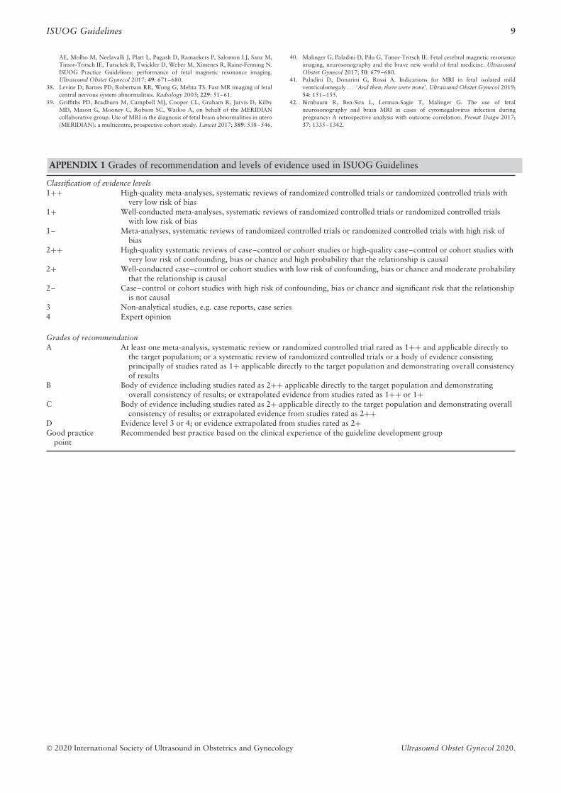
APPENDIX 1 Grades of recommendation and levels of evidence used in ISUOG Guidelines
Classification of evidence levels1++ High-quality meta-analyses, systematic reviews of randomized controlled trials or randomized controlled trials with
very low risk of bias1+ Well-conducted meta-analyses, systematic reviews of randomized controlled trials or randomized controlled trials
with low risk of bias1– Meta-analyses, systematic reviews of randomized controlled trials or randomized controlled trials with high risk of
bias2++ High-quality systematic reviews of case–control or cohort studies or high-quality case–control or cohort studies with
very low risk of confounding, bias or chance and high probability that the relationship is causal2+ Well-conducted case–control or cohort studies with low risk of confounding, bias or chance and moderate probability
that the relationship is causal2– Case–control or cohort studies with high risk of confounding, bias or chance and significant risk that the relationship
is not causal3 Non-analytical studies, e.g. case reports, case series4 Expert opinion
Grades of recommendationA At least one meta-analysis, systematic review or randomized controlled trial rated as 1++ and applicable directly to
the target population; or a systematic review of randomized controlled trials or a body of evidence consistingprincipally of studies rated as 1+ applicable directly to the target population and demonstrating overall consistencyof results
B Body of evidence including studies rated as 2++ applicable directly to the target population and demonstratingoverall consistency of results; or extrapolated evidence from studies rated as 1++ or 1+
C Body of evidence including studies rated as 2+ applicable directly to the target population and demonstrating overallconsistency of results; or extrapolated evidence from studies rated as 2++
D Evidence level 3 or 4; or evidence extrapolated from studies rated as 2+Good practice
pointRecommended best practice based on the clinical experience of the guideline development group
© 2020 International Society of Ultrasound in Obstetrics and Gynecology Ultrasound Obstet Gynecol 2020.