Isoniazid causes heart looping disorder in zebrafish ...
9
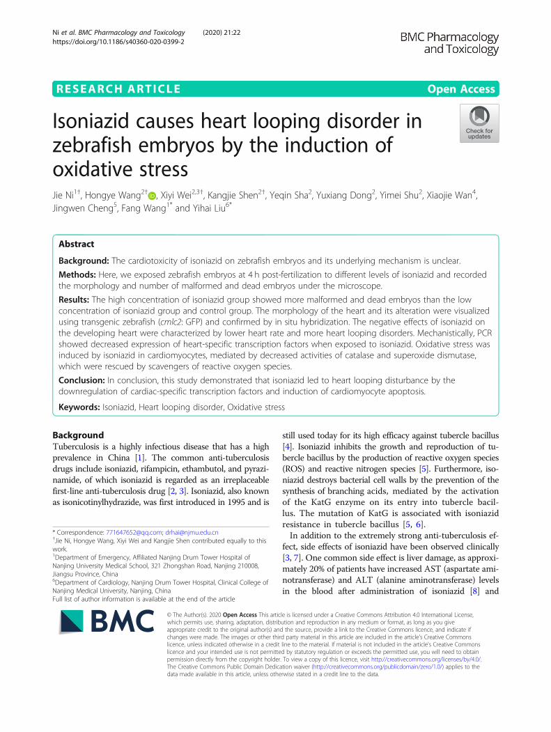
RESEARCH ARTICLE Open Access Isoniazid causes heart looping disorder in zebrafish embryos by the induction of oxidative stress Jie Ni 1† , Hongye Wang 2† , Xiyi Wei 2,3† , Kangjie Shen 2† , Yeqin Sha 2 , Yuxiang Dong 2 , Yimei Shu 2 , Xiaojie Wan 4 , Jingwen Cheng 5 , Fang Wang 1* and Yihai Liu 6* Abstract Background: The cardiotoxicity of isoniazid on zebrafish embryos and its underlying mechanism is unclear. Methods: Here, we exposed zebrafish embryos at 4 h post-fertilization to different levels of isoniazid and recorded the morphology and number of malformed and dead embryos under the microscope. Results: The high concentration of isoniazid group showed more malformed and dead embryos than the low concentration of isoniazid group and control group. The morphology of the heart and its alteration were visualized using transgenic zebrafish (cmlc2: GFP) and confirmed by in situ hybridization. The negative effects of isoniazid on the developing heart were characterized by lower heart rate and more heart looping disorders. Mechanistically, PCR showed decreased expression of heart-specific transcription factors when exposed to isoniazid. Oxidative stress was induced by isoniazid in cardiomyocytes, mediated by decreased activities of catalase and superoxide dismutase, which were rescued by scavengers of reactive oxygen species. Conclusion: In conclusion, this study demonstrated that isoniazid led to heart looping disturbance by the downregulation of cardiac-specific transcription factors and induction of cardiomyocyte apoptosis. Keywords: Isoniazid, Heart looping disorder, Oxidative stress Background Tuberculosis is a highly infectious disease that has a high prevalence in China [1]. The common anti-tuberculosis drugs include isoniazid, rifampicin, ethambutol, and pyrazi- namide, of which isoniazid is regarded as an irreplaceable first-line anti-tuberculosis drug [2, 3]. Isoniazid, also known as isonicotinylhydrazide, was first introduced in 1995 and is still used today for its high efficacy against tubercle bacillus [4]. Isoniazid inhibits the growth and reproduction of tu- bercle bacillus by the production of reactive oxygen species (ROS) and reactive nitrogen species [5]. Furthermore, iso- niazid destroys bacterial cell walls by the prevention of the synthesis of branching acids, mediated by the activation of the KatG enzyme on its entry into tubercle bacil- lus. The mutation of KatG is associated with isoniazid resistance in tubercle bacillus [5, 6]. In addition to the extremely strong anti-tuberculosis ef- fect, side effects of isoniazid have been observed clinically [3, 7]. One common side effect is liver damage, as approxi- mately 20% of patients have increased AST (aspartate ami- notransferase) and ALT (alanine aminotransferase) levels in the blood after administration of isoniazid [8] and © The Author(s). 2020 Open Access This article is licensed under a Creative Commons Attribution 4.0 International License, which permits use, sharing, adaptation, distribution and reproduction in any medium or format, as long as you give appropriate credit to the original author(s) and the source, provide a link to the Creative Commons licence, and indicate if changes were made. The images or other third party material in this article are included in the article's Creative Commons licence, unless indicated otherwise in a credit line to the material. If material is not included in the article's Creative Commons licence and your intended use is not permitted by statutory regulation or exceeds the permitted use, you will need to obtain permission directly from the copyright holder. To view a copy of this licence, visit http://creativecommons.org/licenses/by/4.0/. The Creative Commons Public Domain Dedication waiver (http://creativecommons.org/publicdomain/zero/1.0/) applies to the data made available in this article, unless otherwise stated in a credit line to the data. * Correspondence: [email protected]; [email protected] † Jie Ni, Hongye Wang, Xiyi Wei and Kangjie Shen contributed equally to this work. 1 Department of Emergency, Affiliated Nanjing Drum Tower Hospital of Nanjing University Medical School, 321 Zhongshan Road, Nanjing 210008, Jiangsu Province, China 6 Department of Cardiology, Nanjing Drum Tower Hospital, Clinical College of Nanjing Medical University, Nanjing, China Full list of author information is available at the end of the article Ni et al. BMC Pharmacology and Toxicology (2020) 21:22 https://doi.org/10.1186/s40360-020-0399-2
Transcript of Isoniazid causes heart looping disorder in zebrafish ...
RESEARCH ARTICLE Open Access
Isoniazid causes heart looping disorder inzebrafish embryos by the induction ofoxidative stressJie Ni1†, Hongye Wang2† , Xiyi Wei2,3†, Kangjie Shen2†, Yeqin Sha2, Yuxiang Dong2, Yimei Shu2, Xiaojie Wan4,Jingwen Cheng5, Fang Wang1* and Yihai Liu6*
Abstract
Background: The cardiotoxicity of isoniazid on zebrafish embryos and its underlying mechanism is unclear.
Methods: Here, we exposed zebrafish embryos at 4 h post-fertilization to different levels of isoniazid and recordedthe morphology and number of malformed and dead embryos under the microscope.
Results: The high concentration of isoniazid group showed more malformed and dead embryos than the lowconcentration of isoniazid group and control group. The morphology of the heart and its alteration were visualizedusing transgenic zebrafish (cmlc2: GFP) and confirmed by in situ hybridization. The negative effects of isoniazid onthe developing heart were characterized by lower heart rate and more heart looping disorders. Mechanistically, PCRshowed decreased expression of heart-specific transcription factors when exposed to isoniazid. Oxidative stress wasinduced by isoniazid in cardiomyocytes, mediated by decreased activities of catalase and superoxide dismutase,which were rescued by scavengers of reactive oxygen species.
Conclusion: In conclusion, this study demonstrated that isoniazid led to heart looping disturbance by thedownregulation of cardiac-specific transcription factors and induction of cardiomyocyte apoptosis.
Keywords: Isoniazid, Heart looping disorder, Oxidative stress
BackgroundTuberculosis is a highly infectious disease that has a highprevalence in China [1]. The common anti-tuberculosisdrugs include isoniazid, rifampicin, ethambutol, and pyrazi-namide, of which isoniazid is regarded as an irreplaceablefirst-line anti-tuberculosis drug [2, 3]. Isoniazid, also knownas isonicotinylhydrazide, was first introduced in 1995 and is
still used today for its high efficacy against tubercle bacillus[4]. Isoniazid inhibits the growth and reproduction of tu-bercle bacillus by the production of reactive oxygen species(ROS) and reactive nitrogen species [5]. Furthermore, iso-niazid destroys bacterial cell walls by the prevention of thesynthesis of branching acids, mediated by the activationof the KatG enzyme on its entry into tubercle bacil-lus. The mutation of KatG is associated with isoniazidresistance in tubercle bacillus [5, 6].In addition to the extremely strong anti-tuberculosis ef-
fect, side effects of isoniazid have been observed clinically[3, 7]. One common side effect is liver damage, as approxi-mately 20% of patients have increased AST (aspartate ami-notransferase) and ALT (alanine aminotransferase) levelsin the blood after administration of isoniazid [8] and
© The Author(s). 2020 Open Access This article is licensed under a Creative Commons Attribution 4.0 International License,which permits use, sharing, adaptation, distribution and reproduction in any medium or format, as long as you giveappropriate credit to the original author(s) and the source, provide a link to the Creative Commons licence, and indicate ifchanges were made. The images or other third party material in this article are included in the article's Creative Commonslicence, unless indicated otherwise in a credit line to the material. If material is not included in the article's Creative Commonslicence and your intended use is not permitted by statutory regulation or exceeds the permitted use, you will need to obtainpermission directly from the copyright holder. To view a copy of this licence, visit http://creativecommons.org/licenses/by/4.0/.The Creative Commons Public Domain Dedication waiver (http://creativecommons.org/publicdomain/zero/1.0/) applies to thedata made available in this article, unless otherwise stated in a credit line to the data.
* Correspondence: [email protected]; [email protected]†Jie Ni, Hongye Wang, Xiyi Wei and Kangjie Shen contributed equally to thiswork.1Department of Emergency, Affiliated Nanjing Drum Tower Hospital ofNanjing University Medical School, 321 Zhongshan Road, Nanjing 210008,Jiangsu Province, China6Department of Cardiology, Nanjing Drum Tower Hospital, Clinical College ofNanjing Medical University, Nanjing, ChinaFull list of author information is available at the end of the article
Ni et al. BMC Pharmacology and Toxicology (2020) 21:22 https://doi.org/10.1186/s40360-020-0399-2
approximately 1% of patients have severe hepatocyte dam-age [9]. However, some studies discovered that isoniazidcan lead to skin rash [10] and endocrine disturbance [11].Although isoniazid is regarded as a Pregnancy Category Cdrug by the Food and Drug Administration [12], somestudies have demonstrated potential toxic effects to afetus. Zhang et al. discovered that isoniazid affects liverdevelopment in zebrafish embryos and causes liver injuryby the construction of fabp10a:mcherry transgenic zebra-fish [13]. Additionally, the abnormal development of thenervous system, skull bone, and cartilage in a zebrafishembryo due to isoniazid has been demonstrated in vitroand in vivo [14]. However, there is no definitive report inregard to whether isoniazid has toxic effects on the devel-oping heart of a zebrafish embryo.According to epidemiological statistics, the occurrence of
congenital heart disease is approximately 3–4% in infants[15–17] and is now one of the most common causes ofneonatal death [18]. The types of congenital heart diseaseinclude ventricular septal defect, atrioventricular valve de-fect, transposition of the aorta, and constriction of theaorta. The occurrence of congenital heart disease is attrib-uted to many factors, such as genetic factors, environmentalfactors, pathogenic microorganism infections, and drug tox-icity, among which drug toxicity plays an important role incardiac development defects [19, 20]. For example, tilmico-sin [21], doxorubicin [22], cisplatin [23], and diclofenac so-dium [24] have been reported to induce cardiotoxicity bythe dysregulation of oxidative stress. However, the terato-genicity and cardiotoxicity of isoniazid on fetal heart devel-opment are rarely reported. The zebrafish is a widely usedanimal model due to short spawning periods and transpar-ent embryos. Zebrafish embryos are often used to investi-gate drug toxicity and the molecular mechanisms ofdevelopment [25, 26]. In our study, we utilized cmlc2:GFPheart transgenic and wild type zebrafish embryos. We ob-served that isoniazid disturbed the cyclization process dur-ing heart development of zebrafish embryos and causedcongenital cardiac malformation. Mechanically, it was me-diated by inducing oxidative stress and cardiomyocytesapoptosis and alleviation of oxidative stress can protect car-diac development under the exposure to isoniazid.
Materials and methodsZebrafish husbandryThe wide type zebrafish (AB strain) was purchased fromthe Institute of Hydrobiology of the Chinese Academy ofScience (Wuhan, China) and transgenic zebrafish (ABcmlc2:GFP) were gifted by Dr. Lou in the NanjingUniversity Animal Model Institute. These were kept inthe Animal Model Center in the Jiangsu Province KeyLaboratory of Human Functional Genomics. Recycledwater at 28 ± 0.5 °C and a 14 h light cycle per day wereset to mimic the circadian rhythm. Zebrafish were fed
with brine shrimp twice a day. At 17:00, we transferredthe male and female adult zebrafish into the mating tankin a ratio of 2:1 and segregated them with a partition.The next morning, we moved the partition at 9:00 andthe mating process lasted for 2 h. After spawning, theembryos were collected in clean fish water. The experi-ment protocols were approved by the Animal EthicsCommittee of Nanjing Medical University.
Chemical exposure experimentWe used 0mM, 0.01 mM, 0.04 mM, 0.16 mM, 0.64 mM,2.56 mM, and 10.24 mM doses of isoniazid (I3377-50G;Sigma) for the dose-effect assay according to the thera-peutic range of 3–5 μg/mL (0.01–0.04 mM) of isoniazid[27]. No precipitation was observed at room temperature25 °C. The embryos were cultured in sterile water com-posed of 137 mM/L NaCl, 0.54 mM/L KCl, 0.025 mM/LNa2HPO4, 0.044 mM/L KH2PO4, 1.3 mM/L CaCl2,1mM/L MgSO4, and 0.42 mM/L NaHCO3. N-acetyl-L-cysteine (NAC; A7250-50G; Sigma), a ROS scavenger,was added (1 mM) in advance to clean out ROS in theembryos in the NAC group for the rescue experiments.After 4 h post-fertilization (hpf), 80 well-grown embryosin each group were selected under the microscope andkept in one 100 mm plate (CORNING; Corning; UnitedStates) for embryo culture. The culture water thatcontained isoniazid was changed every 12 h and theseembryos were kept in the incubator at 28 °C and 5%CO2. We recorded the number and morphology ofmalformed and dead embryos and removed dead em-bryos every 12 h after exposure. Each chemical exposuretreatment was performed in triplicate.
Morphology observationWe observed the morphology of embryos in differentstages of development (24 hpf, 48 hpf, and 72 hpf) underwhite light with a fluorescent microscope (Nikon, Tokyo,Japan). In each stage, the total number of dead, un-hatched, and malformed embryos was recorded. The indi-cation of embryo death was defined based on opacity, eggclotting and stopping of heartbeat. We randomly selected20 larvae and recorded the heart rate in 1min in each ex-perimental group. A transgenic zebrafish strain (cmlc2:GFP), where a green fluorescent protein gene was insertedunder the promoter of a cardiomyocyte-specific gene ofcardiac myosin light chain 2 (cmlc2), was applied to ob-serve the change in morphology during heart develop-ment. All experiments were performed in triplicate.
Real-time PCRAfter 48 h of chemical exposure, the total RNA of 20larvae were isolated using Trizol reagent (Thermofisher,Waltham, United State). The total RNA in each group
Ni et al. BMC Pharmacology and Toxicology (2020) 21:22 Page 2 of 9
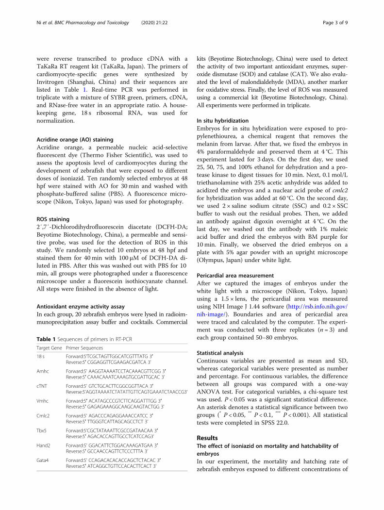
were reverse transcribed to produce cDNA with aTaKaRa RT reagent kit (TaKaRa, Japan). The primers ofcardiomyocyte-specific genes were synthesized byInvitrogen (Shanghai, China) and their sequences arelisted in Table 1. Real-time PCR was performed intriplicate with a mixture of SYBR green, primers, cDNA,and RNase-free water in an appropriate ratio. A house-keeping gene, 18 s ribosomal RNA, was used fornormalization.
Acridine orange (AO) stainingAcridine orange, a permeable nucleic acid-selectivefluorescent dye (Thermo Fisher Scientific), was used toassess the apoptosis level of cardiomyocytes during thedevelopment of zebrafish that were exposed to differentdoses of isoniazid. Ten randomly selected embryos at 48hpf were stained with AO for 30 min and washed withphosphate-buffered saline (PBS). A fluorescence micro-scope (Nikon, Tokyo, Japan) was used for photography.
ROS staining2′,7′-Dichlorodihydrofluorescein diacetate (DCFH-DA;Beyotime Biotechnology, China), a permeable and sensi-tive probe, was used for the detection of ROS in thisstudy. We randomly selected 10 embryos at 48 hpf andstained them for 40min with 100 μM of DCFH-DA di-luted in PBS. After this was washed out with PBS for 10min, all groups were photographed under a fluorescencemicroscope under a fluorescein isothiocyanate channel.All steps were finished in the absence of light.
Antioxidant enzyme activity assayIn each group, 20 zebrafish embryos were lysed in radioim-munoprecipitation assay buffer and cocktails. Commercial
kits (Beyotime Biotechnology, China) were used to detectthe activity of two important antioxidant enzymes, super-oxide dismutase (SOD) and catalase (CAT). We also evalu-ated the level of malondialdehyde (MDA), another markerfor oxidative stress. Finally, the level of ROS was measuredusing a commercial kit (Beyotime Biotechnology, China).All experiments were performed in triplicate.
In situ hybridizationEmbryos for in situ hybridization were exposed to pro-pylenethiourea, a chemical reagent that removes themelanin from larvae. After that, we fixed the embryos in4% paraformaldehyde and preserved them at 4 °C. Thisexperiment lasted for 3 days. On the first day, we used25, 50, 75, and 100% ethanol for dehydration and a pro-tease kinase to digest tissues for 10 min. Next, 0.1 mol/Ltriethanolamine with 25% acetic anhydride was added toacidized the embryos and a nuclear acid probe of cmlc2for hybridization was added at 60 °C. On the second day,we used 2 × saline sodium citrate (SSC) and 0.2 × SSCbuffer to wash out the residual probes. Then, we addedan antibody against digoxin overnight at 4 °C. On thelast day, we washed out the antibody with 1% maleicacid buffer and dried the embryos with BM purple for10 min. Finally, we observed the dried embryos on aplate with 5% agar powder with an upright microscope(Olympus, Japan) under white light.
Pericardial area measurementAfter we captured the images of embryos under thewhite light with a microscope (Nikon, Tokyo, Japan)using a 1.5 × lens, the pericardial area was measuredusing NIH Image J 1.44 software (http://rsb.info.nih.gov/nih-image/). Boundaries and area of pericardial areawere traced and calculated by the computer. The experi-ment was conducted with three replicates (n = 3) andeach group contained 50–80 embryos.
Statistical analysisContinuous variables are presented as mean and SD,whereas categorical variables were presented as numberand percentage. For continuous variables, the differencebetween all groups was compared with a one-wayANOVA test. For categorical variables, a chi-square testwas used. P < 0.05 was a significant statistical difference.An asterisk denotes a statistical significance between twogroups (* P < 0.05, ** P < 0.1, *** P < 0.001). All statisticaltests were completed in SPSS 22.0.
ResultsThe effect of isoniazid on mortality and hatchability ofembryosIn our experiment, the mortality and hatching rate ofzebrafish embryos exposed to different concentrations of
Table 1 Sequences of primers in RT-PCR
Target Gene Primer Sequences
18 s Forward:5’TCGCTAGTTGGCATCGTTTATG 3′Reverse:5′ CGGAGGTTCGAAGACGATCA 3’
Amhc Forward:5’ AAGGTAAAATCCTACAAACGTTCGG 3′Reverse:5′ CAAACAAATCAAAGTGCGATTGCAC 3’
cTNT Forward:5’ GTCTGCACTTCGGCGGTTACA 3′Reverse:5’AGGTAAAATCTATATTGTTCAGTGAAATCTAACCG3’
Vmhc Forward:5′ ACATAGCCCGTCTTCAGGATTTGG 3′Reverse:5′ GAGAGAAAGGCAAGCAAGTACTGG 3’
Cmlc2 Forward:5’ AGACCCAGAGGAAACCATCC 3′Reverse:5′ TTGGGTCATTAGCAGCCTCT 3’
Tbx5 Forward:5’CGCTATAAATTCGCCGATAACAA 3′Reverse:5′ AGACACCAGTTGCCTCATCCAG3’
Hand2 Forward:5’ GGACATTCTGGACAAAGATGAA 3′Reverse:5′ GCCAACCAGTTCTCCCTTTA 3’
Gata4 Forward:5’ CCAGACACACACCAGCTCTACAC 3′Reverse:5′ ATCAGGCTGTTCCACACTTCACT 3’
Ni et al. BMC Pharmacology and Toxicology (2020) 21:22 Page 3 of 9
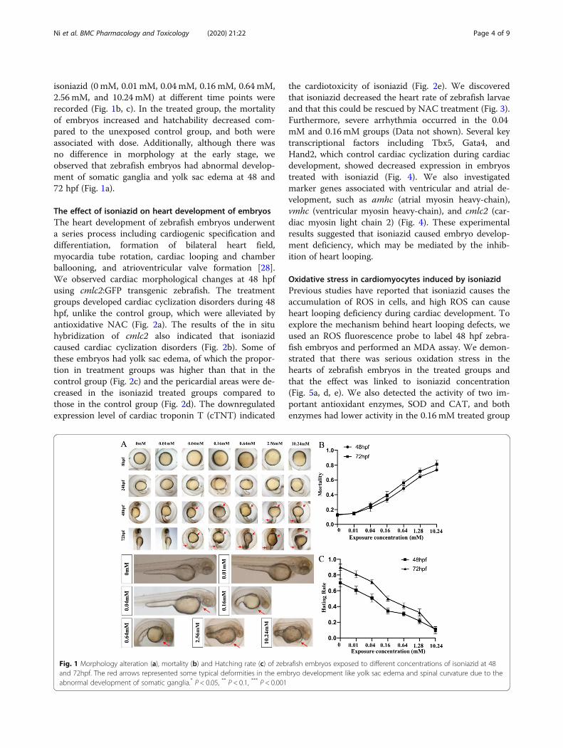
isoniazid (0 mM, 0.01 mM, 0.04mM, 0.16 mM, 0.64 mM,2.56 mM, and 10.24 mM) at different time points wererecorded (Fig. 1b, c). In the treated group, the mortalityof embryos increased and hatchability decreased com-pared to the unexposed control group, and both wereassociated with dose. Additionally, although there wasno difference in morphology at the early stage, weobserved that zebrafish embryos had abnormal develop-ment of somatic ganglia and yolk sac edema at 48 and72 hpf (Fig. 1a).
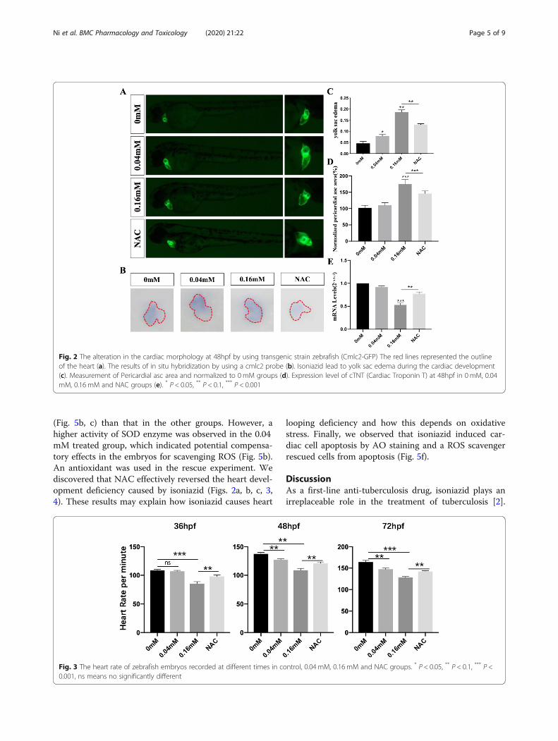
The effect of isoniazid on heart development of embryosThe heart development of zebrafish embryos underwenta series process including cardiogenic specification anddifferentiation, formation of bilateral heart field,myocardia tube rotation, cardiac looping and chamberballooning, and atrioventricular valve formation [28].We observed cardiac morphological changes at 48 hpfusing cmlc2:GFP transgenic zebrafish. The treatmentgroups developed cardiac cyclization disorders during 48hpf, unlike the control group, which were alleviated byantioxidative NAC (Fig. 2a). The results of the in situhybridization of cmlc2 also indicated that isoniazidcaused cardiac cyclization disorders (Fig. 2b). Some ofthese embryos had yolk sac edema, of which the propor-tion in treatment groups was higher than that in thecontrol group (Fig. 2c) and the pericardial areas were de-creased in the isoniazid treated groups compared tothose in the control group (Fig. 2d). The downregulatedexpression level of cardiac troponin T (cTNT) indicated
the cardiotoxicity of isoniazid (Fig. 2e). We discoveredthat isoniazid decreased the heart rate of zebrafish larvaeand that this could be rescued by NAC treatment (Fig. 3).Furthermore, severe arrhythmia occurred in the 0.04mM and 0.16 mM groups (Data not shown). Several keytranscriptional factors including Tbx5, Gata4, andHand2, which control cardiac cyclization during cardiacdevelopment, showed decreased expression in embryostreated with isoniazid (Fig. 4). We also investigatedmarker genes associated with ventricular and atrial de-velopment, such as amhc (atrial myosin heavy-chain),vmhc (ventricular myosin heavy-chain), and cmlc2 (car-diac myosin light chain 2) (Fig. 4). These experimentalresults suggested that isoniazid caused embryo develop-ment deficiency, which may be mediated by the inhib-ition of heart looping.
Oxidative stress in cardiomyocytes induced by isoniazidPrevious studies have reported that isoniazid causes theaccumulation of ROS in cells, and high ROS can causeheart looping deficiency during cardiac development. Toexplore the mechanism behind heart looping defects, weused an ROS fluorescence probe to label 48 hpf zebra-fish embryos and performed an MDA assay. We demon-strated that there was serious oxidation stress in thehearts of zebrafish embryos in the treated groups andthat the effect was linked to isoniazid concentration(Fig. 5a, d, e). We also detected the activity of two im-portant antioxidant enzymes, SOD and CAT, and bothenzymes had lower activity in the 0.16 mM treated group
Fig. 1 Morphology alteration (a), mortality (b) and Hatching rate (c) of zebrafish embryos exposed to different concentrations of isoniazid at 48and 72hpf. The red arrows represented some typical deformities in the embryo development like yolk sac edema and spinal curvature due to theabnormal development of somatic ganglia.* P < 0.05, ** P < 0.1, *** P < 0.001
Ni et al. BMC Pharmacology and Toxicology (2020) 21:22 Page 4 of 9
(Fig. 5b, c) than that in the other groups. However, ahigher activity of SOD enzyme was observed in the 0.04mM treated group, which indicated potential compensa-tory effects in the embryos for scavenging ROS (Fig. 5b).An antioxidant was used in the rescue experiment. Wediscovered that NAC effectively reversed the heart devel-opment deficiency caused by isoniazid (Figs. 2a, b, c, 3,4). These results may explain how isoniazid causes heart
looping deficiency and how this depends on oxidativestress. Finally, we observed that isoniazid induced car-diac cell apoptosis by AO staining and a ROS scavengerrescued cells from apoptosis (Fig. 5f).
DiscussionAs a first-line anti-tuberculosis drug, isoniazid plays anirreplaceable role in the treatment of tuberculosis [2].
Fig. 2 The alteration in the cardiac morphology at 48hpf by using transgenic strain zebrafish (Cmlc2-GFP) The red lines represented the outlineof the heart (a). The results of in situ hybridization by using a cmlc2 probe (b). Isoniazid lead to yolk sac edema during the cardiac development(c). Measurement of Pericardial asc area and normalized to 0 mM groups (d). Expression level of cTNT (Cardiac Troponin T) at 48hpf in 0 mM, 0.04mM, 0.16 mM and NAC groups (e). * P < 0.05, ** P < 0.1, *** P < 0.001
Fig. 3 The heart rate of zebrafish embryos recorded at different times in control, 0.04 mM, 0.16 mM and NAC groups. * P < 0.05, ** P < 0.1, *** P <0.001, ns means no significantly different
Ni et al. BMC Pharmacology and Toxicology (2020) 21:22 Page 5 of 9

Liver toxicity and gastrointestinal reaction were predom-inant side effects observed during clinical drug use andseveral cases indicate that isoniazid causes cardiotoxicity[29, 30]. In our study, we discovered that isoniazidcaused a deficiency in cardiac development via a ROSdependent manner. High levels of isoniazid reduced thehatch and survival rates of zebrafish embryos, which waslinked to heart development using heart-specific trans-genic zebrafish. By combining ROS and AO staining,and oxidant enzyme and antioxidant enzyme assays, wedemonstrated that isoniazid induced oxidative stress andcardiomyocyte apoptosis during heart development. Ithas been reported that isoniazid causes oxidative stressand apoptosis in tumor cell line Hep3B, which may berelated to its high metabolic demand and strong prolifer-ative ability [31]. Thus, the differential response toisoniazid between neonatal myocytes and adult cardio-myocytes is accounted for. Embryonic cells have a strongproliferative capacity, whereas mature cardiac cells havea more mature antioxidant system.During embryonic development, immature cells are
stem-like and have a strong ability for proliferation anddifferentiation. Therefore, they can be easily affected bythe external environment and drugs via alterations totranscriptional regulation. The heart is the first organ toform during embryonic development. It forms in the fol-lowing stages: the formation of cardiogenic zones fromcardiac progenitor cells, with the first cardiogenic zoneand the second cardiogenic zone [32, 33]; the
appearance of a linear cardiac tube due to separationand migration of cardiogenic zones at the proto-intestinal stage, with the heart starting to beat; the loop-ing of the linear cardiac tube; the arising of primitiveatrium and ventricle structure from endocardial endo-thelial cells, with undergoing endothelial mesenchymaltransformation into endocardial cushions and a heartcomes into being after cardiac cell remolding [34–36].Heart development is regulated by a combination oftranscription factors, in which downregulation leads todefects in heart development, teratogenicity, and evendeath of embryos [37]. In our experiment, transcriptionfactors related to cardiac development were downregu-lated in the high concentration group, as explained bythe cardiotoxicity of isoniazid at the molecular level.ROS are a series of chemicals with strong oxidative
capacities that are produced during cell metabolism, in-cluding superoxide radicals, hydroxyl radicals, andhydrogen peroxides. A high concentration of ROS in-duces oxidative stress and mitochondrial dysregulationin cells, which causes the oxidation of lipids, proteins,and nucleic acids and destroys their biological functions.ROS are signal molecules and play important roles inthe process of embryonic development. ROS participatein the development of the nervous system, the differenti-ation and formation of the nerve, and the developmentof lung and vascular tissue [38]. Recently, an article re-ported that high levels of ROS lead to heart looping dis-order during heart development [39, 40]. There are also
Fig. 4 The expression of four cardiac-specific transcript factors in zebrafish embryos after 48 hpf of isoniazid exposure. * P < 0.05, ** P < 0.1, *** P <0.001, ns means no significantly different
Ni et al. BMC Pharmacology and Toxicology (2020) 21:22 Page 6 of 9

many reports that environmental poisons and drugssuch as doxorubicin [41], chlorpyrifos [42], and dinitro-benzene cause abnormal development of various organsby the induction of ROS accumulation. In our study, weobserved similar consequences, that ROS was highlyconcentrated in the heart and cardiac looping was im-paired in the treatment group, which suggested that iso-niazid caused cardiac dysplasia by the upregulation ofROS. In the high concentration group, the atrial andventricle became smaller than those in the middle con-centration group and the control group, which may havebeen due to apoptosis and cell cycle arrest of developingcardiomyocytes caused by ROS. However, our studyhave some limits. First, the molecular mechanism andsignaling pathway that underlie the cardiotoxicity of iso-niazid need to be further investigated. Second, the chor-ion effect may lead to the low concentration of drugexposure, which need a future experiment by utilizingdechorionated embryos [43].
ConclusionsWe observed that isoniazid disturbed the cyclizationprocess during heart development of zebrafish embryos
and caused congenital cardiac malformation. Mechanic-ally, it was mediated by inducing oxidative stress andcardiomyocytes apoptosis and alleviation of oxidativestress can protect cardiac development under the expos-ure to isoniazid.
Abbreviationsamhc: Atrial myosin heavy-chain; AO: Acridine orange; CAT: Catalase; cmlc2;: Cardiac myosin light chain 2; cTNT: Cardiac troponin T; hpf: Hours post-fertilization; MDA: Malondialdehyde; NAC: N-acetyl-L-cysteine;PBS: Phosphate-buffered saline; ROS: Reactive oxygen species;SOD: Superoxide dismutase; SSC: Saline sodium citrate; vmhc: Ventricularmyosin heavy-chain
AcknowledgmentsNot applicable.
Authors’ contributionsHWY, JN, XYW, and KJS contributed equally to this work. All experimentswere performed by HYW, NJ, and XYW. HYW, KJS, and YQS wrote themanuscript. YMS,YXD and XYW were in charge of data analysis and technicalgraphics and also helped revise the manuscript. XJW and JWC helped revisethe manuscript. Professor YHL and FW guided the experimental design andhelped revise the manuscript. The authors read and approved the finalmanuscript.
FundingNot applicatable.
Fig. 5 The ROS level in control, 0.04 mM, 0.16 mM and NAC groups (a). The activity of CAT (b) and SOD (c) induced by isoniazid. Theconcentration of MDA (d), ROS (e) and apoptosis level (f) in different treated groups. The unit of SOD and CAT were ul/mg protein while MDAnmol/mg protein. The concentration of ROS was normalized to 0 mM groups. The red arrows represented the pericardial areas. * P < 0.05, ** P <0.1, *** P < 0.001, ns means no significance
Ni et al. BMC Pharmacology and Toxicology (2020) 21:22 Page 7 of 9
Availability of data and materialsAll data generated and analyzed during this study are included in this article.The datasets used and/or analyzed during the current study are availablefrom the corresponding author on reasonable request.
Ethics approval and consent to participateAnimal experiments were conducted by the Animal Ethic Review Board ofNanjing Medical university and accomplished in line with guildlines ofNational Health and Medical Research Council of China.
Consent for publicationNot applicatable.
Competing interestsThe authors declare that they have no competing interests.
Author details1Department of Emergency, Affiliated Nanjing Drum Tower Hospital ofNanjing University Medical School, 321 Zhongshan Road, Nanjing 210008,Jiangsu Province, China. 2The First Clinical Medical School, Nanjing MedicalUniversity, Nanjing, China. 3Department of Urology, The First AffiliatedHospital of Nanjing Medical University, Nanjing, China. 4Clinical School ofImaging, Nanjing Medical University, Nanjing, China. 5The Medical School ofPediatrics, Nanjing Medical University, Nanjing, China. 6Department ofCardiology, Nanjing Drum Tower Hospital, Clinical College of NanjingMedical University, Nanjing, China.
Received: 7 September 2019 Accepted: 27 February 2020
References1. Wang L, Zhang H, Ruan Y, Chin DP, Xia Y, Cheng S, Chen M, Zhao Y, Jiang
S, Du X, et al. Tuberculosis prevalence in China, 1990-2010; a longitudinalanalysis of national survey data. Lancet. 2014;383:2057–64..
2. Schito M, Migliori GB, Fletcher HA, McNerney R, Centis R, D'Ambrosio L,Bates M, Kibiki G, Kapata N, Corrah T, et al. Perspectives on Advancesin Tuberculosis Diagnostics, Drugs, and Vaccines. Clin Infect Dis. 2015;61Suppl 3:S102–18.
3. Arbex MA, Varella MCL, Siqueira HR, Mello FAF. Antituberculosis drugs: druginteractions, adverse effects, and use in special situations-part 1: first-linedrugs. J Bras Pneumol. 2010;36:626–40.
4. Sarich TC, Zhou T, Adams SP, Bain AI, Wall RA, Wright JM. A model ofisoniazid-induced hepatotoxicity in rabbits. J Pharmacol Toxicol Methods.1995;34:109–16.
5. Timmins GS, Master S, Rusnak F, Deretic V. Nitric oxide generated fromisoniazid activation by KatG: source of nitric oxide and activity againstmycobacterium tuberculosis. Antimicrob Agents Chemother. 2004;48:3006–9.
6. Suarez J, Ranguelova K, Jarzecki AA, Manzerova J, Krymov V, Zhao X, Yu S,Metlitsky L, Gerfen GJ, Magliozzo RS. An Oxyferrous Heme/protein-basedradical intermediate is catalytically competent in the catalase reaction ofmycobacterium tuberculosis catalase-peroxidase (KatG). J Biol Chem. 2009;284:7017–29.
7. Nahid P, Dorman SE, Alipanah N, Barry PM, Brozek JL, Cattamanchi A,Chaisson LH, Chaisson RE, Daley CL, Grzemska M. Executive summary:official American Thoracic Society/Centers for Disease Control andPrevention/Infectious Diseases Society of America clinical practiceguidelines: treatment of drug-susceptible tuberculosis. Clin Infect Dis.2016;63:e147.
8. Mitchell JR, Zimmerman HJ, Ishak KG, Thorgeirsson UP, Timbrell JA,Snodgrass WR, Nelson SD. Isoniazid liver injury: clinical spectrum, pathology,and probable pathogenesis. Ann Intern Med. 1976;84:181..
9. Ahmad J, Bach N, Bansal M, Barnhart HX, Beavers K, Bonkovsky H, Calvo FO,Chalasani N, Chang C, Conjeevaram H. Features and Outcomes of 899Patients With Drug-Induced Liver Injury: The DILIN Prospective Study.Gastroenterology. 2015;148:1340–52 e1347.
10. Yashika G, Rajeshwari G, Sourabh J, Arun K. A rare case of isoniazid-inducederythroderma. Indian J Pharmacol. 2015;47:682–4.
11. Li F, Wang P, Liu K, Tarrago MG, Lu J, Chini EN, Ma X. A high dose ofisoniazid disturbs endobiotic homeostasis in mouse liver. Drug MetabDispos. 2016;44:1742.
12. Mathad JS, Gupta A. Tuberculosis in pregnant and postpartum women:epidemiology, management, and research gaps. Clin Infect Dis. 2012;55:1532–49.
13. Zou Y, Zhang Y, Han LW, Qiu-Xia HE, Zhi-Ping LI, Hou HR, Han J, Wang XM,Juan-Juan LI, Cen J. Impact of isoniazid on developmental toxicity andbehavior of zebrafish larvae. Shandong Sci. 2016;5:47–53.
14. Strecker R, Weigt S, Braunbeck T. Cartilage and bone malformations in thehead of zebrafish ( Danio rerio ) embryos following exposure to disulfiramand acetic acid hydrazide. Toxicol Appl Pharmacol. 2013;268:221–31.
15. Bjornard K, Riehle-Colarusso T, Gilboa SM, Correa A. Patterns of congenitalheart defects, metropolitan Atlanta, 1978 to 2005. Birth Defects Res Part AClin Mol Teratol. 2013;97:87–94.
16. Moons P, Sluysmans T, De Wolf D, Massin M, Suys B, Benatar A, Gewillig M.Congenital heart disease in 111 225 births in Belgium: birth prevalence,treatment and survival in the 21st century. Acta Pã¦diatrica. 2009;98:472–7..
17. Shah GS, Singh MK, Pandey TR, Kalakheti BK, Bhandari GP. Incidence ofcongenital heart disease in tertiary care hospital. Kathmandu Univ Med J.2008;6:33–6.
18. Sanchez-Castro M, Pichon O, Briand A, Poulain D, Gournay V, David A,Caignec CL. Disruption of the SEMA3D gene in a patient with congenitalheart defects. Hum Mutat. 2015;36:30–3.
19. Källén BA, Otterblad OP. Maternal drug use in early pregnancy and infantcardiovascular defect. Reprod Toxicol. 2003;17:255–61.
20. Mladěnka P, Applová L, Patočka J, Costa VM, Remiao F, Pourová J, MladěnkaA, Karlíčková J, Jahodář L, Vopršalová M. Comprehensive review ofcardiovascular toxicity of drugs and related agents. Med Res Rev. 2018;38:1332–403.
21. Ibrahim AE, Abdel-Daim MM. Modulating effects of Spirulina platensisagainst Tilmicosin-induced Cardiotoxicity in mice. Cell J. 2015;17:137–44.
22. Abushouk AI, Ismail A, Salem AMA, Afifi AM, Abdel-Daim MM.Cardioprotective mechanisms of phytochemicals against doxorubicin-induced cardiotoxicity. Biomed Pharmacother. 2017;90:935–46.
23. Abdellatief SA, Galal AA, Farouk SM, Abdel-Daim MM. Ameliorative effect ofparsley oil on cisplatin-induced hepato-cardiotoxicity: A biochemical,histopathological, and immunohistochemical study. Biomed Pharmacother.2017;86:482–91.
24. Singh BK, Pathan RA, Pillai KK, Haque SE, Dubey K. Diclofenac sodium, anonselective nonsteroidal anti-inflammatory drug aggravates doxorubicin-induced cardiomyopathy in rats. J Cardiovasc Pharmocol. 2010;55:139–44.
25. Schwerte T, Pelster B. Digital motion analysis as a tool for analysing theshape and performance of the circulatory system in transparent animals. JExp Biol. 2000;203:1659–69.
26. Development. Test No.203 : Fish, Acute Toxicity Test. Oecd Guidelines forthe Testing of Chemicals. 1992;1(10):1–10.
27. Shah I, Jadhao N, Mali N, Deshpande S, Gogtay N, Thatte U.Pharmacokinetics of isoniazid in Indian children with tuberculosis on dailytreatment. Int J Tuberc Lung Dis. 2019;23:52–7.
28. Bakkers J. Zebrafish as a model to study cardiac development and humancardiac disease. Cardiovasc Res. 2011;91:279–88.
29. Ray A, Nangia V, Chatterji R, Dalal N, Ray RS. Recurrent heart failure inpulmonary tuberculosis patients on antitubercular therapy: a case ofprotector turning predator! Egyptian J Bronchol. 2017;11:288.
30. Ray A. Recurrent heart failure in pulmonary tuberculosis patients onantitubercular therapy: A case of protector turning predator! Egypt JBronchol. 2017;11:288–91.
31. Zhang Y, Zhang W, Tao L, Zhai J, Gao H, Song Y, Qu X. Quercetin protectedagainst isoniazide-induced HepG2 cell apoptosis by activating the SIRT1/ERKpathway. J Biochem Mol Toxicol. 2019;33:e22369.
32. Buckingham M, Meilhac S, Zaffran S. Building the mammalian heart fromtwo sources of myocardial cells. Nat Rev Genet. 2005;6:826–35.
33. Vincent SD, Buckingham ME. How to make a heart: the origin andregulation of cardiac progenitor cells. Curr Top Dev Biol. 2010;90:1–41.
34. Bruneau BG. Signaling and transcriptional networks in heart developmentand regeneration. Cold Spring Harb Perspect Biol. 2013;5:a008292.
35. Olson EN. Gene regulatory networks in the evolution and development ofthe heart. Science. 2006;313:1922–7.
36. Waardenberg AJ, Ramialison M, Bouveret R, Harvey RP. Genetic networksgoverning heart development. Cold Spring Harb Perspect Med. 2014;4:a013839.
37. Bruneau BG. The developmental genetics of congenital heart disease.Nature. 2008;451:943–8.
Ni et al. BMC Pharmacology and Toxicology (2020) 21:22 Page 8 of 9
38. Olguin-Albuerne M, Moran J. ROS produced by NOX2 control in vitrodevelopment of cerebellar granule neurons development. ASN Neuro.2015;7.
39. Daugaard M, Nitsch R, Razaghi B, McDonald L, Jarrar A, Torrino S, Castillo-Lluva S, Rotblat B, Li L, Malliri A, et al. Hace1 controls ROS generation ofvertebrate Rac1-dependent NADPH oxidase complexes. Nat Commun. 2013;4:2180.
40. Razaghi B, Steele SL, Prykhozhij SV, Stoyek MR, Hill JA, Cooper MD,McDonald L, Lin W, Daugaard M, Crapoulet N, et al. hace1 Influenceszebrafish cardiac development via ROS-dependent mechanisms. Dev Dyn.2018;247:289–303.
41. Zennaro C, Mariotti M, Carraro M, Pasqualetti S, Corbelli A, Armelloni S, Li M,Ikehata M, Clai M, Artero M, et al. Podocyte developmental defects causedby adriamycin in zebrafish embryos and larvae: a novel model of glomerulardamage. PLoS One. 2014;9:e98131.
42. Jin Y, Liu Z, Peng T, Fu Z. The toxicity of chlorpyrifos on the early life stageof zebrafish: a survey on the endpoints at development, locomotorbehavior, oxidative stress and immunotoxicity. Fish Shellfish Immunol. 2015;43:405–14.
43. Alvarez G, Perdomo C, Coronel C, Aguilera E, Varela J, Aparicio G, Zolessi FR,Cabrera N, Vega C, Rolon M, et al. Multi-anti-parasitic activity of Arylideneketones and Thiazolidene Hydrazines against Trypanosoma cruzi andLeishmania spp. Molecules. 2017;22:709.
Publisher’s NoteSpringer Nature remains neutral with regard to jurisdictional claims inpublished maps and institutional affiliations.
Ni et al. BMC Pharmacology and Toxicology (2020) 21:22 Page 9 of 9