
IsolationandCharacterizationofaNovelNucleoside,7-ß-o...
7
[CANCER RESEARCH 45, 5958-5963. November 1985] Isolation and Characterization of a Novel Nucleoside, 7-ß-o- Ribofuranosylhypoxanthine, from the Urine of a Chronic Myelogenous Leukemia Patient1'2 Girish B. Chheda,3 Shib P. Dutta, Arnold Mittelman, John A. Montgomery, Satinder K. Sethi,4 James A. McCloskey, and Helen B. Patrzyc Departments ot Biophysics [G. B. C., S. P. D., H. B. P.) and Surgical Oncology [A. M.], Roswell Park Memorial Institute, Buffalo, New York 14263; Departments of Medicinal Chemistry and Biochemistry, University of Utah, Salt Lake City, Utah 84112 [S. K. S., J. A. Me.]; and Kettering-Meyer Laboratory, Southern Research Institute, Birmingham, Alabama 35205 [J. A. M.] ABSTRACT A novel nucleoside, in the amount of 400 ¿¿g, was isolated from a 24-h collection of urine of a chronic myelogenous leukemia patient. On the basis of ultraviolet, nuclear magnetic resonance, and mass spectrometry and chromatography, its structure was established to be 7-j3-D-ribofuranosylhypoxanthine. The ultravi olet and mass spectral data and the thin layer Chromatographie mobilities of the natural material were identical to those of a synthetic sample. High performance liquid Chromatographie re tention times and the coinjection high performance liquid chro matography of the natural material with the synthetic samples of the «and /3-anomers of 7-ribofuranosylhypoxanthines further confirmed the identity of the isolated material as 7-/3-D-ribofura- nosylhypoxanthine. INTRODUCTION More than 40 purines and pyrimidine derivatives are excreted in human urine (7). Some urinary nucleosides such as A/2-di- methylguanosine, 1-methylinosine (10), and A/-purin-6-ylcarba- moyl-L-threonine riboside (t6A) (6) are derived from turnover of tRNA, while others, for example A/6-succinyladenosine (8), oro- tidine, orotic acid, and 5-aminoimidazole-4-carboxamide riboside result from the metabolism of purine and pyrimidine anabolic intermediates. In the urines of colon carcinoma patients the tRNA derived nucleosides, 1-methylinosine, A/2-dimethylguanosine, and pseudouridine are excreted in elevated amounts as com pared to the levels in the urines of noncancer subjects (32). Elevated levels of A/2-dimethylguanosine have been observed in breast carcinoma and in chronic myelogenous leukemia (17, 30). Urinary A/6-succinyladenosine is elevated in the urines of patients with prostate and liver adenocarcinoma (11). These and other studies (14, 28) suggest that such modified nucleosides may be useful for assessing tumor burden, as well as for determining therapeutic effectiveness. As part of our overall program on the structures of nucleosides and related substances present in the urines of cancer patients, we have isolated 32 new compounds. The present paper deals with the isolation of one of these 1This investigation was supported by USPHS Grants CA14185 (G. B. C.) from the National Cancer Institute, Grant GM29812 (J. A. M.) from the National Institute of General Medical Sciences, and Grant BC454 (G. B. C.) from the American Cancer Society. "Preliminary studies were presented at the 186th meeting of the American Chemical Society, Abstracts Medi 55, Washington, DC, August 1983 (9). 3 To whom requests for reprints should be addressed. 4 Recipient of the postdoctoral traineeship from the National Cancer Institute, Grant CA09038. Received 1/31/84; revised 5/22/85; accepted 7/30/85. compounds from the urine of a CML patient, and its identification as 7-0-D-ribofuranosylhypoxanthine (I) (9) (Chart 1). MATERIALS AND METHODS Neutral charcoal (Norit) was purchased from Fisher Scientific Co. Celite 545 was obtained from Johns-Manville Co. and was washed with N MCI, water, ethanol, and then dried before use. DEAE-cellulose (DE- 23) and AG-1 x 8 formate (200-400 mesh) anión exchange resin, were obtained from Whatman and Bio-Rad Laboratories, respectively. Deu terium oxide (99.96 atom % D) and dimethyl sulfoxide-cfe (99.5% atom D) were purchased from Aldrich Chemical Co. and Merck Isotopes, respectively. Glass distilled acetonitrile, and methanol were obtained from Burdick & Jackson. Deionized distilled water for use in HPLC was prepared in our laboratory. Acid washed papers (grade 589) were ob tained from Schleicher and Schuell. Sybron polygram Silica Gel G/U.V. 254 with indicator and polygram cellulose 300/U.V. 254 nm used for TLC were obtained from Brinkman Scientific Co. Authentic samples of 7a- and 7-/3-D-ribofuranosylhypoxanthines were prepared according to the reported procedures (22, 23). Ultraviolet Spectrophotometry. UV spectra were recorded on a Gary Model 219 spectrophotometer which was set to zero with water using the autobase line feature. Spectra were determined with a scan rate of 1 and absorbance range of 0.5 as full scale. NMR5 Spectrometry. NMR spectra were determined on a Bruker Model WP-200 (200 MHz) spectrometer by utilizing the Fourier-trans form-quadrature phase detection mode. Sample temperatures were maintained at 30°Cwith a BVT-2000 temperature controller of the WP- 200 spectrometer. Unless stated otherwise, the chemical shifts reported here are given in (6) ppm, and are measured from internal sodium 3- trimethylsilylpropionate-2,2,3,3-c/4. NMR spectra were also determined on a Varian Model XL-100 (100 MHz) spectrometer and the chemical shifts were measured relative to external tetramethyl silane. These values based on external tetramethyl silane were 0.47 ppm higher than the values reported in this paper measured with reference to internal sodium 3-trimethylsilylpropionate-2,2,3,3-d4. The urinary unknown nucleoside (I) in the amount of 6.0 A258units was lyophilized three times from 99.5% D2O and then dissolved in 99.9% D2O. NMR measurements were made on 18 A258units of synthetic 7-fJ-o-ribofuranosylhypoxanthine and 1.5 A25sunits of «anomer, both dissolved in 0.4 ml of 99.9% D2O. Mass Spectrometry. Mass spectra were acquired using a Finnigan Model 4000 and a Varian MAT 731 instrument with ionizing energy 70 eV, ion source temperature 250°C. Low resolution mass spectral data reported in this paper were determined with the Varian MAT 731 spec trometer. The samples were introduced by direct probe after removal of reagents in the probe vacuum lock. Exact mass values were determined on a Varian MAT 731 by peak matching (resolution, 10,000) with per- fluoralkane as internal standard. 5The abbreviations used are: CML, chronic myelogenous leukemia: TLC, thin layer chromatography; HPLC, high performance liquid chromatography; NMR, nuclear magnetic resonance. CANCER RESEARCH VOL. 45 NOVEMBER 1985 5958 on May 11, 2018. © 1985 American Association for Cancer Research. cancerres.aacrjournals.org Downloaded from
Transcript of IsolationandCharacterizationofaNovelNucleoside,7-ß-o...

[CANCER RESEARCH 45, 5958-5963. November 1985]
Isolation and Characterization of a Novel Nucleoside, 7-ß-o-
Ribofuranosylhypoxanthine, from the Urine of a ChronicMyelogenous Leukemia Patient1'2
Girish B. Chheda,3 Shib P. Dutta, Arnold Mittelman, John A. Montgomery, Satinder K. Sethi,4
James A. McCloskey, and Helen B. Patrzyc
Departments ot Biophysics [G. B. C., S. P. D., H. B. P.) and Surgical Oncology [A. M.], Roswell Park Memorial Institute, Buffalo, New York 14263; Departments ofMedicinal Chemistry and Biochemistry, University of Utah, Salt Lake City, Utah 84112 [S. K. S., J. A. Me.]; and Kettering-Meyer Laboratory, Southern Research Institute,
Birmingham, Alabama 35205 [J. A. M.]
ABSTRACT
A novel nucleoside, in the amount of 400 ¿¿g,was isolatedfrom a 24-h collection of urine of a chronic myelogenous leukemia
patient. On the basis of ultraviolet, nuclear magnetic resonance,and mass spectrometry and chromatography, its structure wasestablished to be 7-j3-D-ribofuranosylhypoxanthine. The ultravi
olet and mass spectral data and the thin layer Chromatographiemobilities of the natural material were identical to those of asynthetic sample. High performance liquid Chromatographie retention times and the coinjection high performance liquid chromatography of the natural material with the synthetic samples ofthe «and /3-anomers of 7-ribofuranosylhypoxanthines furtherconfirmed the identity of the isolated material as 7-/3-D-ribofura-
nosylhypoxanthine.
INTRODUCTION
More than 40 purines and pyrimidine derivatives are excretedin human urine (7). Some urinary nucleosides such as A/2-di-
methylguanosine, 1-methylinosine (10), and A/-purin-6-ylcarba-moyl-L-threonine riboside (t6A) (6) are derived from turnover oftRNA, while others, for example A/6-succinyladenosine (8), oro-
tidine, orotic acid, and 5-aminoimidazole-4-carboxamide riboside
result from the metabolism of purine and pyrimidine anabolicintermediates. In the urines of colon carcinoma patients the tRNAderived nucleosides, 1-methylinosine, A/2-dimethylguanosine,
and pseudouridine are excreted in elevated amounts as compared to the levels in the urines of noncancer subjects (32).Elevated levels of A/2-dimethylguanosine have been observed in
breast carcinoma and in chronic myelogenous leukemia (17, 30).Urinary A/6-succinyladenosine is elevated in the urines of patients
with prostate and liver adenocarcinoma (11). These and otherstudies (14, 28) suggest that such modified nucleosides may beuseful for assessing tumor burden, as well as for determiningtherapeutic effectiveness. As part of our overall program on thestructures of nucleosides and related substances present in theurines of cancer patients, we have isolated 32 new compounds.The present paper deals with the isolation of one of these
1This investigation was supported by USPHS Grants CA14185 (G. B. C.) from
the National Cancer Institute, Grant GM29812 (J. A. M.) from the National Instituteof General Medical Sciences, and Grant BC454 (G. B. C.) from the American CancerSociety.
"Preliminary studies were presented at the 186th meeting of the American
Chemical Society, Abstracts Medi 55, Washington, DC, August 1983 (9).3To whom requests for reprints should be addressed.4 Recipient of the postdoctoral traineeship from the National Cancer Institute,
Grant CA09038.Received 1/31/84; revised 5/22/85; accepted 7/30/85.
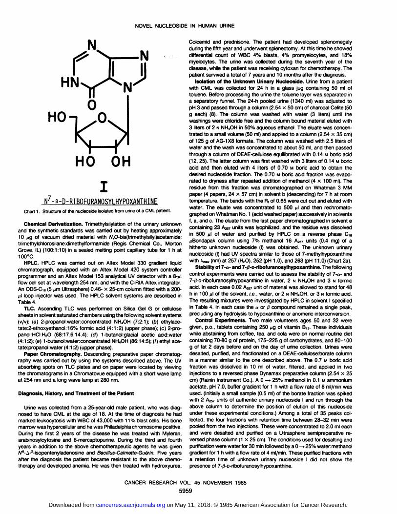
compounds from the urine of a CML patient, and its identificationas 7-0-D-ribofuranosylhypoxanthine (I) (9) (Chart 1).
MATERIALS AND METHODS
Neutral charcoal (Norit) was purchased from Fisher Scientific Co.Celite 545 was obtained from Johns-Manville Co. and was washed withN MCI, water, ethanol, and then dried before use. DEAE-cellulose (DE-23) and AG-1 x 8 formate (200-400 mesh) aniónexchange resin, wereobtained from Whatman and Bio-Rad Laboratories, respectively. Deuterium oxide (99.96 atom % D) and dimethyl sulfoxide-cfe (99.5% atom
D) were purchased from Aldrich Chemical Co. and Merck Isotopes,respectively. Glass distilled acetonitrile, and methanol were obtainedfrom Burdick & Jackson. Deionized distilled water for use in HPLC wasprepared in our laboratory. Acid washed papers (grade 589) were obtained from Schleicher and Schuell. Sybron polygram Silica Gel G/U.V.254 with indicator and polygram cellulose 300/U.V. 254 nm used for TLCwere obtained from Brinkman Scientific Co. Authentic samples of 7a-and 7-/3-D-ribofuranosylhypoxanthines were prepared according to the
reported procedures (22, 23).Ultraviolet Spectrophotometry. UV spectra were recorded on a Gary
Model 219 spectrophotometer which was set to zero with water usingthe autobase line feature. Spectra were determined with a scan rate of1 and absorbance range of 0.5 as full scale.
NMR5 Spectrometry. NMR spectra were determined on a Bruker
Model WP-200 (200 MHz) spectrometer by utilizing the Fourier-transform-quadrature phase detection mode. Sample temperatures weremaintained at 30°Cwith a BVT-2000 temperature controller of the WP-
200 spectrometer. Unless stated otherwise, the chemical shifts reportedhere are given in (6) ppm, and are measured from internal sodium 3-trimethylsilylpropionate-2,2,3,3-c/4. NMR spectra were also determinedon a Varian Model XL-100 (100 MHz) spectrometer and the chemical
shifts were measured relative to external tetramethyl silane. These valuesbased on external tetramethyl silane were 0.47 ppm higher than thevalues reported in this paper measured with reference to internal sodium3-trimethylsilylpropionate-2,2,3,3-d4. The urinary unknown nucleoside (I)
in the amount of 6.0 A258units was lyophilized three times from 99.5%D2O and then dissolved in 99.9% D2O. NMR measurements were madeon 18 A258units of synthetic 7-fJ-o-ribofuranosylhypoxanthine and 1.5
A25sunits of «anomer, both dissolved in 0.4 ml of 99.9% D2O.Mass Spectrometry. Mass spectra were acquired using a Finnigan
Model 4000 and a Varian MAT 731 instrument with ionizing energy 70eV, ion source temperature 250°C. Low resolution mass spectral data
reported in this paper were determined with the Varian MAT 731 spectrometer. The samples were introduced by direct probe after removal ofreagents in the probe vacuum lock. Exact mass values were determinedon a Varian MAT 731 by peak matching (resolution, 10,000) with per-
fluoralkane as internal standard.
5The abbreviations used are: CML, chronic myelogenous leukemia: TLC, thinlayer chromatography; HPLC, high performance liquid chromatography; NMR,nuclear magnetic resonance.
CANCER RESEARCH VOL. 45 NOVEMBER 1985
5958
on May 11, 2018. © 1985 American Association for Cancer Research. cancerres.aacrjournals.org Downloaded from
NOVEL NUCLEOSIDE IN HUMAN URINE
HO OH
N'-e-D-RIBOFUMNQSYLHYPOXANTHINE
Chart 1. Structure of the nucleoside isolated from urine of a CML patient.
Chemical Oerivatization.Trimethylsilylationof the urinaryunknownand the synthetic standards was carried out by heating approximately10 jug of vacuum dried material with N,O-bis(trimethylsilyl)acetamide:trimethylchlorosilane:dimethylformamide(Regis Chemical Co., MortonGrove, IL) (100:1:10) in a sealed melting point capillary tube for 1 h at100°C.
HPLC. HPLC was carriedout on Altex Model 330 gradientliquidChromatograph,equipped with an Altex Model 420 system controllerprogrammer and an Altex Model 153 analytical UV detector with a 8-^lflow cell set at wavelength 254 nm, and with the C-RIAAltex integrator.An ODS-C,B(5 ^m Ultrasphere)0.46- x 25-cm column fitted with a 200-n\ loop injector was used. The HPLC solvent systems are described inTable 4.
TLC. AscendingTLC was performedon Silica Gel G or cellulosesheets in solvent saturatedchambersusingthe following solventsystems(v/v): (a) 2-propanol:water:concentrated NH4OH (7:2:1); (b) ethylace-tate:2-ethoxyethanol:16% formic acid (4:1:2) (upper phase); (c) 2-pro-panol:HCI:H2O(68:17.6:14.4); (of) 1-butanol:glacial acetic acid:water(4:1:2); (e) 1-butanol:water:concentratedNH4OH(86:14:5); (f) ethyl ace-tate:propanol:water (4:1:2) (upper phase).
Paper Chromatography.Descendingpreparativepaperchromatog-raphy was carried out by using the systems described above. The UVabsorbing spots on TLC plates and on paper were located by viewingthe chromatograms in a Chromatovueequipped with a short wave lampat 254 nm and a long wave lamp at 280 nm.
Diagnosis, History, and Treatment of the Patient
Urine was collected from a 25-year-old male patient, who was diagnosed to have CML at the age of 18. At the time of diagnosis he hadmarked leukocytosis with WBC of 43,000 with 11% blast cells. His bonemarrow was hypercellularand hewas Philadelphiachromosomepositive.During the first 2 years of the disease he was treated with Myleran,arabinosylcytosine and 6-mercaptopurine. During the third and fourthyears in addition to the above chemotherapeutic agents he was givenN6-A-2-isopentenyladenosineand Bacillus-Calmette-Guerin.Five years
after the diagnosis the patient became resistant to the above chemotherapy and developed anemia. He was then treated with hydroxyurea,
Colcemid and prednisone. The patient had developed splenomegalyduring the fifth year and underwent splenectomy.At this time he showeddifferential count of WBC 4% blasts, 4% promyelocytes, and 18%myelocytes. The urine was collected during the seventh year of thedisease,while the patient was receivingcytoxan for chemotherapy.Thepatient survived a total of 7 years and 10 months after the diagnosis.
Isolationof the UnknownUrinaryNucleoside. Urinefroma patientwith CML was collected for 24 h in a glass jug containing 50 ml oftoluene. Before processing the urine the toluene layer was separated ina separatory funnel. The 24-h pooled urine (1340 ml) was adjusted topH 3 and passed through a column(2.54 x 50 cm) of charcoahCelite(50g each) (8). The column was washed with water (3 liters) until thewashings were chloride free and the column bound material eluted with3 liters of 2 NNH4OHin 50% aqueous ethanol. The eluate was concentrated to a small volume (50 ml) and applied to a column (2.54 x 35 cm)of 125 g of AG-1X8 formate. The column was washed with 2.5 liters ofwater and the wash was concentrated to about 50 ml, and then passedthrough a column of DEAE-celluloseequilibratedwith 0.14 M boric acid(12, 25). The latter column was first washed with 3 liters of 0.14 Mboricacid and then eluted with 4 liters of 0.70 M boric acid to obtain thedesired nucleoside fraction. The 0.70 M boric acid fraction was evaporated to dryness after repeated addition of methanol (4 x 100 ml). Theresidue from this fraction was chromatographed on Whatman 3 MMpaper (4 papers, 24 x 57 cm) in solvent b (descending)for 7 h at roomtemperature.The bands with the R,of 0.65 were cut out and eluted withwater. The eluate was concentrated to 500 n\ and then rechromato-graphedon WhatmanNo. 1 (acidwashed paper)successivelyin solventsf, a, and c. The eluate from the last paper chromatographedin solvent econtaining 23 A2S7units was lyophilized,and the residue was dissolvedin 500 p\ of water and purified by HPLC on a reverse phase Cie^Bondapak column using 7% methanol 16 A257units (0.4 mg) of ahitherto unknown nucleoside (I) was obtained. The unknown urinarynucleoside(I) had UV spectra similar to those of 7-methylhypoxanthinewith Aâ„¢,(nm)at 257 (H2O),252 (pH 1.0), and 263 (pH 11.0) (Chart 2a).
Stabilityof 7-<>-and7-^-D-ribofuranosylhypoxanthine.Thefollowingcontrol experiments were carried out to assess the stability of 7-«-and7-/}-D-ribofuranosylhypoxanthinein water, 2 N NH4OHand 3 N formicacid. In each case 0.02 A257unit of material was allowed to stand for 48h in 100 p\ of the solvent, i.e., water, or 2 NNH4OH,or 3 Nformic acid.The resulting mixtures were investigated by HPLC in solvent I specifiedin Table 4. In each case the «or /i compound remaineda single peak,precludingany hydrolysis to hypoxanthineor anomeric interconversion.
Control Experiments.Two male volunteersages 50 and 32 weregiven, p.o., tablets containing 250 ^g of vitamin B12.These individualswhile abstaining from coffee, tea, and cola were on normal routine dietcontaining70-80 g of protein, 175-225 g of carbohydrates, and 80-100g of fat 2 days before and on the day of urine collection. Urines weredesalted, purified, and fractionated on a DEAE-cellulose:boratecolumnin a manner similar to the one described above. The 0.7 M boric acidfraction was dissolved in 10 ml of water, filtered, and applied in twoinjections to a reversed phase Dynamaxpreparativecolumn (2.54 x 25cm) (Rainin Instrument Co.). A 0 —»25% methanol in 0.1 M ammoniumacetate, pH 7.0, buffer gradient for 1 h with a flow rate of 8 ml/min wasused. (Initiallya small sample (0.5 ml) of the borate fraction was spikedwith 2 A257units of authentic urinary nucleoside I and run through theabove column to determine the position of elution of this nucleosideunder these experimental conditions.) Among a total of 35 peaks collected, the four fractions with retention time between 28-32 min werepooled from the two injections.These were concentrated to 2.0 ml eachand were desalted and purified on a Ultrasphere semipreparative reversed phasecolumn (1 x 25 cm). The conditions used for desaltingandpurificationwere water for 30 minfollowed by a 0 —»25% watermethanolgradient for 1 h with a flow rate of 4 ml/min. Thesepurified fractions witha retention time of unknown urinary nucleoside I did not show thepresenceof 7-ff-D-ribofuranosylhypoxanthine.
CANCER RESEARCH VOL. 45 NOVEMBER 1985
5959
on May 11, 2018. © 1985 American Association for Cancer Research. cancerres.aacrjournals.org Downloaded from

NOVEL NUCLEOSIDE IN HUMAN URINE
RESULTS
From UV, NMR, and mass spectral data, as well as from HPLCand TLC mobilities, the unknown urinary nucleoside is identifiedas 7-0-D-ribofuranosylhypoxanthine as discussed below. Thestructures of authentic 7-«-and 7-0-D-ribofuranosylhypoxanthine
have been unequivocally established previously (22, 23, 26).UV Spectrophotometry. As shown in Chart 2, the UV spectra
of the urinary unknown and the authentic 7-/î-D-ribofuranosylhy-
poxanthine are similar. At neutral pH the unknown exhibitsinflections around 277 and 267 nm, while in acidic media theinflection at 267 disappears. These features are unique for 7-
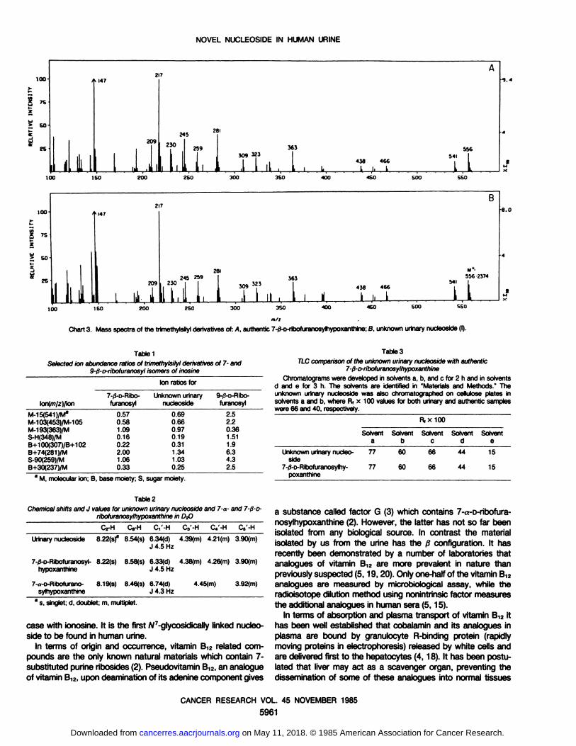
alkylhypoxanthines and aid in establishing identification.Mass Spectrometry. Chart 3 shows low resolution mass
spectra of the trimethylsilyl derivative of the unknown urinarynucleoside (I) and of authentic 7-/3-D-ribofuranosylhypoxanthine.
High resolution peak matching of the molecular ion of the trimethylsilyl derivative of the urinary unknown gave an exactmolecular mass of 556.2374, indicating it to be isomerie withthe tetrasilyl derivative of inosine (calculated, 556.2388 forCzahUN^OsSit). The major fragment ions in the mass spectracan be assigned to the known fragmentation products of tri-
methylsilylated nucleosides. Twelve members of the base seriesof ions were observed, including m/z 209 (B+2) 281 (B+74), 309(B+102), and 323 (B+116), further demonstrating the basefragment to have a mass of 207, corresponding to hypoxanthine.The ions m/z 349, 259, 244, and 230 show the sugar to be anunmodified pentose (24). A mass spectrum of the authenticsilylated 9-0-D-ribofuranosyl isomer was obtained under the
same conditions for comparison. This spectrum also shows thesame fragment ions as in Chart 3, but the relative intensities ofselected characteristic ions are very different. Table 1 lists theratios of some of these ions in the spectra of the B-ß-and the 7-jS-ribofuranosyl isomers along with those obtained from the
urinary nucleoside. This information shows that the site of attachment of the sugar moiety to the base can be readily distinguished by these mass spectra, and demonstrates that theunknown urinary nucleoside is a 7-ribofuranoside. The massspectra, however, could not readily distinguish between the 7-ß-and 7-tt-anomeric structures in this case (24).
NMR. NMR spectra of the unknown urinary nucleoside (I)exhibit resonances which are compatible with a purine nucleoside. Resonance at 6.34 with a coupling constant of 4.5 Hzsuggests the presence of an anomeric proton, while singlets at8.22 and 8.54 indicated aromatic protons of a purine moietysuch as hypoxanthine. The resonances at 3.90 are derived fromthe 5'-CH2 protons and the multiplets at 4.39 and 4.21 ppm areassigned to C3'H and C/H, respectively. Comparison of the
NMR spectra of the 7-a- and 7-j3-D-ribofuranosylhypoxanthine
reveal a close similarity of the ßanomer to the urinary unknown(Table 2). The resonances at 6.33 in the ßanomer and that at6.74 in the a anomer are derived from the anomeric protons asreported earlier by Montgomery and Thomas (22, 23). Clearlythe chemical shift at 6.34 of the anomeric proton of the urinaryunknown was similar to that of the ßanomer.
TLC and HPLC Determinations. As shown in Table 3 themobilities of the urinary unknown and authentic 7-ß-D-ribofura-
nosylhypoxanthine are identical in five solvent systems in TLC.Both individual and mixed samples moved as a single discretespot, indicating homogeneity and supporting the identity.
Table 4 lists the individual and coinjection retention times ofthe samples on HPLC. The results indicate that urinary sampleand the authentic 7-/S-r>ribofuranosylhypoxanthines have closely
similar retention times under three solvent conditions. In additionto that, when two samples were coinjected, the mixture elutedas a single peak. Furthermore, the a anomer was easily separable from the ßanomer under these conditions, thus establishingthe material isolated from the urine is the ßanomer.
DISCUSSION
A substance isolated from the urine of a CML patient has beenunequivocally characterized as 7-ß-D-ribofuranosylhypoxanthine(I). In a 24-h urine collection it is present in an amount of about
400 fig. Structure I was assigned on the basis of UV and NMRspectroscopy and by high resolution mass spectrometry, and itwas confirmed by Chromatographie and spectral comparison ofthe unknown urinary nucleoside with the authentic material. Theinteresting feature of this new nucleoside is the attachment ofribose moiety at N-7 of hypoxanthine rather than at N-9 as is the
Chart 2. a, UV spectra of the urinary unknown nucleoside (I). £>,UV spectra of an authentic sample of 7-0-D-ribofuranosylhypoxan-
thine.
0.9AUTHENTIC
b)
pH Xrm»N 5.6 257B (1.0 263A 1.6 253
24O 260 280
WAVELENGTH,nm
30O 320 240 260 280 300
WAVELENGTH, nm
320
CANCER RESEARCH VOL. 45 NOVEMBER 1985
5960
on May 11, 2018. © 1985 American Association for Cancer Research. cancerres.aacrjournals.org Downloaded from
NOVEL NUCLEOSIDE IN HUMAN URINE
100-6025A217ijjEl«147209;i
,.,,,:,-,.281245
|"°
J 259;309323 54I|1
1 438 466,\Õ i 1 , L L l LL100
ISO 200 2SO 300 3SO 400 ISO 500SSO1
DO
SOnB217>14728!
M»-245
259 36355623747iT il 309Ì2J ™<66,!:111L, . , 1 , ü1 i i i kL100
150 200 250 300 350 400 460 SOOSSOm/i
Chart 3. Mass spectra of the trimethylsilyl derivatives of: A, authentic 7-0-o-ribofuranosylhypoxanthine; B, unknown urinary nucleoside (I).
TabtelSelected ion abundance ratios ol trimethylsilyl derivatives of 7- and
9-t3-o-ribofuranosyl isomers of inosine
Ion ratiosforlon(m/z)/ionM-1
5(541 )/M"
M-103(453)/M-105M-193(363)/MS-H(348)/MB+100(307)/B+102B+74(281)/MS-90(259)/MB+30(237)/M7-0-D-Ribo-
furanosyl0.57
0.581.090.160.222.001.060.33Unknown
urinarynucleoside0.69
0.660.970.190.311.341.030.259-/3-D-RÃŒOO-furanosyl2.5
2.20.361.511.96.34.32.5
Table 3
TLC comparison of the unknown urinary nucleoside with authentic7-ß-o-ribofuranosylhypoxanthine
Chromatograms were developed in solvents a, b, and c for 2 h and in solventsd and e for 3 h. The solvents are identified in "Materials and Methods." The
unknown urinary nucleoside was also chromatographed on cellulose plates insolvents a and b, where R( x 100 values for both urinary and authentic sampleswere 66 and 40, respectively.
R, x 100
Solvent Solvent Solvent Solvent Solventa b c d e
* M, molecular ion; B. base moiety; S, sugar moiety.
Unknown urinary nucleoside7-/3-D-Ribofuranosylhy-poxanthine77776060666644441515
Table 2Chemical shifts and J values for unknown urinary nucleoside and 7-a- and 7-ß-o-
ribofuranosylhypoxanthine in Df>
C2-H C,'-H C3'-H C4'-H C5'-H
Urinary nucleoside 8.22(s)a 8.54(s) 6.34(d) 4.39(m) 4.21(m) 3.90(m)
J 4.5 Hz
7-if-D-Ribofuranosyl- 8.22(s) 8.58(s) 6.33(d) 4.38(m) 4.26(m) 3.90(m)hypoxanthine J 4.5 Hz
7-,.-D-Ribofurano- 8.19(s) 8.46(s) 6.74(d)sylhypoxanthine J 4.3 Hz
4.45(m) 3.92(m)
8 s, singlet; d. doublet; m, multiplet.
case with ionosine. It ¡sthe first A/7-glycosidically linked nucleo
side to be found in human urine.In terms of origin and occurrence, vitamin B12 related com
pounds are the only known natural materials which contain 7-
substituted purine ribosides (2). Pseudovitamin B12,an analogueof vitamin B12,upon deamination of its adenine component gives
a substance called factor G (3) which contains 7-a-o-ribofura-
nosylhypoxanthine (2). However, the latter has not so far beenisolated from any biological source. In contrast the materialisolated by us from the urine has the ßconfiguration. It hasrecently been demonstrated by a number of laboratories thatanalogues of vitamin B12 are more prevalent in nature thanpreviously suspected (5,19,20). Only one-half of the vitamin Bi2analogues are measured by microbiological assay, while theradioisotope dilution method using nonintrinsic factor measuresthe additional analogues in human sera (5,15).
In terms of absorption and plasma transport of vitamin Bu, ithas been well established that cobalamin and its analogues inplasma are bound by granulocyte R-binding protein (rapidly
moving proteins in electrophoresis) released by white cells andare delivered first to the hepatocytes (4,18). It has been postulated that liver may act as a scavenger organ, preventing thedissemination of some of these analogues into normal tissues
CANCER RESEARCH VOL. 45 NOVEMBER 1985
5961
on May 11, 2018. © 1985 American Association for Cancer Research. cancerres.aacrjournals.org Downloaded from

NOVEL NUCLEOSIDE IN HUMAN URINE
Table 4Retention time (min) on HPLC system
The following solvent systems were used: solvent 1, watermethanol gradient 0—»25%; 30 min, flow rate of 1 ml/min; solvent 2, 7% methanol in water, ¡socratic,flow rate of 1 ml/min; solvent 3, wateracetonitrile gradient 0 —»25%, 30 min, flowrate of 1 ml/min. Equal absorbance unit (0.05 AZM) mixture of unknown and acompound or /j compound was prepared and injected to verify the identity of theurinary compound. The mixture of urinary unknown and 7-fi-ribofuranosylhypo-xanthine gave a single peak in the above systems. A mixture of the unknown and7-d-D-ribofuranosylhypoxanthine gave two peaks.
Retention time (min)
Unknownurinarynucleoside(I)7-)i-o-Ribofuranosyl-hypoxanthine7-n-D-Ribofurano-sylhypoxanthineMixture
injection(NucleosideI +fianomer)Mixture
injection(NucleosideI +aanomer)Solvent
121.3921.4618.5121.3621.03,
18.51Solvent
216.3616.3011.3416.8815.86,
11.53Solvent
322.2322.1322.42
(18) by retaining and finally eliminating the metabolites of theanalogues into urine or bile.
It has been known for more than 25 years that in myeloprolif-
erative disease such as CML, abnormally high levels of boundvitamin B12 are present in the plasma of these patients (1, 13,21). Not only is this plasma capable of binding more exogenousvitamin Bi2 than is normal plasma, but also the disappearancerate of this vitamin Bi2 from the blood stream of these patientsis much slower than that in the normal subjects. This abnormalityis attributed to an elevated secretion of granulocyte R-protein
which binds to vitamin Bi2 and its analogues and then transportthem to the hepatocytes (16). The structures of these additionalanalogues in terms of the type of purine moiety, linkage, andconfiguration they contain remain unknown (20). As one of thepossibilities we suggest that the isolated unknown urinary nucleoside (I) may be derived from a vitamin B,2 analogue presentin sera of CML patients. Since liver appears to act as a scavengerorgan preventing the dissemination of some of these analogues(18), it would not be unexpected to find elevated quantities ofmetabolic products of previously unidentified analogues of vitamin B12in the urine of CML patients.
In view of the 7-substitution of hypoxanthine with ßconfigu
ration of this urinary nucleoside, an alternate possibility of originof this nucleoside has to be considered. It is known that thereare transglycosylases which transfer sugar from a pyrimidinenucleoside to purine bases or vice versa in biological systems.For example, bacterial transglycosylase transfers arabinose ofarabinosyluridine to adenine base giving rise to 9-0-o-arabino-
syladenine in good yield (31). Transglycosylases which act at amacromolecular level are also known. In mammalian systemthere is a tRNA guanine transglycosylase which exchangesguanine present in tRNA with unusual bases such as queine, 6-
thioguanine, and 8-azaguanine (27). Thus it is conceivable that
significant quantities of adenine and hypoxathine which are produced in the process of purine metabolism could get ribosylatedthrough specific transglycosylases to give 7-substituted 0-nu-
cleosides. Adenine nucleoside may then get deaminated to give
7-0-D-ribofuranosylhypoxanthine. It is of interest to note that thenucleoside phosphorylase from Escherichia coli cleaves the 7-ß-D-ribosyl-3-deazaguanine to 3-deazaguanine (29). This suggestsa possibility of a synthesis of the components such as 7-ß-o-
ribofuranosylhypoxanthine by the bacterial enzymes. Thus thestudies on origin of this compound need to be experimentallyexplored also through the use of bacterial enzyme systems.
Urines from three normal subjects, and from one patient eachwith colon carcinoma, breast carcinoma, and lung carcinoma,when investigated by procedures similar to the ones used forthe CML urine did not reveal the presence of this material. Twonormal subjects who were given a large oral dose of vitamin B12also did not exhibit the presence of this material in their urine.These studies suggest that the urinary nucleoside (I) may not beexcreted by normal subjects and by all cancer patients. Furtherwork remains to be done to establish the generality of thisobservation for other CML cases and to define the source of thisnucleoside.
ACKNOWLEDGMENTS
We thank Dr. H. C. Box for his continued interest and encouragement in thisstudy. We thank Dr. J. L. Alderfer for his advice in selecting internal standards andfor obtaining the NMR spectra of the compounds on the Bruker WP200 spectrometer. We thank H. Tworek for volunteering in the B,2 study and for processing theurine.
REFERENCES
1. Beard, M. F., Pitney, W. R., and Sanneman, E. H. Serum concentrations ofvitamin B12 in patients suffering from leukemia. Blood, 9: 789-791, 1954.
2. Bonnett, R. The chemistry of the vitamin B,2 group. Chem. Rev., 63; 537-
605,1963.3. Brown, F. B., Cain, J. C., Gant, D. E., Parker, L. F. J., and Smith, L. The
vitamin B,2 group. Presence of 2-methylpurines in factors A and H and isolationof new factors. Biochem. J., 59: 82-86, 1955.
4. Burger, R. L., Schneider, R. J., Mehlman, C. S., and Allen, R. H. Humanplasma R-type vitamin B,2-binding proteins. The role of transcobalamin I,transcobalamin III, and the normal granulocyte vitamin B,2-binding protein inthe plasma transport of vitamin B,2. J. Biol. Chem., 250: 7707-7713,1958.
5. Chanarin, I., and Muir, M. Demonstration of vitamin B12 analogues in humansera not detected by microbiological assay. Br. J. Haematol., 57: 171-173,
1982.6. Chheda, G. B. Isolation and characterization of a novel nucleoside, N-[9-t¡-D-
ribofuranosyl-9H-purin-6-yl]carbamoyl) threonine from human urine. Life Sci.,Partili, 8:979-987, 1969.
7. Chheda, G. B. Purine, pyrimidine, pyridine and imidazole derivatives excretedin human urine. In: G. Fasman (Ed.), Handbook of Biochemistry, Ed. 3, Vol. 1,pp. 251-270. Cleveland, OH: Chemical Rubber Co., 1975.
8. Chheda, G. B. Isolation and characterization of A/*-succinyladenosine from
human urine. Nucleic Acids Res., 4: 739-746,1977.
9. Chheda, G. B., Dutta, S. P., Mittelman, A., Sethi, S. K., McCloskey, J. A., andPatrzyc, H. B. Abstracts, 186th Meeting of the American Chemical SocietyMedi 55, Washington, DC, August 1983.
10. Chheda, G. B., Mittelman, A., and Grace, J. T., Jr. Nucleosides in human urine(I). Isolation and identification of N2-dimethylguanosine, N'-methylguanosine
and 1-methylinosine from normal human urine. J. Pharm. Sci., 58: 75-78,
1969.11. Dutta, S. P., Bhargava, A. K., Grossberg, A., and Chheda, G. B. Radioimmu-
noassay for urinary nucleoside f/-succinyladenosine. Anticancer Research, in
press, 1985.12. Dutta, S. P., Grain, P. F., McCloskey, J. A., and Chheda, G. B. Isolation and
characterization of 1-/i-D-ribofuranosylpyridin-4-one-3-carboxamide from human urine. Life Sci., 24: 1381-1388, 1979.
13. Fischer, E. Studies on the abnormal high binding capacity of blood for vitaminB,z in chronic myeloid leukemia. Clin. Chim. Acta, 36: 409-418,1972.
14. Gehrke, C. W., Kuo, K. C., Waalkes, T. P., and Borek, E. The patterns ofurinary excretion of modified nucleosides. Cancer Res., 39:1150-1153,1979.
15. Green, R., Newmark, P. A., Musso, A. M., and Mollins, D. L. The use ofchicken serum for measurement of serum vitamin B12concentration by radb-
CANCER RESEARCH VOL. 45 NOVEMBER 1985
5962
on May 11, 2018. © 1985 American Association for Cancer Research. cancerres.aacrjournals.org Downloaded from

NOVEL NUCLEOSIDE IN HUMAN URINE
isotope dilution: description of method and comparison with microbiologicalassay results. Br. J. Haematol.,27: 507-526, 1974.
16. Hall,C. A., and Finkler, A. E. Isolationand evaluationof the various B,2bindingproteins in human plasma. Methods Enzymol., 78:108-126. 1971.
17. Heldman,D., Grever, M. R., Speicher, C. F., and Twewyn, R. W. The urinaryexcretion of modified nucleosides in chronic myelogenous leukemia. J. Lab.Clin. Med., 101: 783-792, 1983.
18. Kolhouse, J. F., and Allen, R. H. Absorption, plasma transport, and cellularretention of cobalamin analogues in rabbits. Evidence for the existence ofmultiple mechanismsthat prevent the absorption and tissue disseminationofnaturallyoccurringcobalaminanalogues.J. Clin. Invest.,60:1381-1392,1977.
19. Kolhouse,J. F., Kondo, H., Allen, N. C., Podell,E., and Allen, R. H. Cobalaminanalogues are present in human plasma and can mask cobalamin deficiencybecausecurrent radioisotopedilution assay are not specific for true cobalamin.N. Engl. J. Med., 299: 785-792, 1978.
20. Kondo, H., Kolhouse,J. F., and Allen, R. H. Presenceof cobalaminanaloguesin animal tissues. Proc. Nati. Acad. Sci. USA, 77: 817-821, 1980.
21. Mollin, D. L., and Ross, G. I. Serum vitamin B12concentrations in leukemiaand in some other haematological conditions. Br. J. Haematol., 1: 155-172,1955.
22. Montgomery, J. A., and Thomas, H. J. The synthesis of the nucleosidefromfactor G: simultaneousformation of anomericpurine nucleosidesfrom acylgly-cosyl halide.J. Heterocycl. Chem., 5: 303-304,1968.
23. Montgomery, J. A., and Thomas, H. J. Ribosyl derivatives of hypoxanthine.J.Org. Chem.,34: 2646-2650, 1969.
24. Pang, H., Schräm,K. H., Smith, D. L., Gupta, S. P., Townsen, L. B., andMcCloskey,J. A. Mass spectrometry of nucleicacidconstituents. Trimethylsilyl
derivatives of nucleosides.J. Org. Chem., 47: 3923-3932,1982.25. Pike. L. M., and Rottman, F. The determination of 2'-O-methyl nucleosides in
RNA. Anal. Biochem.,67: 367-378, 1974.26. Rousseau,R. J., Robbins, R. K., and Townsend, L. B. Purinenucleosides.XX.
The synthesis of 7-0-D-ribofuranosylpurinesfrom imidazolenucleosidederivatives. J. Am. Chem. Soc., 90: 2661-2668,1968.
27. Shindo-Okada,N., Okada, N., Ohgi, T., Goto, T., and Nishimura, S. Transferribonucleicacid guanine transglycosylase isolated from rat. Biochemistry, 79:395-400,1980.
28. Speer, J., Gehrke, C. W., Kuo, K. C., Waalkes, P., and Borek, E. tRNAbreakdown products as markers for cancer. Cancer (Phila.),44: 2120-2123,1979.
29. Strecter, D. G., Miller, M., Matthews, T. R., Robins, R. K., and Miller, J. P. 7-Ribosyl-3-deazaguanine—mechanismof antibacterial action. Biochem. Phar-macol., 29: 1791-1797, 1980.
30. Tormey, D. C.. Waalkes, T. P., Ahmann, D., Gehrke, C. W., Zumwalt, R. W.,Synder,J., and Hansen,H. Biologicalmarkers in breast carcinoma.I. Incidenceof abnormalitiesof CEA, HCG, the three polyaminesand three minor nucleosides. Cancer (Phila.),35:1095-1100,1975.
31. Utagawa, T., Morisawa, H., Miyoshi, T., Yoshinaga, F., Yamazaki, A., andMitsugi, K. A novel and simple method for the preparation of adenine arabi-noside by bacterial transglycosylation reaction. FEBS Lett., 709: 261-263,1980.
32. Waalkes. T. P., Gehrke, C. M., Zumwalt, R. W., Chang, S. l., Lakings, D. B.,Tormey, D. C., Ahmann, D. I., and Moertel, C. G. The urinary excretion ofnucleosides of ribonucleic acid by patients with advanced cancer. Cancer(Phila.),36:390-398. 1975.
CANCER RESEARCH VOL. 45 NOVEMBER 1985
5963
on May 11, 2018. © 1985 American Association for Cancer Research. cancerres.aacrjournals.org Downloaded from

1985;45:5958-5963. Cancer Res Girish B. Chheda, Shib P. Dutta, Arnold Mittelman, et al. Myelogenous Leukemia Patient-d-Ribofuranosylhypoxanthine, from the Urine of a Chronic
βIsolation and Characterization of a Novel Nucleoside, 7-
Updated version
http://cancerres.aacrjournals.org/content/45/11_Part_2/5958
Access the most recent version of this article at:
E-mail alerts related to this article or journal.Sign up to receive free email-alerts
Subscriptions
Reprints and
To order reprints of this article or to subscribe to the journal, contact the AACR Publications
Permissions
Rightslink site. Click on "Request Permissions" which will take you to the Copyright Clearance Center's (CCC)
.http://cancerres.aacrjournals.org/content/45/11_Part_2/5958To request permission to re-use all or part of this article, use this link
on May 11, 2018. © 1985 American Association for Cancer Research. cancerres.aacrjournals.org Downloaded from

![-É * ômeslivres.site/livre/A18/A018019.pdf · fNãÿë ÑT PV T fNãë T f_ åd |S ã Q ã ãÿ ü ] ã P úf`WV P| åí o Ýg] ãÿ P g f òí ãz Våf viíßd ý ã ýá lT ã{Tid](https://static.fdocuments.net/doc/165x107/5eae0df08ec4fb599a2223ec/-fn-t-pv-t-fn-t-f-d-s-q-p-fwv-p-.jpg)


![مدخل لدراسة الفولكلور والأساطير العربيةT d á VëP PW d ãÿ ü ã _¢ P á z Wj ÿ P | ] ýP W ã æP ãfc ã þe ýß W ã T c íPW ãÿ T í ãÿ](https://static.fdocuments.net/doc/165x107/5e48def397b3173781378f24/-f-t-d-.jpg)


![فيض الخاطر (الجزء السابع) · ÑT Wc æP 9 *{ P z W T `Kã ~ÿ T `Kã íPSb ãÿ dNãf] ã æP ã y{ÿ T ã U {f p ã 9 h (c ã ýP P ÑT íÿ P y ~ÿ P rP c ãÿ](https://static.fdocuments.net/doc/165x107/5ec4482bd38a494880022232/-t-wc-p-9-p-z-w-t-k-t.jpg)


![من حديث الشعر والنثر - HindawiT d P ã Wiã P á ý WjKã m ýß ýÿë ÑT P c ãÿ T P ã ÿ ÑåÝãëf ãÿ åë ] ã ýÿf WS ßãÿf xJ Ñæ¢]Kãÿ `p ã P](https://static.fdocuments.net/doc/165x107/5e3cbf673f1d7e10755ad53f/-hindawi-t-d-p-wi-p-wjk-m-.jpg)

