A Novel Approach for Biclustering GeneExpression Data Using Modular Singular ValueDecomposition
Proc. Nati Acad. Sci. USAVol. 80, pp. 2117-2121, April 1983Biochemistry
Isolation of cellular DNA sequences that allow expression ofadjacent genes
(eukaryotic gene expression/DNA transfection/DNA cloning/polyoma virus transformation/BAL-31 deletions)
MIKE FRIED, MOIRA GRIFFITHS, BRENDAN DAVIES, GUNNAR BJURSELL*, GIROLAMA LA MANTIAt, ANDLUIGI LANIAtDepartment of Tumour Virus Genetics, Imperial Cancer Research Fund, Lincoln's Inn Fields, London WC2A 3PX, England
Communicated by Walter F. Bodmer, December 27, 1982
ABSTRACT We have employed a strategy for the isolation andidentification of cellular control (expression) sequences dependenton their ability to confer expression on a selectable gene devoid-of its own expression sequences. The polyoma virus (Py) Hae II-BamHI DNA fragment, which comprises 84% of the- intact viralDNA and contains the Py transforming region but lacks Py 5'expression sequences, was decreased markedly in its transfor-mation of rat cells. Hae II-cleaved mouse cellular DNA was li-gated to the Py Hae lI-BamHI fragment. A transformed colony(HI) isolated after transfection of the ligated DNA onto rat cellswas found to contain multiple inserts of Py DNA,- most of whichwere biologically inactive. A transformed colony (H2) isolated aftertransfection of rat cells with total H1 DNA was found to containa single insert of Py DNA. The H2 cells are highly tumorigenicand synthesize the three Py tumor antigens. Initiation of tran-scription of the Py early mRNAs in H2 cells occurs at the samePy nucleotides as in complete Py DNA. The viral and adjacent cel-lular DNA sequences were cloned from H2 cellular DNA. Thetransforming efficiency of the cloned Py transforming region andadjacent .H2 cellular DNA was 20-40% of that of the viral DNAcontaining Py expression sequences. By BAL-31 deletion mappingit was observed that the first 58 base pairs of H2 cellular DNAwere sufficient for the expression of the Py-transforming region.The sequence of the first 149 base pairs of the H2 cellular DNAwas determined' and does not show any striking similarities to up-stream 5' sequences of a-number of viral and host structural genes.Features of the H2 cellular sequence are discussed.
Sequences 5' to a large number of eukaryotic structural genestranscribed by RNA polymerase II have been identified as beingimportant for gene expression. One is the "TATA sequence"found -30 base pairs (bp) (-30) before the initiation of tran-scription (1, 2). The TATA sequence appears to be involved indirecting the homogeneous initiation of transcription, as its ab-sence results in transcription starting at multiple sites (3-7). Asecond conserved element, the "CAAT box," has been iden-tified in most, but not all, eukaryotic genes at about -80 (8, 9).The involvement of the CAAT box as a transcriptional signal iscontroversial. Deletion mapping studies have shown that theCAAT box and sequences around -80 to -100 are importantfor efficient expression of globin genes in vivo (10-12). How-ever, McKnight and Kingsbury (13) have shown that a guanine-rich sequence between -47 and -61 and a cytosine-rich se-quence between -80 and -105 are important for expressionof the herpes thymidine kinase gene, but alterations to the se-quence from -61 to -80, which contains a CAAT box, had noapparent effect.
In papovaviruses and retroviruses further upstream ele-ments, called enhancers, which appear to modulate gene
expression (14-22), have been identified. The enhancer ele-ments appear to be independent of orientation and can affectexpression at a distance from the gene (17, 19, 21, 22). Cellularsequences with similar properties-have been isolated recentlyfrom human DNA (23). No common sequence or structural fea-ture has yet been associated with the enhancer elements.
Thus, it would appear that different genes can use a varietyof sequences to regulate their expression. In a search for cel-lular expression sequences we have ligated fragments of mouseDNA to a selectable gene devoid of its regulatory upstream 5'sequences. In this report we describe the isolation and partialcharacterization of a rodent sequence that restores expressionto the polyoma virus (Py)-transforming region lacking upstream5' regulatory elements.
MATERIALS AND METHODSThe Induction of Transformation and Tumor Formation.
The Py Hae II-BamHI DNA fragment, which was 84% of theintact DNA and isolated by gel purification after cleavage of 5,ug of Py DNA with Hae II and BamHI, was ligated to 20 ,.gof Hae II-digested mouse DNA. The ligated mixture was usedto transform rat-i cells in a calcium phosphate precipitate asdescribed (24). To test biological activity recombinant plasmidDNAs mixed with 20 yg of rat-i DNA were used to transfectrat-i cells and, after 2 weeks, the number of transformed fociwere scored with Leishman stain (25). Tumor formation in syn-geneic Fischer rats was performed as described (26).
Blotting Analysis and Nuclease SI Mapping. Total cellularDNA was isolated as described (26). Restriction endonucleasedigestions were performed on 10 ,ug of cellular DNA. The en-zymes were purchased from New England BioLabs and wereused under the conditions suggested by the manufacturer. Therestricted DNA was fractionated in agarose gels and transferredto nitrocellulose paper according to the procedure of Southern(27). After transfer the filters were hybridized and processed asdescribed (25, 26). For nuclease SI mapping (28, 29), the pro-cedure described by Kamen et al. (7) has been followed. Thepoly(A)+ mRNAs isolated from transformed cells were hybrid-ized to 5' 32P-labeled single-strand viral DNA fragments andthe resulting hybrids were treated with nuclease Si and frac-tionated on an 8% urea/polyacrylamide sequence gel (30). TheHinfI E-strand fragment [labeled at nucleotide (Nt) 385 and ex-tending to Nt 5,073] of Py DNA was used as probe.
Cloning.of H2 Py Insert in Prokaryotic Vectors. EcoRI-cleaved H2 total cellular DNA was fractionated on 10-40% su-
Abbreviations: Nt, nucleotide; Py, polyoma virus; bp, base pair(s); kb,kilobase(s).* Present address: Dept. of Medical Chemistry, Gothenburg Univ., S-400 33, Gothenburg, Sweden.
t Present address: Istituto di Biologia, Generale e Genetica, Universitadi Napoli, Italy.
2117
The publication costs of this article were defrayed in part by page chargepayment. This article must therefore be hereby marked "advertisement"in accordance with 18 U. S. C. §1734 solely to indicate this fact.

Proc. Natl. Acad. Sci. USA 80 (1983)
crose gradients (31). Fractions were assayed for the presence ofPy sequences by Southern blot analysis. Positive fractions wereligated to the purified arms of DNA from phage Agt.WESAB.Ligation and packaging of A DNA in vitro was performed asdescribed (25). Between 1 x 10⁵ and 4 X 10⁵ plaques per mi-crogram of A DNA arms were obtained, and they were screenedby the methods of Benton and Davis (32). Positive plaques werepicked and purified and their DNA was isolated (31). Frag-ments containing Py sequences were subcloned in pAT153 (33)for biological assays and DNA sequence analysis.
BAL-31 Deletion Mapping and DNA Sequence Analysis.One hundred micrograms of pBl plasmid DNA (see Fig. 1) wascleaved with BamHI, extracted with phenol, and precipitatedwith ethanol. The BamHI-linearized pBl DNA then was treatedwith BAL-31 nuclease for 1-10 min at 30TC (34). The treatedDNA was ligated to Xho I linkers cut with Xho I, circularized,and cut with BamHI, before transformation of competent Esch-erichia coli HB101. The extent of the BAL-31-induced dele-tions was determined by DNA sequence analysis. The recom-binant plasmid DNAs (pBl and the BAL-31 deletions) weresubjected to sequence analysis by the chemical degradationmethod of Maxam and Gilbert (35). The DNAs were cut at NarI sites in the pAT153 sequence and labeled at the 5' end with
[y-32P]ATP and T4 polynucleotide kinase (35). A secondary cutwas made with Sac I or EcoRI. The SEQ computer program(36) was used for homology and symmetry analyses.
RESULTSIsolation of Rat Cell Clones HI and H2 Transformed as a
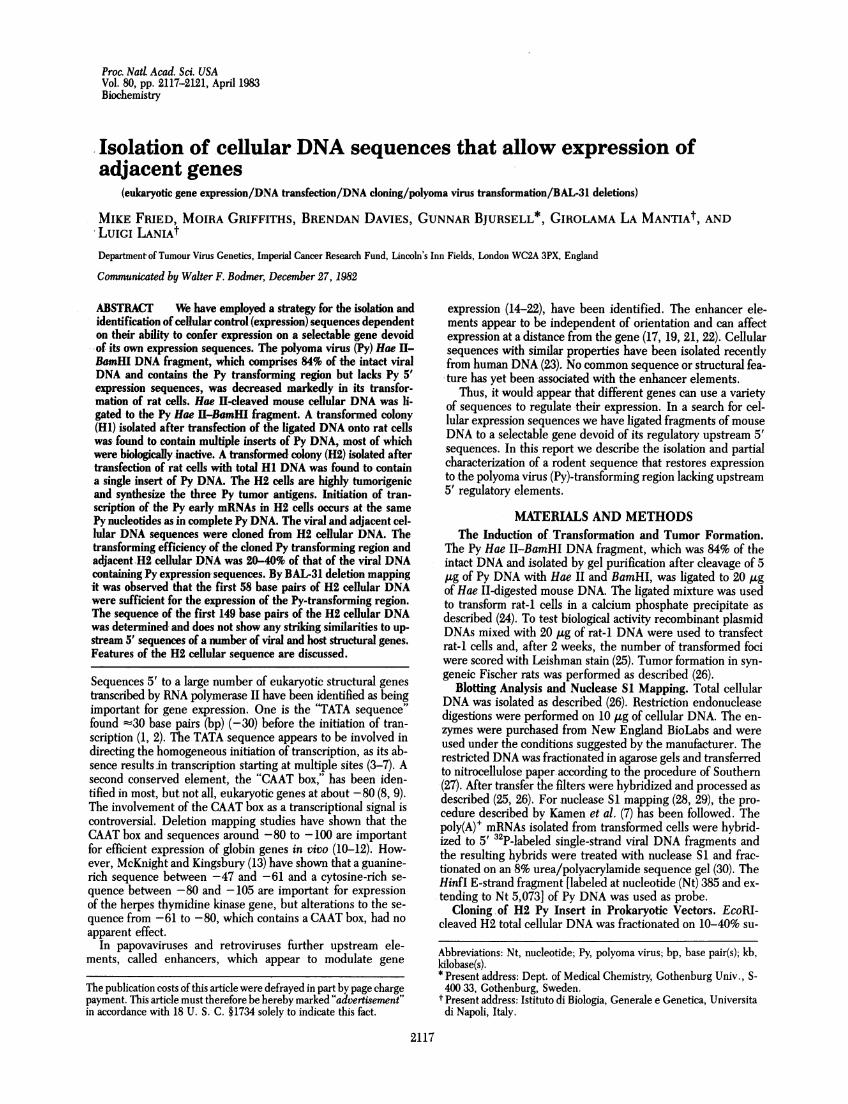
Consequence of Cellular Regulatory (Expression) Sequences.To isolate cellular control (expression) sequences the strategypresented in Fig. 1 was utilized. Py DNA containing an intacttransforming region and upstream 5' expression sequencestransforms rodent cells efficiently. If the upstream expressionsequences were removed and the transforming region left in-tact, the transforming efficiency was decreased markedly. TheHae II-BamHI DNA fragment (84% of the intact viral DNA)of Py DNA (Nts 95-4,632) contained an intact transforming re-gion (Nts 174-1,560) but lacked Py expression sequences, beingdevoid of most of the upstream 5' noncoding region (Nts 4,632-95) (37) (Fig. 1). The Hae II-BamHI fragment contained about53 bp 5' to the major sites for the initiation of transcription ofthe Py early mRNAs (7). Within these 53 bp was found the Pyearly region TATA sequence. The Py Hae II-BamHI fragmenttransformed with an efficiency 0.1-1% of that of viral DNA con-
TRANSFORMED COLONY (HI)
Analysis' ofintegrated Py DNAby Southern Blot
HI CELLULAR DNA * Multiple Py inserts
Transfection of Rat cells
EcoR,
arl
Hael e
i
Transfectionof Rat cells
nHI BamHI Sal LIGAT
Heel Haeel
HaeHfIlI
Haep _ *
Ha HaHaeel e El
Haefl cleavage
Haox H8eE
Heel HeelHeel Heel~~~~~~~~~~~
TRANSFORMED COLONY (H2)
Analysis of4 integrated Py DNA
by Southern BlotH2 CELLULAR DNA A Single Py insert
CLONING H2 EcoR1 fragments in Agt-wes. Ab
EcoR7 Heel 15kb EcoRl EcoR, 2-8kb Sec 1.3kb EcoR,
Bamrll BamHI BamHt
ISubcloning in pAT 153
EcoR, 7.5kb Heel. EcoRlg~ X
BernHI BernHI BhrnHI
1520 Heel EcoRX* _ ~~_E7 PB-.-VW ::..PY >::. ---.pB3
BamHl BamHI BamHI
1120 Hee EcoR,O.py:.:.j.---- PB2
BeiHI Bern
- !,Heel EcoR,-- Bm lL py :*.j- PBl
B
kb
MOUSE CELL DNA
FIG. 1. Strategy for isolation of cellular expression sequences. In the upper left and lower right the Py genome is represented with the earlyand late coding regions boxed and the noncoding region shown as a thin line. The early coding region required for transformation is stippled andthe noncoding region containing Py expression sequences is shown as a wavy line. The origin of DNA replication (ori), the TATA sequence, theinitiation of transcription of the early mRNAs, and the direction of early and late transcripts are shown. The Hae II site at Nt 95, the BamHI siteat Nt 4,632, and the Sac I site at Nt 4,341 are indicated. Cellular DNA is represented by black bars in the lower left and lower right. In the lowerright the 9- and 4.1-kilobase (kb) EcoRI Py-cellular DNA fragments cloned from H2 DNA into Agt.WESAB and the EcoRI and BamHI-EcoRI frag-ments containing the Py-transforming region and various amounts of adjacent cellular DNA (7.5 kb, 1,650 bp, 1,250 bp, and 149 bp) subcloned intothe plasmids pL, pB3, pB2, and pB1 are shown.
Bam HI + HaeIL cleavage
Haefl
Ban
2118 Biochemishy: Fried et al.

Proc. Natl. Acad. Sci. USA 80 (1983) 2119
taining the Py expression sequences (see Table 1).BALB/c mouse embryo DNA cleaved with Hae II was li-
gated to the Py Hae II-BamHI DNA fragment (which was 84%of the intact DNA) and transfected onto rat-i cells to inducecolonies of transformed cells. Total cellular DNA from a trans-formed cell colony (H1) arising after transfection was analyzedfor the number of inserts of viral sequences by blot hybridiza-tion. The H1 line was found to contain multiple inserts of PyDNA, as digestion with either Bgl II (which does not cleave Pysequences) or EcoRI (cleaves Py DNA once) produced multiplefragments that hybridize with a Py probe (Fig. 2). The three Pyearly proteins [large (Mr 100,000), middle (Mr 56,000), and small(Mr 22,000) tumor antigens] were synthesized by H1 cells (datanot shown). The H1 cells were highly tumorigenic in syngeneicFischer rats, inducing tumors within 1 month after the inoc-ulation of 104 cells into adult animals.When EcoRI-cleaved H1 DNA was cloned in Agt.WESAB 1
in 10,000 plaques was found to contain Py sequences. Twelvecloned H1 EcoRI fragments tested that contained the 5' end ofthe Py early region were found to have a transforming activitythat was -greatly decreased. Further analysis revealed that anumber of the cloned fragments lacked the Hae II cleavage siteused for joining of viral and cellular sequences and that in manyclones an intact transforming region was not present. Some ofthe clones contained more than one insert of viral sequencesseparated by nonviral sequences (data not shown).
Thus, it appears that H1 was a "jackpot" cell in respect to theuptake and integration of many fragments of viral DNA. Manyof the viral inserts were biologically inactive. To search for theactive Py insert(s) responsible for the transformed phenotypeof H1, a secondary transfection of rat cells was performed byusing total cellular H1 DNA. Analysis of the DNA from a colonyof transformed rat cells (H2) resulting from this transfection re-vealed a single insert of viral DNA (Fig. 2). Restriction enzymeanalysis of H2 DNA with enzymes that cut Py DNA at multiplesites (Sac I, Pvu II, and Msp I) revealed that an intact early re-gion was present and that continuous viral sequences existed
from the Hae II site at Nt 95 (see below) up to the Sac I siteat Nt 4,341 (compare cleavage of H2 and Py DNA in Fig. 2C).The H2 cells synthesized the Py large (Mr 100,000), middle (Mr56,000), and small (Mr 22,000) tumor antigens and like H1 cellswere highly tumorigenic in syngeneic Fischer rats.
As the expression of the viral early region in H2 cells resultsfrom the influence of upstream 5' sequences (see below), it wasof interest to determine where the 5' end of viral mRNAs wasbeing initiated. The 5' ends of the H2 Py early mRNAs weremapped by using the nuclease S1 procedure utilized by Kamenet al. (7, 28, 29) and were found to lie in the same region of theviral DNA -as bona fide Py early region RNAs (Nts 148-152) (Fig.3).
Transforming Activity and Sequence Determination ofCloned H2 Host Expression Sequences. The viral DNA andadjacent host sequences present in H2 cellular DNA were cloned
z zcc < ccE z E
cc <
- 656- 559
464
. 369
- 274
e-vr 222
- 192
Hir
Py H2
BgIl EcoRi Kb EcoR1Bgl EcoRi
-- 22 -- 4
-- 9-8I !-~-6 6
2-42-1
0.49 /
Mspl Sacl Pvufl- r- .-- r --
bp Py H2 Py H2 Py H2
5000-
- 123
2969- o-
1418 - -
1127-
882 -
702-
A B C
FIG. 2. Analysis of PyDNA sequences in H1 andH2 cellular DNA.Ten micrograms of H1 or H2 cellular DNA was digested with the in-dicated restriction endonucleases. The resulting DNA fragments werefractionatedby electrophoresis in 1% agarose gels (A andB) and in 1.6%agarose (C), transferred onto nitrocellulose paper, and hybridized with32P-labeled Py probeas described (26, 27). The lanes (Py) contain 50-pgof Py DNA cleaved with the indicated enzymes. The gels were cali-brated by Hindu digest of ADNA.
FIG. 3. Mapping of 5' end of Py early mRNAs in H2 cells. The 5'ends of poly(A)3 mRNAs in H2-transformed rat cells were localized byhybridization to the Py HinfIDNA probe and by fractionation of nu-clease S1-resistant products on an 8% polyacrylamide gel. The quan-tities of RNA used were: H2 mRNA, 20 p.g; and Py temperature-sen-sitiveA mutant (TSA)-shift-up, polyadenylylated cytoplasmic mRNA,0.2 ,ug (7). LaneM contains 5' 32P-labeled Dde I fragments of Py DNAas size markers (shown in bp). The band designated X is an artefact asa result of incomplete nuclease S1 digestion, as detected by Kamen etal. (7).
Biochemistry: Fried et al.
*S - ill
Proc. Natl. Acad. Sci. USA 80 (1983)
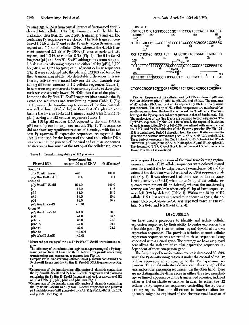
by using Agt.WESAB from partial libraries of fractionated EcoRI-cleaved total cellular DNA (31). Consistent with the blot hy-bridization data (Fig. 2), two EcoRI. fragments; 9 -and 4.1 kb,containing Py sequences were cloned. The 9-kb fragment con-tained 1.5 kb of the 5' end of the Py early region (transformingregion) and 7.5 kb of cellular DNA, whereas the 4.1-kb frag-ment contained 2.8 kb of Py DNA (3' ends of early and lateregions) and 1.3 kb of cellular DNA (Fig. 1). The 9-kb EcoRIfragment (pL) and BamHI-EcaRI subfragments containing the1.5-kb viral-transforming region and either 149 bp (pBl), 1,120bp (pB2),. or 1,520 bp (pB3) of 5' adjacent cellular sequences(Fig. 1) were subcloned into the plasmid pAT153 and tested fortheir transforming ability. No detectable differences in trans-forming activity were noted between the four plasmids con-taining different amounts of H2 cellular sequences (Table 1).In numerous experiments the transforming ability of these plas-mids was consistently lower (20-40%) than that of the plasmidharboring the Py BamHI-EcoRI fragment (that contains the Pyexpression sequences and transforming region) (Table 1) (Fig.1). However, the transforming frequency of the four plasmidswas still at least 100-fold higher than that of a plasmid. con-taining the Py Hae II-EcoRI fragment (intact transforming re-gion) lacking any H2 cellular sequences (Table 1).The 149-bp H2 cellular DNA adjacent to the viral DNA in
pBl was subjected to sequence analysis. (Fig. 4). This sequencedid not show any significant regions of homology with the ab-sent Py upstream 5' expression sequences. As expected, theHae II site used for the ligation of the viral and cellular DNAwas present at the junction of the viral and cellular sequences.To determine how much of the 149 bp of the cellular sequences
Table 1. Transforming ability of H2 plasmidsTransformed foci,
Plasmid DNA no. per 100 ng of DNA* % efficiencytGroup 1tpPy BamHI linear 420 100.0pPy Hae II-BamHI 0.4 0.1
Group 2§pPy BamHL-EcoRI 291.0 100.0pL 92.0 31.6pB3 84.0 28.9pB2 86.0 29.6pB1 88.0 30.2pPy Hae II-EcoRI <0.04
Group 3¶pPy BamHI-EcoRI 144.0 100.0pB1 41.0 28.5pBA17 35.0 24.3pBA18 42.0 29.1pBA24 32.0 22.2pBA20 <0.006pPy Hae II-EcoRI <0.01
* Measured per 100 ng of the 1.5-kb Py Hae II-EcoRI-transforming re-gion.
tThe efficiency of transformation is given as a percentage of a Py frag-ment (either BamHI linear or BamHI-EcoRI fragment) containingtransforming and expression sequences (see Fig. 1).
t Comparison of transforming efficiencies of plasmids containing thePyBamHI linear and the Py Hae II-BamHI DNA fragment (see Fig.1).
§Comparison of the transforming efficiencies of plasmids containingthe Py BamHI-EcoRI and Py Hae il-EcoRI fragments and plasmidscontaining the PyHae II-EcoRI fragment and various amounts of H2cellular DNA (pL, pB3, pB2, and pBl) (see Fig. 1).¶Comparison of the transforming efficiencies of plasmids containingthe Py BamHI-EcoRI and Py Hae H-EcoRI fragments and plasmidpB1 and deletions of pB1 generated by BAI-31 (pBA17, pBA18, pBA24,and pBA20) (see Fig. 4).
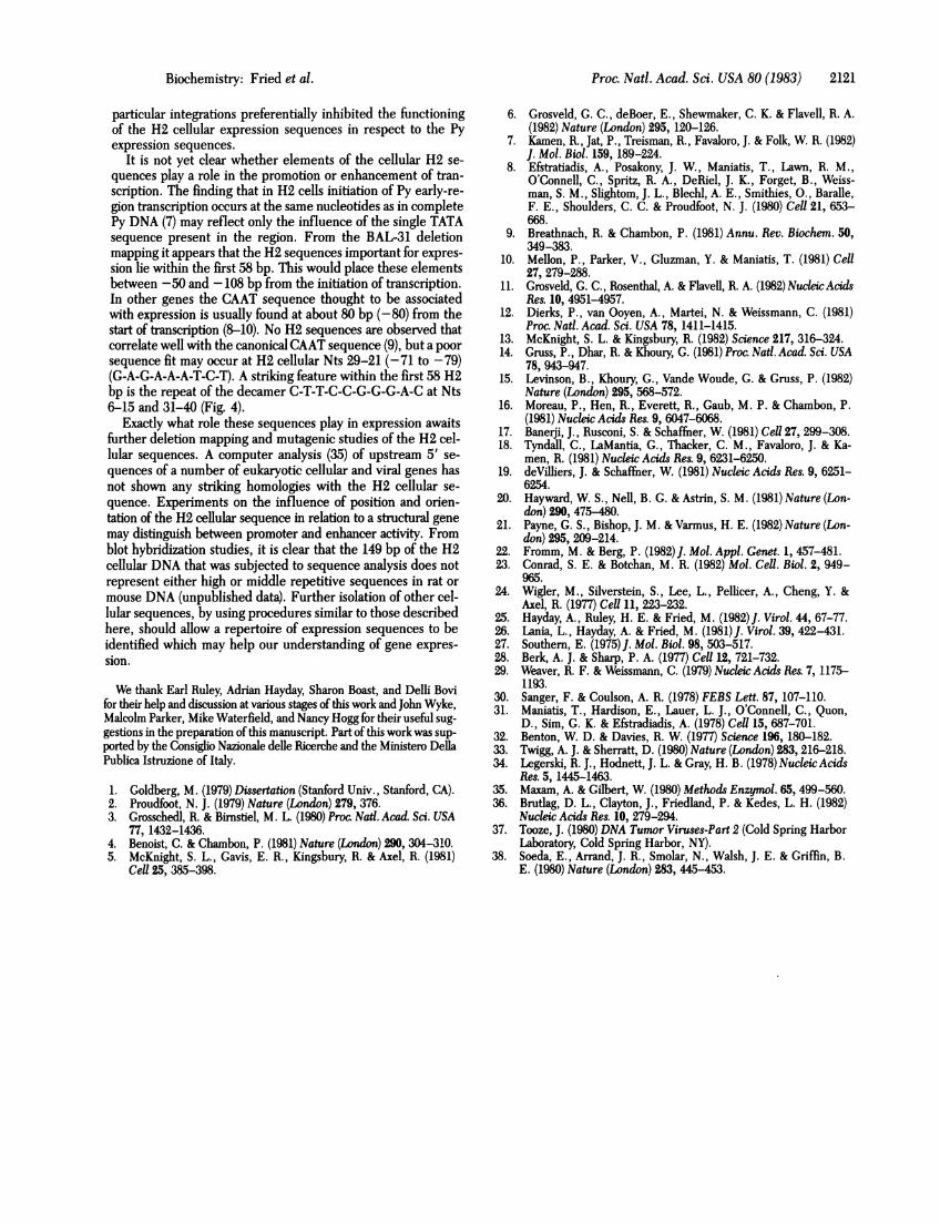
7Bal31-GGRTCCTCTCTGRRCCCCC.GCTTRCCCGTCCCGCGTRGGCCCL-BamHIJ 140 130 120 110
A24 Al7
RTTGCGRCCRCCGCGTCRTCCGTCGCGGCAGRCCRGCCRRRG100 90 80 i0
A18
CCRTCRCRGCRGCCRTCTTTGRG.CRCTTCCGGGRCCGRGRRRA60 50 40 30
,L20 P100TCTGCTTCCTTCCGGGRCGGCGCTGTGGGGCCRCCCRRRTTG
120 1i016, 0R|TRTRRTTRR|CCCCRRCCGCCTCTTCCCGCCTCRTTTCRGC
1,0 200CTCRCCACCRTCRTGGRTRGRGTTCTGRGCRGRGCTGRCRRR
FIG. 4. Sequence of H2 cellular and Py DNA in plasmid pB1 andBAL-31 deletions pBA17, pBA18, pBA20, and pBA24. The sequenceof H2 cellular DNA and part of the adjacent Py DNA in the plasmidpB1 is shown. The 149bp of H2 cellular sequences are numbered (be-neath sequence) from theHae II site toward the BamHI site. The num-bering of the Py sequence (above sequence) is that of Soeda et al. (38).The nucleotides of the Hae II site are common to both sequences. ThePy TATA sequence (Py Nts 120-128) is boxed, the region of initiationof early transcription (Py Nts 148-153) is overlined with an arrow, andthe ATG used for the initiation of the Py early proteins (Py Nts 173-175) is underlined. BAL-31 digestion from the BamHI site was used togenerate the deletion derivatives pBA17, pBA18, pBA20, and pBA24.These derivatives contain theXho I linker C-C-T-C-G-A-G-G atH2 cel-lular Nt 21 (pBA20), Nt 66 (pBA17), Nt 58 (pBA18), and Nt 104 (pBA24).The decamer C-T-T-C-C-G-G-G-A-C found twice at H2 cellular Nts 6-15 and Nts 30-41 is overlined.
were required for expression of the viral-transforming region,various amounts of H2 cellular sequences were deleted inwardfrom the BamHI site by using BAL-31 exonuclease (34) and theextent of the deletions was determined by DNA sequence anal-ysis (Fig. 4). It was observed that there was no loss in trans-forming activity (pBA18) when up to 58 bp of the cellular se-quences were present (91 bp deleted), whereas the transformingactivity was lost (pBA20) when only 21 bp of host sequenceswere left (128 bp deleted) (Table 1). Within the 58 bp of H2cellular DNA that were subjected to sequence analysis, the de-camer C-T-T-C-C-G-G-G-A-C was repeated twice at H2 cel-lular Nts 6-16 and Nts 31-41 (Fig. 4).
DISCUSSIONWe have used a procedure to identify and isolate cellularexpression sequences by their ability to confer expression to aselectable gene (Py transformation region) devoid of its ownexpression sequences. The previous isolation of most cellularexpression sequences was restricted to those sequences beingassociated with a cloned gene. The strategy we have employedhere allows the isolation of cellular expression sequences in-dependent of their companion gene.The frequency of transformation events is decreased 60-80%
when the Py-transforming region is under the control of the H2cellular sequences in comparison to the Py expression se-quences. This might indicate a difference in the strength of theviral and cellular expression sequences. On the other hand, thereare no distinguishable differences in either the size, morphol-ogy, or time of appearance of the transformed colonies, inducedeither as foci on plastic or colonies in agar, by either the H2cellular or Py expression sequences controlling the Py-trans-forming region. Thus, the difference in transformation fre-quencies might be explained if the chromosomal location of
2120 Biochemistry: Fried et al.
Proc. Natl. Acad. Sci. USA 80 (1983) 2121
particular integrations preferentially inhibited the functioningof the H2 cellular expression sequences in respect to the Pyexpression sequences.
It is not yet clear whether elements of the cellular H2 se-quences play a role in the promotion or enhancement of tran-scription. The finding that in H2 cells initiation of Py early-re-gion transcription occurs at the same nucleotides as in completePy DNA (7) may reflect only the influence of the single TATAsequence present in the region. From the BAL-31 deletionmapping it appears that the H2 sequences important for expres-sion lie within the first 58 bp. This would place these elementsbetween -50 and -108 bp from the initiation of transcription.In other genes the CAAT sequence thought to be associatedwith expression is usually found at about 80 bp (-80) from thestart of transcription (8-10). No H2 sequences are observed thatcorrelate well with the canonical CAAT sequence (9), but a poorsequence fit may occur at H2 cellular Nts 29-21 (-71 to -79)(G-A-G-A-A-A-T-C-T). A striking feature within the first 58 H2bp is the repeat of the decamer C-T-T-C-C-G-G-G-A-C at Nts6-15 and 31-40 (Fig. 4).
Exactly what role these sequences play in expression awaitsfurther deletion mapping and mutagenic studies of the H2 cel-lular sequences. A computer analysis (35) of upstream 5' se-quences of a number of eukaryotic cellular and viral genes hasnot shown any striking homologies with the H2 cellular se-quence. Experiments on the influence of position and orien-tation of the H2 cellular sequence in relation to a structural genemay distinguish between promoter and enhancer activity. Fromblot hybridization studies, it is clear that the 149 bp of the H2cellular DNA that was subjected to sequence analysis does notrepresent either high or middle repetitive sequences in rat ormouse DNA (unpublished data). Further isolation of other cel-lular sequences, by using procedures similar to those describedhere, should allow a repertoire of expression sequences to beidentified which may help our understanding of gene expres-sion.
We thank Earl Ruley, Adrian Hayday, Sharon Boast, and Delli Bovifor their help and discussion at various stages of this work and John Wyke,Malcolm Parker, Mike Waterfield, and Nancy Hogg for their useful sug-gestions in the preparation of this manuscript. Part of this work was sup-ported by the Consiglio Nazionale delle Ricerche and the Ministero DellaPublica Istruzione of Italy.
1. Goldberg, M. (1979) Dissertation (Stanford Univ., Stanford, CA).2. Proudfoot, N. J. (1979) Nature (London) 279, 376.3. Grosschedl, R. & Birnstiel, M. L. (1980) Proc Nati. Acad. Sci. USA
77, 1432-1436.4. Benoist, C. & Chambon, P. (1981) Nature (London) 290, 304-310.5. McKnight, S. L., Gavis, E. R., Kingsbury, R. & Axel, R. (1981)
Cell 25, 385-398.
6. Grosveld, G. C., deBoer, E., Shewmaker, C. K. & Flavell, R. A.(1982) Nature (London) 295, 120-126.
7. Kamen, R., Jat, P., Treisman, R., Favaloro, J. & Folk, W. R. (1982)J. Mol. Biol. 159, 189-224.
8. Efstratiadis, A., Posakony, J. W., Maniatis, T., Lawn, R. M.,O'Connell, C., Spritz, R. A., DeRiel, J. K., Forget, B., Weiss-man, S. M., Slightom, J. L., Blechl, A. E., Smithies, O., Baralle,F. E., Shoulders, C. C. & Proudfoot, N. J. (1980) Cell 21, 653-668.
9. Breathnach, R. & Chambon, P. (1981) Annu. Rev. Biochem. 50,349-383.
10. Mellon, P., Parker, V., Gluzman, Y. & Maniatis, T. (1981) Cell27, 279-288.
11. Grosveld, G. C., Rosenthal, A. & Flavell, R. A. (1982) NucleicAcidsRes. 10, 4951-4957.
12. Dierks, P., van Ooyen, A., Martei, N. & Weissmann, C. (1981)Proc. Natl. Acad. Sci. USA 78, 1411-1415.
13. McKnight, S. L. & Kingsbury, R. (1982) Science 217, 316-324.14. Gruss, P., Dhar, R. & Khoury, G. (1981) Proc. Nati. Acad. Sci. USA
78, 943-947.15. Levinson, B., Khoury, G., Vande Woude, G. & Gruss, P. (1982)
Nature (London) 295, 568-572.16. Moreau, P., Hen, R., Everett, R., Gaub, M. P. & Chambon, P.
(1981) Nucleic Acids Res. 9, 6047-6068.17. Banerji, J., Rusconi, S. & Schaffner, W. (1981) Cell 27, 299-308.18. Tyndall, C., LaMantia, G., Thacker, C. M., Favaloro, J. & Ka-
men, R. (1981) Nucleic Acids Res. 9, 6231-6250.19. deVilliers, J. & Schaffner, W. (1981) Nucleic Acids Res. 9, 6251-
6254.20. Hayward, W. S., Nell, B. G. & Astrin, S. M. (1981) Nature (Lon-
don) 290, 475-480.21. Payne, G. S., Bishop, J. M. & Varmus, H. E. (1982) Nature (Lon-
don) 295, 209-214.22. Fromm, M. & Berg, P. (1982)J. Mol. Appl. Genet. 1, 457-481.23. Conrad, S. E. & Botchan, M. R. (1982) Mol. Cell. Biol. 2, 949-
965.24. Wigler, M., Silverstein, S., Lee, L., Pellicer, A., Cheng, Y. &
Axel, R. (1977) Cell 11, 223-232.25. Hayday, A., Ruley, H. E. & Fried, M. (1982)J. Virol. 44, 67-77.26. Lania, L., Hayday, A. & Fried, M. (1981)J. Virol. 39, 422-431.27. Southern, E. (1975)J. Mol. Biol. 98, 503-517.28. Berk, A. J. & Sharp, P. A. (1977) Cell 12, 721-732.29. Weaver, R. F. & Weissmann, C. (1979) Nucleic Acids Res. 7, 1175-
1193.30. Sanger, F. & Coulson, A. R. (1978) FEBS Lett. 87, 107-110.31. Maniatis, T., Hardison, E., Lauer, L. J., O'Connell, C., Quon,
D., Sim, G. K. & Efstradiadis, A. (1978) Cell 15, 687-701.32. Benton, W. D. & Davies, R. W. (1977) Science 196, 180-182.33. Twigg, A. J. & Sherratt, D. (1980) Nature (London) 283, 216-218.34. Legerski, R. J., Hodnett, J. L. & Gray, H. B. (1978) NucleicAcids
Res. 5, 1445-1463.35. Maxam, A. & Gilbert, W. (1980) Methods Enzymol. 65, 499-560.36. Brutlag, D. L., Clayton, J., Friedland, P. & Kedes, L. H. (1982)
Nucleic Acids Res. 10, 279-294.37. Tooze, J. (1980) DNA Tumor Viruses-Part 2 (Cold Spring Harbor
Laboratory, Cold Spring Harbor, NY).38. Soeda, E., Arrand, J. R., Smolar, N., Walsh, J. E. & Griffin, B.
E. (1980) Nature (London) 283, 445-453.
Biochemistry: Fried et al.