Isolation and structural determination of polyphenols in ...of the series would have proved useful...
191
ISOLATION AND STRUCTURAL DETERMINATION OF POLYPHENOLS IN TASMANIAN EUCALYPTS. by RICHARD BARRY BROWN A thesis submitted in partial fulfilment of the requirements for the degree of -MASTER OF SCIENCE UNIVERSITY OF TASMANIA, HOBART. AEEILL_1222-
Transcript of Isolation and structural determination of polyphenols in ...of the series would have proved useful...
ISOLATION AND STRUCTURAL DETERMINATION
OF
POLYPHENOLS IN TASMANIAN EUCALYPTS.
by
RICHARD BARRY BROWN
A thesis submitted in partial fulfilment
of the requirements for the degree of
-MASTER OF SCIENCE
UNIVERSITY OF TASMANIA,
HOBART. AEEILL_1222-

Signed:
Except as stated herein, this thesis contains
no Material which has been accepted for the award of
any other degree or diploma in any university and, to
the best of my knowledge, this thesis contains no
copy nor paraphrase of material previously published
or written, except when due reference is made in the
text of the thesis.
4t-

iii
TABLE OF CONTENTS
Page.
ABSTRACT.
ACKNOWLEDGEMENTS. vii
CHAPTER 1. INTRODUCTION. 1
3
4
7
12
1.0. EUCALYPT CLASSIFICATION AND CHEMOTAXONOMY.
1.1. HILLIS' SURVEY OF EUCALYPT POLYPHENOLS.
1.2. HILLIS' KNOWN AGLYCONES AND GLYCOSIDES.
1.3. HILLIS' UNKNOWNS.
CHAPTER 2. ISOLATION - EXPERIMENTAL. 17
2.0. ISOLATION - GENERAL PROCEDURES. 19
2.0.1. Sources of leaves. 19
2.0.2. Liquid-liquid extraction. 19
2.0.3. Chromatography. •19
2.1. ISOLATION - SPECIAL PROCEDURES. 27
2.1.1. Liquid-liquid extraction. 27
2.1.2. Chromatography. 31
CHAPTER 3. IDENTIFICATION - EXPERIMENTAL. 48
3.0. SPECTRAL DETERMINATIONS. 50
3.1. SPECTRA. 52
3.2. DERIVATIVES. 100
3.3. ROTATIONS, ANALYSES, MELTING POINTS. 103
3.4. EXPERIMENTAL DATA. 104
iv
TABLE OF CONTENTS (continued)
Page.
CHAPTER 4. IDENTIFICATION - DISCUSSION. 122
4.0. POLYPHENOLS OF KNOWN STRUCTURE. 123
4.0.1. Hydroxybenzoic acids. 123
4.0.2. Flavonols. 126
4.0.3. Flavonol glycosides. 128
4.0.4. Ellagic acid. 132
4.0.3. (+)-Catechin. 13 6
4.1. POLYPHENOLS OF UNKNOWN STRUCTURE 139
(HILLIS' UNKNOWNS).
4.1.1. Unknown D - Flavanones. 139
4.1.2. Unknown D Flavones. 143
4.1.3. Unknown A (4-0-methylellagic acid) . 153
4.1.4. Unknown B (1,5-dimethy1-2,6-bis- 160
(trihydroxyphenyl) furo [1,5-c] -
furan).
4.1.3. Unknown F (a tetrahydroxydibenzofuran- 166
dicarboxylic acid.isomer).
APPENDIX. CHROMATOGRAPHIC BEHAVIOUR - SUMMARY. . 1.71
1. POLYPHENOLS OF KNOWN STRUCTURE. 172
2. POLYPHENOLS OF UNKNOWN STRUCTURE.
173
REFERENCES. 174
. PUBLICATION. fg3

ABSTRACT.
Chapter 1 contains a brief review of
chemotaxonomy as it relates to the classification of
eucalypts.
Particular reference is made to a chromatographic
survey by Hillis of the low molecular weight
polyphenols on eucalypt leaves.
That survey provisionally identified a number of
polyphenols of known structure but labelled other
compounds "Unknowns", because of the lack of evidence
for the identification of their structure.
The present work recounts the isolation and
examination of the polyphenols in the leaves of three
Eucalypt species important to Tasmania: E. delegatensis,
E. sieberi and E. coccifera.
Chapter 2 gives experimental details of the
isolation of polyphenols from these species especially
the isolation of some of Hillis' unknowns.
Cha ter 3 describes the experimental techniques
by which evidence for the identity of the isolated
polyphenols has been gathered.
Chapter 4 then discusses the identity of these
polyphenols.
First to be identified are a number of
known polyphenols: gallic, gentisic and

vi
protocatechuic acids; quercetin, myricetin,
kaempferol; the flavonol glycosides, afzelin, rutin,
quercitrin, isoquercitrin, hyperin and cannabiscitrin;
ellagic acid and (+)-catechin.
Secondly, some of Hillis' unknowns have been
isolated and identified.
Unknown D has been isolated
(1) from E. sieberi and is identified as a mixture
of flavanones: pinocembrin, alpinetin and a
new natural product, 0,0-.dimethylpinocembrin;
(2) from E. coccifera and is identified as a mixture
of flavones: apigenin and two C-methylflavones,
sideroxylin and 4',5-dihydroxy-7-methoxy-6-
methylflavone, the last identified provisionally.
Unknown A is identified as 4-0-methylellagic acid,
a new ellagic acid ether.
Unknown B is provisionally identified as a new
lignan, 1,5-dimethy1-2,6-bis (trihydroxyphenyl) furo -
[1,5-c] furan.
Unknown F is tentatively given the structure of
a tetrahydroxydibenzofuran-dicarboxylic acid isomer.
Throughout the discussion the use of chromatography
of mixtures to provide evidence for ch,emotaxonomy
is critically considered.

ACKNOWLEDGEMENTS.
The author wishes to thank his supervisor,
Dr. I.R.C. Bitk, Chemistry Department, University of
Tasmania, for continued advice and encouragement during
the course of this work.
Gratitude is also extended to Associate Professor
J.B. Polya and Dr. J.B. Bremner of the same Department
and University for the supervision of the final draft .
of this thesis.
Dr. W.E. Hillis, Senior Principal Research
Scientist, C.S.I.R.O., Division of Applied Chemistry,
Officer-in-Charge, Wood and Forest Science Section,
suggested the project, gave continual encouragement and
advice and supplied many authentic samples; to him
special thanks are given.
Dr. T.J. Batterham, John Curtin School of Medical
Research, Australian National University, gave the use
of his laboratory and facilities during a two-week
stay in that laboratory, together with his interest and
advice.
Finally, thanks are due to Mr. L. Brasch,
Librarian, Associated Pulp and Paper Mills Ltds
Library, Burnie and to the Librarian, Forest Products
Laboratory Library, Melbourne, for the use of the
facilities of their respective libraries.
vii

POLYPHENOLS OF TASMANIAN EUCALYPTS.
CHAPTER 1.
1
INTRODUCTION.

CONTENTS.
Page.
100. EUCALYPT CLASSIFICATION AND CHEMOTAXONOMY. 3
1.1. HILLIS' SURVEY OF EUCALYPT POLYPHENOLS. 4
1.2. HILLIS' KNOWN POLYPHENOL AGLYCONES AND 7
GLYCOSIDES.
1. 3. HILLIS' UNKNOWN POLYPHENOLS. 12
2

3
CHAPTER 1.
1.0. EUCALYPT CLASSIFICATION AND CHEMOTAXONOMY.
The classification of eucalypt species in use
'today is Blakely's "A Key to the Eucalypts"16
based
on the so-called antheral system of Bentham. A
second classification used by Blakely was based
on general morphology but in some cases it
contradicted the first.
While the original key has anomalies it remains
the only complete system evolved and.is the key
upon which any classification used in this study is
based.
The reliable classification of this difficult
genus requires as many taxonomic criteria as
possible.
• In recent years increasing study has been made
of wood anatomy, bark bark anatomy, 23 cytology, 101
pollen grains,91
seed coat anatomy,34
and floral
morphology2021
as taxonomic criteria.
Further it has been proposed that the chemical
composition of various plant tissues could also
be a useful criterion in taxonomy,48
particularly as it
expresses its results in different terms from those of
botany and thus provides an independent check upon

4
11 classification.. Bate-Smith, Hasegawa, Horn Horn et al. and and
47 more recently and extensively, Hillis have studied the
chemical composition of a variety of plants and plant
tissues.
7 Baker and Smith - first brought the focus of
chemotaxonomy to bear on the Eucalypt genus in 1890
concentrating on the essential oil composition in
eucalypt leaves.
Various plant tissues have been studied to decide
which Would provide the most reliable chemotaxonomic guide.
Wood and phloem extractives have proved of limited
usefulness although heartwood extractives have shown
43 more promise, while the greatest attention has been
given to leaf extractives which seem to provide a more
reliable guide to phenotype than extractives from other
plant tissues.
Different groups of compounds found in leaf tissue
have been studied from the chemotaxonomic viewpoint.
In addition to the study of essential oils by
Baker and Smith 7 and their successors, leaf Waxes have been -
examined by Horn and his co-workers59
who found them to
be composed mainly of long-chain /3-diketones with little
promise of taxonomic significance.
1.1. HILLIS 1 SURVEY OF EUCALYPT POLYPHENOLS.
Hillis47
and others have suggested that compounds
of low molecular weight in the leaves of eucalypts could
provide a more useful taxonomic criterion.

Hillis' extensive survey of over 300 eucalypt
species used the pattern of low Molecular weight-
polyphenols in conjunction with other criteria to draw
conclusions about eucalypt classification.
This examination was largely chromatographic,
as far as possible using standard markers, and Hillis
pointed out the need of extending the work by employing
isolation and standard procedures of identification. 48
He proposed thereby to verify the identity of key
polyphenols as well as to identify other polyphenols to
which not even provisional identity was ascribed but which
were simply labelled "Unknowns".
The present work takes the results Of Hillis'
survey as they apply to Tasmanian eucalypts, in particular
to three species, and by isolation and techniques of
structural analysis confirms the provisional identity of
many of Hillis.' polyphenols. It establishes the identity
of some unknowns and isolates and identifies an additional
polyphenol hitherto unrecorded by Hillis.
Further, the most abundant glycosides in each species
are characterised to complete the chemotaxonomic picture.
As a result of this work a question is raised
about the advisability of using chromatography alone
as a guide even to provisional identity of compounds
where overlying spots on the chromatogram are common.
Especially j: this true- when soMe significance.is .attached

to such provisional identity in classification.
This is not to deny the validity of the use of
chemical criteria in taxonomy in conjunction with other
criteria but to suggest that the identity of compounds
which are put forward as playing a significant role in
classification must be accurately known if the
conclusions based on their identity are to be reliable.
Two species of the Fraxinales or Ash series were
examined for comparison of their polyphenolic content,
E. dele atensis and E. sieberi. While not endemic both
species have particular significance for Tasmania.
16a E. delegatensis (R.T. Baker) or E. gigantea
1.6a (Hook,t.) is particularly important to the State as a
Source of paper pulp and as a timber, Tasmanian Oak.
16a E. sieberi (L. Johnson) or Tasmanian Ironbark
grows locally in the north-east from Coles Bay to
Georges River between the coast and the headwaters of
the South Esk River. Attempts to grow E. sieberi in
large stands in other parts of the State for paper-making
purposes have failed.
Although E. sieberi grows also in the sub-alpine
regions of Victoria and southern New South Wales, Hillis
found in his survey that the chemical composition of the
Tasmanian samples was significantly different from that
of the samples gathered on the mainland.
16b coccifera (Hook f.) or Tasmanian Snow Gum
grows in sub-alpine conditions at altitudes of 2000-4500
6
49

feet. A Tasmanian endemic, it is widespread except in
the north-east of the State. 17.
Time permitted examination of only one of the
' Piperitales or Peppermint series, E. coccifera, although
comparison of the polyphenol content of a second Species
of the series would have proved useful for chemotaxonomy.
1.2. HILLIS' KNOWN POLYPHENOL AGLYCONES AND GLYCOSIDES.
Table i lists the aglycones chromatographically
identified by Hillis together with those isolated and
identified during the course of this current work. The
flavonol glycosides isolated are listed in Table ?.
Other polyphenols, provisionally called caffeic,
chlorogenic and sinapic acids and Hillis' unknown.H;
were observed in chromatograms in the course of this study
but were not isolated, so they are not listed in Table 1.
The structures corresponding to Hillis' known
aglydones listed in Table 1 are given in Chart 1, while
the structures of the known aglycones and glycosides
actually isolated here are given in. Chart 2 on
page'll. _
As Charts (1) and (2) show, there is substantial
agreement between the known polyphenols identified by
Hillis and those isolated in. this work. But one
should note the isolation of a third hydroxybenzoic
acid, protocatechuic acid, which is not recorded by Hillis
throughout his survey, although it is known to be widely
distributed among the higher plants.62

0,0-dimethylpinocembrin
pinocembrin
) )
.alpinetin
) )
0,0-dimethylpinocembrin )
E. delegatensis.
E. sieberi.
unknown D
TABLE 1.
1. Polyphenol • aglycones chromatographically identified by Hillis (H) and identified after isolation by Brown B .
kaempferol
quercetin
Myricetin
gallic acid (H & B)
gentisic acid (H & B)
protocatechuic acid (B)
a catechin (H) (+)-catechin (B)
chlorogenic acid (H)
ellagic acid (H & B)
2. Poly henel a•1 ones called unknowns by Hillis and structurally identified by Brown.
HILLIS
unknOwn A
unknown B
BROWN
SPECIES
4-0-methylellagic acid E. sieberi.
1,5-dimethy1-2,6-bis(3,4,5- trihydroxyphenyi furo-[1,5-c] furan E.•sieberi.
yc
unknown F
) )
sideroxylin (4',5-dihydroxy , ) 7-methoxy-6,8-dimethylflavoneD E. coccifera.
) 4',5-dihydroxy-7-methoxy- )
6-methylflavone )
3,4,7,8-tetrahydroxy-dibenzofuran,1 9 5-dicarboXylic E. sieberi.
apigenin
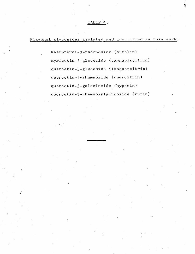
TABLE 2 .
Flavonol glycosides isolated and identified in this work.
kaempferol-3-rhamnoside (afzelin)
myricetin-3-glucoside (cannabiscitrin)
quercetin-3-glucoside (isoquercitrin)
quercetin-3-rhamnoside (quercitrin)
quercetin-3-galactoside (hyperin)
quercetin-3-rhamnosylglucoside (rutin)
9

Ho HO
OH 0
10
R1'
R2
= H/R3= Rhamnosyl; Afzelin
R1' R 2 = H ; Kaerripf erol
Ri =OH,R2 =H I R3 =Rhamnosylgtucosyl, Rutin R1
=OH,R2
=H ; Quercetin
R2
=0H ; Myricetin R3 = Rhamnosyl ; Quercitrin
R3= Glucosyl: Isoquercitrin
R3 =Galactosyl, Hype rin
R11
R = OH R3 = GI ucosyl; Cannabiscitrin
OH
OH
Ellagic acid
COOH
R11 R2 = OH,R3 = H ; Gallic acid
R1 ,R3 = OH,R 2 =H ; Gent isic acid
R2 = OH,Rv R3 H ; Protocatechuic acid
OH
OH
HO
OH
OH
(4-)-Catechin (2R: 3S)
CHART 1. HILLIS' KNOWN POLYPHENOL S and GLYCOSIDES ISOLATED from TASMANIAN EUCALYPTS.

11
HO
OH
RI = OH,R2=H: Cyanidin
R1 ,R
2 =OH; Oelphinidin
OH
OH 0
R1' R 2 =H: Kaempferol
- R 1 =OH, R 2 =H: Quercetin
R1 R 2 = OH: Myricetin
Chlorogenic acid
OH
OH
Cat ec-hin
HO
OH
CHART 2. HILLIS' MAIN PHENOLIC COMPONENTS IN SOLUTION AFTER
ACID' TREATMENT OF EUCALYPT LEAVES.
HO
COOH
2
R1
. = OH,R2=H; Gallic acid
R2 = OH,R 1 =H; Gentisic acid

In addition to the identification of
protocatechuic acid ( 1 ), a more detailed identification
has been made of the catechin isolated.
COOH
12
OH
1.3. HILLIS' UNKNOWN POLYPHENOLS.
The major concern of the present study has been
the isolation and identification of unknown compounds in
. these species. The structures of the compounds isolated
from the ashes with chromatographic behaviour similar to
that of Hillis' unknown Dare given in Chart 3 and are
seen to be flavanones, one of which is a new compound.
Whereas the compounds isolated from the peppermint and
also called unknown D by Hillis are shown in the same
chart to be flavones, of which two are rather rare
C-methylflavones.
Hillis stresses the importance of unknown D in
chemotaxonomy, states that it is found only in the
49 Renantherae section of Blakely's classification and

1 3
uses the compound to indicate association between
numerous renantherous species. 49 He also states this
unknown is present in the peppermint, E. coccifera,
although he points out the anomalous chemical
composition of this species. '
In proposing that unknown D is not a single
component but a mixture and indeed a different mixture
in the ashes and the peppermint examined, the present
work raises doubts as to the taxonomic significance of
unknown D.
Unknown A has been identified as a monomethyl
ether of ellagic acid (Chart 3), previously
unreported in the literature. This is in substantial
agreement with the tentative identification made by
Hillis who referred to it as an "ellagic acid-like"
compound. 48 It was found in his survey that about half
of the species examined contained the compound in varying
amounts but the species were spread throughout the genus
in such a random manner that the presence of this
compound was apparently not significant to classification.
Unknown B was also described by Hillis as
"ellagic acid-like" because of its-chromatographic
behaviour.48
A compound from E. sieberi isolated and
reported upon in this thesis has properties on paper
chromatograms similar to those of unknown B, but on spectral
evidence seems to be not a derivative of ellagic acid but
a new lignan.

R 0 1
UNKNOWN D 14
E.detegatensis E.sieberi
Me0 o R 0 2 o
0 0- Dimethylpinocembrin / Ft17 R 2 =H ; Pinocembrin
II / =1-1 1 R 2 =Me ; Alpinetin
R1 = R2 = Me; 010-Dimethylpinocembrin
E.coccif era
R 0 4 0
RR2,R3 IR4 = H: Apigenin
R1 ,R21R3 = Me, R4 =H ; Sideroxylin
R27 R3 = Me, R1 ,R4 = H; 5/4'-Dihydroxy-7-methoxy-6-methytflavone
UNKNOWN A
E.sieberi
4- 0 -Methytellagic acid
CHART 3. UNKNOWNS Aj D JSOLATED from TASMANIAN EUCALYPTS.

Finally, unknown F is tentatively identified now
as a dibenzofuran shown in Chart 4. Hillis found that
this compound was spread erratically throughout the
Eucalypt genus and he based no chemotaxonomic
conclusions upon its presence or absence. 48
It was a particular difficulty in the isolation
of this polyphenol that ellagic acid •tended to remain
as an impurity. Frequently unknown F spots on
chromatograms showed not as an authentic yellow but
as an ambiguous brown colour, highlighting the problem -
of provisional chromatographic identification.
G.1.c. methods of trimethylsilyl-ether separation44
'
45199
f this unknown were not attempted, as explained later.
15

HO
HO
HO
3
2 4 CH CH 2
\ 1 5 H C-C—C CH 3
3
CH2 HC 8 zy 6
0 7
OH
OH
OH
UNKNOWN B
1 ,5 -dimethyl- 2,6-bis(3,4,5-frihydroxyphenyl)furo[1,5-c]furan
OH
COOH
HO
OH
COOH
OH
UNKNOWN F ' 3,4,7,8 -tetrahydroxydibenzofurem-1,5-dicarboxylic acid
CHART 4. UNKNOWNS B, F ISOLATED from E.sieberi.

CHAPTER 2 .
17
ISOLATION - EXPERIMENTAL

CONTENTS.
2.0. ISOLATION - GENERAL PROCEDURES.
2.0.1. SOURCES OF LEAVES.
2.0.2. LIQUID-LIQUID EXTRACTION.
2.0.3. CHROMATOGRAPHY.
2.0.3.1. Paper Chromatography.
2.0.3.2. Thin Layer Chromatography.
2.0.3.3. Column Chromatography.
2.0.3.4. Gel Filtration.
2.0.3.5. Gas-Liquid Chromatography.
2.1. ISOLATION - SPECIAL PROCEDURES.
2.1.1. LIQUID-LIQUID EXTRACTION.
2.1.1.1. E. delegatensis.
2.1.1.2. E. sieberi.
2.1.1.3. E. coccifera.
2.1.2. CHROMATOGRAPHY.
2.1.2.1. E. delegatensis.
2.1.2.2. E. sieberi.
2.1.2.3. E. coccifera.
Page.
19
19
19
19
23
2 5
26
26
2 6
27
27
27
27
29
31
31
35
42

CHAPTER 2.
2.0. ISOLATION - GENERAL PROCEDURES.
Since the isolation techniques of liquid-liquid
extraction and chromatography followed for all three
species were similar, these techniques are given first
as general procedures.
Small departures from these general processes
as well as additions to them for a particular species
are then given separately as special procedures, : -
. (2.1, page 27 ).
2.0.1. SOURCES OF LEAVES.
Samples of leaves of E. delegatensis examined
were .obtained from Associated Pulp and Paper Mills Ltdis
forests at Meander. Leaves of E. sieberi were collected
at Fingal and those of E. cOccifera on Mt. Fenton,
Mt. Field National Park, Hobart.
2.0.2. LIQUID-LIQUID EXTRACTION.
Dried milled leaves of each eucalypt (2 kg)
were exhaustively extracted with methanol and the extract,
concentrated in vacuo to 2 1, was poured in a thin
streaminto 10 1 of vigorously stirred water. The green
waxy material which precipitated was filtered, re-
dissolved in methanol and re-precipitated as before.
The process was repeated until paper chromatograms showed
the precipitate showed no polyphenols.
The combined aqueous methanolic extracts,
concentrated in vacuo to 3 1, were extracted with
1 9

petroleum ether (400 -60 0 ) until chromatograms showed
that all chlorophylls, waxes and oils had been
removed.
The aqueous methanolic concentrate was carried
through a series of liquid-liquid extractions,
summarised in Chart 5, to be read with the legend in
Chart 6 9 pages 21 9 22.
2.0.3. CHROMATOGRAPHY.
Many of the chromatographic techniques used
in this work have found application not only in the
isolation but also the identification of polyphenols.
The materials used are grouped here for convenience
but their use is detailed in Chapter 2.1, Special
Procedures.
References to this section will also be made
in Chapter 3 which concerns identification of
polyphenols.
20

21
Legend overleaf. I or IH
(II) Ether extraction.
(III) Bicarbonate extraction.
(VII) Ethyl acetate extraction.
(vi ) (IV) Acidification plus ether extraction.
Not examined further.
(v) (VIII) Bicarbonate
extraction.
(XII) n-Butanol extraction.
•
(IX) Acidification plus ethyl acetate extraction.
Bicarbonate (XIII) extraction.
Not examined further.
(xvi) (XVII) Not
examined further.
(XIV) Acidification
plus butanol extraction
Not examined further.
CHART 5. LIQUID-LIQUID EXTRACTION FLOW CHART.

CHART 6 LEGEND CHART 6. LEGEND TO ACCOMPANY FLOW CHART 5.
22
(I) An aqueous methanolic extract of eucalypt leaves
was concentrated under vacuum and extracted with
petroleum ether to remove chlorophylls, waxes and oils.
(IH) Portion of the aqueous methanolic extract from
(I) was hydrolysed with 2M hydrochloric acid.
ETHER EXTRACTION.
(II) The extract from (I) or (Da was extracted
continuously with ether and the ether extract
concentrated to 500 ml.
(III) The ether concentrate from (II) was extracted with
5% aqueous sodium bicarbonate (6 x 100 ml. portions
(IV) The aqueous bicarbonate extract from (III) was
acidified, re-extracted with ether and this ether
extract concentrated, becoming (V).
(VI) The ether extract from (III), i.e. after
bicarbonate extraction, was concentrated.
ETHYL ACETATE EXTRACTION.
(VII) The aqueous methanolic extract from (II), i.e.
after ether extraction, was extracted continuously
with ethyl acetate and the ethyl acetate extract
concentrated.
(VIII) The ethyl acetate extract from (VII) was extracted
with 5% aqueous sodium bicarbonate.
(IX) The aqueous bicarbonate extract from (VIII) was
acidified, re—extracted with ethyl acetate and
the ethyl acetate extract concentrated, becoming (X).
(XI) The ethyl acetate extract from (VIII), i.e. after
bicarbonate extraction was concentrated.
n-BUTANOL EXTRACTION.
(XII) The aqueous rnethanolic extract from (VII) was
hand extracted with n-butanol and the butanol
extract concentrated.
(XIII) The butanol extract from (XII) was extracted with
5 % aqueous sodium bicarbonate.
(XIV) The aqueous bicarbonate extract from (XIII) was
acidified, re-extracted with n-butanol and the
butanol extract concentrated, becoming (XV).
(XVI) The butanol extract from (XIII), i.e. after
bicarbonate extraction, was concentrated,
becoming (xvT)
(XVII) The aqueous nnethanolic extract from (XII), i.e.
(XVII).was not examined further.

2 3
2.0.3.1. PAPER CHROMATOQRAPHY.
Paper chromatography was carried out on either
Whatman No 1 or Whatman No 3MM paper using the following
solvent systems:
Volume Abbreviation Solvent system. ratio. adopted.
1. n-butanol- acetic acid- water48
(6:1:2) BAW (612) _
2. n-butanol- acetic acid- water39 (4:1:5
BAW (415) upper phase)
3. t-butanol- acetic acid- water75a
(3:1:1) TBA
4. benzene- acetic acid- water54
(6:3:7) BeAW
5. acetic acid- water48
(6:94) 6H0Ac.
6. acetic acid- water75a (15:85) 15H0Ac,
7. hydrochloric p.cid- acetic acid- water 5 1 (3:30:10) Forestal = F
8. phenol- water39
57 ethyl acetate- formic acid- water (10:2:3) EFW
Systems 1-9 were used in the development of
either two-dimensional analytical chromatograms or
one-dimensional preparative chromatograms of aglycones
and glycosides.
(saturated, upper PhOU phase) •

24
Solvent system. Volume Abbreviation ratio, adopted.
10. ethyl acetate- pyridine- wateri0
(12:5:4) EPW
11. n-butanol- pyridine- wateri0
(6:4:3) BPW
12. acetone- water39 (80:20) AcW
Systems 10-12 were used in the one-dimensional
analytical chromatography of sugars.
The chromatograms were examined under u.v. light
at wavelengths of 365 nm and 254 nm. before and after
exposure to concentrated ammonia vapour.
Polyphenol spots and bands on paper were
visualised by spraying with: Class of
'Spray'. Abbreviation, compound detected,
1. ferric chloride (1%)- potassium
ferricyanide (1%) 10 FeFe
2. diazotised 2.-nitroaniline (0.05%)
103 in aqueous 20% sodium acetate pNA
3. vanillin in conc. hydrochloric
acid (1%) 13
4. ethanolic 2-toluenesulphonic
acid (3%) 96
sodium borohydride in isopropyl
alcohol (1%)3 followed by acid
fuming
85 6. diazotised sulphanilic acid
7. chlorine vapour followed by 10%
aqueous sodium sulphite80
all phenols
all phenols
flavan-3-ols
flavan-3-ols
flavanones
lignans
lignans

25
Sugar spots were visualised with:
8. aniline hydrogen phthalate73
(1% aqueous)
9. aniline phosphate 3 (1% aqueous)
10. 2.-anisidine hydrochloride in butanol (1%) 17,75b
followed by heat (13e)
2.0.3.2. THIN LAYER CHROMATOGRAPHY.
Chromatoplates were prepared both in analytical
(0.1 mm) and preparative thickness (0.3 mm) using the
following adsorbents:
1. Camag silica, KieselgeI DSF-5, and Merck G.F.
254 Kieselgel.
2. Camag Cellulosepulver DF.
1.
2.
3.
4.
3. Polyamide (Woelm) - silica- rice
Solvent systems used in thin
82 starch.
layer
Volume Abbreviation
chromatography were:
Solvent system. ratio. adopted.
methanol- chloroform- petroleum
ether (60 °-80° ) 54
54 chloroform- acetic acid
chloroform- ethyl acetate-
54 formic acid
toluene- ethyl formate-
r4 formic acid)
58 benzene- pyridine- formic acid
(2:4:7)
(6:1)
(5:4:1)
(5:4:1)
(36:9:5)
MCP
CA
CEF
TEF
BPF
26
Spots and bands on chromatoplates were
visualised by spraying with:
1. antimony (III) chloride (10% in CHC1 3 ) followed
by heating. 58
2. diazotised E-nitroaniline (0.05%) in 20%
aqueous sodium acetate. 36
3. basic lead acetate (25% aqueous).
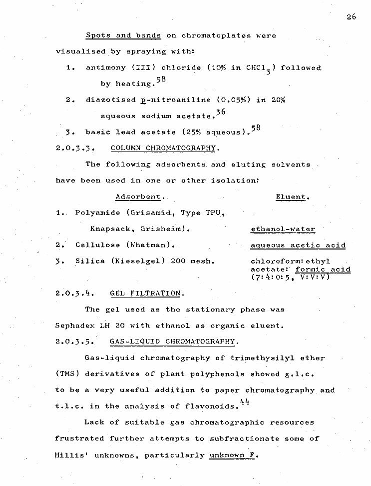
2.0.3.3. COLUMN CHROMATOGRAPHY.
The following adsorbents. and eluting solvents -
have been used in one or other isolation:
Adsorbent. Eluent.
1. Polyamide (Grisamid, Type TPU„
Knapsack, Grisheim). ethanol-water
2. Cellulose (Whatman). aqueous acetic acid
3. Silica (Kieselgel) 200 mesh. chloroform: ethyl acetate: formic acid (7:4:0:5, V:V:V)
2.0.3.4. GEL FILTRATION.
The gel used as the stationary phase was
Sephadex LH 20 with ethanol as organic eluent.
2.0.3.5. GAS-LIQUID CHROMATOGRAPHY.
Gas-liquid chromatography of trimethysilyl ether
(TMS) derivatives of plant polyphenols showed g.l.c.
to be a very useful addition to paper chromatography and
t.l.c. in the analysis of flavonoids. 44
Lack of suitable gas chromatographic resources
frustrated further attempts to subfractionate some of
Hillis' unknowns, particularly unknown F.

2 7
2.1. ISOLATION - SPECIAL PROCEDURES.
The following isolation methods for each species
were followed in conjunction with, or as departures from,
the General Procedures in Charts 5,6, pages 21,22.
2.1.1. LIQUID-LIQUID EXTRACTION.
2.1.1.1. E. delegatensis.
Hydroxybenzoic acids, quercetin, ellagic acid.
The concentrated ether extract (VI) chromatographed
two-dimensionally showed the presence of mainly
hydroxybenzoic acids, quercetin and ellagic acid.
Upon further concentration, of (VI) in vacuo
impure ellagic acid precipitated which, after filtering,
was crystallized from aqueous ethanol (75 mg).
Rutin.
The unhydrolysed aqueous methanolic extract (I)
after concentration and standing yielded a yellow
precipitate of impure rutin (200 mg) which was crystallized
from aqueous ethanol.
The filtrate (I) was extracted immediately with
ethyl acetate (VII) and the strong acids removed
as outlined • (VIII).:
2.1.1.2. E. sieberi.
Unknown D.48.
An off-white.precipitate settled in the
concentrated ether solution from (II) and was found to
be a mixture of flavanones (Hillis' unknown D) together
with minor components.

28
Unknown F.48
The ether extract remaining after removal of
the above flavanones was extracted repeatedly with 5%
aqueous sodium bicarbonate until no appreciable colour
showed in the bicarbonate layer. After neutralisation
and re-extraction with ether, this extract showed the
presence of Hillis' unknown F48
together with other
components.
After 'several weeks of standing in the cool room
a yellow-white precipitate (500 mg) settled in the
ether extract of the bicarbonate solubles. A paper
chromatogram of this precipitate in two dimensions
showed that it contained traces of Hillis' unknown D,
later identified as a flavanone mixture, some ellagic
acid and Hillis' unknown F.
Trituration of the yellow-white precipitate
with ether removed the traces of unknown D. Subsequent
repeated trituration with ethyl acetate monitored on
two-dimensional chromatograms showed that unknown F
was more soluble in ethyl acetate than was ellagic acid
so that the triturates gradually concentrated the
proportion of unknown F.
However after five triturations and re-
crystallizations unknown F could not be obtained pure,
ellagic acid persistently re-crystallizing as an impurity, 9

2 9
Unknown A48
The aqueous methanolic extract (I) after
continuous extraction with ether (II) was then hydrolysed
with 2M hydrochloric acid until paper chromatograms
showed all glycosides had been ruptured.
This acid hydrolysate was then extracted
continuously with ethyl acetate (VII). and ributanol
as in the General Procedure previously given on pages 21,22.
An off-white precipitate in the ethyl acetate
extract of the hydrolysed solution. after Step (VII) later
proved to be a mixture of two components, the minor
component being ellagic acid and the major one4 Hillis'
unknown A.
2.1.1.3. E. coccifera.
Hydroxybenzoic Acids, flavonols ., ellagic acid,
Hillis' unknown H:48
Portion of the original aqueous methanolic solution
was hydrolysed with 2M hydrochloric acid until paper
chromatograms showed the hydrolysis of glycosides was
complete, the solution becoming (IH) for the liquid-
liquid extractions in Charts 5 .,6, pages 21 22.
Figure 1 shows that the ether extract of the
neutralised bicarbonate solution contained caffeic (Spot 5),
gallic (9), gentisic (6), chlorogenic (8) and ellagic
acids -(1) together with a mixture of kaempferol (4),
quercetin (3) and myricetin (2) and finally a spot which
would correspond to Hillis' unknown H (7).

cm
Fig.l. POLYPHENOLS of E.coccif era extracted by 5% NaHCO 3 30 </\\\\\\
PDLYPHENOL
1.
Z .
3.
5. blue-wh. blue-white
6. blus-wh bl-gr-white
7. pink orange
8. blue intense green
4.
365nrri MH3 365nm rrauve dull yellow
orange intense or.
yellow intense y.
p-gr, . intense y-gr.
CM
9. mauve dark mauve (254-nm) (254m)
I I
2 4 6 8 cm 15% HOAc
Abbreviations: or, = orange
y. . yellow
yellow-green
blue-who = blue-white
bl-gr-white . blue-
green,-white.
12
10
8
BAW 4:1:5
6
F1g.2. POLYPHENOLS in E.coccifera ETHER EXTRACT after NaHCO3 extraction.
4 15% HOAc
POLYPHENOL 365nm NH 365nm 3
1. - mauve dull yellow
2. orange intense or.
3. yellow intense y,
4. Y400 . intense y-gr.
10. purple purple-blue
Abbreviations:
or. = orange
y, = yellow
y-gr, . yellow-green
cm
8
BAW 4:1:5
4

48 Unknown D.
The ether solution from (III), i.e. after
bicarbonate extraction of it, was shown on paper
chromatograms (Figure 2) to contain a mixture of compounds,
mainly unknown D (Spot 10), a flavonol mixture (4,3,2)
and ellagic acid (1).
Glycosides.
A second portion of the original aqueous methanolic
solution (I) was carried through the liquid-liquid
_extractions in Charts 5,6, pages 21,22, without hydrolysis.
Figure 3 shows a paper chromatogram of the
ether extract from Step (II) in Chart 5.
2.1.2. CHROMATOGRAPHY.
Applications of the chromatographic materials
listed on pages 23-26 are now given in more detail for
each species examined.
2.1.2.1. E. delegatensis.
Extracts chromatographed are referred to by
numerals corresponding to Charts 5,6 on pages21,22.
The ether extract of neutralised bicarbonate
solution (V) was reduced to dryness and dissolved in 3%
aqueous acetic acid. This mixture was applied to a
cellulose column and eluted first with 3% then 6%
aqueous acetic acid. Gentisic and protocatechuic acids
• were eluted as a mixture (50 mg) followed by impure gallic
acid (50 mg). Quercetin, unknown F and ellagic acid
were removed only when eluted with 50% aqueous ethanol.

32 Fig.3. POLYPHENOLS in ETHER EXTRACT of E.coccif era oci:Me0H solution.
6
BAW 4:15
PO LYPH EN OL
1. ella.gic acid
2. myricetin
3. quercetin.
4. kaempferol
5. caffeic acid
6. gentisic acid.
7. Hillis' 'Unknown H
8. chlorogenic acid.
9. gallic acid.
10.Hillis' Unknown, D
it. afzelin.
12.qu.ercitrio
13. isoQuerci trin
14.hyperin
15, sinapic acid. (trace)
4 6 15% HOAc colour (365nm)
'mauve
orange
yellow
yellow-green
blue-white
blue-white
pink
blue
dark nauve (254m)
purple-blue
purple
purple
purple-
purple
blue-green.
colour/NH.3(365nm)
dull yellow
intense orange
intense yellow
intense yellow-green.
white-blue
blue-green-white
orange
intense green_
intense, dark mauve (251414
purple-blue
yellow-green
yellow
yeLlow
yellow
light -green

Column eluates were monitored on paper
chromatograms which showed that while gentisic and
protocatechuic acids had approximately identical Rf
values when developed with 6% acetic acid,, they had
different Rf _values in BAW (415). .
Repeated preparative paper chromatography on
Whatman No 3 paper using BAW (415) as developing solvent,
with drying between developments, isolated sufficient
gentisic (35 mg) and protocatechuic acids (20 mg)
for their identification.
The ether extract remaining after extraction
with bicarbonate (VI) was concentrated and a portion
(2 ml) applied to a preparative thickness cellulose thin
layer plate and the plate developed with CEF solvent
yielding impure quercetin (100 mg), which crystallized
as yellow needles from dilute alcohol (85 mg), and a
compound (7 mg) corresponding in chromatographic
properties to Hillis' unknown D48
crystallized as
white needles from methanol (3 mg).
The unhydrolysed methanolic aqueous extract (I)
after concentration and standing yielded a yellow
precipitate of impure rutin (200 mg) which crystallized
from dilute alcohol.
The ethyl acetate solution after bicarbonate
extraction (XI) was taken to dryness onto polyamide
powder, which was applied to a polyamide column eluting
first with water and then with ethanol-water mixtures
33
31j
of increasing ethanol concentration. Bands were
observed in u.v. light and the ethanol concentration
increased until a satisfactory movement of bands was
achieved.
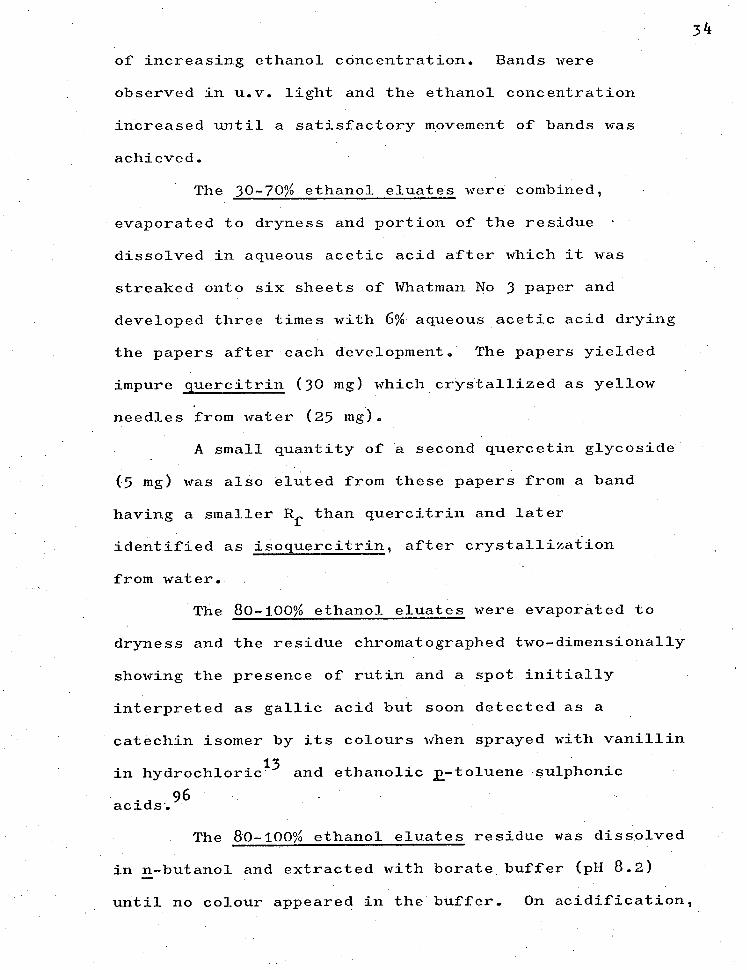
The 30-70% ethanol eluates were combined,
evaporated to dryness and portion of the residue •
dissolved in aqueous acetic acid after which it was
streaked onto six sheets of Whatman No 3 paper and
developed three times with 6% aqueous acetic acid drying
the papers after each development. The papers yielded
impure quercitrin (30 mg) which crystallized as yellow
needles from water (25 mg).
A small quantity of a second quercetin glycoside
(5 mg) was also eluted from these papers from a band
having a smaller Rf than quercitrin and later
identified as isoquercitrin, after crystallization
from water.
The 80-100% ethanol eluates were evaporated to
dryness and the residue chromatographed two-dimensionally
showing the presence of rutin and a spot initially
interpreted as gallic acid but soon detected as a
catechin isomer by its colours when sprayed with vanillin
in hydrochloric13. and ethanolic 2 .-to1uene sulphonic
acids•. 96
The 80-100% ethanol eluates residue was dissolved
in n-butanol and extracted with borate buffer (pH 8.2) —
until no colour appeared in the buffer. On acidification,

35
concentration and standing the buffer extract yielded a
precipitate of impure catechin isomer (90 mg) which
crystallized from dilute acetic acid (70 mg).
2.1.2.2. E. sieberi.
Preparative paper chromatography of the
bicarbonate extractives (III) using the descending method
in 6% aqueous acetic acid separated the glycosides
(20 mg) from other polyphenols with a minimum of hydrolysis.
Dissolved in methanol the mixture of glycosides
was spotted onto sheets of Whatman No 1 paper and the
sheets developed in BAW (415) in the ascending direction
with the chromatograms being dried and re-developed in
the same solvent a second time. The same papers were
then developed in the second dimension using 15% aqueous
acetic acid as developing solvent and again the
development was repeated.
In this way the separation of the glycosides
shown in .Chart 7 was achieved.
Spots with corresponding Rf values were eluted
with ethanol containing 30% water and the eluate
evaporated and spotted on Whatman No 1 paper for final
purification by two-dimensional development in the
same solvents as above.
Final elution and evaporation yielded glycosides
which after crystallization were identified as quercitrin
(4 mg), isoquercitrin (4 mg), hyperin (3 mg) and
cannablscitrin (3 mg).
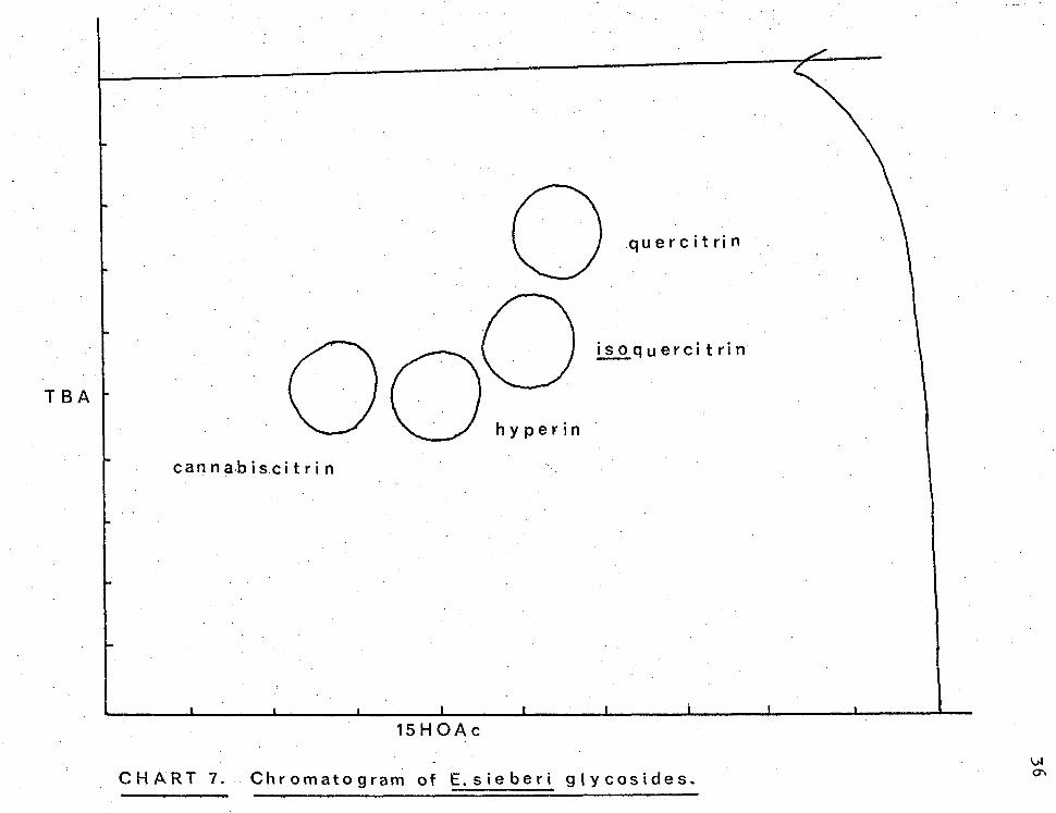
0 quercitrin
isoquercitrin
can nabiscitrin
TBA
15HOAc
CHART 7. Chromatogram of Esieberi glycosides.

The butanol extract obtained from Ste (XII) was
extracted five times with borate buffer of pH 8 and
then by 5% aqueous sodium bicarbonate solution.
Chromatographic survey of the neutralised bicarbonate
solution which had been re-extracted into butanol showed
the probable presence of quercetin, myricetin as well
as gallic, gentisic and ellagic acids (Chart 8, overleaf).
After evaporation under vacuum the crude mixture
shown in Chart 8 (350 mg) was dissolved in methanol,
applied to a Sephadex LH 20 column and was allowed to be
adsorbed onto the gel before elution began using ethanol
as eluent. Fractions were monitored on thin layer
chromatoplates using TEF (541) as solvent.
After bulking of like fractions the separation
of compounds obtained is shown in Chart_24, page 39.
- The compounds isolated from the Sephadex column
followed by preparative t.l.c. and crystallization
are listed in Table 3.
Fractions.
TABLE 3.
Crystallizing solvent. Compound isolated.
1-14 galliC acid (50 mg) methanol.
15-30 quercetin (60 mg) aqueous ethanol.
31-50 gentisic acid (30 mg) methanol.
51-200 quercetin (23 mg) aqueous ethanol.
201-240 myricetin (18 mg) aqueous ethanol.
241-300 ellagic acid (95 mg) aqueous ethanol.
37

gent isic acid
quercetin
gallic acid SAW 4:1:5
myricetin
ellagic acid
A
15H0Ac
CHART 8. Chromatogram of n-butanol extract of neut. NaHCO3 so tut ion.

SEPHADEX LH 20 COLUMN
TLC toluene :ethyl acetate: formic acid
gentisic acid SILICA 5 1
gall ic acid
quercet in
0 ° 0 0
0 0 0
myricetin el lagj. c acid.
ellagic acid and polymetic material
. P P 0 11) CI)
TEF Rf
fractions 1 -14 15
30 37-50 51 200 201 240 241-300
CHART 9. SE PHADE X 'COLUMN ELUATES CHRO MATOPL ATE.

An off-white precipitate (350 mg) settled in the
ethyl acetate extract (VII) of the hydrolysed solution
which had been left to stand in the refrigerator.
Paper chromatography showed this precipitate to
be in the main a mixture of two components, the minor
component being ellagic acid and the major component
having colours in u.v. light similar to those of
Hillis' unknown A48
although not having identical Rf
values, possibly because of slightly different solvent
systems.
A chromatoplate of silica gel to which the
mixture (125 mg) had been applied was developed with the
15HOAC solvent system. In this way unknown A was
separated from ellagic acid and crystallized from
aqueous ethanol to give a pure compound (60 mg) which
has been identified as 4-0-monomethya ellagic acid.
The ethyl acetate extract remaining after removal
of the above precipitate was paper chromatographed and
showed in addition to the above . compounds the presence
of hydroxybenzoic acids and a spot with properties
similar to that of Hillis' unknown B.48
Repeated extraction of the ethyl acetate solution
with 5% aqueous sodium bicarbonate with subsequent
neutralisation of the bicarbonate solution showed by
two-dimensional chromatograms that the unknown B compound
had been extracted by the bicarbonate extraction.
Re-extraction of the neutralised bicarbonate
40

solution with ethyl acetate and. evaporation was
followed by application of the mixture (150 mg) to a.
silica plate developed With BPF (.3:9 -:5). The top band
of the plate contained impure unknown B.
- After elution and taking to dryness, the
component in the top band (k5 mg) was applied .to sheets
of Whatman.No 3 MM paper which were developed with 15%
aqueous acetic acid, dried and re-developed with the
same solvent until the characteristic diffuse mauve bands
of Unknown B were resolved.
Elution of these bands with aqueous ethanol
and evaporation gave a residue which crystallized from
aqueous ethanol, to give a compound (28 mg) with
chromatographic properties similar to those of unknown B.
Whatman cellulose powder was formed into a small
column,the unknown F mixture obtained by trituration
of an ether precipitate was, applied in 15%'
aqueous acetic acid and this solvent used in column
development. Fractions collected showed on paper
chromatograms that separation had still not been achieved.
Finally a.portion (150 mg) of the unknown F.
mixture was applied to a silica .chromatoplate - using CEF
as developing solvent. Paper chromatogram Monitoring
Showed there had. notbeen a clean separation but there
seemed a greater concentration of - unknown F in two bands
which were elUted,evaporated and re-run - several times.

4 2
In this way sufficient unknown F (16 mg)
was obtained after three re-crystallizations to obtain
spectral analyses.
2.1.2.3. E. coccifera.
Roman numerals used in the fifillOwini-pararaPhs
refer to Charts 5,6:onpages 21,22.
The ether solution remaining after extraction
with bicarbonate (VI) was shown by paper chromatography
to contain a mixture of unknown D, flavonols and
ellagic acid.
The unknown D mixture spots on paper did not
give red colours when sprayed with sodium borohydride and
subsequently exposed to acid vapour33
as the E. sieberi
flavanones had done. This prompted the isolation of the
E. coccifera unknown D mixture.
Analytical thin layer chromatography using
Camag silica gel as adsorbent, and chloroform; ethyl acetate;
formic acid (7:4:1, CEF) as solvent suggested that
column chromatography using the same adsorbent and
developing solvent might separate the unknown D mixture.
The ether after bicarbonate extraction was taken
to dryness (3 g) and this was dissolved in CEF (7:4:0.5),
placed on a silica column and developed and eluted with
the same solvent system. Fractions collected were —
monitored on analytical t.l.c. plates with CEF (7:4:1)
as developing solvent system.

Figure 4 summarises the separation after
fractions of similar composition had been joined.
Fraction A contained a mixture of apparently
two compounds fluorescing purple under u.v. light
(365 urn) and a minor component of higher Rf value
fluorescing blue under u.v. light. Preparative t.l.c.
(silica, CEF) then separated the blue component from the
purple components mentioned above.
The compounds in Fraction A which fluoresced
purple were separated into two spots on analytical
silica plates using . CEF as solvent.
Preparative t.l.c. using the same adsorbent and
solvent separated the purple-fluorescent materials into
( two bands which when eluted gave apigenin as the band
of lower Rf and what appeared to be another single
-compound of higher Rf .
In all, six solvent systems were used before
one of them, BPF, showed that in fact the apparently
single compound was a mixture of a major and minor
component.
Figure 5 shows the analytical t.1. c. separation
of this mixture using BPF, into components later
identified as (4), (5) and (6).
It now appeared that the purple fluorescing
spot in Fraction A was a mixture of three compounds,
one of lower Rf whose isolation has already been
mentioned (apigenin (4)) and a mixture of two presumably
43

Fig.4. SILICA COLUMN SEPARAHON of E.coccifera POLYPHENOLS.
10
SOLVENT C E F
8
0
c3) f i I I I I
A B C D E F 0 FRACTIONS
A. Unidentified compound and. Unknown D mixture.
B. Unknown D mixture and kaempferol
C. Kaempferol and quercetin
D. Kaempferol, quercetin and. myricet5_n_
E. Querceti-n, nwricetin
F. Quercetin, myricetin
G. Traces of quercetin, myricetim and. ellagic acid.
H. "ellagic acid.

HO
(5) 01-1
( )
E. coccifera FLAVONES.
Fig. 5. TLC SEPARATION of FLAVONE MIXTURE
USING CEF and BPF SOLVENTS.
Rf
0.74 0.72
0.67
R f
0.73
0.69 4 7
CEF BPF CHCI:EtOACHCOOH C
6 H IC
5 H5 N:HCOOH
3 6 7 : 4 : 1 36 : 9 : 5
closely related compounds of higher Rf .
Repeated preparative t.l.c. on silica using
BPF and involving the procedure of developing, drying
and re-developing, separated the major component of
the mixture. Several crystallizations from ethanol gave
a chromatographically pure compound, identified on
evidence .recorded in the discussion section as a rare
C-methylflavone, found only once before in nature,
sideroxylin or 4',5-dihydroxy-7-methoxy-6,8-dimethyl
flavone (25 mg) (6).
The minor component (5) in this mixture was
present in too small an amount to enable isolation in
sufficient quantity for complete spectral analysis.
However enough material (0.1 mg) was obtained by
eluting spots from six analytical silica chromatoplates
developed three times with BPF for u.v. spectral
analysis, discussed in Chapter 4 1 page 150.
Fraction C from the column was shown by two-
dimensional paper chromatography to be a mixture mainly
of kaempferol and quercetin. Kaempferol was isolated
using preparative silica t.l.c. plates with MCP as
developing solvent; kaempferol (20 mg) crystallized
from aqueous ethanol.
An ether extract of the original unhydrolysed
aqueous methanolic solution (I) contained four glycosides.
Quercitrin, isoquercitrin and hyperin were
identified using known markers on paper chromatograms.
46

The remaining glycoside, different from others
previously found in this work, was isolated by
preparative paper chromatography by repeated
two-dimensional development using first 6H0Ac and then
BAW (612) as developing solvents.
In this way sufficient of tliis glycoside was
obtained (2 mg) to identify it as afzelin.
47

CHAPTER 3.
48
IDENTIFICATION - EXPERIMENTAL.

CONTENTS.
3.0. SPECTRAL DETERMINATIONS.
3.0.1. U.v. SPECTRA.
3.0.2. N.m.r. SPECTRA.
3.0.3. I.r. SPECTRA.
3.0.4. MASS SPECTRA.
3.1. SPECTRA.
3.1.1. POLYPHENOLS OF KNOWN STRUCTURE.
3.1.2. POLYPHENOLS OF UNKNOWN STRUCTURE
(HILLIS' UNKNOWNS).
3.2. DERIVATIVES.
3.2.1. TRIMETHYLSILYLATION.
3.2.2. METHYLATION.
3.2.3. TRIDEUTERIOMETHYLATION.
3.2.4. ACETYLATION.
3.2.5. PEROXIDE OXIDATION OF FLAVONOL-3
GLYCOSIDES.
3.2.6. ACID HYDROLYSIS OF GLYCOSIDES.
3.3. ROTATIONS, ANALYSES, MELTING POINTS.
3.4. EXPERIMENTAL DATA.
POLYPHENOLS OF KNOWN STRUCTURE.
3.4.2. POLYPHENOLS OF UNKNOWN STRUCTURE
(HILLIS' UNKNOWNS).
49
Paze.
50
50
51
51
52
52
52
66
100
100
101
101
102
102
103
103
104
104
114

50
CHAPTER 3.
3.0. SPECTRAL DETERMINATIONS.
3.0.1. U.v. SPECTRA.
3.0.1.1. Stock diagnostic reagents used throughout this
75b work and prepared according to the methods of Mabry et al.
were sodium methoxide (Na0Me), aluminium (III) chloride
(A1C13), hydrochloric• acid (HC1), sodium acetate (Na0Ac)
and boric acid (H3B0
3)0 The reagents above were applied
and spectra run according to the procedures given in
detail by the same authors.
3.0.1.2. U.v. spectra were run on a Hitachi Perkin-Elmer
124 double beam spectrophotometer or on a Perkin-Elmer 400
spectracord in methanol alone or in methanol with the
reagents given above; stock solutions of the polyphenols
being prepared in either of the following ways: 75b
3.0.1.3. The solutions used were made from re-crystallized
compound (0.1 mg) in Analar methanol (10 ml) and the
concentration adjusted so that maximum absorbance
between 240-420 nm occurred between 0.6 - 0.8. When log E
values were obtained the concentration was accurately
determined.
3.0.1.4. Alternatively, the polyphenols were purified
by one-or two-dimensional paper chromatography. Spots,
when sufficiently discrete, were viewed under u.v. light,
their zones cut out of the chromatogram and eluted with
Analar methanol. The compound was dried by evaporation,

• 51
re-dissolved in a minimum of methanol and its spectrum
was recorded as above.
Since methanol may elute u.v.-absorbing
compounds from the paper itself, when spectra were run
in this way a reference solution was made by extracting
a piece of blank chromatogram which had been developed
in the same way as the chromatogram containing the
polyphenol(s).
3.0.2. N.m.r. SPECTRA.
N.m.r. spectra were recorded on a Jeolco JNM4H-100
spectrometer.at 100 MHz or on a Varian A60 spectrometer
at 60 MHz for 5% solutions in DMSO-d6 with HMDS or TMS
as external standard or in the case of the 60 MHz spectra,
as internal standard, or using C5D5N or CDC13
or
mixtures of the first two and the last, with TMS as
internal standard. In the case of spectra run to
analyse trimethylsilyl ethers of polyphenols a drop each
of hexamethyldisilazane and trimethylchlorosilane was 75c
added to ensure anhydrous conditions. It was
• standard procedure to run additional spectra after
addition of deuterium oxide for detection of hydroxyl
groups.
3.0.3. I.r. SPECTRA.
These were determined in Nujol and
hexachlorobutadiene mulls or using KBr discs, on a
Perkin-Elmer 221 spectrophotometer.

52
3.0.4. MASS SPECTRA.
Mass spectra were measured on an Ae.I.MS9
instrument at 70 eV. or on an EA1 QUAD 300 quadrupole
mass spectrometer.
3.1. SPECTRA.
Spectra are presented in two sections, first
(3.2.1.), those referring to polyphenols of known
structure and the second (3.2.2.), those of polyphenols •
of unknown structure (Hillis' unknowns). 48
The spectra given are referred to in discussion
in Chapter 4.
3.1.1. SPECTRA OF POLYPHENOLS OF KNOWN STRUCTURE.
3.1.1.1. Hydroxybenzoic acids.
Chart 10. N.m.r. spectra of gallic, gentisic
and protocatechuic acids.
11. Mass spectra.
3.1.1.2. Flavonols.
Chart 12. N.m.r. spectrum of quercetin.
13. N.m.r. spectrum of quercetin
compared with that of myricetin.
14. Mass spectra.
15. Mass fragmentation.

53
3.1.1.3. Ellagic acid.
Chart 16. Mass spectrum and fragmentation.
17. N.m.r. spectrum (C55
N/Et0H).
18. N.m.r. spectrum of TMS ether
(). CDC13
3.1.1.40 (+)-Catechin.
Chart 19. N.m.r. spectrum (DMSO-d6
20. N.m.r. spectrum (C55
N).
21. Mass fragmentation.

47-
6
HO 2H
l 11 TUT-17111 111111111T -111T11111111- 1111
6
COOH 1•72
6 OH H-Z
*\\ HO 3
2H
GENTISIC ACID
11111111111
H-3
1H
sir
11 1 1 11 li1111
8 7 ilit
2H
H-2,6
7 6 5
CHART 10. N.M.R. SPECTRA of HYDROXYBENZOIC ACIDS .
7
( p.p.m.)
5
PROTOCATECHUIC ACID
111111111111 ■ 11
7.29
OH
GALLIC ACID
COOH
2 x OH
11.04

GENTISIC ACID
PROTOCATECHUIC ACID GALLIC ACID
100
110
137
136 125
108
(m+) .153 170
110
rn/e CHART ii. MASS SPECTRA of HYDROXYBENZOIC ACIDS.

(Scale 6} . I .... 6 P.P.m. 6 9 8
tIli tit I. I IIII.t.,,,,,„. 7
QUERCETIN
6
S.
DMSOd (int.TMS) 6
CHART 12. N.M.R. SPECTRUM of qUERCETIN.

8 H
6 OH 0
H6' H2' OH
5
2'
QUERCETIN
H2' H6'
MYRICETIN
OH <-0H
HO. ° 0H 0 1 WOH
OH 0
H8 H6
S 8
7
CHART 13. N.M.R. SPECTRA of FLAVONOLS.

318
302
100—
80
60 oh
40
209 (base peak) MYRICETIN
153
iS1 137
125
20
0
100-
80
60 Oh
40 1
20
.145
(base peak)
.137
.128
.142
.302
.153
256 274
284
QUERCETIN
100 140 180 220 260 CHART 14. MASS SPECTRA of E.sieberi FLAVONOLS.
300

OH 0
HO OH
OH 0
OH
OH R 1 R2
m/e 137,153
HO
OH 0+ m/e 137
R1 =1-1 7 QUERCETIN R?:OH, MYRICETIN
HO
R1R2 HO OH
OH 64. OH R1 R 2
m/e 153 0+
m/e 137,153
17 - CO
M -18 142 7 150 2
-CO -CO
m/e 109,125 m/e 109
m/e 109,125
.Q1-1ART 15. MASS FRAGMENTATION of E.sieberi FLAVONOLS.

100
( base peak ) 202
(M4 ) ELLAGIC ACID
80
-0 -0
CHO —200
— CO —CO —CO — CHO
— CHO
60
40
20
27o 266
162 189 190
218 2f7 228 .246
273
0 me 100 120 140 160 180 200 220 240 260 280•
CHART 16. MASS SPECTRUM and FRAGMENTATION of ELLAGIC ACID
300

1111 11•111 1 1II I111111i I
r\vPi\vw,pl,tytivAftchtv\■vjLeAra.",
8 1. 3 2 7 6
OH
ELLAGIC ACID
P..C5D5N
OH cHa
8-01
P
A
Yi Vp,AA) '1.'44014.0.,0\vni-N,yvovi\ANNI\AA,^v
P.P.rn.)
Et0H
CHART 17. N.M R. SPECTRUM of ELLAG1C ACID.

7.63
2H
1 III I Il I I I IlfIlI1
ELLAGIC ACID TMS ETHER
CDCC
NAMe )401))1 feltOlverNtgYs.thrtAithiltellAWV,14/\AVVW.15A- leVAAA''\'11\i"PAm.N"ii
(.6p.P.rni
9
7 6 5 . 4
3
1
CHART 18. N.M.R. SPECTRUM of ELLAGIC ACID TMS ETHER.

(-0—Catechin
21-1 SOH
701-1
4 .0H 30H 3.0H
H2/ H5/
DMSOd6
H8 H6 H2 H3 •
2,xH4
9 CHART 19.
6 5 7 N.M.R. SPECTRUM of (±)-CATECH1N. (DmSo-d 6 )

6.76 6.64 8+1 6-H
113 6 7
illirliiiii.rf,
a-ax
-4-"Lt-J • 9 3.4 ax OH
-44
'leg
P .Ct%N
3-H
_L._ 22Hz
i3.4eq
1" -13.4q-ax
AB
3.70 3.31
Heq 4Hq. ax
5.20 241
1H
2H
( p.p.m.)
OH
(4)-CATECHIN
8 7 6 5 4 3 2
C I-1AR T 20. N.M.R. SPECTRUM of (+)--CAT EC El I N (C5D5N)

OH •> OH
OH W . = m/e 290
— CHO
OH
m/e 123
rDA
HO HO
HO OH
CH 2 OH m/e 139
CH 2 OH
m/e 138
CH OH + 2
m/e 139
CHART. 21. MASS FRAGMENTATION of (+)-CATECHIN.
66
3.1.2. SPECTRA OF POLYPHENOLS OF UNKNOWN STRUCTURE.
3.1.2.1. Unknown D48
- Flavanones.
Chart 22. N.m.r. spectra and configuration.
23. I.r. spectral data.
N.m.r. spectral data.
25. N.m.r. spectrum of pinocembrin.
26. N.m.r. spectrum of alpinetin.
27. N.m.r. spectrum of 0,0-dimethyl-
pinocembrin.
28. Mass spectra.
29. Mass fragmentation.
3.1.2.2. Unknown D48
- Flavones.
Chart 30. N.m.r. spectrum of apigenin.
31. Mass spectrum of apigenin.
32. . Mass fragmentation of apigenin.
33. Mass spectrum of sideroxylin.
34. Mass fragmentation of sideroxylin.
35. N.m.r. spectrum of sideroxylin
(Dmsod6 /CDC13).
36. N.m.r. spectrum of sideroxylin
(c5D5N/cDc1
3).
37. N.m.r. spectrum of sideroxylin mixture.
38. N.m.r. spectrum of TMS ether of
sideroxylin mixture.
39. Mass fragmentation of second
C-methyl flavone.
40. Mass spectrum of sideroxylin mixture.

3.1.2.3. Unknown A 48 - 4-0-methyl ellagic acid. 67
Chart 41. N.m.r. spectrum.
42. N.m.r. spectrum of methylated
unknown A (CDC13).
43. N.m.r. spectrum of methylated
unknown A (C6D6 .
44. U.v. spectra of unknown A and
ellagic acid in methanol.
45. U.v. spectra of the same in methanol
plus solid sodium acetate.
46. U.v. spectra of the same in methanol
plus sodium methoxide solution.
47. Mass spectrum and fragmentation
of unknown A.
3.1.2.4. Unknown B48
- (1,5-dimethy1-2,6-bis(3,4 9 5-
Chart 48.
119.
trihydroxyphenyl) furo ,[1 1 5-c] furan.).
U.v. spectrum of unknown B in
methanol.
U.v. spectrum in methanol plus
sodium methoxide solution.
Mass spectrum.
50. N.m.r. spectrum.
3.1.2.5. Unknown F48
- (a tetrahydroxydibenzofuran-
dicarboxylic acid).
Chart 51. N.m.r. spectrum.
52. I.r. spectra of ellagic •acid and
unknown F.
53. Mass spectrum.

H 3ax
H 3eq 3eq H 3ax
NMR SPECTRA and CONFIGURATION of E.sieberi FLAVANONES.
42-30 . -1.- 16 Cos2 154 ° 4.. 13 . 0 Hz
JH2-3eq 1000S2 57° 3.0Hz
-J 4 12 Hz J 2.5 Hz OBSERVED ..F.13 Hz
CHART 22.N.M.R. SPECTRA of E.sieberi .FLAVANONES and CONFIGURATION.
Pheq 2 eq
2S
H 2ax
H — H aX a X H H eq. ax
DIHEDRAL ANGLES
H 2a x
57 ° H3eq
154 °
H 3 ax

MET HOXYL CARBONYL 3000 1700 1600 1500 1300 1200 1100 , 1000 GOO 700 600 cm
PHENYL
Ph
Ph
3400 3200 HYDROXYL,
3600
CHART 23. I.R. SPECTRAL DATA for Esieberi FLAVANONES.

H2ax ALPINETIN PINOCEMBRIN
appm 5-90 11-20 7.79
HO
00 \H--"
12-48
H 3•07 1-1
3-60
H3 ) 2SO4
Acetone
CH2N2
(CH3)2SO
NMR SPECTRAL DATA for E.sieberi FLAVANONES.
3.78 Me0
7-40 310
Me()
5,7-DIMETHOXYFLAVANONE
CHART 24. N.M.R. SPECTRAL DATA for •E.sieberi FLAVANONES•

11 1 1 11 T 11- 1- 1 I I 1111- 11111 1 - 1 I 11
HO
8 7 6 5 4
Soo 00 1300 too 400 J700 ?OD 200 Ti 111 11
1120
7.'0H
vovr,r0401
tiltlit111111i tilt] 111111111111111111111111111111111111 1 1 3
iiiimimil
12.48
5-0H
5H
P1NOCEMBRIN
CHART 25 , N.M.R. SPECTRUM of PINOCEMBRIN.

7-0H
11.20
• P.Prn . 4 3 2 1
lit
8 7 I 1 1 1 1 1
till
So goo doo • foo Woo .foo • .20o 100
1 111111j111111T -1111 III 1- 11IIT•11-11-11 - 11111 -11111- TATT1- 111111{1 -1- TTi111111 . IITI11 1 T1 111111 1 11i
ALPINETIN
X 1i I ) \ I \ I I
jut 'reittiMpvPhereky
(11/1 41̀ !
144°41/MANIVAlftir4413*1 *410401(0/1 Nti V\MoWel'IN*A1
3H
HS H6
5H
OMe 5
Ph H
DMS0d 6
2H
X 1
wirkfirwov4,4
H2
A0\441444* ; ‘f l i 14 ,04
rrvh
YTY E-1!T'
CHART 26. N.M.R. SPECTRUM of ALPINETIN.
600 foe 1111111
400 Joe I
,
200 II III
Joe IlIjil
I r Il l I
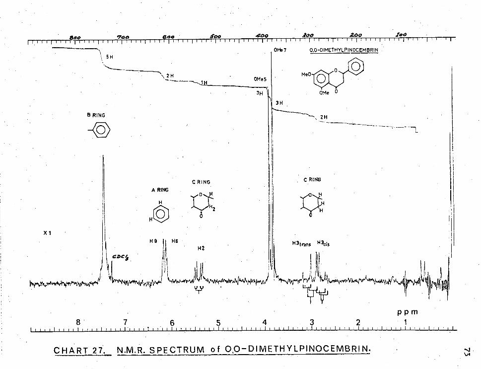
OMe7 0.0-DIMETHYLPINOCEMBRIN + 5 H ‘,
\ 2H OMe5
3H 3H
2H
C RING
A RING
H
X 1
CDC/
tkrovo4Osoryle"*.'$0wHei
H 1 H6 •
•
PI ?Ilk) Veff4,1' NItrA4k1 4‘1V4) v
1 i i
H2 1 H3 trans H3cis
III
1 i! 1
ilV1
41,Wkr-ivs**446A11400V) '"N;mtniwnrAti 104/0,ymmA1/41v4^v.,44nivekAfry kh
YTY
B RING
C RING
P P m 8 7 6 5 4 3 2 1
CHART 27. N.M.R. SPECTRUM of 0,0—DIMETHYLPINOCEMBRI N.

124 152
266'
.256 179
PINOCEMBRIN
104
100
0
166
270 ALPINETtN
(M+ )
193
138 •
100
0/0
-104
100
269
X8o 0,0-DIMETHYL-PINOCEMBRIN
2.94.
162 233
0 rve '100 120 140 160 180 200 220 240 260 280 300
CHART 28. MASS SPECTRA of E.siebefi FLAVANONES.

M+.= 256 ,R R 2 = H =A
M4-= 270 , =H, R2 =Me =B
M+.= 284. R 1 = Me , R2= Me C
a
RI
R20 R20 OH R20 OH
A , m/e =152 m/e=104 m/e = B, 166
..c., 180 N.00 ). 124,138,152
CHART29. FRAGMENTATION of E.sieberi FLAVANONES,
179 , m/e = 255
193 269 207 283

2.H DMSOd6
/ int.TMS
H
1
OH
H-6 H-2',6'
H-3
. H-8
H-35'
J =9 Hz J =3Hz
SPPm 8 7 6 D'A ':Nro ll°.1r14"1714 .
APIGENIN
CHART 3O. N.M.R. SPECTRUM of APIGENIN .

APIGENIN 100 96 (base peak)
80
.721 118
.2o7
60
0/0
40
124
153
152 20 270 _791
242
193 176178
0 90 100 120
CHART 31. 140 160 180 200 220 240 260 280
MASS SPECTRUM,

- CO HO --
0
OH m/e = 11 8
m/e = 96
4CH
HO
C=0 HON
OH m/e = 124
HO ONNI
01-4 0 OH
M 4- m/e = 270 m/e = 242 m/2e = 121
rDA
CHART32.MASS FRAGMENTATION of API GENIN.

SIDEROXYLI N 80 -
251
60-
12%
3/2
170 164
196
142 .193
207' 135
239
292
93
297
105
0 mie 100 120
CHART 33.
140 160 180 200 220 MASS SPECTRUM.
267
260 240
20-
280 300

M-28
rDA m/e= 84
m/2e=142
Me
ge0
Me Cc) A
m/e= 194 -Me
A-15 -Me m/e=179
-CO \ A- 2 x15 m/e =164
A-15-28 we =151
OH 13 m/e =118
-CH
B - 13 m/e = 105
-C
B-13-12 m/e = 93
.(C)H Me I
-CO
tie n
0 Me0 ON Me0/-
Z (r1 Me
Me'* OH
M* m/e = 312
/-Me
OH
m/e=121
Me() /N/1 Me N
OH 0 • M - 15 m/e= 297
-Me
M- 2 x15 m/e = 282
-Me
M- 3 x15 m/e= 267
-CO
M- 3 x15-2 8 m/e= 239
-Me I
A- 2x15-28 m/e= 136
- Me A- 3 15-28
•CHART34, MASS FRAGMENTATION of SIDE R OX YL.. 1 N.

' T 1 1- I 1 III V
7 5 9 a 4 3
SI DEROXYL TiN
5-ow zw
12.96
ZH
8-C1-1 6•C14 2.39 2.21
CACI
114
CHART 35. N.M.R. SPECTRUM of SIDE ROXY LIN. (DMS0-d6 /CDC1 3)

1111 11111
OH
CDC/A 2A4
9 8 7 6 5 4 3 2
III I I I I I 1 111111111111
?
SIDEROXYLIN
11111111111111111
211
P • Ct%N
114
CHART 36. N.M.R. SPECTRUM of S1DEROXYLIN. (C 5D5N/CDCI 3)

7 6 3. 2
2.50
DMS0d6 I 6- CH3
13•4o
5-0H
CHART 37.N.M.R. SPECTRUM of MIXTURE of SIDEROXYLIN C-METHYLFLAVONE.

!MIMI rill I f Ifir Illf 711 lillr1(1111 liII 111111111
9 7 6 5 4 3 2
180
2.21
6-C1
•72.
H -3:5'
CDC
607 1-4-3
J•13.6
OrIWAV'e,44tit Wviolvi?'\vm ( 6p p m
12.64
5-0H
CHART38.N.M.R. SPECTRUM of TMS ETHER of SIDEROXY LI N +C-METHYLFLAVONE.

OH
Me°
CH
0
H C O A, .
m/e=179
/CO
A728 Ar15 m/e =151 rrve =164
Me
AT 15 -28 m/e=136
/- Me
B, m/e = 118
- CH
B,- 13 m/e = 105
B,- 13 - 12 m/e= 93
Me0 Me
A,- 2x15 -28 m/e =121
CHART 39. MASS FRAGMENTATION of SECOND C-METHYLFLAVONE.
•13H \I-, I /
M e 0
Me
rDA OH M, - 28
m/e= 270 m/2e = 135
Me 0 Me
OH M;*
m/e= 298
/-me
OH
m/e = 121
-CO
M- 2 x15-28 m/e =2/.0
OH 0 M7-15
m/e=283
-Me
M,- 2x15 m/e= 268

.27o
280
300
312
(frfif 298
297
282
283
SIDEROXYL I N MIXTURE
93
207
/93 /94
Iill! "5. 160 180 200 220
MASS SPECTRUM.
268 .24,0
240 260
80 -
60
4 0
20-
105
.Z42
0 rWe 100 120
CHART 40.
118
121
135
136
.951
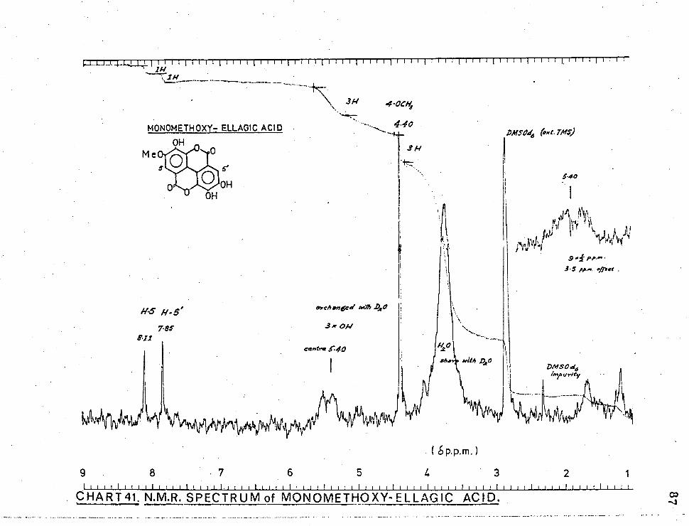
MONOMETHOXY— ELLAG1C ACID OH
Dm5016 (exc. TMS)
Me
• p.p.m.)
4 3 9 5 8 7 6 2 1
5 s' OH
OH
540
t
TIT
CHART41. N.M.R. SPECTRUM of MONOMETHOXY-ELLAGIC ACID.

OMe Me0 0 ■,0
OMe OM e
0
111111 111 1
3.36
OMe ls
( SPP.rn
9 8
7
6
5
4
3
2 1111111•1111111111111111 1111111111111 1 1 1 1 111111111J11 . 111•111111 I 111111 I
CHART 42.N.M.R. SPECTRUM of METHYLATED UNKNOWN A.
I 111 1.j 1111 1 11 1 11 111 -111111ErTIF1111 - 111111 - 1, • - 1 T I LITTIT 1- 11'11111
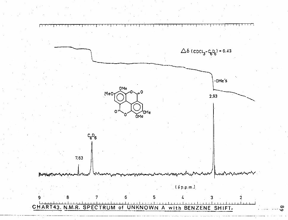
.L6 CDCI. -C.6D6) = 0.43
7
5
4
3
CHART43. N.M.R. SPECTRUM of UNKNOWN A with BENZENE SHIFT.

Methanol. ellagic acid
--4— 4-monomethyl ellagic acid
OD
220 250 300 350 400 X nm CHART 44 U.V. SPECTRA of UNKNOWN A and ELLAGIC ACID. .0
<0
-

250 • 350 300 220
Me0H/Na0Ac
e.a.
m.m. e.a.
400 nm CHART 45. U.V. SPECTRA of UNKNOWN A and ELLAGIC ACID. (Na0Ac)

Met hana1lNa0M
e, a. m.m. e.a.
220 250 300 350 400 X nm CHART 46; U.V. SPECTRA of UNKNOWN A and EL LAGIC ACID. (Na0Me)

MONOMETHOXY-ELLAGIC ACID ( base peak) ( M + *) 3.16
-CO -CO -CO -CO -CH
3o1
161 160
.189 218
217
245-
273
100
80
60
40
20
0 mie 100 120 140 160 180 200 220 240 260 280 300
CHART 47. MASS SPECTRUM and FRAGMENTATION of ELLAGIC ACID ETHER.
320

Ale Oil
Meazifraavie ir;:merei.
/146011fra0/Vie after to min,
HO
HO
0.5 OH / I 0
/C / HO C I C OH I CH I H2 3 H
OH
OD
0.4
a? oo 250 aoo CHAR T 48.11,V. SPECTRA f UNKNQ1NN B.

100
80
60
°A 40
20
180 200 220 240 260 280 300 320 340 360 . 380390 0
rrve 160
( base peal)(m+)
CHART 49. MASS SPECTRUM of UNKNOWN B.

11111111,1 1 11 111 . 111111111IITTTITT11111 { -111111- 1 - 111- 11.1111- 11--1111111111
(SP.P.M.) 7
5
4
2
OHART 50. N.M.R. SPECTRUM of UNKNOWN B.

DMSO-d6 (ext.TMS)
3.92
6H g x 2 width
2H UNKNOWN F
Removed with D20
'`YMPoebiAtA14
( 6 P.P.m.) 7
6
5 4
3
2
II 1 1111111111.
CHART 51. N.M.R. SPECTRUM of UNKNOWN F.
8
I.
r\tv
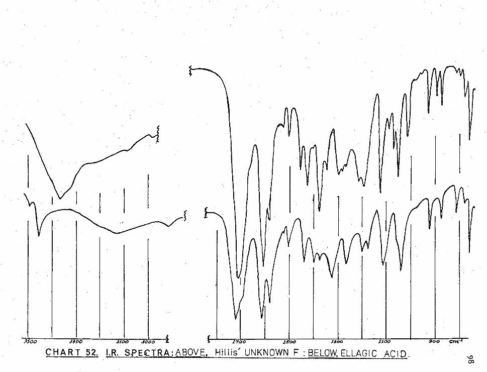
3Soo 3390 31190 3000 & 1700 2500 1300 1100 900 Cin
CHART 52. I.R. SPECTRA:ABOVE, Hillis' UNKNOWN F : BELOW, ELLAGIC ACID.

100 150
80
276, 60 /77
/3.2
40
20
(base) 276
(peak)
(M +. ) 320
232 ..50
m/e 0100 .120 140 160 180 200 220
CHART 53. MASS SPECTRUM of UNKNOWN F.
Zoo
240 260 280 300 320

100
3.2. DERIVATIVES.
Derivatives of various polyphenols have been
made during the course of this study using one or other
of the following procedures.
3.2.1. TRIMETHYLSILYLATION. 75c
A suitable quantity of the polyphenol (20 mg)
was dissolved in pyridine (1 ml) (dried by storing over
KOH pellets) in a 50 ml round-bottomed quickfit flask
and hexamethyldisilizane (HMDS) (0.25 ml) and
trimethylchlorosilane (TMCS) (0.25 ml) were added and
the flask stopped and allowed to stand for 10-20 min.
at room temperature. In the •case of 5-hydroxy
flavonoids the silylating solution was allowed to stand
overnight before further work-up. Solvent and excess
reagents having been evaporated under vacuum and the
dry residue extracted with Analar chloroform with
subsequent filtration of salts, the clear CC1 4 solution
was taken to dryness, re-dissolved in a quantity of
solvent suitable for n.m.r. analysis with addition of
drop of each of the silylating reagents to ensure
• anhydrous conditions.
The original compound was regenerated unchanged
in most instances by allowing the trimethylsilyl ether
to stand overnight in 20% aqueous methanol with a drop
of glacial acetic acid. •

1 01
3.2.2. METHYLATION.
Diazomethane was generated in a fume hood either
from nitrosomethylurea prepared according to the method
of Arndt2 or by distillation from an ether solution
of 2-tolylsu1phonylmethylnitrosamide.18 The
diazomethane ether solution was added several times to
a methanolic solution, or in some cases suspension,
of the polyphenol over a period of several days, the
solution being cooled to minimise the loss of
diazomethane by evaporation.
Methylation of pinocembrin was carried out with
dimethyl sulphate according to the method of Thomas. 104
3.2.3. TRIDEUTERIOMETHYLATION.
Trideuteriomethylation of phenolic hydroxyls in
certain polyphenols was attempted using the method
devised by Bick et al.8 for alkaloid hydroxyls. A
diazomethane solution in light petrol dried over KOH
pellets was extracted once with an equal volume of
dimethylsulphoxide dried over molecular sieve, which
removed some 75% of the diazomethane and the sulphoxide-
diazomethane solution added to the polyphenol in dry
dimethylsulphoxide containing a few drops of heavy water.
The addition was repeated daily for several days.
This method of trideuteriomethylation achieved only
a limited success with phenolic hydroxyls. The 5-hydroxyl
of pinocembrin and the hydroxyls of unknown A were only
partially trideuteriomethylated and the yields in both
cases were poor.
102
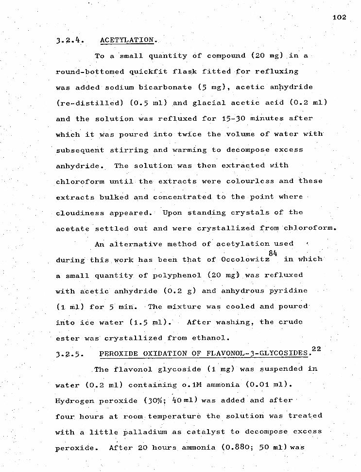
3.2. 4. ACETYLATION.
To a 'small quantity of compound (20 mg) in a
round-bottomed quickfit flask fitted for refluxing
was added sodium bicarbonate (5 mg) acetic anhydride
(re-distilled) (0.5 ml) and glacial acetic acid (0.2 ml)
and the solution was refluxed for 15-30 minutes after
which it was poured into twice the volume of water with
subsequent stirring and warming to decompose excess
anhydride. The solution was then extracted with
chloroform until the extracts were colourless and these
extracts bulked and concentrated to the point where
cloudiness appeared. Upon standing crystals of the
acetate settled out and were crystallized from chloroform.
An alternative method of acetylation used
84 during this work has been that of Occolowitz in which
a small quantity of polyphenol (20 mg) was refluxed
with acetic anhydride (0.2 g) and anhydrous pyridine
(1 ml) for 5 min. The mixture was cooled and poured
into ice water (1.5 ml). After washing, the crude
ester was crystallized from ethanol.
3.?.5. PEROXIDE OXIDATION OF FLAVONOL-3-GLYCOSIDES 22.
The flavonol glycoside (1 mg) was suspended in
water (0.2 ml) containing o.1M ammonia (0.01 ml).
Hydrogen peroxide (30%; 40 ml) was added and after
four hours at room temperature the solution was treated
with a little palladium as catalyst to decompose excess
peroxide. After 20 hours ammonia (0.880; 50 ml)was

103
added and the solution warmed for five minutes.in a
boiling water bath. The resulting solution was first
used directly for paper chromatographic analysis and
then extracted withether to separate the aglycone and
the concentrated ether solution and remaining aqueous
solution separately co-chromatographed with authentic
samples of appropriate compounds.
3.2.6. ACID HYDROLYSIS OF GLYCOSIDES.83
The glycoside (1 mg) was heated at 100 0C with
0.5M hydrochloric acid (0.5 ml) for 30 minutes and the
resultant solution examined by paper chromatography,
with solutions of authentic samples.
3. 3. ROTATIONS, ANALYSES AND MELTING POINTS.
Optical rotations of the flavanones, the
flavan-3-ol and the rotation of rutin (quercetin-3-
rhamnosylglucoside) were found using a Bellingham-
Stanley polarimeter on solutions of 2% or 5%
concentration depending on the quantity of material
available, in a cell of 10 cm length and volume 2 ml.
Microanalyses are by the Australian Microanalytical
Service, Melbourne, and the Australian National University.
Melting points are uncorrected.
Samples were dried at 0.05 mm Hg over phosphorus
pentoxide at temperatures specified in the text. All
evaporations were carried out under reduced pressure.

104 3.4. EXPERIMENTAL DATA.
In purely qualitative tests in dilute solution the
location of maxima in u.v. spectra is sufficient so
extinction values are not reported for every compound.
The data are given first for polyphenols of known
structure; second, for polyphenols called by Hillis unknowns.
3J .1. POLYPHENOLS OF KNOWN STRUCTURE.
3.4.1.1. Hydroxybenzoic acids.
3.4.1.1.1. Gallic acid (3,4,5-trihydroxybenzoic acid
Gallic acid had Rf
0.58 BAW (612), 0.65 in F,
0.42 in 6H0Ac. It crystallized from water in long white
needles, m.p. 240 0-42 0 (dec.) (lit 9c 225 °-250° dec.),
undepressed on admixture with an authentic sample;
1max210, 272 3 , in Me0H/Na0Me:370, in Me0H/A1C1 :308,
in Me0H/A1C1 3+ HC1:273, in Me0H/Na0Ac:261, in Me0H/Na0Ac
+ H3B0
3:286
nm;Vmax. 3457, 3349, 3256, 1698 (carboxyl C=0),
1610, 1536, 1247, 1025, 865, 765 cm -1 ; 6 (DMSO-d6 ) 7.29
(s, 2H, ArH), 10.0 (broad multiplet, 411, OH, CO 2H,
removed with D20); the mass spectrum is shown in Chart 11 1
page 55 . (Found: C, 49.0; H, 3.4 Cale. for C 7 H6 05 . C,
49,4; H, 3.6%).
3.4.1.1.2. Gentisic acid (2,5-dihydroxybenzoic acid).
Gentisic acid had Rf
0.82 in BAW (612), 0.86 in
BAW (415), 0.86 in F 0.62 in 6H0Ac, 0.66 in 15H0Ac. It
crystallized as needles from water, m.p. 197 0-200 0
69b 0 0 (lit. 199 - -200 - );
Na0Me:342, in MeOH/Na0Ac + H3 BO
3 :335 nm;V max 3290, 3130,
1660 (carboxyl C=0), 1610, 1585, 1270, 1230, 1175, 930,
Amax in Me0H 237 (sh) 335, in Me0H/

1 05
840, 785 cm-1 ;8(DMSO-d6 ) 7.08 (d, 1H, H3 , meta= 3 Hz)
7.28 (q, 1H, 14, jmeta= 3 Hz ; jortho 9.5 Hz), 7.55=
1H, 4, jmeta= 3 Hz), 11.04 (m, 3H, OH, CO2H, removed
with D 20); the mass spectrum is shown in Chart 11
page 55. (Found: C 54.4; H, 4.0. Calc. for C H6 04 • '
CI 5 4 -5; HI 3.9%).
3.4.1.1.3. Protocatechuic acid (3,4-dihydroxybenzoic
Protocatechuic acid had R 0.68 in BAW (612),
0.70 in BAW (415), 0.70 in F, 0.60 in 6H0Ac and 0.66 in
15H0Ac. It crystallized from water in off-white:_needles,
69a 00 m.p. 194 ° ° -196 (dec.) (lit. 195
...149e dec.); ) max in
MeOH: 259, 294 in Me0H/Na0Me: 276, 299, in Me0H/A1C1 • . 3 .
274, 388 (sh), in Me0H/A1C1 3+HC1: 261, 295, in Me0H/
Na0Ac: 251, 289, in Me0H/Na0Ac+H3 BO3'• 267, 302 nm; max
3327, 3182, 1672 (carboxyl C=0), 1605, 1590, 1522, 1290,
1127; 1090, 921, 820, 764 cm -1 ; (DMSO-d6 ), 7.16 (
1H,
, 7.69 (d, 1H, H_, 116' jortho Hz) = 9 5 jortho= 9 Hz), 7.73
(s, 1H, H2), 10.34 (multiplet, 3H, OH, COa H, removed with -
D2' 0)• the mass spectrum is shown in Chart1l i page 55.
(Found: C, 54.3; H, 4.1. Cale. for C 7H6 04 : C, 54.5;
H7 3.9%).
3.4.1.2. FLAVONOLS:
3.4.1.2.1. Quercetin (3'74'7577-tetrahydroxyflavon -
3-01).
Quercetin had Rf 0.64 in BAW (612); 0.40 in F,
0.56 in TBA and 0.03 in 15H0Ac. It crystallized from

106
dilute alcohol as yellow needles, m.p. 314 °-316 °
(lit.35h 316 0-318 0 ); the penta-acetate recrystallized from
methanol, m.p. 198 °-199 ° (lit.35h 200 0 ); quercetin had
Amax 255, 259 (sh) 300 (sh) 370, in Me0H/Na0Me: 250 (sh),
320 (dec.), in Me0H/A1C1 3 : 272, 304 (sh), 332, 458, in
Me0H/A1C1 3 +HC1: 265, 300 (sh), 360, 428, in Me0H/Na0Ac:
255 (sh), 274, 329, 390 (dec.), in Me0H/Na0Ac+H 3B03 :
260, 303 (sh), 388 nm: V max 3400, 3310, 1666 (chelated
C=0), 1610, 1560, 1270, 1170, 864, 794 cm-1 ;8(1)Ms0-d6 ),
6.22 (d, 1H, lics , jmeta= 3 Hz), 6.42 (d, 1H, Hz , jmeta
3 Hz), 6.90 (d, 1H, H5 ,,
jmeta= 3 Hz ' J ortho = 9 Hz), 7.80 (m ' 1H, 21-2' ), 9.06
(broad m, 2H, OH 314 removed with D20), 9.42 (m, 1H, _,"
OH removed with D20), 10.86 (m, 1H, OH removed with
D20), 12.60 (s, 1H, OH5' partially removed with D
2 0);
— for the penta-acetateS(CDC1 3 ), 6.15 (d, 1H, 116, jmeta=
3 Hz), 6.45 (d, 1H, Hz , jmeta= 3 Hz), 6.75 (d, 1H, H5 ,,
Jortho = 9 Hz), 7.55 (q, 1H,11, 3 Hz, —b" jmeta = jortho =
9 Hz) 7.72 (d, 1H, H J = 2" meta 3 Hz); mass spectrum m/e --
(rel. intensity) 302 (24), 152 (15), 153 (32), 137 (46).
(Found: C, 59.4; H, 3.3. Calc. for C 15H1007 : c, 59.6;
H, 3.3%).
3.4.1.2.2. Myricetin (3' ,5,5',7-pentahydroxy- flavon-3-401).
Myricetin had Rf 0.41 in BAW (612), 0.43 in BAW
(415), 0.31 in TBA, 0.00 in 6H0Ac and 0.01 in 15H0Ac.
It crystallized as yellow-green needles from aqueous
ortho 9 Hz), 7.62 (1, 1H, E6 ,, j=

1 07
alcohol, m.p. 354 °-357 ° (lit.7 51357 °-360 0 ), undepressed
on admixture with an authentic sample; Amax
in MeOH:
254, 270 (sh), 300 (sh), 372, in Me0H/Na0Me: 262 (sh),
284 (sh), .322,.420 (dec.), in Me0H/A1C1 3 : 270, 315 (sh),
450, in Me0H/A1C1 3+HC1: 266, 275 (sh), 307 (sh), 358 (sh),
in Me0H/Na0Ac: 270, 335 (dec.), in Me0H/Na0Ac+H3 BO
3'• 256,
304 (sh), 392 nm; max 3445 ' 3315' 1655 (chelated C=0),
1605, 1550, 1270, 1170, 858 (isolated ArH) cm -1 : 6 (DMS0-d6),
6.25 (d, 1H, E6 , jmeta= 2.5 Hz), 6.48 (d, 1H, 118 , jmeta =
2.5 Hz), 7.35 (s, 2H, H2 , 6 ,), 9.35 (m, 1H, 0114 „ removed
with p20), 9.88 (m, 1H, 01_13 ,, removed with D 20), 10.87
(m, 1H, OH removed with D2 • 0), 12.55 (s. 1H, OH
5 chelated,
-
partially removed with D 20); mass spectrum m/e (rel.
intensity) 318 (72), 302 (24), 153 (67), 150 (16), 137
(45), 125 (28), 109 (100) corresponding to the
fragmentation shown in Chart 15, page 59. (Found:
C, 56.4; H, 3.1. Calc. for C 15H 1008 : C, 56.6; H, 3.2%).
3.4.1.2.3. Kaempferol (4',5,7-trihydroxyflavon-3-01).
Kaempferol had Rf
0.82 in BAW (612), 0.80 in BAW
(415), 0.55 in F, 0.00 in 6H0Ac and 0.04 in 15H0Ac. It
crystallized as pale yellow needles from aqueous alcohol,
m.p. 274 °-275 ° (1 5g277 279o ), undepressed on
admixture with an authentic sample; Xmax
in MeOH: 253 (sh
295 (sh), 322 (sh), 366, in Me0H/Na0Me: 277, 316, 416,
in Me0H/A1C13 ' • 261 (sh), 267, 302 (sh), 350, 422, in
Me0H/A1C1 3+HC1: 256 (sh), 268, 303 (sh), 349, 422, in
Me0H/Na0Ac: 275, 303, 388, in Me0H/Na0Ac+H3 BO
3 ' • 265, 297

1 08
(sh), 320 (sh), 370 nm;%ax 3420, 3300, 1664 (chelated
C=0), 1605, 1558, 1510, 1270, 1170, 864 (strong) cm-1 ;
6(DMSO-d6 , int. TMS), 6.26 (d, 111, E6 , jmeta= 2.5 Hz),
6.48 (d, 1H, Hz,jmeta 2.5 Hz), 7.00 (d, 2H, H31,51 , =
8.5 Hz), . 8.5 Hz), 8.12 (d, , ortho 2H j = ortho
9.45 (m, 1H, OH removed with D20), 10.18 (m, 1H,
oli4 „ removed with D 20), 10.90 (m, 111, OH7 , removed with
D 20), 12.60 (s, 1H, OH5' partially removed with D 2
0); —
the trimethylsilyl ether gave g(CDC1 3 ), 6.15 (d, 1H, H6 ,
Jmeta= 2.5 Hz), 6.45 (d, 1H, Hz ,
jmeta = 25 Hz), 6.84
(d, 2H, E31,5' ,5 ortho= 8.5 Hz), 7.95 (d, 2H, H2161 ,
ortho= 8.5 Hz), 12.40 (s, 1H, OH
5' not trimethylsilylated);
— mass spectrum m/e (rel. intensity) 286 (24), 152 (32),
121 (44), 118 (35), 105 (36), 93 (29). (Found: C,
62.7; H, 3.6. Cale. for C 15H 1006 : C, 62.9; H, 3.5%).
3.4.1.3. FLAVONOL GLYCOSIDES.
3.4.1.3.1. Riitin (quercetin-3-rhamnosylglucoside).
Rutin had Rf 0.41 in BAW (415), 0.46 in TBA,
0.40 in EPW, 0.42 in 15H0Ac and 0.52 in PhOH. It
crystallised as pale yellow needles from dilute alcohol, 6g
m.p. 1900-1920 (dried at 100 0 in vacuo) (l e
it. 188 °-190° ),
undepressed on admixture with an authentic sample, and
2 = _30 (c, had[oC]0 0 1.0 in Me0H) (lit3.5f-300 ) • X x in
ma
MeOH: 255, 265 (sh), 295 (sh), 356, in Me0H/Na0Me:
272, 326, 409 (no dec.), in Me0H/A1C1 3 : 273, 300 (sh),
432, in Me0H/A1C1 +HC1: 271, 300, 362 (sh), 402, in

109
Me0H/Na0Ac: 270, 324, 390, in Me0H/Na0Ac+H3 BO
3 . • 260,
298, 386 nm; 1)max 3374 (broad), 1658 (chelated C=0),
1605, 1550, 1204, 1200, 1030, 900, 600 cm -1 ; 8(DMSO-d6 ),
1.0 (d, 3H, CH3rhamnose), 3.25 (m. 10H, sugar protons),
4.45 (s, 1H, H rhamnosyl), 5.40 (m, 1H, H glucosyl),
6.25 (d, 1H, = 0 3 Hz), 6.45 (d, 1H, H,, J meta = .116' jmeta
3 Hz), 6.92 (d, 1H, H..,'jortho= 9 Hz), 7.68 (m, 2H,
9.05 (m, 2H, °.!!3t 1k'' removed with D 20), 10.78 (m, 111,
OH7'
removed with D 20), 12.60 (s, 1H, OH5' chelated,
- -
partially removed with D20); the trimethylsilyl ether
had 6(CDC1 3 ), 0.90 (d, 3H, CH3rhamnose), 3.30 (m, 10H,
sugar protons), A.15 (s, 1H, H rhamnosyl), 5.75 (m, 1H,
H glucosyl), 6.10 (d, 1H, meta= H0p, 3 Hz), 6.35 (d,
j
1H, H„, meta 3 Hz), 6.80 (d, 1H 9 Hz),
j= , 11_„ j ortho =
7.38 (m, 2H, H2 , ,6 ,)• (Found: C, 48.5; H, 5.3: Calc.
for C27 H30 016 .3H 2
0: c, 48.8; H, 5.5%).
3.4.1.3.2. Quercitrin (quercetin-3-rhamnoside).
Quercitrin had Rf 0.70 in BAW (415), 0.68 in
TBA, 0.48 in 15H0Ac and 0.56 in PhOH. It crystallized
from water as yellow needles, m.p. 180 0-181 0 ( it .6 9d
1820-184 0 ), undepressed on admixture of an authentic
sample; A max in MeOH: 255, 265 (sh), 293 (sh), 348, in
Me0H/Na0Me: 270, 324, 393 (no dec.), in Me0H/A1C13 .• 274,
304 (sh), 333, 432, in Me0H/A1C1 3+HC1: 272, 303 (sh),
350, 400, in Me0H/Na0Ac: 272, 320 (sh), 372, in Me0H/
Na0Ac+H3 BO
3 • 260, 300 (sh), 365 nm;ax 3385
(broad),

1645, (chelated C=0), 1602, 1550, 1200, 900, 600 cm -1 ;
8(DMSO-d6), 1.1 (d, 3H, CE3 rhamnosyl), 3.40 (m, 3H,
x3,4,5rhamnosyl), 4.60 (m, 1H, H rhamnosyl), 5.35 (s, 1H, El rhamnosyl), 6.22 (d, 1H, H,
--ob' jmeta= 3 -1z) '. 6.38 (d, 111 i 11.8 ,
jmeta= 3 Hz), 6.89 (d, 111, H5 ,, jortho =
7.30 (m, 1H, OH7 , removed with D20), 12.58 (s. 1H, OH5 ,
partially removed with D20); the trimethylsilyl ether
had S(CDC1 3 ), 0.85 (d, 3H, CH3 rhamnosyl), 3.30 (m, 3H,
—
113,4,5rhamnosyl), 4.20 (m, 1H, E2 rhamnosyl), 5.05 Cs ,
1H, H 1 rhamnosyl), 6.05 (d, 1H 1,15 , — jortho= 9 Hz),
7.25 (m, 2H, H2',6' ). (Found: C, 51.9; H, 4.9. Calc.
for C21 H20 0 1
2H20: C, 52.1; H, 4.9%).
3.4.1.3.3. Isoquercitrin (quercetin-3-glucoside).
Isoquercitrin had Rf 0.58 in BAW (612), 0.50 in
• TBA, 0.20 in 6H0Ac, 0.40 in 15H0Ac and 0.50 in PhOH.
• It crystallized as pale yellow needles.from water, m.p.
222 °-224 ° 228°-229°); Xmax in MeOH: 258, 268 (sh),
357, in Me0H/Na0Me: 272, 326, 410 (no dec.), in Me0H/
AlC13 : 273, 301 (sh), 330, 426, in Me0H/A1C1 3+HC1: 269,
301 (sh), 349, 403, in Me0H/Na0Ac: 257 (sh), 273, 315,
368, in Me0H/Na0Ac+H 3B03 : 261,- 297 (sh), 370 nm;
oxidation products quercetin and glucose had identical
R when co-chromatographed with authentic samples.
(Found: C, 51.1; H, 4.7. Calc. for C21 H20 0 1
.11/2H20:
C, 51.3;: H, 4.7%).
110
9 Hz),

111
Hyperin (quercetin-3-galactoside).
Hyperin had Rf 0.55 in BAW (415), 0.42 in TBA,
0.33 in 15H0Ac and 0.56 in PhOH. It recrystallized
from aqueous alcohol as pale yellow needles, m.p.
235 0-23 _o / (dec.) (lit 5d237
0..2380)
267, 293 (sh), 356, in Me0H/Na0Me: 272, 328, 400 (no
dec.), in Me0H/A1C13 "• 274, 304 (sh), 425, in Me0H/A1C1
3
+HC1: 271, 304, 355 (sh), 398, in Me0H/Na0Ac: 268, 320
(sh), 370 (sh), in Me0H/Na0Ac+H3 BO
3.• 279, 310 (sh),
376 nm; peroxide oxidation yielded quercetin with melting
.point undepressed on admixture with an authentic sample
and galactose with Rf in two solvents identical with
an authentic sugar and located by spraying with
2.-anisidine hydrochloride followed by heating.
.4.1.3.5. Cannabiscitrin (myricetin-3-glucoside).
Cannabiscitrin had R 0.47 in BAW (415), 0.43 in
TBA, 0.24 in 15H0Ac and 0.32 in PhOH. It crystallized
from aqueous alcohol as yellow needles, m.p. 218 0-220 0 ;
Xmax in MeOH: 267, 290 (sh), 367 (sh), in Me0H/Na0Me:
272 (sh), 321, 396 (no dec.), in Me0H/A1C1 3 : 271, 307
(sh), 408, in Me0H/A1C1 3+HC1: 271,305 (sh), 355, 400,
in Me0H/Na0Ac: 270, 300 (sh), 340, 425, in Me0H/Na0Ac+
H3 BO
3 : 262, 296 (sh), 395 nm; peroxide oxidation yielded
myricetin with melting point undepressed on admixture
with an authentic sample and glucose identified by Rf
values with an authentic sugar.
;Xmax in MeOH: 259,

3..1.3.6. Afzelin (kaempferol-3-rhamnoside).
Afzelin had Rf 0.80 in BAW (612), 0.82 in BAW
(415), 0.33 in 6H0Ac and 0.68 in PhOH. It crystallized
o o 71 from alcohol as yellow needles, m.p. 170 -172 (lit.
172°-174°); 'max in MeOH: 267, 315 (sh), 348, in Me0H/
Na0Me: 272, 300 (sh), 350 (sh), 399 (no dec.), in
255 (sh), 274, 300, 346, in Me0H/A1C13+HC1: Me0H/A1C1 3 .•
274 2 298 (sh), 348, 398, in Me0H/Na0Ac: 273, 3 16 (sh),
in Me0H/Na0Ac+H3 BO
3 ' • 269, 316 (sh), 352 nm; peroxide
oxidation of afzelin yielded kaempferol and rhamnose
identified by co-chromatography with authentic samples.
3.4.1.4. Ellagic acid.
Ellagic acid h d Rf 0.32 in BAW (415), 0.34 in
BAW (612), 0.02 in 6H0Ac and 0.05 in 15H0Ac. It
crystallized as a fine yellow powder from aqueous alcohol,
m.p. > 360 ° (lit?3b> 360° ), undepressed on admixture of
an authentic sample;\ 111ax in MeOH: 244 (sh), 255, 265
(sh), 366, in Me0H/Na0Me: 245, 284, 350 (sh) (no dec.),
in Me0H/A1C1 3 : 244 (sh), 269, 385, in Me0H/A1C1 3 +HC1:
254, 360, in Me0H/Na0Ac: 244 (sh), 254, 277, 365, in
Me0H/Na0Ac+H3 BO
3'• max
244, 266, 318 (sh), 378 nm; N) 3460, .
3140, (bound OH), 1720 (non-chelated C=0), 1612, 1580,
1510, 1270,1200, 1114 (C-0 stretch), 1043 (C-0 stretch),
928, 880 (isolated ArH), 818 cm; 80 0 ), 8.01
(s, sH, H5,51 ), 5.08 (d, 2H, 0E4,4 „ removed with lyo,
J = 25 Hz), 4.25 (d, 211, OL13,31 , J.= 25 Hz, removed with
112

D20); for the trimethylsilyl ether, 8(CDC13), 7.63 (s,
2H, H55'
); mass spectrum m/e (rel. intensity) 302 -,
(100), 286 (4), 273 (7), 246 (9), 228 (7), 190 (7), 162
(10), corresponding to the fragmentation shown in
Chart 16, page 60. (Found: C, 55.4; H, 2.0. Calc.
for C14H08: C, 55.6; H, 2.0%).
3.4.1.5. (+)-Catechin -pentahydroxyflavan).
(+)-Catechin had Rf 0
'68 in BAW (612), 0.60 in
F, 0.45 in 6H0Ac and 0.60 in 15H0Ac. It crystallized as
fawn needles from dilute acetic acid,
69h (lit. 175 °-177 ° ), undepressed on admixture with an
authentip sample, and had [oC] j168 = +16.5 (c. 1.0 in 50%
acetone, water); A max in MeOH: 280, in Me0H/Na0Me: 292,
430 (dec.), in Me0H/A1C1 3 : 280, 286, in Me0H/A1C1 3 -1-HC1:
280, in Me0H/Na0Ac: 283, 295 (sh), in Me0H/Na0Ac+H 3B03 :
287 rim; )max 3488, 3386, 3210, 1610, 1520, 1290, 1152,
1032, 868, 828, 798 cm -1 ; 8(DMS0-d6 ), 3.70 (m, 2H, 114eq'
, ), 4.96 (d, , H J 114ax ) ' 4 ' 50 (m' 1H H 1H -2' 2,3trans
=
8 Hz), 5.72 (d, 1H, J meta= 3 Hz), 6.02 (d, 1H, E8 ,
Jmeta= 3 Hz), 6(C5D5N), for ABMX signals, 3.31 (q, 1H,
-124ax' 3,4ax= 9 Hz, J4eq,4ax= 16.3 Hz), 3.70 (q,
1H,
Li4eq' j 3,4eq = 5 Hz,
E3' j3,4eq+ j3,4ax= 14 Hz, J 2,3trans = 8 Hz) , 5.20 (
1H, H2' 2,3trans J = 8 Hz); mass spectrum m/e (rel.
-
intensity) 290 (27), 152 (55), 149 (16), 139 (100),
138 (10), 123 (92), according to the fragmentation
pattern in Chart 21, page 65. (Found: C, 61.8;
113
m.p. 173 ° - 175 °
= 4eq,4ax 16.3 Hz), 4.59 (octet, 1H,
j

114
H, 4.9. Calc. for C 15H 406 : C, 62.1; H, 4.9%).
3.4.2. POLYPHENOLS OF UNKNOWN STRUCTURE (HILLIS' UNKNOWNS). 48
3.4.2.1. Unknown D - Flavanones.
3.4.2.1.1. ' Pinocembrin (E. delegatensis and E. sieberi).
Pinocembrin had R1 0.91 in BAW, 0.05 in HOAc,
0.86 in CEF, and 0.68 in MCP. It crystallized from
methanol in short white needles, m.p. 192 0-193 ° 69f n
(lit. 194 - -195 ° ), undepressed on admixture with
synthetic pinocembrin, and had[oC] 1D5 -45.3 ° (c, 0.9 in
acetone) (lit.25 -46 ° ) ; X max 289, 324 (sh), nm, log
S 4.33, 4.02; A .
in Me0H/A1C1 310, in Me0H/Na0Ac: max max 3 323 nm; (53.07 (H3eq , q), 3.52 (H3ax , 1), 5.90 (H2 , 1),
6.28 (16 +.116 , s), 7.80 (ring-B H, m); 11.20, 12.48
(7-0H, m, 5-0H, s, removed with D 20); J2,3eq 3.0,
J 2,3ax 13.0, j33 17.0. (Found: C, 70.1; H, 4.7. eq,ax
Cale. for C 1511 1204 : C, 70.3; H, 4.7%).
On methylation with methanolic dimethyl sulphate
and potassium carbonate, pinocembrin gave 0,0-dimethyl-
pinocembrin, m.p. 156 °-157 ° .
3.4.2.1.2. Alpinetin (E. sieberi).
Alpinetin had 1/i. 0.87 in BAW, 0.04 in HOAc,
0.60 in CEF, and 0.62 in MCP. It. crystallized from 69g
acetone in short white needles, m.p. 218 0-2200 (lit.
223 ° ), and had[0(] 1 /531 -46.0 (c, 0.9 in CHC1 3 ); A max 2 85,
322 (Sh.), nm, log Elmax 4.37, 3.94; A max in Me0H/Na0Ac:

1 15
321 nm;S2.783eq' q), 3.16 (H 1), 3.92 (Me0, s),
5.66 (1F12 ,q) 6.16 (_16 , d),6.24 (43 , d), 7.63 (ring-B
H, m), 11.20 (-OH7 , m, removed by D20);
J2,3eg 3.2, _
2,3ax jeq,3ax 17. 0, J6,8 3.0. (Found: C, 71.1; j 13 ' 1 ' j
H, 5.3. Cale. for C 16 1-1 1404 : C, 71.1; H, 5.2%).
On methylation with diazomethane in DMSO,
alpinetin gave 0,0-dimethylpinocembrin, m.p. 155 °-156 °
undepressed on admixture with a synthetic sample.
3.4.2.1.3. 0,0-dimethylpinocembrin (E. sieberi).
Isolated as described above from E. sieberi
extracts, (3) had Rf 0.94 in BAW, 0.07 in HOAc, 0.75 in
CEF, and 0.79 in MCP. It crystallized from acetone in
long white needles, m.p. 159°-160° undepressed by
admixture with a synthetic sample, and hadf.4 1 -45.8 °
(c, 2.0 in 50% Me0H/CHC13 ); X
283, 322 (sh) mm, log max
.6 max 4.39, 3.66; 6(CDC13' TMS) 2.77 (H q), 3.02 (H •
q), 3.74 (7-Me0, s), 3.89 (5-Me0, s), 5.40 (H2 , q), 6.10
(H d), 6.18 (H d), 7.42 (ring-B H, s) J 3.2, _6, _8, 2,3eq
J , J 2,3ax
13.0 3eq,3ax 17 ' 0 ' j6,8 3'0. 6(CDC13-C6D6):
+ 0.43 (5-Me0), + 0.64 (7-Me0). (Found: C, 72.0; H,
5.6. C 17H 16 04 requires C, 71.8; H, 5.7%).
The major peaks of the mass spectra of all three
of the above flavanones are combined in Chart 28, page 74
and the suggested fragmentation pattern in Chart 29,
page 75.

3.4.2.2. Unknown D - Flavones (E. coccifera).
3.4.2.2.1. Apigenin (4', 5,7-trihydroxyflavone).
Apigenin had Rf 0.90 in BAW (415), 0.87 in TBA,
0.05 in 6H0Ac and 0.08 in 15H0Ac. It crystallized as
yellow needles from ethanol, m.p. 345 °-346 ° (lit.35f
348 ° - 3500 ); Xmax in MeOH: 266, 295 (sh), 335, in Me0H/
Na0Me: 275, 325, 393 (no dec.), in Me0H/A1C1 3 : 274, 300,
346, 385, in Me0H/A1C13 +HC1: 275, 298, 338, 382, in Me0H/
Na0Ac: 274, 300, 375, in Me0H/Na0Ac+H3 BO3.• 267, 302 (sh),
340 nm: max 3330, (broad), 3280, (broad), 1655 (chelated ' -
C=0), 1610, 1590, 1560, 835 (strong) cm-1 ; IS(DMSO-d6 , int.
TMS), 6.31 (d, 1H, E6 ,
Jmeta = 3 Hz, partly obscured), 6.88 (s, 1H, H
3 ), 6.98
-
(d, 2H, 431,5„ ortho 9 Hz), 7.99 (d, 2H, H j=
ortho 9 Hz), 10.42 (broad m, 1H, 0114 „ removed with j= D20), 10.60 (broad m 1H, OH77 remOved with D2
0), 12.86 _ (s, 1H, 0H
5 chelated, removed partially with D 20); mass -'
spectrum m/e (rel. intensity) 270 (18), 153 (30), 152
(20), 121 (70), 118 (67) with suggested fragmentation
in Chart 32, page 78.
3.4.2.2.2. Sideroxylin (4',5-dihydroxy-7-methoxy-
6,8-dimethylflavone).
Sideroxylin had Rf 0.92 in BAW (612), 0.90 in
BAW (415), 0.05 in 6H0Ac, 0.07 in 15H0Ac and 0.92 in
PhOH. It crystallized from ethanol as yellow needles,
m.p. 285 °-286 ° (lit undepressed on admixture
with an authentic sample;Amax in MeOH: 277,295 (sh),
116
"Tmeta= 3 Hz), 6.67 (d, 1H, Ez ,

1 17
328, in MeOH/Na0Me: 276, 320, 390 (no dec.), in Me0H/
AlC13 : 282, 306, 351, in Me0H/A1C1 3 +HC1: 283, 306, 345,
in Me0H/Na0Ac: 276, 295 (sh), 332, 385, in Me0H/Na0Ac
+H3 BO
3 : 276, 295 (sh), 327 nm; 3495, 1640 (chelated
max
C=0), 1600, 1570, 1540, 1250 (0Me), 1450, (CH 3 ), 1375
(OH3 ), 1100, 840 cm-1 ; S(DmS0-deCOC13 ), 2.21 (s, 311,
CH3 ' -6) 2.39 (s, CH -8), 3.81 (s, 311, OCH
3 -7), 6.59
- -
-3 ortho= 8.5Hz), 7.80 (s, 1H, H ) 9 7.00 (d, 2H, H
(d i 2H, IL I, 6', j ortho = 8'5 Hz),
8.68 (broad m, 1H, OH 4 ,
removed with D 20), 12.96 (s 1 311, CH3 -6), 2.38 _
(s, 311, CH3 -8), 3.78 (s, 3H, OCH3 -7), 6.60 (s, 111, H 3 ),
7.03 (d, 211, H._ 19 3 5 1 „ j ortho= 8 '
Jortho
= 8.5 Hz), 8.75 (s, 111, OH 4 , 1 removed with
13.00 (s, 111, OH5' partially removed with D 20); -
RCM13-C
6D6
)+0.60 (OCH3 -7); mass spectrum m/e (rel.
-
intensity) 312 (22), 297 (21), 284 (7), 194 (11), 193 (14),
179 (29), 164 (36), 151 (74), 142 (18), 136 (27), 121
(45), 118 (33), 105 (37) with fragmentation shown in
Chart 34, page 80. (Found: C, 69.3; H, 3.3. Cale. for
: C, 69.2; H i 5.2%). C 18H 16° 5
3.4.2.2.3. 4',5-dihydroxy-7-methoxy-6-methylflavone.
It has not been possible to isolate sufficient of
this compound for tests other than Rf values and a u.v.
spectrum but some conclusions may be drawn from other
spectra of the mixture of this compound and sideroxylin.
(See discussion, pages 151-153). The compound had R f (P.C.)
Hz), 7.81 (s, 211,
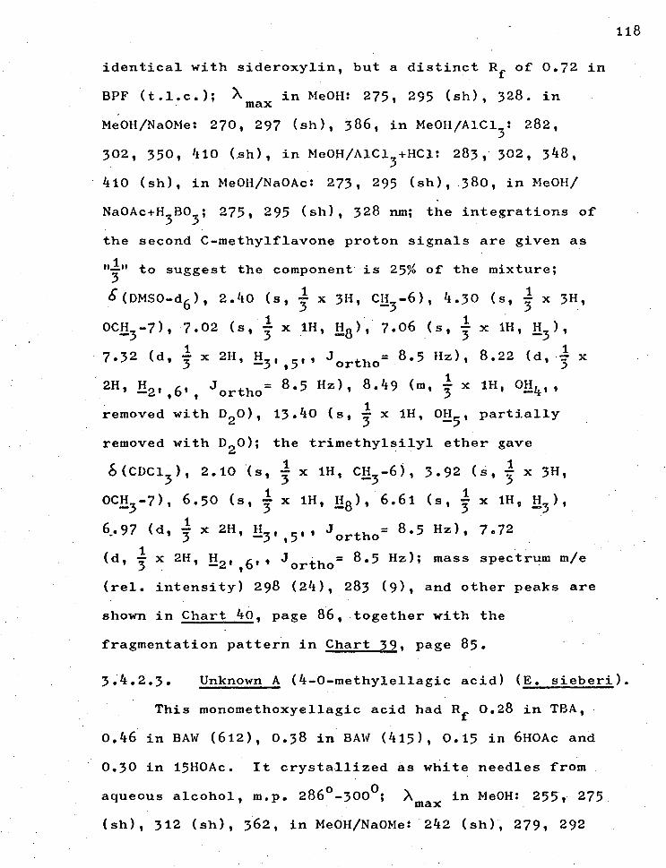
118
identical with sideroxylin, but a distinct Rf of 0.72 in
BPF (t.l.c.); X imax in MeOH: 275, 295 (sh), 328. in
Me0H/Na0Me: 270, 297 (sh), 386, in Me0H/A1C1 3 : 282,
302, 350, 410 (sh), in Me0H/A1C1 5 +HC1: 283, 302, 348,
410 (sh), in Me0H/Na0Ac: 273, 295 (sh), 380, in Me0H/
Na0Ac+H3 BO
3' • 275, 295 (sh), 328 nm; the integrations of
the second C-methylflavone proton signals are given as
to suggest the component is 25% of the mixture; 3
S(DMSO-d6 ), 2.40 (s, x 3H, CH3 -6), 4.30 (s, x 3H,
1 OCH3 -7), 7.02 (s, x 1H, H8 ), 7.06 (s, x 111, 113 ) 1
7.32
2H, H (d
1 , 3 x 2H, E319 _,,
o
jortho= 8.5 Hz), 8.22 (d, x
1 2 , , ,, ,
j ortho 1
removed with D 20), 13.40 (s, 3 x 1H, 0E51 partially
removed with D 20); the trimethylsilyl ether gave
6(CDC13 ), 2.10 (s, x 1H, CE3 -6), 3.92 (s, x 3H,
OCH3 -7), 6.50 (s, x 1H, H8 ), 6.61 (s, x 1H, 113 ) 1
1 6.97(d, 3 x 211, H5t,_11ortho= 8.5 Hz), 7.72 j
1 (d, x 2H, H2 ,
,6,, ortho 8°5 Hz); mass spectrum m/e j=
(rel. intensity) 298 (24), 283 (9), and other peaks are
shown in Chart 40, page 86, together with the
fragmentation pattern in Chart 39, page 85.
3.4.2.3. Unknown A (4-0-methylellagic acid) (E. sieberi).
This monomethoxyellagic acid had Rf 0.28 in TBA,
0.46 in BAW (612), 0.38 in BAW (415), 0.15 in 6H0Ac and
0.30 in 15H0Ac. It crystallized as white needles from
aqueous alcohol, m.p. 286 0-3000; X triax in MeOH: 255, 275
(sh), 312 (sh), 362, in Me0H/Na0Me: 242 (sh), 279, 292
. 8.5 Hz), 8.49 (n, x 111, 0H 41 ,
119
(sh), 325 (sh), 383, in Me0H/A1C13 . 265, 305 (sh), •
352 (sh), 372, in Me0H/A1C1 3+HC1: 254, 300 (sh), 352 (sh),
363, in Me0H/Na0Ac: 255, 275, 295 (sh), 368 (sh), 395
(sh), in Me0H/Na0Ac+H3B03
: 260, 276 (sh), 312 (sh),
370 nm; ymax 3420, 3322, 3160 (bound OH's), 1720 (non-
chelated C=0), 1610, 1576, 1288 (OCH3 stretch), 1077
- (OCH
3 stretch), 1236, 1206, 1106 (lactone C-0 stretch),
- 1027 (lactone C-0 stretch), 916, 886 (isolated ArH),
0 cm y. 805 0(DMS0-d ex. , .s, 1, H 6' t TMS) 811 ( H - -5 ), 7.85
(s, 1H, H51 ), 5.40 (m, 311, 0E3,3 , ,41 removed with D 20), - 4.40 (s, 3H, OCE3 4): for the methyl etherE(CDC1 3 ),
7.63 (s, 2H, H55'
), 3.36 (s, . 12H, OCH 3,3',4,4'), -,
6 (c6 D6 ), 7.65 (s, 21-1 , L15,5 1), 2 .93 (s, c. 1211, OCH
3,3',4,4'), 6(CDC1 3-C6D6) = +0.43; mass spectrum m/e
(rel. intensity) 316 (100), 301 (38), 273 . (19), 245 (7),
218 (8), 189 (11), 161 (6), corresponding to the
fragmentation shown in Chart 47, page 93. (Found:
C. , 56.7; H, 2.4. C 15H808 requires C, 56.9; H, 2.5%).
3.4.2.4. Unknown B (1,5-dimethy1-2,6-bis(3,4,5-
trihydroxyphenyl) furo [1,5-c] furan).
This compound had Rf 0.75 in SAW (612), 0.72 in
BAW (415), 0.66 in TBA, 0.08 in 6H0Ac and 0.15 in 15H0Ac.
It crystallized as off-white prisms from ethanol, m.p.
120o-122 o and had [ 441 15 79.1
o (c.1.0 in CHC1 )- X
3 max
in MeOH: 235 (sh), 283, 370 (sh), in Me0H/Na0Me: 300
(broad) (dec.)„ inMe0H/A1C1 3 . • 286, 310 (sh), 358 (sh),
in Me0H/A1C1 3+HC1: 286, 360, 375 (sh) 375 (sh), 430 (sh),
120
in Me0H/Na0Ae: 287, 360,380 (sh), in Me0H/Na0Ac+H3 BO
3 :
285, 360, 382 (sh), max 3 4 10 (broad), 3130 (broad),
1720 (broad), 1600, 1510, 1416, 1280, 1220, 1130,
1110 cm-1 ; 8(pms0-d6 , ext. Tms), 1.55 (s, 311,
2.25 (s, 311, cH3 ), 3.72 (m, 211, furofuran H), 3.98
(m, 211, furofuran H), 4.40 Cm, 2H, furofuran H), 7.45
(m, 2H, ArH), 7.93 (m, 2H, ArH), 8.52 (m, 211, Ar0H,
removed with D20), 9.02 (m, 4H, Ar0H, removed with D20);
mass spectrum m/e (rel. intensity) 390 (100), 304 (13),
302 (14), 287 (19), 285 (19), 255 (26), 254 (26), 206
(11), 204 (11), 157 (19) as in Chart 49, page 95.
3.4.2.5. Unknown F (a tetrahydroxydibenzofuran
dicarboxylic acid). (E. sieberi).
The compound had Rf 0.47 in BAW (612), 0.51 in
BAW (415), 0.37 in TBA, 0.12 in 6H0Ac and 0.24 in 15H0Ac.
It crystallized from ethanol as yellow needles, m.p.
175 °-180 ° ; Xmax in MeOH: 256, 305, 351 (sh), 366, in
Me0H/Na0Me: 256 (sh), 283, 355 (sh), in Me0H/A1C1 3 : 241
(sh), 267, 320 (sh), 382 (sh), in Me0H/A1C1 3+HC1: 256,
310 (sh), 352 (sh), 365, in Me0H/Na0Ac: 258, 274,
360 (sh), in Me0H/Na0Ac+H3 BO3'• 260, 305, 360 (sh),
375 nm; Vmax 3370 (bonded OH), 3340 (carboxylic OH),
1712 (strong) (carboxylic C=0), 1605, 1520, 1280-1300
(acid), (1120, 1040, 1010) (C-0 stretch), 925, 888
(isolated ArH), 868 cm-1 ; So(DMS0-d6 , ext. TMS), 7.94
(s, 2H, ArH), 10.30 centre (broad m, 6H, Ar0H, ArCOOH,

removed with D 20); mass spectrum rue (rel. intensity)
320 (10), 304 (6), 276 (100), 232 (10), 200 (5),
176 ( .70), 177 (20), 150 (100), 131 (60) as in Chart 53,
page 99.
121

CHAPTER 4.
122
IDENTIFICATION - DISCUSSION.
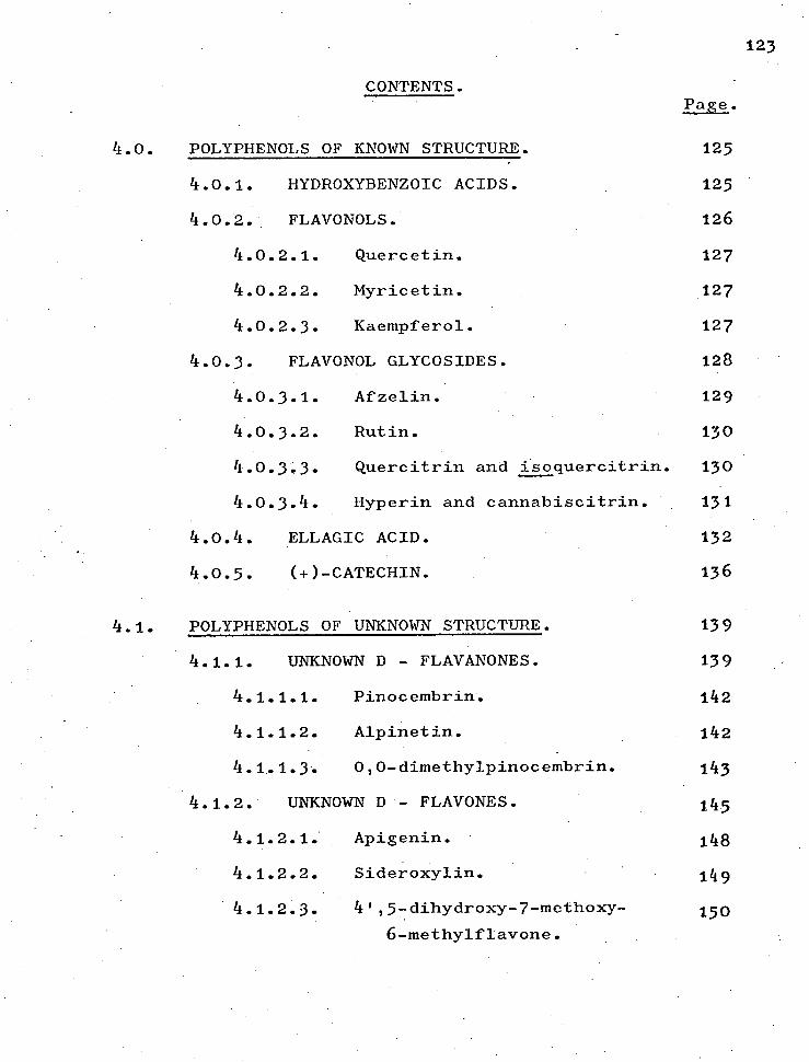
CONTENTS. Page.
4.0. POLYPHENOLS OF KNOWN STRUCTURE. 125
4.0.1. HYDROXYBENZOIC ACIDS. 125
4.0.2. FLAVONOLS. 126
4.0.2.1. Quercetin. 127
4.0.2.2. Myricetin. 127
4.0.2.3. Kaempferol. 127
4.0.3. FLAVONOL GLYCOSIDES. 128
4.0.3.1. Afzelin. 129
4.0.3.2. Rutin. 130
4.0.3.3. Quercitrin and isoquercitrin. 130
4.0.3.4. Hyperin and cannabiscitrin. 131
4.0.4. ELLAGIC ACID. 132
4.0.5. (+)-CATECHIN. 136
4.1. POLYPHENOLS OF UNKNOWN STRUCTURE. 139
4.1.1. UNKNOWN D - FLAVANONES. 139
4.1.1.1. Pinocembrin. 142
4.1.1.2. Alpinetin. 142
4.1.1.3. 0,0-dimethylpinocembrin. 143
4.1.2. UNKNOWN D - FLAVONES. 145
4.1.2.1. Apigenin. 148
4.1.2.2. Sideroxylin. 149
4.1.2.3. 4',5-dihydroxy-7-methoxy- 150 6-methylflavone.
1 23

CONTENTS (continued). Page.
• 4.1.3. UNKNOWN A (4-0-methylellagic 153
acid).
4.1.4. UNKNOWN B (1,5-dimethy1-2,6-bis 16o
(trihydroxyphenyl) furo
[1,5-c furan).
4.1.5. UNKNOWN F (a tetrahydroxy- 166
dibenzofuran-dicarboxylic
acid isomer).
12 4

CHAPTER 4.
4.0. POLYPHENOLS OF KNOWN STRUCTURE.
The evidence for the identification of the
polyphenols of known structure isolated in this work
is now discussed.
4.0.1. HYDROXYBENZOIC ACIDS.
125
cOOH COOH
OH
5 3 Ho
( 2 )
( 3 )
Gallic (2) and gentisic (3) acids were readily 81
identified from their n.m.r., and mass spectra with
chromatographic behaviour corresponding to that
48 recorded by Hillis. The melting point of each
agreed with literature values. 69b,69c
The identity of protocatechuic acid (1, page 12) seems
certain from comparison of its n.m.r. spectrum with
those of the above acids. (Chart 10, page 54 ).
In combination with the mass spectra shown in (Chart 11,
page 55 ), the u.v. spectral shift obtained with boric
64 acid in acetate and a chromatogram showing separate
spots for the two dihydroxyacids, together with a

melting point in agreement with the literature,69a
completed the identification.
4.0.2. FLAVONOLS.
OH
126
Ri R2
OH , H ,
OH OH •
H , H
Quercetin ( 8 )
Myricefln ( 9 )
Kaempferot (10 )
The flavonols quercetin (8), myricetin (9)
and kaempferol (10) have been isolated and the
evidence for their identity is.now given.
On paper chromatograms all three compounds
had the yellow colour characteristic of flavonols with
a free 3-hydroxyl12 when viewed under u.v. light and
their u.v. spectra showed the two bands typical of the
35a conjugated benzol-cinnamoyl system (7).
4.0.2.1. Quercetin.
68 The shifts induced with sodium acetate,
acidified aluminium chloride64
and boric acid in
acetate64
in u.v. spectra- of quercetin showed its
hydroxyl substitution which was confirmed by the n.m.r.
spectrum. (Chart 12 page 56 )• Co-chromatography
and melting point of the mixture with an authentic
sample without depression, together with a satisfactory
elemental analysis completed the identification of
quercetin.
4.0.2.2. Myricetin.
Myricetin was identified first from its n .m.r.
spectrum which, when compared with that of quercetin
(Chart 13, page 57), showed that the quercetin
5'-proton is no longer present in myricetin; in
addition the myricetin spectrum shows a two-proton
singlet for the protons in positions-2',6'.
Identification of myricetin was completed by
comparison of its mass spectrum and proposed
fragmentation with those of quercetin (Charts 14,15
pages 58,59 ) and finally - by a mixed melting point
without depression using an authentic sample, together
with a satisfactory elemental analysis.
4.0.2.3. Kaempferol.
It is noteworthy that Hillis found
chromatographically that E. coccifera was the only
127

49 peppermint to contain kaempferol (10).
, The presence of kaempferol in E. coccifera
has been verified in this work by the isolation of
the aglycone as well as by the oxidation of afzelin.
Spectral evidence, especially the n.m.r.
signals of ortho-protons in positions-3', 5' and
positions-2', 6' and meta-protons in positions-6,8
together with the chromatographic comparison with
an authentic sample and melting point determination
seemed unambiguous evidence for the presence of
kaempferol.
4.0.3. FLAVONOL GLYCOSIDES.
128
HO
OH 0 R1' R2 = H, R3=
Rhamnosyl Afzelin
R1 =OH, R2
= H, R3
= RhamnoSylgi ucosyl : Rutin
R3
= Rhamnosyl Quercitrin
R3 =Glucosyl : Isoquercitrin
R :Gala c t o syl : Hyperin 3
R1' = OH;R 3 = Glucosyl : Cannabiscitrin

129
4. 0.3. Afzelin (kaempferol-3-rhamnoside). 22
Peroxide oxidation of this glycoside
isolated from E. coccifera released the sugar as
22 shown by paper chromatography. Chandler and Harper
found that controlled oxidation of glycosides with
peroxide released only sugar residues from the
3-position and proved more reliable for glycoside
differentiation than the standard method of exhaustive
methylation applied by King and Acheson 71 and adapted
by Nordstrom to the microscale.83
The products of the oxidation were identified
by their Rf values in several solvents using the 102
chromogenic spray of diazotised-k-nitroaniline for 75h
kaempferol and 2.-anisidine hydrochloride for
rhamnose.
The oxidation evidence was supported by u.v.
spectral analysis of the glycoside, showing the
presence of a free 7-hydroxyl by a bathochromic shift 68
of the shorter wavelength band in sodium acetate.
The spectrum of the glycoside with sodium methoxide
reagent showed no decomposition when run again some ten
minutes after basification, confirming the absence of
an alkali sensitive grouping of hydroxyls, such as a
3,3 1 ,4'-group."
The similarity between the u.v. spectra run
in aluminium chloride and acidified aluminium chloride
showed the shift was due to a chelating 5-hydroxyl,

with no acid-labile complexes being formed with
ortho-di-hydroxyls. 37,103
The glycoside was thus identified as afzelin,
kaempferol-3-rhamnoside, with the reported melting
point. 71
4.0.3.2. Rutin (quercetin-3-rhamnosylglucoside).
Among the flavonol glycosides isolated from
16a E. delegatensis, rutin was identified by comparison
35b of its observed and published u.v. and n.m.r. spectral
14 data, the latter for both rutin and its trimethylsilyl
75g ether. After acid hydrolysis, co-chromatography
and mixed melting point without depression of a
mixture with an authentic sample of quercetin confirmed
the identify of the aglycone.
The sugar residues after hydrolysis were co-
chromatographed with known sugars in two solvents and
their R values showed them to be rhamnose and glucose
and the identity of the diglycoside as quercetin-3-
rhamnosylglucoside.
4.0.3.3. Quercitrin (quercetin-3-rhamnoside) and
isoquercitrin (quercetin-3-glucoside).
The other two glycosides isolated from
E. delegatensis appeared from Rf values to
76 monoglycosides and peroxide oxidation, according to
the method of Chandler and Harper,22
gave in both cases
the aglycone quercetin identified by comparison with
an authentic sample. After oxidation one glycoside
13 0

.(asoonTB-C-uTleoTaAm)
hyperin ( quercetin-3 - gal act oside)
( 9) , ( quercet in-3 - gluc oside) .
( quercetin-3 - gal act o si de)
( myric et in-3- gluco se) .
0 H
.
5 Z
Z H
.
V
si
H.
(. -)0
P
tn
11
CD
rt-
1 1
CD
11
0
O0
0' H
.
Ptf
H
M
et•
0
0
CD
p
A)
H.
rt-
(D
X
CD
1 c+
eF
1-1
) 0'
c+
H.
I\.5
CD
0
H.
0
H.
fa.
IV C
L
1-3
cm
M
M
a
p
1-3
V
II
si
co
c+
0,
It
OA)
Cr
H.
CD
11
CD
It
so
o
,- o
(.4..
o
ocl
0'
w
H)
0
0
M
ti
H
H.
CD
11
M
0
fjq
0
H-
B
LT1
II
co
Pg
co
H.
• 0
O0
H
c+
(D
11
I0
X
Cl)
0
P0'
0 ' C
D
d
CD
Cl)
H.
H.
M
(D
cl-
0
fa
. tS
0
H.
M
CD
0
0
0
(-1-
0
CL
c+
CT1
Cl)
11
H.
0
0
0'
CD
0
H.
CD
0
M
0
P
II
X
cn
tf
c+
0
eF
H.
H-
a)
0
P
Cl)
0
H.
fa.
i 0
P
II
H)
• c+
ce
P
CD
M
H.
H.
0"
t<
c-F
1'0
Cl)
CD
CD
H.
rt.
H.
q H
0
fa.
1-3
rt-
0
0'
eF
H
CD
0
0'
fit
0'
0
g
0.
Cl)
P
CD
g
co
Cl)
• o
Cl)
Crcl
o
So
0
H)
P
P
It
H.
g
1.0
( ri
)1 CD
si
c-F
CD
1-
3 c+
0
I-1
et-
&-
CD
CD
0
CD
1 CD
CD
11
0
(D
0
(D
c+
0CD
H
. 0
0
Cl)
0.,
H.
c-F-
Cl)
11
0
0
H.
Ucl
H)
H.
dg
c+
CD
g.,
0
H
H.
0
CD
NJ
'd
H.
cn
g
H)
,- CD
P
II
0
M
0
fa,
M
11
0
11
C)
0
0
Cl)
h:$
P
co
V
V
f.a
PCr
t <
a)
0
I—,
0
P
H.
M
P
CD
11
0.
(D
11
H
(1)
c+
0
a
a)
rn<
I
H.
w
a'
H
Cl)
•CD
CD
CD
Cl)
CD
Cl)
0.
. CD
11
0
H.
P
0
0
fa.
O..-
-,.
0
t4
O1.0
0
H.
M
0
I m
It
a)
(")
H
So
II
•co
Il
co
H.
CD
0
CI,
Oc+
M
cfc
l H
. 1-3
P
II
0 V c+
V
H.
I (D
0
0
M.
C11'1
0
c+
I 4.)
11
0
P
0
Fj-
c-1-
PL)..
g
.1j.
(DM
•g
crl
,. 1--,
-. P
Co k()
-
rf- v
r I-I- 0.
In
0"
0
c+
CD
O(D
H
. 0
§
,.......
M
0
fa.
0
c+
0'
M .
c-t-
CD
In
H.
0.
. 11
O
rt.
(D
M
Cl)
CD
0
CA
H
H.
. M
H
il
et:
5 CD
CL
, 0
0
.0
eF
0
cn
Cu P
c+
CD
g CD
O
H.
1-1
Oct
0.. •
et
- (D
P
0"
it
li
0
I:
tn
0 I•
0
0
CD
0 H)
4
CD
H.
0
0 M
0
Cl)'
H.
CD 0
ri-
C) H-
II H.
a'
0 H-
t- CC
)

132
4.o.4. ELLAGIC ACID.
OH
HO
OH
OH
( 11
The final compound isolated from E. delegatensis
was identified as ellagic acid (11) from its Rf values
and characteristic chromatographic colours, being
violet-blue (u.v. light, 365 nm) and dull yellow when
53b fumed with ammonia. When sprayed with ferric
ferricyanide and diazotised 2 .-nitroaniline, paper
chromatograms of the compound showed blue and yellow-
53b brown respectively. Co-chromatography with an
authentic sample showed no separation of spots.
It was necessary to examine the spectral
properties of ellagic acid in some detail because two
48 of Hillis' unknowns, (viz. A B), were provisionally
identified by him as "ellagic 'acid-like compounds".
In methanol, ellagic acid gave a characteristic
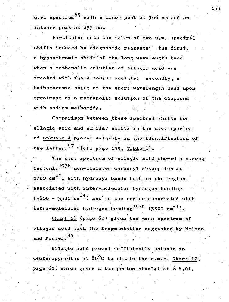
u.v. spectrum65 with a minor peak at 366 nm and an
• intense peak at 255 urn.
Particular note was taken of two u.v. spectral
shifts induced by diagnostic reagents; the first,
a hypsochromic shift of the long wavelength band
when a methanolic solution of ellagic acid was
treated with fused sodium acetate; secondly, a
bathochromic shift of the short wavelength band upon
treatment of a methanolic solution of the compound
with sodium methoxide.
Comparison between these spectral shifts for
ellagic acid and similar shifts in the u.v. spectra
of unknown A proved valuable in the identification of
the latter. 97 / xcf. page 159, Table 4).
The i.r. spectrum of ellagic acid showed a strong
lactonic107b
non-chelated carbonyl absorption at
-1 1720 cm, with hydroxyl bands both in the region
associated with inter-molecular hydrogen bonding
- (3600 - 3500 cm 1 ) and in the region associated with
-1, intra-molecular hydrogen bonding 107a (3300 cm /.
Chart 16 (page 60) gives the mass spectrum of
ellagic acid with the fragmentation suggested by Nelson
• 81 and Porter.
Ellagic acid proved sufficiently soluble in
deuteropyridine at 80 °C to obtain the n.m.r. Chart 17,
page 61 which gives a two-proton singlet at 8 8.01,
133

Deshielding
and doublets at 65.08 and 64.25, although retaining
signals due to solvent of crystallization.
The singlet signal due to the 5,5 -protons
of ellagic acid is .confirmed by the n.m.r. spectrum of
its trimethylsilyl ether (Chart 18, page 62) with
the two-proton signal at 67.63 p.p.m.
While no work has been published on the n.m.r.
spectra of ellagic acid-type dilactones, work on
ketones (A)38
and coumarins (B)38
seems to allow
inferences to be drawn for the dilactones.
Use is made of these inferences in the
identification of unknown A, page 154, so further
discussion is apposite.
Reference plane
0
R I
Shielding I Deshielding
13 4
( A )
Shielding Reference
plane
( B )
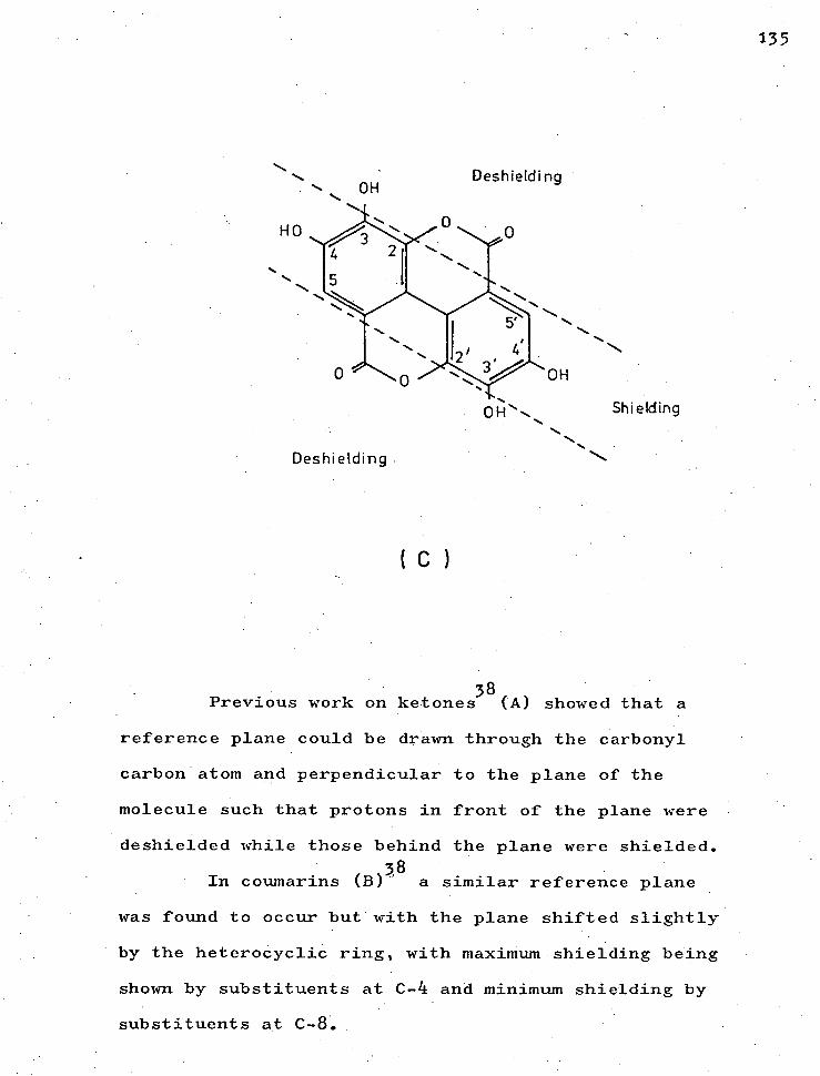
Deshielding OH
Shielding
Deshielding
HO
C
Previous work on ketones38 (A) showed that a
reference plane could be drawn through the carbonyl
carbon atom and perpendicular to the plane of the
molecule such that protons in front of the plane were
deshielded while those behind the plane were shielded. 58
In coumarins (B) a similar reference plane
was found to occur but with the plane shifted slightly
by the heterocyclic ring, with maximum shielding being
shown by substituents at C-4 and minimum shielding by
substituents at C-8.
135

By analogy it is proposed in this work that
similar planes can be drawn for ellagic acid type
compounds (C). The hydroxyls that would incur the
maximum shielding would be those resonating at 64.25
namely the 4,4'-hydroxyls, while the hydroxyls
incurring the minimum shielding would be those at
positions-3,3', resonating at 6 5.08 p.p.m.
4.0.5. (+)-CATECHIN.
OH
136
HO OH
HO
OH
OH
( 12 )
A compound isolated from a Sephadex column
gave a red colour on a paper chromatogram when sprayed
13 with methanolic vanillin-hydrochloric acid, a yellow
colour when sprayed with ethanolic E-toluene sulphonic
acid,96 reactions given only by flavans containing.an
'A-ring derived from phloroglucinol (12) i.e. a
5,7-dihydroxyflavan.
Changes in the u.v. ape :ctrum, re-run

137
after some minutes, when a methanolic solution of the
compound was treated with sodium methoxide indicated
the presence of an alkali sensitive group of hydroxyls
--such. as-a. 3,3',4!—group.30 The existende of the 3',4'-
dihydroxyl group being further supported by the
bathochromic shift of the cinnamoyl band, of the U.v.,
spectrum of a methanolic solution of the compound
treated with boric acid and sodium acetate. 75d
The n.m.r. spectrum of the flavan (Chart 19;
page 63 ) run in deuterodimethylsulphoxide to show
the aromatic proton signals indicated that the flavan
was an isomer of 3' 4',5,7-tetrahydroxyflavan-3-ol (13).
An additional spectrum (Chart 20, page 64 ) run in
deuteropyridine gave signals for the methylene protons
at C-4 and the methine protons at C-3 and C-4 which
26 were analysable as a first-order ABMX system.
The size of the coupling between the C-2, C-3
protons (8 Hz) showed them to be trans-26 and thus the
isolated compound to be a catechin rather than an
epicatechin. The specific rotation was measured
as positive, similar to that of an authentic sample,
and the catechin was identified as the ( )-enantiomer
of the catechins (Figure 6, overleaf).

138
(+).- CATECHIN 2R : 3S
HO
OH OH
Figure
The mass spectrum of the flavan showing a
molecular ion of 290 and a base peak due to a typical
o-hydroxybenzyl cation24
at 139 supported the above
identification and as Chart 21, page 65 illustrates,
the major peaks correspond to a retro-Diels-Alder fission
4 of the heterocyclic ring accompanied by hydrogen
transfer mainly from the 3-hydroxyl.87
The above evidence was supported by an i.r.
spectrum which showed strong hydroxyl absorption
indicative of both inter-molecular and intra-molecular
hydrogen bonding but no absorption attributable to
a carbonyl group.
A melting point in agreement with the literature

and undepressed on admixture with an authentic sample,
together with a satisfactory elemental analysis'
completed the identification of the flavan as
(+)-catechin on 3', 4', 5,7-tetrahydroxy-2,3-trans-
flavan-3-ol.
4.1. POLYPHENOLS OF UNKNOWN STRUCTURE.
4.1.1. UNKNOWN D - FLAVANONES. 15
From a leaf extract of E. sieberi (page 27)
were isolated three closely related substances with
chromatographic behaviour similar to compound D, which
have properties corresponding to flavanones.
In particular, all three compounds showed a
dull red colour when sprayed with sodium borohydride
followed by exposure to concentrated hydrochloric acid
fume's." The i.r. spectra showed the presence of
carbonyl groups, confirmed by reaction with
dinitrophenylhydrazine; the u.v. spectra, with strong
peaks around 286 bra, were consistent with those of
flavanones, and likewise the mass spectra, 9 which
furthermore had strong M-77 ions indicating unsubstituted
B. rings.
The latter inference was supported by five-
proton multiplets in the aromatic region of the
n.m.r. spectra and by characteristic i.r. peaks
around 765 and 700 cm -1 . 19
139
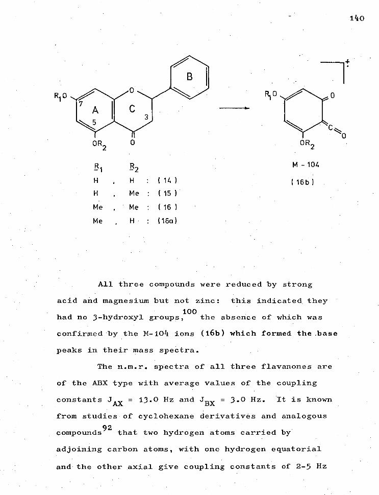
140
OR 2
-1
H
H
Me
Me
•
•
,
,
-R2 H
Me
Me
H
:
:
:
:
( 14 )
( 15 )
( 16 )
(16a)
M - 104
(16b)
All three compounds were reduced by strong
acid and magnesium but not zinc: this indicated they
100 had no 3-hydroxyl groups, the absence of which was
confirmed by the M-104 ions (16b) which formed the base
peaks in their mass spectra.
The n.m.r. spectra of all three flavanones are
of the ABX type with average values of the coupling
constants JAx = 13.0 Hz and JBx = 3.0 Hz. It is known
from studies of cyclohexane derivatives and analogous
compounds92
that two hydrogen atoms carried by
adjoining carbon atoms, with one hydrogen equatorial
and the other axial give coupling constants of 2-5 Hz

a 141
whereas when both hydrogens are axial the coupling
constant is larger (6-11 Hz).
In the case of the flavanones examined here the
coupling constant of 13.0 Hz is so high that it can
only represent an axial-axial coupling so that H-2
would be axial and the 2-phenyl equatorial or vice versa.
Further, assuming the Karplus formula, 27
modified by Williamson and Johnson l08predicting a
relation between the dihedral angle ( ) and the vicinal
coupling constant, is always true for flavanones, 79
it is possible to calculate the dihedral angles which
the two C-3 hydrogen atoms make with the C-2 hydrogen:
= 13.0 = 16 cos 2 14 = 154° (nearest
- H3a degree)
= 3.0 = 10 cos it 570 (nearest
2 - H
3e degree)
Chart 22 9 page 68 , shows the application of
n.m.r. spectral data to determine relative
configuration between C-2 and C-3 hydrogen atoms in
flavanones.
Each of the flavanones was found to be
laevo-rotatory with the one optical centre at C-2;
hence they belong to the same configurational series. 1
Chart 23 , page 69 , shows a comparison of the •
important portions of the infra-red spectra of the
three flavanones and Chart 24, page 70 , summarises
the n.m.r. spectral signals of Charts 25 1 26,27 1 pp. 71,72175.

142
with their assignments.
4.1.1.1. Pinocembrin.
The compound of lowest molecular weight, which
was present in least amount, reacted with ferric •
chloride and coupled with diazotized E-nitroaniline.48
Its u.v. spectrum showed large bathochromic
66 shifts with aluminium chloride and with sodium
acetate, 66 indicating the presence of hydroxyl groups
at positions 5 and 7 respectively. The broad i.r.
band around 3120 cm -1 and the carbonyl absorption at
1629 cm-1 supported the presence of a 5-hydroxyl group,
and the data indicated that the compound was
pinocembrin (14).
Its identity was confirmed by a comparison of
its properties with those of a synthetic specimen.
4.1.1.2. Alpinetin.
The second flavanone also gave a ferric chloride
48 test, but coupled slowly with diazotized
Its u.v. spectrum showed a bathochromic shift with
66 100 sodium acetate but not with aluminium chloride,
indicating that the compound had a 7-hydroxyl but no
5-hydroxyl group. This was consistent with the presence
of a methoxyl proton resonance in the n.m.r. spectrum,
and with an i.r. hydroxyl absorption at 3475 cm-1 and a
carbonyl at 1660 cm-1 .
95 The substance thus has the structure of alpinetin (15)
a rather rare flavanone not previously encountered in
eucalypts.

143
Its melting point agreed with that reported
for alpinetin, but was over 80 ° higher than that of
69g the isomeric pinostrobin (16e); moreover, an authentic
sample of the latter showed a u.v. shift with
aluminium chloride but not with sodium acetate.
The identity was confirmed by conversion into
its 0-methyl derivative, which proved identical with
the 0,0-dimethyl derivative prepared from pinocembrin.
4.1.1.3. 0,0-dimethylpinocembrin.
The third flavanone gave none of the phenolic
tests or u.v. shifts described for (14) or (15), but
-1s it had an i.r. carbonyl absorption at 1671 cm, and
its n.m.r. spectrum showed two methoxyl proton
resonances with benzene shifts corresponding to
locations at positions 5 and 7. 109
Its identity as 0,0-dimethylpinocembrin (16),
which does not appear to have been found previously
in nature, was confirmed by a comparison of its
properties with those of an authentic sample
prepared from pinocembrin.

144
Mass spectral fragmentation of the flavanones
followed a pattern shown by Audier4
to be a retro-
Diels-Alder fission, illustrated below:
A fuller interpretation of the mass spectra of
the flavanones and assignment of peaks is shown in
Charts 28,29 (pages 74,75 ).
It should be noted for chemotaxonomy that,
using similar evidence, a compound identified as
pinocembrin (14') was isolated from E. delegatensis.
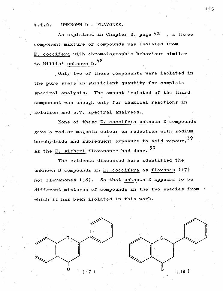
1 45
4.1.2. UNKNOWN D - FLAVONES.
As explained in Chapter 2, page 42 , a three
component mixture of compounds was isolated from
E. coccifera with chromatographic behaviour similar
to Hillis' unknown D. 48
Only two of these components were isolated in
the pure state in sufficient quantity for complete
spectral analysis. The amount isolated of the third
component was enough only for chemical reactions in
. solution and u.v. spectral analyses.
None of these E. coccifera unknown D compounds
gave a red or magenta colour on reduction with sodium
borohydride and subsequent exposure to acid vapour,39
90 as the E. sieberi flavanones had done.
The evidence discussed here identified the
unknown D compounds in E. coccifera as flavones (17)
not flavanones (18). So that unknown D appears to be
different mixtures of compounds in the two species from
which it has been isolated in this work.
( 1 7 )

146
Evidence structurally significant for all
components is given first followed by evidence
peculiar to each.
All three compounds gave positive ferric chloride
tests but inconclusive phenylhydrazine reactions: this
relative unreactivity of the carbonyl group in flavones
6 is reported by Gripenberg
35e and Baker et al.
The i.r. spectra confirmed the presence of
carbonyl groups in the major components.
The u.v. spectra of all three components
showed some similar features:: first, strong bands
around 275 nm and 325 nm, consistent with a flavone
conjugated benzoyl-cinnamoyl system; 35a
secondly,
bathochromic shifts of the longer wavelength band upon
addition of sodium acetate, in accord with the presence
of a 4'-hydroxyl in ring B;35a
thirdly, bathochromic
shifts with aluminium chloride unaffected by addition
67 of hydrochloric acid, indicating the presence of a
chelated 5-hydroxyl but no adjacent hydroxyls capable
of labile complexes; fourthly, the lack of adjacent
hydroxyls was confirmed by the failure of the methanolic
spectra to shift with the boric acid/acetate reagent.64
The i.r. spectra of the major components
supported the inference of a 4'-hydroxyl substitution
in the B-ring by the presence of a strong peak around 74
840 cm-1 , together with hydroxyl absorption around

-1 3330 cm , found by Briggs19 to be common among
flavones with 4'-hydroxyl groups.
The n.m.r. spectra of the major components
were similar: first, two doublets (J = 9 Hz) consistent
with two pairs of ortho- protons in a para-substituted
benzene, confirming the 4'-hydroxyl group; secondly,
a sharp singlet at low field ( E6.6 - 86.8 p.p.m.)
was typical of the 3-proton of flavones.109
The 4'-hydroxyl pattern of the B-ring was
also indicated by the mass spectra of the compounds
which showed peaks corresponding to M-93 ions. Peaks
at M-118 supported the n.m.r. evidence for the
absence of 3-hydroxyls.
147

148
H o
OH
I 21 ) 4.1.2.1. Apigenin.
Considering each flavone in particular, the
spot of lowest Rf' corresponded to a compound whose u.v.
spectrum showed, in addition to the above shifts, a
bathochochromic shift of the shorter wavelength band
upon addition of sodium acetate, indicating the
68 presence of a 7-hydroxyl group.
This inference was supported by hydroxyl
-1 absorption around 3280 cm in the i.r. spectrum.19
The n.m.r. spectrum of this compound showed
peaks assignable to meta-coupled 6,8-protons (J = 3 Hz)
in addition to the signals already discussed.
(Chart 30, page 76).
Chart 31, page 77 , shows a molecular ion of
270 and the fragmentation coming from the retro-Diels-
Alder fission of the heterocyclic ring4
is suggested

149
in Chart 2a, page 78 . The melting point compared favourably with the
literature35f and completed the identification of the
compound as apigenin, (19), 4',5,7-trihydroxyflavone.
The mixture of higher Rf
than that of apigenin
appeared to be a mixture of C-methylflavones,
53a,55 considered to be comparatively rare plant extractives.
Although Hillis did not record C 7methylflavones
in his earlier survey he has since discovered their 55
presence in eucalypts.
One component of the mixture was isolated in
the pure state and its identity is discussed first.
The second component was isolated in sufficient
quantity for u.v. spectral analysis but further evidence
for its identity is discussed by reference to the
n.m.r. and mass spectra of the mixture of the
C-methylflavones.
4.1.2.2. Sideroxylin.
Unlike the u.v. spectrum of apigenin, that of
the isolated C-methylflavone showed no bathochromic
shift of the shorter wavelength band with sodium acetate,
68 indicating the absence of a 7-hydroxyl in ring A.
The n.m.r. spectrum revealed the presence of
methyl groups at positions -6,8. It is characteristic
of these that the 6-methyl signal is upfield from the
8-methyl. 78 The n.m.r. spectrum also showed a
7-methoxyl group shifted characteristically (+0.60 p.p.m.)
150
109 when a spectrum was run in benzene.
Charts . 5,54, pages 79,80, give the mass
spectrum and a suggested fragmentation of the major
C-methylflavone. This evidence, together with an
undepressed melting point on admixture of an authentic
sample, confirmed the identity of the compound as
sideroxylin (21), 4 ,5-dihydroxy-7-methoxy-6,8-
dimethylflavone, reportedly found only once before
in nature.55
4.1.2.3. 4',5-dihydroxy-7-methoxy-6-methylflavone.
The second component of the mixture of
components of higher Rf than that of apigenin appears
also to be a C-methylflavone although the evidence
for its identity is given tentatively.
The u.v. spectral shifts for this compound were
identical with those of sideroxylin, suggesting a
similar chromophore system and hydroxyl pattern.
When the n.m.r. spectra of pure sideroxylin
and impure sideroxylin were compared and the only signal
to disappear on purification was noted with its
integration, it seemed to be in a position consistent
• 78 with a 6-methyl group. (Charts 35,36,37, pages 81,82,83).
The lone 8-proton signal which might be
expected to appear in a compound without the 8-methyl
of sideroxylin has been shown to appear in the same
spectral region as the 3-proton, 75f in compounds of
similar substitution. 78

151
A similar comparison of the n.m.r. speatra of
sideroxylin and the trimethylsilyl ether of the
sideroxylin mixture also showed, upon purification of
the sideroxylin, the disappearance of only one signal,
which was assignable to a 6-methyl group. (Charts 35,36,38
pages 80,81,84).
The i.r. spectra of pure sideroxylin and the
mixture were identical, suggesting only .a minor
structural difference between the two compounds in the
mixture.
The mass spectra of flavonoids have been
94 4 Reed and Wilson, and Audier
made separate studies of the fragmentation of flavones
but there has been no detailed study of the mass spectra
of C-methylflavones.
From a study of the mechanism of fission of
ring C, the heterocyclic ring, these workers have
concluded that the major fragmentation mode in flavones
is a retro-Diels-Alder fission as illustrated in
Chart 52, page 78.
Audier4
found that the nature of the substituents
had a great influence on the relative intensities of
the peaks which resulted from Diels-Alder rupture and
that substituents displace the peaks of flavone itself
as a function of their mass.
By analogy, Chart 34,page 80 , suggests a
mass fragmentation of sideroxylin corresponding to major
reported recently.

15 2
peaks of its mass spectrum in Chart 33 , page 79 .
Chart 39 , page 85 , suggests a similar
mass fragmentation for the second C-methylflavone
indicated by the peaks •298 (M+ ), 283 (M-15) and
M-28 2--) which seem to arise mainly from the impurity
in Chart 4 , page 86 which shows the mass
spectrum of the mixture.
These peaks could be explained by the
C-methylflavone structure (20)proposed also on n.m.r.
spectral evidence.
While isolation of this second component in
the pure state has not been achieved, except enough
for u.v. spectral analysis, evidence from n.m.r., i.r.
and mass spectra of the mixture suggests the
provisional identity of this component as 4',5-dihydroxy-
7-methoxy-6-methylflavone (20), a compound not
reported in the literature.
OH
(20)

153
4.1.3. UNKNOWN A (4-0-methylellagic acid).
OH
Me()
OH
OH OMe
( 22 ) HO
OH
OH
( 23 )
In this work the compound isolated with
chromatographic behaviour similar to Hillis' unknown A48
has properties corresponding to a monomethyl derivative
of ellagic acid, a compound that seems not to have been
found previously in nature.
The n.m.r. spectrum of unknown A showed a
three-proton methoxyl singlet at 84.40 p.p.m., the
downfield shift being due in part to the use of
deuterodimethylsulphoxide with tetramethylsilane
as external standard. In contrast to the two-proton
154
singlet for the aromatic protons of ellagic acid,
the n.m.r. spectrum of the monomethyl derivative shows
one proton singlet at 67.85 and 68.11 p.p.m.
(Chart 41, page 87).
The separate n.m.r. signals for the 5,5'-protons
in unknown A showed the difference in environment of
each. Although data for analogous compounds is not
reported in the literature, the magnitude of the shift
difference for these signals (0.25 p.p.m.) suggested
the environment difference was due to ortho- rather
107c than meta- groups, favouring structure (22) for
unknown A rather than structure (23).
107c Chemical shifts found by Williams for
mono-substituted benzene protons relative to benzene
are upfield by 0.23 p.p.m. for an o-OCH3 and upfield by
-
0.37 p.p.m. for an o-OH, a shift difference of 0.16
p.p.m. These figures suggest the assignment of the
signal at 67.85 p.p.m. to the proton ortho- to the
4'-hydroxyl and the signal at 68.11 p.p.m. to the
proton ortho- to the 4-methoxyl in structure (22).
Further, the hydroxyl multiplet in the n.m.r.
spectrum is in the region ( 6 5.00 p.p.m.) previously
assigned to 3,3'-hydroxyls in the discussion of ellagic
acid on page 136 supporting the deduction that
3,3'-hydroxyls are present in unknown A.i.e. structure (22).
In an attempt to secure unambiguous evidence
for the location of the methoxyl in unknown A, the
155
compound was methylated and n.m.r. spectra run in
deuterochloroform and deuterobenzene, 104 anticipating
a shift difference between 4,4'-methoxyl signals and
3,3'-methoxyl signals, distinguishable according to
the discussion of ellagic acid on page 136
It was planned to trideuteriomethylate 8
another sample of unknown A and by comparison of n.m.r.
spectra in the same solvents locate the original
methoxyl unambiguously.
However the spectra of the methylated ether
in deuterochloroform and in deuterobenzene showed
the same shift for all methoxyls, a reasonable inference
despite the partial methylation. (Charts 42,45 pages 88,89).
Support for the identity of unknown A as
4-0-methylellagic acid rather than 3-0-methylellagic
acid was obtained from analysis of its u.v. spectra.
The comparison of the u.v. spectra of ellagic
acid with those of its monomethyl ether in methanol
alone, in methanol with fused sodium acetate added
and in methanol with sodium methoxide added, is shown
in Charts 44,42216, pages 90,91,92.
Addition of fused sodium acetate to a methanolic
solution of the monomethyl ether ionized the more acidic
hydroxyls para- to the carbonyls35c to give a new band
at 275 nm and a spectrum similar to that of ellagic
acid after treatment with the same reagent.
(Chart 45, page 91).

OMe
136
HO
OH
OMe
( 24 )
OMe OMe
HO HO
OMe
( 25 )
( 26)
Other workers97
have found no similar shift of
the short wavelength band of the spectrum of the
3,3'-dimethylether (24). Of course both structures
(22) and (23) of the monomethyl ether (page 153) have
at least one ionizable hydroxyl para- to a carbonyl
group so the evidence is inconclusive. But the size
of the shift in the spectrum of the ether compares with
the size of the shift in that of ellagic acid with two
hydroxyls para- to carbonyls, suggesting free 3,3'-
hydroxyls in the ether as in structure (22), the

157
4-0-methyl ether.
Further, in methanolic sodium methoxide solution,
which is known to ionize all free hydroxyl groups, 35c
the spectra of ellagic acid and the ether were
identical with the spectra of the compounds in acetate
solution.
In contrast Scott97
records that a sodium
methoxide solution of the 3,3'-dimethyl ether ( 25)
gives a distinctly different u.v. spectrum with an
intense band at about 428 urn, tentatively assigned to
transitions which result in a symmetrical chromophore
system such as (25) to (26).
It seems reasonable that the ether structure
(23'), with a methyl group in the 3-position should also
show this band at about 420 nm which would correspond
to the analogous transition (27) to (28). But there is
no evidence of such a band.
OMe
HO
OH
OH
( 23 )

158
OMe
OMe
HO HO
O H
( 27)
( 28 )
On the contrary the maxima of the spectrum
are similar to those of the acetate spectrum for the
4,4'-dimethyl ether given by Scott. 97 This evidence
supports the deduction that the methyl group in
unknown A is at position -4 not position -3.
The above argument is summarised in Table 4
overleaf.
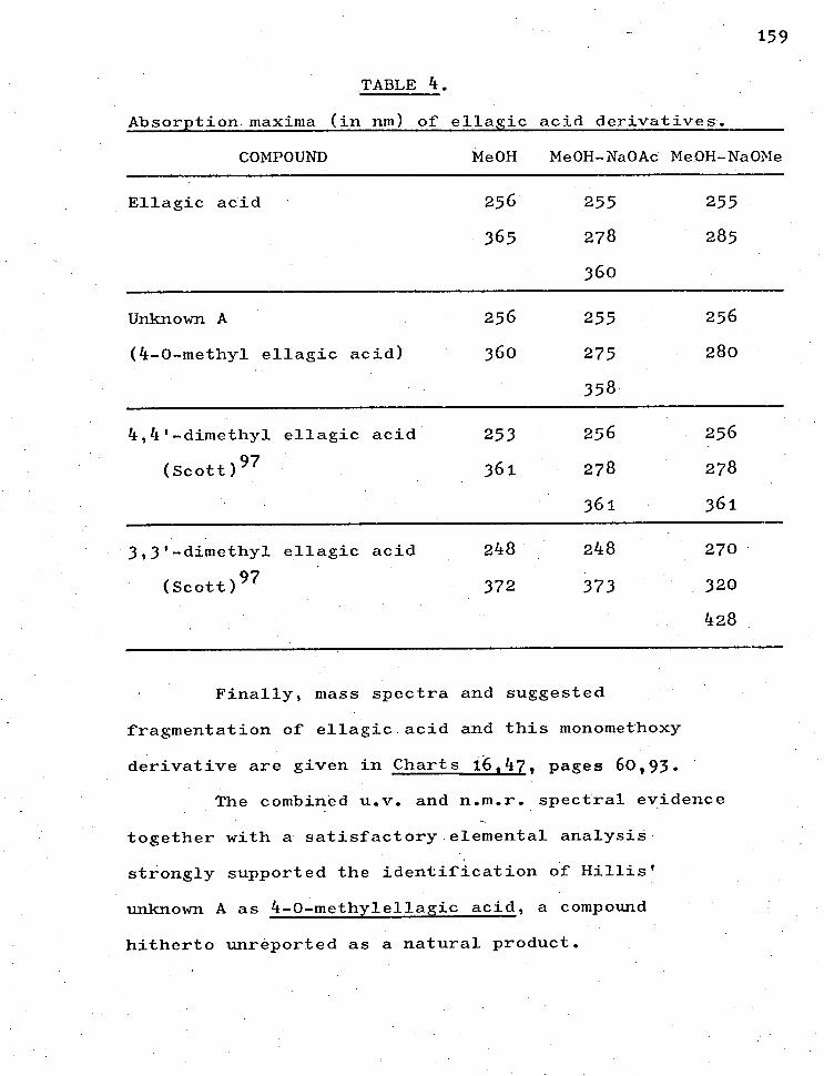
159
TABLE 4.
Absorption maxima (in nm) of ellagic acid derivatives.
COMPOUND Me0H Me0H-Na0Ac Me0H-Na0Me
Ellagic acid • 256 255 255
365 278 285
360
Unknown A 256 255 256
(4-0-methyl ellagic acid) 360 275 280
358
4,4'-dimethyl ellagic acid 253 256 256
(Scott) 97 361 278 278
361 361
3,3'-dimethyl ellagic acid 248 248 270
(Scott) 97 372 373 320
428
Finally, mass spectra and suggested
fragmentation of ellagic acid and this monomethoxy
derivative are given in Charts 16,47, pages 60,93.
The combined u.v. and n.m.r. spectral evidence
together with a satisfactory elemental analysis
strongly supported the identification of Hillis'
unknown A as 4-0-methylellagic acid, a compound
hitherto unreported as a natural product.

Me0
HO
HO CH2 CH 2 / OMe
HC--- CH
H,C HC OH
4 07
160
4.1.4. UNKNOWN B (1,5-dimethy1-2,6-bis (trihydroxyphenyl)
furo I 1,5-c ) furan)
Hillis48 suggested that the compound he termed
unknown B should be provisionally identified as
belonging to the ellagic acid or dilactone class
because of its chromatographic behaviour.
From E. sieberi a compound has been isolated in
this work with Rf values and u.v. colours comparable to
those of unknown B but the spectral properties observed
61 for this compound are those of lignans rather than
dilactones.
The u.v. spectrum of the E. sieberi compound
with maxima about 215 nm and 280 nm, shown in Chart 48 1. 61
page 94 , resembled.the published spectra of the
lariciresinol (29) class but was not comparable to the
u.v. spectra of dilactones examined on preceding
pages of this thesis.
Lariciresinol.
( 29 )
Other common classes of polyphenols with u.v.
spectral maxima at ca. 290 rim are flavanones 4041 and
flavanonols40 (3-hydroxyflavanones). However the
isolated unknown B could not be reduced with sodium
borohydride followed by exposure to acid vapour nor
with magnesium in hydrochloric acid solution, so it was
, not a flavanone.39 It could not be reduced with zinc
and aqueous hydrochloric acid, so it was not a
flavanonol. 39
The mass spectrum of unknown B given schematically
in Chart 49, page 95 , showed a molecular ion of 390,
far too large for flavonoids ana greater than that of
even the tetramethyl ether of ellagic acid (358)
together with a fragmentation pattern quite different
from that shown for the dilactone in Chart 16, page 60
supporting the u.v. evidence that unknown B is not an
ellagic acid-like compound. The molecular ion of 390
could correspond to structure (30) shown in
Chart 24, overleaf-.
The isolated unknown B gave a positive red-purple
colour with Mule's test of chlorine-fuming followed by
sodium sulphite spray,8o
confirming the presence of
the syringyl or pyrogallol residue (3 1 ) and gave a
negative result with Pauly's reagent of diazotised
sulphanilic acid85
indicating the absence of a
catechyl (32) or a guaiacyl (33) group, both results
lending support for the proposed structure (30).
161

/0
C / 1-1 2
H-C — C-H
HC CH r 2
0
OMe
OH
OMe
(34 )
162
LIRIORESINOL
(30 )
HO 3 0
2 4
HO-t CH CH 2
\ 1 5 /
HO H3C-C— C-CH3 OH
CH2 HC OH 8 N
o/ 6
9 OH
HILLIS/ UNKNOWN B
( C H 0 = 390 ) 20 22 8
15-dimethyl- 2,6- bis(3.4.5-trihydroxypheny1 )furo(1,5-cifuran
CH .3 SYRINGYL
CATECHYL
GUAIACYL
(31)
(32)
(33)
CHART 54.
a) B
In
II
H-
'-1
(D
CDti
m
g
oc+
CD
Cm
c+
B
V
A
H.
CD
A)
0
H
el-
0
11 c+
X
0
I H
. 1-3
CD
)
in
5
0 H
R
. H
0
CD
0
I-1
tri
in
0
R.
H)
a)
II
H.
0
H
0
0
ON
•
• et
- el
- 0
rh
0
2
0:) 0
R. H
O
0
• cr
N m
sl)
O
H
11
H)
1 II
in
•
0
H-
CD
II
11•0
tl
m
o•
in
11
0
,I
CM
0
)0
H
• CL
(D
H
. el
- 0
H
- U)
0
0
li
In
H
- cD
0
in
rt
. H
. tt
0
0
In
H
. 11
o
CD
rt-
CD
0
rg
g
CD
el-
A)
el-
II
0
I I
CI,
M
0
in
II
I
O g
in
1-
-, 0
et
- H
. U)
it
H
.
g. Cn
0
H
11
0
H. g
H
m
CD
00
E,
CD
cf
Cr'
■,a
,-...
A) 0
In
i H
g
H
R. a) 0 0 .P
- H
.
P. .
•
5 el-11
....
H.
c+
..._.
CD
0
A)
O.
0
Po
CO
CKI
k<
1-3
In
cl-
H
0
11
0,
0'
H.
0'
H.
in
H
m
11
CD
o
a)
•c:1
0'
CD
0
0
O.
0
in
a in
A)
ti
A)
Cr;
CD
CD
It
H
11
ch
0
CD
cD
H
0
1--1.
0,
0
0
c+
0
t. et
- 0
O
. c-t
- 0
O
C
0
/I
0.
O
A)
0
H-
O
B '•
ul 0 o„
c+ 0.,
0'
H.
H.
O
0
H.
Cm
II •
69T a2-eci • 5
ou Inmq ITT uoT4daoscce TXxoapAtt Suoal.s
( 6C) ureaSv-rp
(OC) eart4ortag.s
OcD
A
,zi
eF
CD
H.
0
CD
gX
0
0
CD
)
0
I-4)
X
g
A
ti
0
M
0
A
et-
et.
M
H
CD
in
H.
eh
OA
(C)
in
ct)
CD .1
H.
rh
Po
II
H.
II
II
g
H.
0.
H.
rh
H.
0
09
m
CD
A
0
H.
0
1-3
Pk) 0
V g
0
11 0
et-
0"'
H
0
H
O0
0
in
CD
a)
a)
H
H
- hi
(D
CL
I
H O
q
H
M
H 0
O
. I-L
t.
II
in
0
o
It
et-
- 0
H.
m
11
0
0,
gV
0
VI
0
eF
0
N.'
(t) CD
11
`.
M
H.
Crq
• H
..
g
eh
CS'
H.
in
0
H
Po
et-
0'
rh
(D
0
H-
Cr;
CD
et-
0
CD
(D
0'
CI9
eh
..-.
H.
0
II
/I
CD
et-
in
0
rt)
M
0
O
0
O'
et-
cD
0
0'
CD
0,
0,
It
20
110
,11
0P
H
. H
. I
a)
0
et-
)1
et-
el-
9
Oel
- hi
0
et
- et
- t+
0
H
. CD
2
O
. CD
rh
0
0
. en
0
et-
M
g
,-1
vi
O
i--.
g
ii
0
(D
E\D
0
0.'
in O
H
CD
H C
D
• '
0
H
CD
Ott
rh
H.
%,...)
in
M
,—
. Pt
It
t)
(T)
0'
g
11 o
o V
; 0
. II
)O
SI)
H
. H
CD
0
It
0
A)
0
•
04)
0
0
It
in
it
,--
c+
cr-
t$
0
o'
0'
g•
ot
U)
rh
- H
. 0
•
In
0
OO
' 0
0
2
N.
0 CD
0
X
O
0
0
et-
in
• 0
\ Il
0
H.
rI.
0
0.
2
-0
R.
H
. 0
R.
0
a)
0
• CD
In
0
•
II
H.
rh
O''
0
0,
a)
(D
It
VI
0‘
0
O.
H
0
It
M
t. Q
. H
. a)
ti
H
a)
0
c+
-II
0
o.)
Om
11
csq
Cr
I"
§ 1
0 P
11 cr)
• H)
1-3
o
el-
(+
o CD
C:7
\ H-
9
A
g
0
A
r.n
P)
c'D
t3
ti
H
eh
0
in
in
• a.
0
H.
< p-I
. •O
in
0
H
. Oc
t H
. •
(J)
II
0
O
2
O'
0
0
• H
. 0
H
- It
•
5
in
ri-
m
in
0
rt-
0
0
H- M
H
II
H-
O0
Po
It
II
2
0
ei
- H
- A)
CD
Cr)
0
, et
- et
- in
I-3
CD
0
A
)..
..
I Oq
eh
H
. CD
/tt
II
CD
H-
O
A)
0.
ti
el-
9
in
in
1 A)
to
0
V
CD
in
H
0
0
in
in
g
c+
0
CD
H-
0.
et-
B
CD
H.
H)
0
0
0
Po
H-
CD
OH
II
'd
II)
g CD a
o CD
H)
It
m
1
0
0
H.
0..
II
c+
H
. in
•
H.
Ce
k<
✓g
o, 0 t-
11 A)
a)
et
- 0
O
II
et-
0
0'
X
H.
0
0.
O'
CD
0.
CD
H
H
CD
in
0
g u)
-.1
(r)
et-
0
-.I
ti
0'
0 1-1
CD
o 0
o a)
rt-
in 1
0,
P. H
,1 H-
0 o'
0
0 o
a'
Cr;
II
0
).<
It
H.
et-
H
0
0
O'
•g
0
0
A)
0
II
0
O'
-;
0"
cr
11
. M
H
H
. H
- O
P.)g
R.
• 0
H
CD
(D
OCD
c+
0.
. H
. 0
0 •
En
represented in structure (35), page 165.
A structure such as (30) presents stereochemical
problems, the first being the geometrical isomerism due
to the bridging )6-carbons. Molecular models showed the
cis-form to be comparatively strain-free in contrast to
the severely strained trans-form, supporting the
conclusion of Dickey31 t
hat it is probable that such
compounds exist only in the cis-form.
The lignan structure (30) suggested here for
unknown B has two asymmetric centres with a possible
two pairs of enantiomers. If we exclude the trans-
isomers on the above grounds there is left one pair of
possible isomers represented by configurations (36)
and (37). (Chart 55, overleaf ).
The n.m.r. spectral evidence discussed on
page 163 seems to favour configuration (36) to explain
the different environments for the aromatic protons
and for the methyl groups in structure (30). Unknown B
was dextro-rotatory.
The tentative conclusion o£ the present study is
that unknown B is not a dilactone like ellagic acid
as suggested by Hillis but is rather a lignan of the
substituted furan type for which we suggest the
provisional structure (30), 1,5-dimethy1-2,6-bis
(3,4,5-trihydroxyphenyl) furo[ 1,5-c] furan, a
hitherto unreported lignan.
164

(37)
13 5 )
165
OH
HO
OH
one enantiomer of cis-form of unknown g.
Ar(OH)3
(36)
The &I-pair of the cis-form of unknown B.
CHART 55.

166
UNKNOWN F (a tetrahydroxydibenzofuran-dicarboxylic
acid isomer).
The compound isolated from E. sieberi with
chromatographic behaviour similar to that of Hillis'
48 unknown F gave u.v. spectra with diagnostic reagents
used throughout this work similar to the spectra of
ellagic acid, suggesting that unknown F is a compound
of similar chromophore system.
The n.m.r. spectrum showed no signals assignable
to methoxyl protons expected for an ellagic acid
derivative. The signals were a two-proton singlet at
6 7.94 p.p.m., downfield from the similar singlet in
the spectrum of ellagic acid (Charts 51,17 1 pages 97,61) and
broad multiplets in the region of phenolic and carboxylic
hydroxyls integrating for six protons and removed by
addition of deuterium oxide.
The i.r. spectrum (Chart 52, page 98 ) of
unknown F was similar to that of ellagic acid with
these differences: first, the strong band at ca.
3350 cm-1 characteristic of the hydroxyl absorption
in hydrogen bonded carboxylic acids,49
with a broadening
which suggested inter-molecularly bonded phenolic
hydroxyls; secondly, a very strong band at ca. 1710 cm-1 ,
at a frequency lower than that of the lactonic carbonyl
of ellagic acid, seems attributable to the carbonyl of
a carboxylic acid, undergoing hydrogen bonding.
The presence of one or more carboxyl groups
4.1.5.

was supported by the liberation of carbon dioxide upon
addition of the compound to saturated potassium
bicarbonate solution while a positive reaction with
ferric chloride indicated the presence of phenolic
hydroxyls.
Structures (11) (ellagic acid) and (38), (39),
(40), (41) and (42) were considered likely to have
some spectral properties similar to those observed
for unknown F. (Chart 56 overleaf).
Structure (38) is flavellagic acid, a persistent
88-989 i impurity found by Perkin n his synthesis of ellagic
acid. When an authentic sample of flavellagic acid
was mixed with a sample of authentic ellagic acid and
a mixture of unknown F, unknown B and unknown A,
Chart 57 1 page 169, was the result.
Distinct Rf values and chromatographic colours
and a one-proton singlet in the n.m.r. spectrum made it
certain that unknown F is not flavellagic acid.
Structure (39) has a dioxa- group but the
105 isolated unknown F gave a negative dioxa- test and
gave a molecular ion too small for this structure,
which requires 336 mass units.
The remaining structures (40), (41) and (42),
have structural features which seem to _satisfy the
requirements of unknown F.
The presence of carboxyls and phenolic hydroxyls
is supported by the bicarbonate and ferric chloride
167

OH
1.68 OH
HO
OH OH
(38)
HO
(39)
(40)
t 41)
(42)
CHART 56. Possible structures of Unknown F.

BAW 4;1;5
Unknown B
Unknown F
Unknown A Ellagic acid
Flavel lagic acid
a
15 HOAc
,CHART 57. Chromatogram of Unknowns A.B,F and El lagic, Flavellagic acids.

170
reactions respectively. The n.m.r. two-proton singlet
indicates a chemically equivalent environment and a
symmetrical molecule.
The downfield shift of this singlet relative
to the similar singlet in the spectrum of ellagic acid
can be explained by the greater deshielding afforded by
the ortho- Carboxyls in structures (40), (41) and (42).
Published u.v. data for dibenzofurans 5. showed
clearly resolved bands at ca. 250 nm and ca. 300 nm, in
substantial agreement with the spectrum of unknown F
with bands at ca. 250 nm and ca. 305 nm. The ortho-
dihydroxyls were confirmed by the u.v. shifts with
aluminium chloride and boric acid in acetate solution. 35a
Examination of molecular models of (40), (41)
and (42) suggested that steric hindrance would be large
between the carboxyls of (40), since they angle towards
each other, less between the carboxyls in (42) which
angle away from each other, and least in (41).
However further evidence is needed to
• distinguish between isomers (40), (42) and (42).
Finally the mass spectrum of unknown F showed
a molecular ion of 320 (Chart 53, page 99), supporting the
tentative identification of unknown F as a
tetrahydroxydibenzofuran-dicarboxylic acid.

APPENDIX.
CHROMATOGRAPHIC BEHAVIOUR - SUMMARY.
1. Polyphenols of known structure.
2. Polyphenols of unknown structure
(Hillis' unknowns).
171

17 2
1. Polyphenols of known structure.
Polyphenol agylcones isolated in the present work.
• . •
gallic acid
gentisic acid
+ - protocatechuic acid
- + - myricetin
+ - quercetin
- - + kaempferol
ellagic acid
(+)-catechin
COLOURS ON PAPER CHROMATOGRAMS
Day FeFe
365 nm 254 nm a NI! pNA 3 .65 11171 -) day day
m. s.bu. br. bu.
- grey who s.grey
-
pk. bu. wh.
m. m. • Ine plc. bu.
.p.y. or. b.y. _ bu.
p.y. y. Y. b.y. - bu.
p.y. b.y. b.y. b.y. - bu.
- m.fl. s.m.fl.dull y.- bu.
m . m. bu.
Rf x 100 (Paper)
in various solvents.
F HA BA/HA
65 35 58 1i2
86 59 82 62
70 60 686o
26 4300
40 6400
55 8200
33 34 02
60 45•68 40
DE
LE
GA
TE
NS
IS
SI E
BE
RI
CO
CC
I FE
RA
SOLVENTS: HA =6% acetic acid.
BA/HA = butanol-acetic acid-water: 6:1:2; followed by 6% acetic acid.
F = Forestal: hydrochloric acid: acetic acid-water: 3:30:10.
COLOURS: b = bright; bu. = blue; fl. = fluorescent; gr. = green; 1. = light; m. = mauve; or. = orange; pk. = pink; pu. = purple; s. = strong; wh. = white; y. = yellow.
SPRAYS: FeFe = 1% ferric chloride/1% potassium ferricyanide; pNA = diazotised
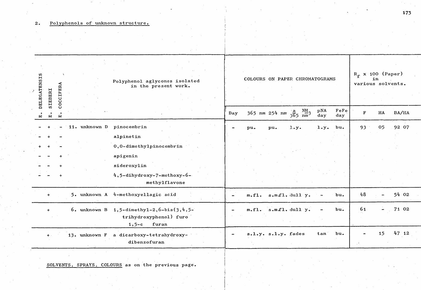
Polyphenol aglycones isolated in the present work.
_ + - 11. unknown D pinocembrin
- + - alpinetin
+ + - 0,0-dimethylpinocembrin
- - + apigenin
- - + sideroxylin
- - + 4,3-dihydroxy-7-methoxy-6-
methylflavone
5. unknown A 4-methoxyellagic acid
6. unknown B 1,5-dimethy1-2,6-bis(3,4,5-
trihYdroxyphenol) furo
1,5-c furan
13. unknown F a dicarboxy-tetrahydroxy-
dibenzofuran
DE
LEG
ATE
NS
IS
SI
EB
ER
I
COCC
IFE
RA
173
2. Polyphenols of unknown structure.
COLOURS ON PAPER CHROMATOGRAMS Rf
x 100 (Paper) in
various solvents.
Day 365 nm a NH
254 nm 3-6.5 1,173 m pNA day
FeFe • day
HA BA/HA
pu. P - 1 .Y- 1.y. bu. 93 05 .9207
m.fl. s.m.fl. dull y. bu. 48 54 02
m.fl. s.m.fl. dull y. bu. 71 02
s.l.y. s.l.y. fades tan• bu. 15 47 12
SOLVENTS, SPRAYS, COLOURS as on the previous page.

- REFERENCES.
174

REFERENCES. 175
1. Arakawa,H. and Nakazaki,M., Chem. & Ind., 73
(1960); Ann., 626, 111 (1960.
2. Arndt,F., in Org.Syntheses (edited by A.H. Blatt),
Coll. Vol. 2, 461 (1943); idem, ibid., 2,
165 (1943).
Ash,A.S.F. and Reynolds,T.M., Aust.J.biol.Sci.,
435 (1954).
4. Audier,H., Bull.Soc.Chim.Fr., 2892 (1966).
5. Badger,G.M. and Christie,B.J., J.Chem.Soc.,
3439 (1956).
6. Baker,W., Harborne,J.B. and 011is,W.D., J.Chem.Soc.,
1303 (1952).
7. Baker,R.T. and Smith,H.G., "The Eucalypts and
their Essential Oils". Govt. Printer,
Sydney, N.S.W., 2nd Ed. (1920).
8. Baldas,J., Bick,I.R.C., Porter,Q.N. and
Vernengo,M.V., Chem.Communications, 132 (1971).
9. Barnes,C.S. and Occolowitz,J.L., Aust.J.Chem.,
17, 975 (1964).
10. Barton,G.M., Evans R.S. and Gardner,J.A.F.,
Nature, 170, 249 (1952).
11. Bate-Smith,E.C., J.Linnean Soc.London (Botany),
21, 95 (1962).
12. Bate-Smith,E.C., Sci.Proc.Roy.Dublin Soc., az, 165 (1956).
13. Bate-Smith,E.C. and Swain,T., Chem. & Ind. (London),
377 (1953),

176
14. Batterham,T.J. and Highet,R.J., Aust.J.Chem., 17,
428 (1964).
15. Bick,I.R.C., Brown,R.B. and Hillis,W.E.,
Aust.J.Chem., 22, 449 (1972).
16. Blakely,W.F., "A Key to the Eucalypts", Forestry
and Timber Bureau, Canberra, 2nd. ed. (1955).
a. page 206. b. page 225.
17. Block,R.J., Durrum,E.L. and Zweig,G., "A Manual
of Paper Chromatography and Paper Electrophoresis"
(2nd. Ed.), Academic Press, New York (1958).
18. Boer,Th.J.de.and Backer,H.J., in OrE4Syntheses,
Coll. Vol. 4, 943 (1963).
19. Briggs,L.H. and Colebrook,L.D., Spectrochimica
Acta, 18, 939 (1962).
20. Carr,D.J. and Carr,S.G.M., Aust.J.Botany, 7,
109 (1959).
21. Carr,D.J. and Carr,S.G.M., Nature, 184, 1549 (1959).
22. Chandler,B.V. and Harper,K.A., Aust.J.Chem., 14,
586 (1961).
23. Chattaway,M.M., Aust.J.Botany, 1, 402 (1953);
2, 21, 28 (1955).
24. Clark-Lewis,J.W., Aust.J.Chem., 21, 3025 (1968).
25. Clark-Lewis,J.W., Rev. pure 12,
96 (1962).
26. Clark-Lewis,J.W., Jackman,L.M. and Spotswood,T.M.,
Aust.J.Chem., 1/, 632 (1964).

177
27. Conroy,H.H., "Advances in Organic Chemistry",
Vol.II, page 265, Interscience, New York (1960).
28. Dadswell,H.E. and Burnell,M., C.S.I.R.Bull.,
62 (1932).
29. Dadswell,H.E., Burnell,M. and Eckersley,A.M.,
C.S.I.R.Bull., 78 (1934).
30. Dichene,E.B., J.Am.Pharm.Assoc., 40, 495 (1951).
31. Dickey,E.E., 1.12Ez.Chem., 12, 179 (1958).
32. Drewes,S.E., J.Chem.Soc. (C), 1140 (1968).
33. Eigen,E., Blitz,M. and Gunsberg,E., Arch.Biochem.
Biophys., 68, 501 (1957).
34. Gauba,E.,and Pryor,L.D., Proc.Linnean Soc.N.S.Wales,
84 278 (1959).
35. Geissman,T.A.(Ed.), "The Chemistry of Flavonoid
Compounds", Pergamon Press, Oxford (1962).
a. pages 107-155. f. page 418.
b. page 110. g• page 419.
c. page 122. h. page 423.
d. page 336. 1. page 429.
e. page 408.
36. Geissman,T.A., in "Modern Methods of Plant Analysis"
(ed. Paech,K. and Tracey,M.Y.) Vol.III
page 475. Springer, Berlin (1955).
37. Geissman,T.A. and Jurd,L., Arch.Biochem.Biophys.,
26, 259 (1955).
38. Grigg,R., Knight,J.A. and Roffey,P., Tetrahedron,
22, 3301 (1966).

178
39. Harborne,J.B., J.Chromatog., 2, 581 (1959).
40. Harborne,J.B. and Geissman,T.A., J.Amer.Chem.
Soc., 21, 831 (1956).
41.
42.
43. Hathaway,D.E., Biochem.J., 82, 80 (1962).
44.. Hemingway,R.W. and Hillis,W.E., J.Chromatog.,
42, 250 (1969).
45. Hemingway,R.W., Hillis,W.E. and Bruerton,K.,
J.Chromatog., 22, 391 (1970).
46.
47.
48.
49.
50.
51.
52.
53. Hillis,W.E.(Ed.), "Wood Extractives"
Academic Press, New York (1962).
a. page 140. b. page 243.
54. Hillis,W.E. and Ishi4ura,N., Aust.J.biol.Sc.„ 22,
1425 (1969).
55. Hillis,W.E. and Isoi,K., Phytochem., 4, 541 (1965).
56. Hbrhammer,L. and Hgnsel,R., Arch.Pharm., 285,
438 (1952).
57. frerhammer,L., Hgnsel,R. and Frank,P., Arch.Pharm.,
286, 481 (1953).
Hasegawa,M.,
Hasegawa,M.,
J.Amer.Chem.Soc., 79, 214,
40,
4202
111
(1957).
(1958). J.Japan.Forestry Soc.,
Heyl,F.W., J.Amer.Chem.Soc., 41, 1289 (1919).
(1966). Hillis,W.E., Phytochem., 2, 541
Hillis,W.E.,
Hillis,W.E.
Hillis,W.E.,
Hillis,W.E.,
Hillis,W.E.,
Phytochem., 2,
6,
6,
6,
6,
1075
259
275
373
845
(1966).
(1967).
(1967).
(1967).
(1967).
Phytochem.,
Phytochem.,
Phytochem.,
Phytochem.,

179
58. HOrhammer,L., Wagner,H. and Hein,K., J.Chromatog.,
12, 235 (1964).
59. Horn,D.H.S., Kranz,Z.H. and Lamberton,J.A.,
Aust.J.Chem., 17, 464 (1964).
60. Horowitz,R.M.„ J.Org .Chem.„ 22, 1733 (1957).
61. Hughes,G.K. and Ritchie,E., Aust.J.Chem., z,
104 (1954).
62. Ibrahim,R.K. and Towers,G.H.N., Arch.Biochem.Biophys.,
82, 125 (1960).
63. Jain,A.C. and Sheshadri,T.R., Quart.Rev., 10,
169 (1956).
64. Jurd,L., Arch.Biochem.Biophia., 62, 376 (1956).
65. Jurd,L., J.Amer.Chem.Soc., 21, 3445 (1956).
66. Jurd,L. and Geissman,T.A., 1.021z2Shem., 21,
1395 (1956).
67. Jurd,L., Geissman,T.A. and Seikel„M.K., Arch.
Biochem.BigEhys., 62, 284 (1957).
68. Jurd,L. and Horowitz,•M• J.Or .Chem., 22, __Pt
1618 (1957).
69. Karrer,W., "Konstitution und Vorkommen der
Esganischen Pflanzenstoffe", Birkhauser
Basel (1958).
a. page 358. e. page 614.
b. page 360. f. page 634.
c. page 361. g. page 635.
d. page 610. h. page 692.

180
70. Kimura,R., J.Pharm.Soc.Japan, 60, 151 (1940).
71. King,F.E. and Acheson,R.M., J.Chem.Soc.,
168 (1930).
72. Kondo,H. and Tomita,M., J.Pharm.Soc.Japan,
21, 139 (1932).
73. Kowkabany,G.N., Advances in Carboh drate Chem.,
2, 303 (1954).
74. Looker,J.H. and Hanneman,W.W., J.Org .Chem.,
27, 381 (1962).
75. Mabry,T.J., Markham,K.R. and Thomas,M.B.,
"The Systematic Identification of Flavonoids",
Springer-Varlag, New York (1970).
a. page 4. e. page 255.
b. page 27. f. page 262.
c. page 35. g. page 296.
d. page 50.
76. Markham,K.R. and Mabry,T.J., Phytochem.,
791 (1968).
77. Markham,K.R. and Mabry,T.J., Phytochem., 2,
1197 (1968).
78. Markham,K.R., Rahman,W., Jahan,S. and Mabry,T.J.,
J.Heteroc clic Chem., 4 61 (1967).
79. Massicot,J. and Marthe,J-P., Bull.Soc.Chim.Fr.,
10, 1962 (1962).
80. Mgule,C., Beitr.wiss.Botan., 4, 166 (1900).
81. Nelson,P.F. and Porter,Q.N., Holzforschung,
Aug., 107 (1967).

181
82. Nordby,H.E., Kew,T.J. and Fisher,J.F.,
J.Chromatog., 24, 257 (1966).
83. NordstrOm,C.G., Acta Chem.Scand 10, 1491 (1956).
84. Occolowitz,J.L. and Wright,A.S., Aust.J.Chem.,
12, 858 (1962).
85. Pauly,H., Z.physiol.Chem., 42, 508 (1904).
86. Pearl,I.A., Beyer,D.L. and Dickey,E.E., J.Org .Chem.,
23, 705 (1958).
87. Pelter,A., Stainton,P. Johnson,A.P. and
Barber M., J.Heterocyclic Chem., 2, 256 (1965).
88. Perkin,A.G., J.Chem.Soc., 1.32, 251 (1906).
89. Perkin,A.G. and Nierenstein,M., J.Chem.Soc.
Trans.Sec., 87, 1412 (1905).
90. Pew P.C. J.Amer.Chem.Soc., 22, 2831 (1955).
91. Pike,K.M., Aust.J.Botany, 4, 13 (1956).
92. Pople,J.A., Schneider,W.G. and Bernstein H.J.,
"High-resolution Nuclear Magnetic Resonance",
pages 392-398, McGraw-Hill, New York (1959).
93. Rao,K.V. and Sheshadri,T.R., Proc.Indian Acad.Sci. (A),
23A, 273 (1946).
94. Reed,R.I. and Wilson,J.M. J.Chem.Soc., 5949 (1963).
95. Robertson,Y.A., Whalley,W.B. and Yates,J.,
J.Chem.Soc., 3117 (1950).
96. Roux,D.G., Nature, 180, 973 (1957).
97. Scott,A.E. "Interpretation of Ultraviolet S ectra
of Natural Products", page 148, Pergamon
Press, London (1964).

182
98. Seikel,M.K. and Geissman,T.A., J.Amer.Chem.Soc.,
72, 5721 (1950).
99. Shain,L. and Hillis,W.E., Eby1222Itaismx, 61,
841 (1971).
100. Shinoda,J., J.Pharm.Soc.Japan, 48, 214 (1928).
101. Smith-White,S., Proc.Linnean Soc.N.S.Wales, 67,
335 (1942).
102. Swain,T., Biochem.J., 22, 200 (1952).
103. Swain,T., Chem. & Ind. (London), 1480 (1954).
104. Thomas,M.B. and Mabry,T.J., Phytochem., 7, 787
(1968).
105. Tomita,M., J.Pharm.Soc.Japan, 22, 138 (1933).
106. Vogel,A.I., "Practical Organic Chemistre, 3rd ed.,
page 970. Longmans, London (1956).
107. Williams, D.H. and Fleming,I., "Spectroscopic
Methods in Organic Chemistry", McGraw-Hill,
London (1966 ).
a. page 54. c. page 89.
b. page 61.
108. Williamson,K.L. and Johnson,W.S. J.Amer.Chem.Soc.,
s2, 4623 (1961).
109. Wilson,R.G., Bowie,J.H. and Williams,D.H.,
Tetrahedron, 24, 1407 (1968).

PUBLICATION.
183
gcoleman
Typewritten Text

This article has been removed for
copyright or proprietary reasons.
gcoleman
Typewritten Text
IC Bick, R Brown, W Hillis, Three Flavanones from leaves of Eucalyptus sieberi, Australian journal of chemistry 1972, 25(2), 449-451.
gcoleman
Typewritten Text