ISOLATION AND OPTIMIZATION OF PROTEASE ... - irjet.net · SPECTROPHOTOMETER FOR MAKING NOVEL...
15
International Research Journal of Engineering and Technology (IRJET) e-ISSN: 2395-0056 Volume: 05 Issue: 02 | Feb-2018 www.irjet.net p-ISSN: 2395-0072 © 2018, IRJET | Impact Factor value: 6.171 | ISO 9001:2008 Certified Journal | Page 900 ISOLATION AND OPTIMIZATION OF PROTEASE PRODUCING BACTERIA FROM SOIL SAMPLE AND ESTIMATE THE ACTIVITY BY SPECTROPHOTOMETER FOR MAKING NOVEL ENZYME-BASED DETERGENTS “GREEN CHEMICALS” N Shobana Sundari 1 1 Coimbatore, Tamil Nadu, India. ---------------------------------------------------------------------***--------------------------------------------------------------------- Abstract - Enzymes are a specialized protein produced in an organism, which is capable in catalyzing a specific chemical reaction. The most important conversions in the context of green chemistry is with the help of enzymes. Enzyme-based detergents also known as ‘green chemicals’ find a wide range of applications in laundry, dishwashing, textile and other such industries. The enzyme preparations like proteases, amylases, lipases and celluloses are considered as indispensable ingredients in these detergents. Among disparate category of enzymes obtained from microbial sources, Protease is an important group of enzyme which conducts proteolysis by hydrolysis Protease production is an inherent capacity of all microorganisms. Bacteria are the predominant group of alkaline protease producers, the genus Bacillus being the most common source. Alkaline proteases have various applications and most studied microbial enzymes at commercial, industrial, pharmaceuticals and also have great significance in Biotechnological studies. The present study was to isolate protease producing bacteria from soil samples collected from Kuthiraimozhi village in Thiruchendure Taluk, Tuticorin district, Tamilnadu, India. The isolation was done by serial dilution and plating methods. All the isolates were screened for proteolytic activity on skim milk agar plate and gelatine hydrolysis method at 37°C for 48 h. Out of 20 bacterial strains screened, maximum protease producing two strains has been selected based on the zone of proteolysis on casein hydrolysed medium Plate. Both the isolated strain has been identified as Bacillus sp and designated as BP2 and BP8, on the basis of morphological and biochemical characters using Bergey s Manual of Determinative Bacteriology. Estimation of enzyme producing activity of bacteria isolate was studied by Spectrophotometry using folin phenol reagent at 660 nm. The growth pattern of the protease producing bacterial culture and the protein degrade activity was analyzed by comparing the crude sample with purified enzyme. Over the period of time the bacterial growth & protease activity was maximum in purified enzyme 46μg/ml/minutes compare with crude sample 16μg/ml/minutes. Optimization of the physiochemical parameters were done by shake flask study. Maximum yield of protease was obtained after 48h of incubation. The optimum enzyme activity & maximum yield of protease was found to be at pH 7 and temperature 37ºC, Carbon source as Starch and Nitrogen source as Beef extract. SDS PAGE were performed to identify the molecular mass of the protease enzyme. Biocleaning and biowashing study was analyzed by compare with the various laundry detergent and enzyme-based detergent mixture on stained blood, egg cloths, cleaning optical lenses & surgical instrument foulded by blood protein. Over the period of time the maximum stained removal and cleaning effect was observed in the enzyme-based detergents mixture as compared to detergent sample. The biocleaning and biowashing process of the above study concluded that the isolated protease enzyme producing Bacillus sp. could be used as a promising agent of ‘Biocleaner’/‘Green chemical’ for the removal of stain effectively. For further process the bacterial strain BP2 & BP8 was submitted to Biotech laboratory for 16s rRNA sequencing. Further experiments were carried out to enhance enzyme production for commercialized purpose. Key Words: 1. Protease producing microorganisms, 2. Bacillus Sp, 3. Proteolytic activity, 4. Casein & Gelatin hydrolysed 5. Spectrophotometer, 6. SDSPAGE, 7. Detergent and Enzyme –based detergent / Green chemical. 1. INTRODUCTION Soil microorganism, particularly bacteria play an pivotal roles in various biogeochemical cycles and are responsible for the cycling of organic compounds. Microorganisms constitute a huge and almost unexplained reservoir of resources likely the most important source of enzyme production & to provide innovative applications useful to man, They represent by far the richest repertoire of molecular and chemical diversity in nature. Many bacteria produce enzymes which they secrete into the world around them. These enzymes breakdown the target molecules outside the bacterial cell and which can then be absorbed by them. Enzymes are the most important substances use day to day life in so many areas either in research, medicine or commonly in industries and are more stable than with plant and animal and obtained cheaply . Protease is an significant group of enzyme which conducts proteolysis by hydrolysis of the peptide bonds that link amino acid composed in the polypeptide chain. Protease synthesis is an immanent range of all microorganisms. Bacteria are the prevalent group of alkaline protease producers, the genus Bacillus being the most common Assistant Professor, Department of Biotechnology, RVS College of Arts & Science, Sulur,
Transcript of ISOLATION AND OPTIMIZATION OF PROTEASE ... - irjet.net · SPECTROPHOTOMETER FOR MAKING NOVEL...

International Research Journal of Engineering and Technology (IRJET) e-ISSN: 2395-0056
Volume: 05 Issue: 02 | Feb-2018 www.irjet.net p-ISSN: 2395-0072
© 2018, IRJET | Impact Factor value: 6.171 | ISO 9001:2008 Certified Journal | Page 900
ISOLATION AND OPTIMIZATION OF PROTEASE PRODUCING BACTERIA
FROM SOIL SAMPLE AND ESTIMATE THE ACTIVITY BY
SPECTROPHOTOMETER FOR MAKING NOVEL ENZYME-BASED
DETERGENTS “GREEN CHEMICALS”
N Shobana Sundari1
1
Coimbatore, Tamil Nadu, India. ---------------------------------------------------------------------***---------------------------------------------------------------------
Abstract - Enzymes are a specialized protein produced in an organism, which is capable in catalyzing a specific chemical reaction. The most important conversions in the context of green chemistry is with the help of enzymes. Enzyme-based detergents also known as ‘green chemicals’ find a wide range of applications in laundry, dishwashing, textile and other such industries. The enzyme preparations like proteases, amylases, lipases and celluloses are considered as indispensable ingredients in these detergents. Among disparate category of enzymes obtained from microbial sources, Protease is an important group of enzyme which conducts proteolysis by hydrolysis Protease production is an inherent capacity of all microorganisms. Bacteria are the predominant group of alkaline protease producers, the genus Bacillus being the most common source. Alkaline proteases have various applications and most studied microbial enzymes at commercial, industrial, pharmaceuticals and also have great significance in Biotechnological studies. The present study was to isolate protease producing bacteria from soil samples collected from Kuthiraimozhi village in Thiruchendure Taluk, Tuticorin district, Tamilnadu, India. The isolation was done by serial dilution and plating methods. All the isolates were screened for proteolytic activity on skim milk agar plate and gelatine hydrolysis method at 37°C for 48 h. Out of 20 bacterial strains screened, maximum protease producing two strains has been selected based on the zone of proteolysis on casein hydrolysed medium Plate. Both the isolated strain has been identified as Bacillus sp and designated as BP2 and BP8, on the basis of morphological and biochemical characters using Bergey s Manual of Determinative Bacteriology. Estimation of enzyme producing activity of bacteria isolate was studied by Spectrophotometry using folin phenol reagent at 660 nm. The growth pattern of the protease producing bacterial culture and the protein degrade activity was analyzed by comparing the crude sample with purified enzyme. Over the period of time the bacterial growth & protease activity was maximum in purified enzyme 46µg/ml/minutes compare with crude sample 16µg/ml/minutes. Optimization of the physiochemical parameters were done by shake flask study. Maximum yield of protease was obtained after 48h of incubation. The optimum enzyme activity & maximum yield of protease was found to be at pH 7 and temperature 37ºC, Carbon source as Starch and Nitrogen source as Beef extract. SDS PAGE were performed to identify the molecular mass of the protease enzyme. Biocleaning and biowashing study was analyzed by compare
with the various laundry detergent and enzyme-based detergent mixture on stained blood, egg cloths, cleaning optical lenses & surgical instrument foulded by blood protein. Over the period of time the maximum stained removal and cleaning effect was observed in the enzyme-based detergents mixture as compared to detergent sample. The biocleaning and biowashing process of the above study concluded that the isolated protease enzyme producing Bacillus sp. could be used as a promising agent of ‘Biocleaner’/‘Green chemical’ for the removal of stain effectively. For further process the bacterial strain BP2 & BP8 was submitted to Biotech laboratory for 16s rRNA sequencing. Further experiments were carried out to enhance enzyme production for commercialized purpose. Key Words: 1. Protease producing microorganisms, 2. Bacillus Sp, 3. Proteolytic activity, 4. Casein & Gelatin hydrolysed 5. Spectrophotometer, 6. SDSPAGE, 7. Detergent and Enzyme –based detergent / Green chemical.
1. INTRODUCTION Soil microorganism, particularly bacteria play an pivotal roles in various biogeochemical cycles and are responsible for the cycling of organic compounds. Microorganisms constitute a huge and almost unexplained reservoir of resources likely the most important source of enzyme production & to provide innovative applications useful to man, They represent by far the richest repertoire of molecular and chemical diversity in nature. Many bacteria produce enzymes which they secrete into the world around them. These enzymes breakdown the target molecules outside the bacterial cell and which can then be absorbed by them. Enzymes are the most important substances use day to day life in so many areas either in research, medicine or commonly in industries and are more stable than with plant and animal and obtained cheaply . Protease is an significant group of enzyme which conducts proteolysis by hydrolysis of the peptide bonds that link amino acid composed in the polypeptide chain. Protease synthesis is an immanent range of all microorganisms. Bacteria are the prevalent group of alkaline protease producers, the genus Bacillus being the most common
Assistant Professor, Department of Biotechnology, RVS College of Arts & Science, Sulur,

International Research Journal of Engineering and Technology (IRJET) e-ISSN: 2395-0056
Volume: 05 Issue: 02 | Feb-2018 www.irjet.net p-ISSN: 2395-0072
© 2018, IRJET | Impact Factor value: 6.171 | ISO 9001:2008 Certified Journal | Page 901
source. Alkaline proteases have various applications and most studied microbial enzymes at commercial, industrial, pharmaceuticals, diagnostic, effluent abatement, environmental bioremediations and also have great significance in Biotechnological studies. Uses of protease are infinite and can be characterized approximately into detergents, dairy industry, tanning, baking and brewing industries. Proteases implement a large variety of functions and have significant biotechnological applications. Bacillus synthesis a wide variety of extra-cellular enzymes, including proteases. Several Bacillus species involved in protease synthesis are e.g. B. cereus, B. sterothermophilus, B. mojavensis, B. megaterium and B. Subtilis. Proteases is in laundry detergents act as a stains removing agent from clothing. Protease treatments can modify the surface of wool and silk fibres to supply novel and exclusive finishes. Proteases have been used in the skin de hairing process, where de hairing is carried out at pH values between 8. and it also used as contact-lens enzyme cleaners and enzymatic deriders. The proteolytic enzymes also offer a moderate and selective debridement, supporting the natural therapeutic process in the successful local management of skin ulcerations by the efficient removal of the necrotic material. With esteem to the factors affecting yield and properties of the proteases, it was measured of interest to isolate new sources of protease, purify proteases and examine the factors affecting their activity. The present study was to isolate protease producing bacteria from soil samples and to study potential application of the proteases for industrial applications. (Dalal Rupali,2015).
2. MATERIAL AND METHODS 2.1 Soil Analysis :
The soil was analysed from tamilnadu agricultural soil testing laboratories, Coimbatore. James L. Walworth, (2006).
2.2 Source of soil sample collection:
The soil samples were collected from in an around Kudhirai mozhi village, Thiruchendur, Tamil Nadu, India. Soil Sample were collected below 15-16 cm depth and stored in sterile plastic bags at 4° C . Date and time were noted on sample collecting bags. Singh, Dhyan, et. al, (1999).
2.3 Isolation of Microorganism:
Collected soil was diluted by serial dilution technique. In this technique sample was prepared by adding 1g of soil in 9 ml distilled water i.e. 10-1 to 10-4 dilution factor. After serial dilution Nutrient Agar media (NAM) plate was prepared for the culture of bacteria and Actinomycetes isolation agar for the culture of actinomycetes. Under aseptic condition 0.1 ml of soil sample from each sites were spread on the agar plate by glass spreader and incubated at 370C for 24 hours. Mixed culture was obtained on the plates after 24 hours. Pure
culture was obtained by streaking technique after incubation at 37 0C for 24 hours by picking single colony from different mixed culture plates. Colony forming units were determined after 2 days of incubation at 370C. (M. Alexander,1977). 2.4 Morphological study of isolated Microorganism: Staining technique was carried for identification of morphological study. 2.4.1 Gram’s staining: Place the culture to cleaned slide. Then prepare the smear and heat fix. Cover the smear with crystal violet and allow it to stand for 20-60 seconds. Wash off the stain for two seconds using a wash bottle of distilled water. Cover the smear with Gram’s iodine for 60 seconds. Decolorize the smear with 95% ethyl alcohol for 10-20 seconds or until no more colour flows from the smear. Immediately wash the smear with distilled water for 2 seconds and drain. Cover the smear with safranin for 20 seconds. Wash with distilled water for 2 seconds. Blot dries the smear. Let the stained slide air dry. Examine the smears 1st under low power and then under oil immersion objectives for size, shape and arrangement of cells. 2.4.2 Negative staining: Take a clean grease free glass slide. Put a small drop of nigrosin close to one end of the slide. Transfer a loopful of broth culture into the drop of stain and mix gently with the loop. Take a spreader slide holds it an angle of 30 degree and move it toward drop of suspension until it contacts the drop causing the liquid to spread along its spreading edge. Allow the smear to air-dry completely without any heat fixation. Observe the stained smear under oil immersion for colour of cells. 2.4.3 Spore staining: Steams fix the smear by placing the slide, smear side up, on the edge of a beaker of boiling water. Cover the heat fixed smear with blotting paper and saturate it with malachite green. The smear is heated over a hot water both for five minutes by adding more malachite green. Allow the smear to cool, remove the paper and rinse the smear with water for 30 seconds. Counter stain the washed smear with safranin for 30 seconds. Wash the smear again gently with water to remove safranin. Blot dries the smear. Observe the blotted dry smear under oil immersion objective for endospores. 2.4.4 Motility: Take a clean cover slip and apply Vaseline to four corners of cover slip using a match stick. Place the cover slip on a clean paper with the Vaseline side up. Transfer one to two loopfuls of culture in the centre of the cover slip with the help of sterile inoculating loop. Take a clean grease free cavity slid. Place the cavity slide over the cover slip so that the drop is

International Research Journal of Engineering and Technology (IRJET) e-ISSN: 2395-0056
Volume: 05 Issue: 02 | Feb-2018 www.irjet.net p-ISSN: 2395-0072
© 2018, IRJET | Impact Factor value: 6.171 | ISO 9001:2008 Certified Journal | Page 902
paced in the centre. Carefully invert the slide so that drops hang in the well. Observe under microscope. 2.5 Biochemical test: Biochemical test was carried for identify the enzymatic activity of bacteria. These tests are use full for identify the unknown bacterium. 2.5.1 Indole test: Inoculate samples to 1% tryptone broth and maintain one uninoculated control tube. Incubate inoculated and un inoculated tubes at 35 degree Celsius for 24-48 hours. Add 1ml of Kovac’s reagent to each tube including control. Shake the tubes gently. Allow the tubes to stand to permit the reagent to come to the top. Observe the tubes for the red violet ring at the interface between the top of the broth and the reagent. Appearance of red ring in the tube indicates the positive result and no ring form indicate the negative result. 2.5.2 Methyl Red test: Pour 5 ml of MR-VP broth in each tube. Inoculate samples to MR-VP tubes and kept as un inoculated control. Incubate the inoculated and un inoculated tubes at 35 degree Celsius for 24-48 hours. After incubation, transfer 2.5 ml of the broth culture to another tube. Add 5 drops of methyl red to each tube between the palms of the hands to disperse the methyl red. Observe the colour change. The medium change to red colour indicates positive result and no colour change indicates the negative result. 2.5.3 Voges-Proskauer test: Inoculate the isolated samples into the MR-VP broth tubes and keep one tube as un inoculated control. Incubate all tubes at 35 degree Celsius for 48 hours. Transfer 2.5 ml of broth culture to another sterilised tube. Add 6 drops of Alpha- napthanol and 2 drops of KOH to the broth culture. Gently mix the contents of tube and observe the colour change. The medium change to red colour indicates positive result and no colour change indicates the negative result. 2.5.4 Citrate Utilization test: Inoculate samples to SC agar slant and keep one tube as un inoculated control. Incubate all the tubes at 35°C for 24-48 hours. Observe the slant culture for growth and colour change of the medium. The medium change to blue colour indicates the positive result and no colour change indicate negative result. 2.5.5 Catalyse test: Take one clean slid then place the small drops of H2O2.Then add sample culture into it streak well observe the changes. Formation of bubble indicates the positive result and no changes indicate the negative result.
2.5.6 Nitrate reduction test: Take 5 ml nitrate broth with a Durham tube in culture tubes and sterilised them by autoclaving. Inoculate the test organisms in the nitrate broth tubes. Incubate at 35°C for 48 hours. Observe the incubated tubes for gas production. Mix an equal volume 0.5 ml of reagent A with 0.5 ml of reagent B just before use. Add o.1 ml of the test reagent to the broth cultures. Observe for any change in colour immediately. Within minutes of positive and negative test if no change develops after adding the test reagents, it is taken as a negative test. 2.5.7 Lactose and Glucose fermentation test: Inoculated the organism into the fermentation medium contain tubes (prepare lactose and glucose fermentation media separately) and incubate the tubes at 35°C for 24-48 hours. Observe for the colour change in the broth and gas production in the Durham tube. 2.5.8 Casein Hydrolysis test: Prepare the skim milk agar plates. Inoculate the sample cultures and keep one plate for control. Incubate the all the plates for 24-48 hours at 37°C in an inverted position. Observe the inoculated plate for any clearing zone the line of growth. The appearance of zone indicate the samples are hydrolyse the casein and no zone indicate the no hydrolyse the casein. 2.5.9 Urease test: Sample was inoculated into the urease agar tube then it was incubated at 37°C for 24 hours. After incubation pink colour indicates the positive result and no change indicate the negative result. 2.5.10 TSI test: Sample was inoculated into the TSI agar tubes then it was incubated at 37°C for 24 hours. After incubation colour change indicates the positive result and no change indicate the negative result. 2.5.11 Mannitol motility agar test: Sample was inoculated into the mannitol motility agar tubes then it was incubated at 37°C for 24 hours. After incubation changes indicates the positive result and no change indicate the negative result. 2.5.12 Oxidase test:
Culture was strike in oxidase disc and sees the violet colour change it indicates the positive result and no change indicate the negative result.

International Research Journal of Engineering and Technology (IRJET) e-ISSN: 2395-0056
Volume: 05 Issue: 02 | Feb-2018 www.irjet.net p-ISSN: 2395-0072
© 2018, IRJET | Impact Factor value: 6.171 | ISO 9001:2008 Certified Journal | Page 903
2.6 Growth curve cycle:
Inoculate aseptically using sterile pipette the LB broth flask with 1ml of inoculum. The tubes used should have matched transmittance fitting to cuvette slot for reading the transmittance directly keeps one flask as blank.
2.7 Optimization of growth using different source and different environment condition:
Optimize the bacterial growth by using of different source such as different Carbon, Nitrogen source and different environment conditions are pH and Temperature. These carried in Protease production medium. Media composition is Glucose (0.5%), Peptone (0.75%), Magnesium sulphate (0.5%), Potassium dihydrogen ortho phosphate (0.5%), ferrous sulphate (0.01%), pH (7.0).Then the Carbone and Nitrogen sources are altered. Arun Kumar Sharma, Vinay Sharma, et. al, (2015.) 2.7.1 Carbon sources:
Change the carbon source from protease production medium then add different types of carbon source such as starch, glucose, fructose, mannitol, sucrose. Then sterilize the medium and inoculate the sample culture into the medium incubate room orbital shaker at 24-48 hours. After 24-48 hours the OD will take from spectrophotometer at 600 nm. (Abebe,et. al 2014). 2.7.2 Nitrogen sources: Change the nitrogen source from protease production medium then add different types of nitrogen source such as ammonium sulphate, ammonium nitrate, and sodium nitrate, and beef extract, yeast extract. Then sterilize the medium and inoculate the sample culture into the medium incubate room orbital shaker at 24-48 hours. After 24-48 hours the OD will take from spectrophotometer at 600 nm. 2.7.3 Different Temperature: Change the incubating temperature like 4-100° C to analyse the growth rate in the medium. This technique carried in protease production medium. Add the culture into the medium then incubate different temperature for 24-48 hours. After incubation the culture was analysed using spectrophotometer at 600 nm. Abebe, Bizuye , A,sago, et. al, (2014). 2.7.4 Different pH: Change the pH from production medium like 2-10 then analyse the growth of bacteria in the medium. Add the culture in to the medium has different pH then incubate room temperature in orbital shaker. After incubation the medium analyse using spectrophotometer at 600 nm. (Abebe,et. al 2014).
2.8 Change the carbon and nitrogen source in production media:
To change the carbon and nitrogen source in protease production media for optimization of growth. Change the glucose then replace the starch as a carbon sours and change the peptone then replace the beef extract as nitrogen sours. Then the culture was inoculated and incubated in 37°C for 48 hours. After 48 hours incubation the OD value taken in spectrophotometer at 600 nm. F Soundra Josephine, Ramya V S, et. al, (2012).
2.9 Qualitative analysis of protease enzyme:
To identify crude sample as protein some test were carried out as biuret test, Ninhydrin test, Xanthoproteic test. This test gives colour reactions on the basis amino acid present (Deb A C, 1996).
2.9.1 Quantitative analysis of protease enzyme by Lowry’s method:
Prepare the protease production medium and inoculate the sample culture into it then incubate at 37° C for 24 hours in orbital shaker. After incubation the cultured medium was take in one clean tube then centrifuge at 5000 rpm for 5-10 minutes. The supernatant was transferred another clean tube. Prepare standard from bovine serum albumin concentration is 1mg/ml. The test tubes were labelled as blank, 0.2, 0.4, 0.6, 0.8, 1and unknown sample. The test tube with 4 ml of distilled water labelled as blank. Micropipette was used to pipette out the require volume of working standard in respected labelled test tube (0.2-1ml) and make up to 4ml with distilled water. Pipette out 0.2ml of unknown sample solution and transferred into respected labelled test tube. Add 2ml of alkaline copper sulphate reagent to the all test tubes. Mix the tubes well and incubate 10 minutes. After incubation add 0.2ml of folin phenol reagents to all the tube and incubate for 30 minutes in dark. Zero the calorie meter with blank take the OD at 660 nm. Plot absorbance protein concentration to get a standard calibration curve. The unknown concentration of crude protease was determined using tyrosine standard curve. Estimate the amount of protein presents in the given sample from standard graph. Lowry. O.H, Rosenbrough, N.J, et. al (1951).
2.10 Purification of enzyme by Acetone precipitating method:
Fermentation broth was taken into a centrifuge tube at the end of 24 h, 48 h and 72 h of incubation and it was centrifuged at 8000 rpm, 4 ºC for 15 minutes. After centrifugation, bacterial cell pellet was removed and bacteria free supernatant was used as a source of purification process 2ml of crude enzyme was taken one tube then added 10 ml of ice cold acetone in to it mix well. Then keep it in 4°C for 24 hours. After 24 hours the mixture was centrifuged at 10000 rpm for 10 minutes in 4°C. the pellet was mixed with PBS and it will use further process. Balakrishnan, padmapriya, et. al, ( 2012).

International Research Journal of Engineering and Technology (IRJET) e-ISSN: 2395-0056
Volume: 05 Issue: 02 | Feb-2018 www.irjet.net p-ISSN: 2395-0072
© 2018, IRJET | Impact Factor value: 6.171 | ISO 9001:2008 Certified Journal | Page 904
2.10.1 Demonstration of enzyme activity:
2.10.1.1 Crude enzyme assay activity:
1ml of crude enzyme was added to 1ml of 1% casein then add 1ml of PBS for stimulate the enzyme activity. Mixed well and incubated 37°C for 30-45 hours. After incubation 5% of TCA 1ml was added into it. TCA is used for stop the further enzyme activity. Then it was filtered by Whatmann’s No 1 filter paper. 1ml filtered was taken in test tube then add 2.5ml of analytical reagent mix well incubate 10 minutes in room temperature. After incubation add 120µl of Folin phenol reagents and incubate 30 minutes in dark. Set the blank without enzyme. After incubation the mixture was taken OD in coloury meter at 660 nm. Grewal, S and Mutha, P,(2010). 2.10.1.2 Purified enzyme assay activity:
1ml of purified enzyme was added to 1ml of 1% casein then add 1ml of PBS for stimulate the enzyme activity. Mixed well and incubated 37°C for 30-45 hours. After incubation 5% of TCA 1ml was added into it. TCA is used for stop the further enzyme activity. Then it was filtered by Whatmann’s No 1 filter paper. 1ml filtered was taken in test tube then add 2.5ml of analytical reagent mix well incubate 10 minutes in room temperature. After incubation add 120µl of Folin phenol reagents and incubate 30 minutes in dark. Set the blank without enzyme. After incubation the mixture was taken OD in coloury meter at 660 nm. Rashesh, Doshi et.al, (2001). 2.11 Identification of Molecular weight of protein sample:
SDS-PAGE method used for identification of protein molecular weight, the protein sample was in polyacrylamide gel in the electrophoresis chamber after running of sample the gel was carefully removed and stained with coomasic brilliant blue for overnight then distained by using of methanol: acetic acid: distilled water (300: 300: 100ml). After distaining molecular weight determined by comparison of protein marker. Barrett A.J, Rawlings ND, Woessner JF, (2003).
2.12 Washing test:
Application of protease enzyme by isolated organism as a detergent additive was studied as per (Sidra Aftab et. al 2006). For this three white cotton clothes (5*5cm) were stained with cumatiand brilliant blue stain separately and following sets were then prepared and studied: coomasie brilliant blue and blood stained cloth dipped in petri plate with distilled water (10ml). coomasie brilliant blue and blood stained cloth dipped in petri plate with distilled water(10ml)+0.5g detergent, coomasie brilliant blue and blood stained cloth dipped in petri plate with distilled water (10ml)+5mlenzyme solution. coomasie brilliant blue and blood stained cloth dipped in petri plate with distilled water (10ml) +0.5ml detergent +5ml enzyme solution. All four
flasks were incubated at room temperature for 24 houres min. After incubation, cloth pieces were taken out, rinsed with water and dried. Visual examination of cloth pieces exhibited the effect of enzymes in removal of stains. Untreated cloth pieces stained with coomasie brilliant blue and blood were taken as control. Sevinc, N and Demirkan, E, (2011).
3. RESULT AND DISCUSSION:
3.1 Soil Analysis Report:
The soil analysed from tamilnadu agricultural soil testing laboratory. Table 3.1.1 were show the result of soil analysis. 3.2 Isolation of Microorganism:
From of collected soil sample, 5 bacterial colonies were isolated by using off pure culture technique. All samples shown clear zone around streaked lines. Sample one given the high zone in skim milk agar plate. Then the 5 sample one was selected for further study. Five bacterial isolates from soil sample were named as S1, S2, S3, S4, S5, respectively. Figure 3.2.1 and Table 3.3.1 show the growth of the bacteria on plate.
Table 3.1.1: Physicochemical analysis of soil
Parameters Result
Soil texture Soil mixed sand soil
Amount of calcium carbonate No
Electric conductivity (dSm-1) 0.18
pH 7.3
Nitrogen (Kg/ekker) 45
Phosphorous (Kg/ekker) 7.0
Potassium (Kg/ekker) 50
Iron (ppm) 5.4
Manganese (ppm) 2.8
Zinc (ppm) 1.0
Copper (ppm) 0.9
Figure 3.2.1 : Ioslation of protease producing bacteria by serial dilution & spread plate method.

International Research Journal of Engineering and Technology (IRJET) e-ISSN: 2395-0056
Volume: 05 Issue: 02 | Feb-2018 www.irjet.net p-ISSN: 2395-0072
© 2018, IRJET | Impact Factor value: 6.171 | ISO 9001:2008 Certified Journal | Page 905
Figure 3.2.2 : Show the result of Pure culture.
3.3 Morphological study of isolated Microorganism:
3.3.1 Gram’s staining Result: Appearance of pink colour rod shaped bacilli indicates the gram negative bacteria in all samples. Table 3.3.1 and Figure 3.3.1.1 show the Gram’s staining result.
Figure 3.3.1.1: show the result of Gram’ s staining.
3.3.2 Negative staining Result:
The Nigrosin stained smear shows blue background and the colourless diblobacilli of the sample bacteria. Table 3.3.1 and Figure 3.3.2.1 show the morphology of the samples.
Figure 3.3.2.1: Show the result of Negative staining
3.3.3 Spore staining result:
Endospores are visible as green oval structures outside and inside the rod shaped red coloured vegetative cells. Table 3.3.1 and Figure 3.3.6 show the morphology of the samples. 3.3.4 Motility test Result:
Movement of the bacteria indicate the positive result of motility test. Table 3.3.1 show the result of motility. 3.4 Characterization of isolated strain: 3.4.1 Indole test Result: No red colour development in all the tube represents an indole negative. Table 3.4.1 and Figure 6 (a) and (b) show the negative result. 3.4.2 Methyl Red test Result: The development of a stable red colour in all the all inoculated tube is due to the metabolism of pyruvic acids resulting in decrease of pH of broth to 4.2 and the methyl red turns red at this pH (methyl red positive test). Table 2 and Figure 8 show the positive result.
Figure 3.4.2.1: Show the result of MR test
3.4.3.Voges-Proskauer test Result:
No red colour development in all inoculated tube indicates the VP negative. Table 3.4.1 and Figure 3.4.3.1(c) negative result.

International Research Journal of Engineering and Technology (IRJET) e-ISSN: 2395-0056
Volume: 05 Issue: 02 | Feb-2018 www.irjet.net p-ISSN: 2395-0072
© 2018, IRJET | Impact Factor value: 6.171 | ISO 9001:2008 Certified Journal | Page 906
Table 3.3.1: Morphological identification of Bacterial strains isolated from Kuthiraimozhi village soil
Figure 3.4.3.1: show the result of VP test
3.4.4 Citrate utilization test result: No color change in all inoculated tubes so it indicates the negative result in citrate utilization test. . Table 4.3.1 and Figure 3.4.4.1 (a) and (b) negative result. 3.4.5 Catalyse test result: Bubble formation indicates the positive result in catalyse test from all the samples. Table 4.3.1 and Figure 3.4.4.1 8 (a)positive result. 3.4.6 Urease test result: No colour change in all inoculated tubes so it indicates the negative result in citrate utilization test. . Table 4.3.1 and Figure 3.4.4.1 6(a) and (b) negative result. 3.4.7 TSI test result: After incubation the bud and slant was changed yellow colour its indicate the positive result. Table 4.3.1 and Figure 3.4.4.1 6 (a) and (b) show the positive result.
Figure 3.4.4.1 show the result of indol, manitol motility agar, TSI, urease, citrate test.
3.4.8 Mannitol motility agar test result: After incubation the medium changed yellow colour and turbidity it indicates the positive result of motility. Table 4.3.1 and Figure 3.4.4.1 6(a) and (b) show the positive result. 3.4.9 Oxidase test result: Changes of violet colour indicate the positive result. Table 4.3.1 and Figure 3.4.4.1 8 (b) show the positive result.
Sample Gram’s staining
Negative staining
Spores staining
Motility
S1 Positive Short chain Endo
spores Motile
S2 Positive Short chain Endo
spores Motile
S3 Positive Short chain Endo
spores Motile
S4 Positive Short chain Endo
spores Motile
S5 Positive Short chain Endo
spores Motile
S6 Positive Short chain Endo
spores Motile
S7 Positive Short chain Endo
spores Motile
S8 Positive Short chain Endo
spores Motile
S9 Positive Short chain Endo
spores Motile
S10 Positive Short chain Endo
spores Motile
S11 Positive Short chain Endo
spores Motile
S12 Positive Short chain Endo
spores Motile
S13 Positive Short chain Endo
spores Motile
S14 Positive Short chain Endo
spores Motile
S15 Positive Short chain Endo
spores Motile
S16 Positive Short chain Endo
spores Motile

International Research Journal of Engineering and Technology (IRJET) e-ISSN: 2395-0056
Volume: 05 Issue: 02 | Feb-2018 www.irjet.net p-ISSN: 2395-0072
© 2018, IRJET | Impact Factor value: 6.171 | ISO 9001:2008 Certified Journal | Page 907
3.4.10 Nitrate reduction test result: Development of red colour and gas indicates the positive result. Table 3.4.1and Figure 3.4.10.1 show the positive result.
Figure 3.4.10.1: show the result of Nitrate reduction test.
3.4.11 Lactose and Glucose fermentation test result: Development of yellow colour in the medium indicates the fermentation of sugars. Table 3.4.1 and Figure 3.4.11.1 show the positive result.
Figures 3.4.11.1 Show the result of Carbohydrade fermentation test
3.4.12 Casein Hydrolysis test result:
A clear area around the all samples growth is a positive reaction for extracellular caseinase secretions. The s
1
was given the high zone so this sample used for further study. (D.Narendra, et al, 2012).
Table 3.4.1: Biochemical Characterization Of Bacterial strain isolated from Kuthiraimozhi village soil
.B
act
eri
al
stra
in
Ind
ole
T
est
MR
T
est
VP
T
est
Cit
rate
T
est
Ca
sein
h
yd
roly
sis
Ox
ida
se
Te
st
cata
lysi
s T
est
Ure
ase
T
est
TS
I
Te
st
MM
A
Te
st
La
cto
se
Te
st
Glu
cose
T
est
Nit
rate
re
du
ctio
n
S1 - + - - + + + - ++ ++ + + +,G S2 - + - - + + + - ++ ++ + + + S3 - + - - - + + - - - - + - S4 + + - - + - + + + - + - + - S5 + + - + + - + + - + - - + + S6 + + - - + + + - - + + + + S7 - + - - + + - - ++ + + + + S8 - - + + + + + + ++,G ++ + + + S9 - - + + + + + - ++ ++ + + -
S10 - - - + - - + - ++, G - - + - S11 - + - + - + + - ++ - - - - S12 + + - + - + + - ++ ++ + + - S13 - + - + + + + - ++ - + + - S14 + + - + + + + - - + - - + - S15 - - - - - + + - +,- + + - + - S16 + - + - - - - - + + - - +
Figure 3.4.12.1: Show the result of Casein hydrolysis

International Research Journal of Engineering and Technology (IRJET) e-ISSN: 2395-0056
Volume: 05 Issue: 02 | Feb-2018 www.irjet.net p-ISSN: 2395-0072
© 2018, IRJET | Impact Factor value: 6.171 | ISO 9001:2008 Certified Journal | Page 908
Table 3.4.12.1: show the positive result and diameter of the zone.
Sample Growth Zone of the
inhibition(mm)
S1 Yes 20
S2 Yes 19
S3 Yes 12
S4 Yes 15
S5 Yes 11
S6 Yes 7
S7 Yes 8
S8 Yes 12
S9 Yes 14
S10 Yes 11
S11 Yes 13
S12 Yes 10
S13 Yes 7
S14 Yes 6
S15 Yes 14
S16 Yes 12.8
3.5 Growth curve cycle Result: The bacterial growth curve on colorimeter is measured by determining the degree of light transmission through the culture. The less light transmitted indicates the presence of more bacteria in the sample. The graph drawn between the OD and time gives the grown curve. Number of cells which varies with the incubation period can be determined by comparing the obtained OD with reference curve drawn between OD and number of cells. (Table and Figure 3.5.1)
Table 3.5.1: show the result of growth curve cycle
Time (minutes) OD (Nano meter)
0 0
60 0
120 0
180 0.02
240 0.09
270 0.18
300 0.39
330 0.59
Figure 3.5.1 : The graph show the result of Bacterial
growth curve cycle
The growth of the isolates were studied. The optical density of Basillus sp. were determined at 600 nm. The growth of the isolate is much better at in the medium which is clearly shown in the Figure:3.5.1.
3.6 Optimization of the various physiochemical parameters by using shake flask study for the maximum production of protease.
3.6.1 Effect of different Nitrogen source on protease
Production:
After 48 hours incubation growth of bacterial cells high in Beef extract and Yeast extract added medium. So the production and growth will increase by add the Beef extract and Yeast extract. Table and graph 3..6.1.1 show the result of production and growth optimization. The OD value takes from spectrophotometer at 600nm.
Table 3.6.1.1 : Effect of different Nitrogen source on protease production by Bacillus cereus after 48 hours:
Figure 3.6.1.1: Effect of Different Nitrogen Sources on Optimization of Bacterial growth and Protease
production.

International Research Journal of Engineering and Technology (IRJET) e-ISSN: 2395-0056
Volume: 05 Issue: 02 | Feb-2018 www.irjet.net p-ISSN: 2395-0072
© 2018, IRJET | Impact Factor value: 6.171 | ISO 9001:2008 Certified Journal | Page 909
The nitrogen source was found to be essential for the growth of Basillus sp. The percentage of Bacterial growth and Protease production was evaluated at different nitrogen sources (Ammonium sulphate, Ammonium nitrate, Sodium nitrate, Beef extract .and Yeast extract). Among these, the percentage of Bacterial growth and Protease production was maximum in Beef extract containing medium and therefore Beef extract was selected for the rest of the experiments. 3.6.2 1 Effect of different Carbon source on protease production :
After 48 hours incubation growth of bacterial cells high in starch and Fructose added medium. So the production and growth will increase by add the Starch, Mannitol and Fructose. Table and graph 3.6.2.1 show the result of production and growth optimization. The OD value takes from spectrophotometer at 600nm.
Table 3.6.2.1: Effect of different Carbon source on protease production by Bacillus cereus after 48 hours:
Carbon source OD value of Positive
control with different source (600 nm)
OD value of sample result (600 nm)
Starch 0.519 2.077
Mannitol 0.129 2.059
Fructose 0.440 2.074
Sucrose 0.034 2.069
Glucose 0.840 2.063
Figure 3.6.2.1: Effect of Different Carbon Sources on
Optimization of Bacterial growth and Protease production.
The carbon source was found to be essential for the growth of Basillus sp. The percentage of Bacterial growth and Protease production was evaluated at different carbon sources (Starch. Glucose, Fructose, Mannitol and Sucrose). Among these, the percentage of Bacterial growth and Protease production was maximum in Starch containing medium and therefore Starch was selected for the rest of the experiments.
3.6.3 Effect of different Temperature on Protease production: After 48 hour’s incubation growth of bacterial cells high in room temperature and 37°C. So the production and growth will increase in room temperature and 37°C. Table and graph 3.6.3.1 show the result of production and growth optimization. The OD value takes from spectrophotometer at 600nm.
Table 3.6.3.1: Effect of different Temperature on
hours:
Temperature OD value of Positive
control with different source (600 nm)
OD value of sample result
(600 nm)
8°C 0.840 0.00
27°C 0.840 1.063
37°C 0.840 1.003
50°C 0.840 0.02
60°C 0.840 0.01
Figure 3.6.3.1: Effect of Different Temperature on
Optimization of Bacterial growth and Protease production.
The Temperature was found to be essential for the growth of Basillus sp. The percentage of Bacterial growth and Protease production was evaluated at different Temperature (8°C, 27 °C, 37°C, 50°C and 60°C). Among these, the percentage of Bacterial growth and Protease production was maximum in 27°C, therefore Temperature 27°C was selected for the rest of the experiments. 3.6.4 Effect of different pH on protease production: After 48 hour’s incubation growth of bacterial cells high in pH 7 and 9. So the production and growth will increase in pH 7 and 9. Table and graph sow the result of production and growth optimization. The OD value takes from spectrophotometer at 600nm.
Protease production by Bacillus cereus after 48

International Research Journal of Engineering and Technology (IRJET) e-ISSN: 2395-0056
Volume: 05 Issue: 02 | Feb-2018 www.irjet.net p-ISSN: 2395-0072
© 2018, IRJET | Impact Factor value: 6.171 | ISO 9001:2008 Certified Journal | Page 910
Table 3.6.4.1: Effect of different pH on protease production by Bacillus cereus after 48 hours:
pH
OD value of Positive control with different
source (600 nm)
OD value of sample result
(600 nm)
3 0.840 0.02
5 0.840 1.01
7 0.840 1.27
9 0.840 1.08
10 0.840 0.95
Figure 3.6.4.1: Effect of Different pH on Optimization
of Bacterial growth and Protease production. The pH was found to be essential for the growth of Basillus sp. The percentage of Bacterial growth and Protease production was evaluated at different pH (pH 3, pH 5, pH 7, pH 9 and pH 10). Among these, the percentage of Bacterial growth and Protease production was maximum in pH 7 therefore pH 7 was selected for the rest of the experiments.
3.7 Modify the concentration of carbon and nitrogen source in production media: Result of changed the carbon and nitrogen source the production was greater than normal protection media. 3.8 Qualitative analysis of protease enzyme result:
To identify crude sample as protein some test were carried out as biuret test, Xanthoproteic test. This test gives colour reactions on the basis amino acid present (Deb A C, 1996). Figure 3.8.1 show the result of qualitative analysis result. 3.9 Quantitative assay of protein:
The total protein content of the samples was determined by Lowry s method. (Nighojkar A, 2007).The protein standard used was Bovine Serum Albumin (BSA) (1mg/ml). OD was taking in calorie meter at 660 nm. Table and Figure 3.9.1 show the result of quantitative test result.
Figure 3.8.1: show the result of Qualitative analysis of protease enzyme
Figure 3.9.1: show the result of Quantitative analysis of protease enzyme
3.10 Crude enzyme assay activity:
Result of crude enzymatic activity is the od value taken from colorimeter and identify the amount of protease enzyme is determined by using tyrosine standard graph. OD value of enzyme activity is 0.024 nm and amount of protease enzyme is 5 mg/ml. Figure 3.10.1 (a) show the result of enzyme activity.
Figure 3.10.1: show the result Crude enzyme assay activity

International Research Journal of Engineering and Technology (IRJET) e-ISSN: 2395-0056
Volume: 05 Issue: 02 | Feb-2018 www.irjet.net p-ISSN: 2395-0072
© 2018, IRJET | Impact Factor value: 6.171 | ISO 9001:2008 Certified Journal | Page 911
3.11 Purified enzyme assay activity:
Result of purified enzymatic activity is the OD value taken from colorimeter and identify the amount of protease enzyme is determined by using tyrosine standard graph. OD value of enzyme activity is 0.119 nm and amount of protease enzyme is 12 mg/ml. Figure 3.11.1 (b) show the result of enzyme activity. 3.12 Identification of Molecular weight of protein sample: Protein molecular weight was determined by using SDS-PAGE.Glucose - peptone medium protein molecular weight is 68Kda and Starch – Beef extract medium protein molecular weight is 54Kda.
Figure 3.11.1: show the result of Purified enzyme assay activity
3.13 Washing test result: After incubation the cloths are removed from solutions result was observed. The enzyme was removed the stain completely it equal to detergent, the detergent enzyme mixture is remove the stain poorly and water remove the stain very poorly. Table and Figure 14 show the result of washing.
Table 3.13.1: Show the result of washing test
Stain Water Detergent Enzyme Detergent + enzyme
Coomassie brilliant blue
+ ++ +++ +
Low concentration of blood stain
+ +++ ++ +++
High concentration of blood stain
+ +++ ++ +++
+ Poor removal of stain, ++Good removal of stains, +++ Very good removal of stain.
Figure 3.13.1: show the Coomassie Brilliant Blue stained cloths before washing.
Figure 3.13.2: show the coomasi brilliant blue stained cloths before washing.
Figure 3.13.3 : show the low blood stained cloths
before washing.

International Research Journal of Engineering and Technology (IRJET) e-ISSN: 2395-0056
Volume: 05 Issue: 02 | Feb-2018 www.irjet.net p-ISSN: 2395-0072
© 2018, IRJET | Impact Factor value: 6.171 | ISO 9001:2008 Certified Journal | Page 912
Figure 3.13.4: show the high blood stained cloths before washing.
Figure 3.13.5: show the high blood stained cloths after Washing.
Figure 3.13.6: show the low blood stained cloths after Washing
Figure 3.13.7: show the cleaning the surgical instrument (Before & After enzyme Washing)
CONCLUSION
This study helps us to conclude that isolated bacterial strain of Basillus sp. can be used for proteolytic activity by by skim milk agar. The above study concluded that the isolated strains could be used as a promising agent for the protease production & making novel enzyme-based detergents “GREEN CHEMICALS” & strain removing purpose. Further studies and optimization could prove the protease to be very useful in industrial processes requiring protease or its hydrolytic products.
[1] Perennial Crop Support Series Jalalabad, Afghanistan Publication No. 2008-001-AFG February 8, 2008. [2] Megha Bhatt, Sejal Patel, Puja Prajapti and Y.T.Jasrai (2015), Isolation and Identification of Soil Microflora of National Parks of Gujarat, India, IJGMAS, 421-429. [3] Aery, N.C. 2010. Manual of environmental analysis, Ane Books Pvt. Ltd. Pp. 424. [4] Atlas, 1998. Microbial ecology: fundamentals and applications, 4th edn., Pearson Education India. Pp. 704. [5] Dalal Rupali*, (2015), Screening and Isolation of Protease Producing Bacteria from Soil Collected from Different Areas of Burhanpur Region (MP) India, IJGMAS, 597-606. [6] Abebe, Bizuye, A,sago, et.al., (2014),Isolation, Optimization and characterization of protease producing bacteria from soil and water in Gondar town, Northwest Ethiopia. International Journal of Bacteriology, Virology and Immunology.Vol 1(3):020-024. [7] Rajni and Hatti Kaul, Enzyme Production. Biotechnology. Vol V.
REFERENCE
International Research Journal of Engineering and Technology (IRJET) e-ISSN: 2395-0056
Volume: 05 Issue: 02 | Feb-2018 www.irjet.net p-ISSN: 2395-0072
© 2018, IRJET | Impact Factor value: 6.171 | ISO 9001:2008 Certified Journal | Page 913
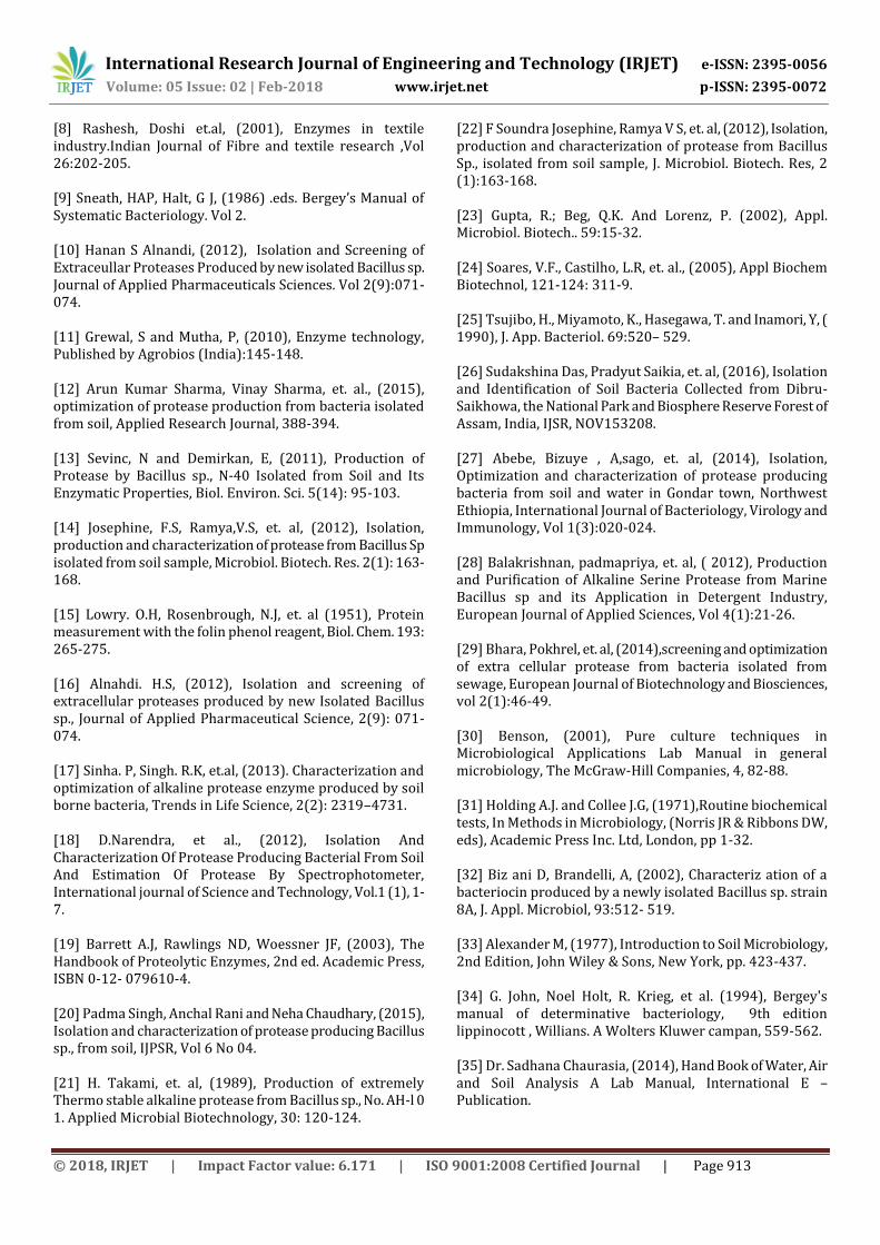
[8] Rashesh, Doshi et.al, (2001), Enzymes in textile industry.Indian Journal of Fibre and textile research ,Vol 26:202-205. [9] Sneath, HAP, Halt, G J, (1986) .eds. Bergey’s Manual of Systematic Bacteriology. Vol 2. [10] Hanan S Alnandi, (2012), Isolation and Screening of Extraceullar Proteases Produced by new isolated Bacillus sp. Journal of Applied Pharmaceuticals Sciences. Vol 2(9):071- 074. [11] Grewal, S and Mutha, P, (2010), Enzyme technology, Published by Agrobios (India):145-148. [12] Arun Kumar Sharma, Vinay Sharma, et. al., (2015), optimization of protease production from bacteria isolated from soil, Applied Research Journal, 388-394. [13] Sevinc, N and Demirkan, E, (2011), Production of Protease by Bacillus sp., N-40 Isolated from Soil and Its Enzymatic Properties, Biol. Environ. Sci. 5(14): 95-103. [14] Josephine, F.S, Ramya,V.S, et. al, (2012), Isolation, production and characterization of protease from Bacillus Sp isolated from soil sample, Microbiol. Biotech. Res. 2(1): 163-168. [15] Lowry. O.H, Rosenbrough, N.J, et. al (1951), Protein measurement with the folin phenol reagent, Biol. Chem. 193: 265-275. [16] Alnahdi. H.S, (2012), Isolation and screening of extracellular proteases produced by new Isolated Bacillus sp., Journal of Applied Pharmaceutical Science, 2(9): 071-074. [17] Sinha. P, Singh. R.K, et.al, (2013). Characterization and optimization of alkaline protease enzyme produced by soil borne bacteria, Trends in Life Science, 2(2): 2319–4731. [18] D.Narendra, et al., (2012), Isolation And Characterization Of Protease Producing Bacterial From Soil And Estimation Of Protease By Spectrophotometer, International journal of Science and Technology, Vol.1 (1), 1-7. [19] Barrett A.J, Rawlings ND, Woessner JF, (2003), The Handbook of Proteolytic Enzymes, 2nd ed. Academic Press, ISBN 0-12- 079610-4. [20] Padma Singh, Anchal Rani and Neha Chaudhary, (2015), Isolation and characterization of protease producing Bacillus sp., from soil, IJPSR, Vol 6 No 04. [21] H. Takami, et. al, (1989), Production of extremely Thermo stable alkaline protease from Bacillus sp., No. AH-l 0 1. Applied Microbial Biotechnology, 30: 120-124.
[22] F Soundra Josephine, Ramya V S, et. al, (2012), Isolation, production and characterization of protease from Bacillus Sp., isolated from soil sample, J. Microbiol. Biotech. Res, 2 (1):163-168. [23] Gupta, R.; Beg, Q.K. And Lorenz, P. (2002), Appl. Microbiol. Biotech.. 59:15-32. [24] Soares, V.F., Castilho, L.R, et. al., (2005), Appl Biochem Biotechnol, 121-124: 311-9. [25] Tsujibo, H., Miyamoto, K., Hasegawa, T. and Inamori, Y, ( 1990), J. App. Bacteriol. 69:520– 529. [26] Sudakshina Das, Pradyut Saikia, et. al, (2016), Isolation and Identification of Soil Bacteria Collected from Dibru-Saikhowa, the National Park and Biosphere Reserve Forest of Assam, India, IJSR, NOV153208. [27] Abebe, Bizuye , A,sago, et. al, (2014), Isolation, Optimization and characterization of protease producing bacteria from soil and water in Gondar town, Northwest Ethiopia, International Journal of Bacteriology, Virology and Immunology, Vol 1(3):020-024. [28] Balakrishnan, padmapriya, et. al, ( 2012), Production and Purification of Alkaline Serine Protease from Marine Bacillus sp and its Application in Detergent Industry, European Journal of Applied Sciences, Vol 4(1):21-26. [29] Bhara, Pokhrel, et. al, (2014),screening and optimization of extra cellular protease from bacteria isolated from sewage, European Journal of Biotechnology and Biosciences, vol 2(1):46-49. [30] Benson, (2001), Pure culture techniques in Microbiological Applications Lab Manual in general microbiology, The McGraw-Hill Companies, 4, 82-88. [31] Holding A.J. and Collee J.G, (1971),Routine biochemical tests, In Methods in Microbiology, (Norris JR & Ribbons DW, eds), Academic Press Inc. Ltd, London, pp 1-32. [32] Biz ani D, Brandelli, A, (2002), Characteriz ation of a bacteriocin produced by a newly isolated Bacillus sp. strain 8A, J. Appl. Microbiol, 93:512- 519. [33] Alexander M, (1977), Introduction to Soil Microbiology, 2nd Edition, John Wiley & Sons, New York, pp. 423-437. [34] G. John, Noel Holt, R. Krieg, et al. (1994), Bergey's manual of determinative bacteriology, 9th edition lippinocott , Willians. A Wolters Kluwer campan, 559-562. [35] Dr. Sadhana Chaurasia, (2014), Hand Book of Water, Air and Soil Analysis A Lab Manual, International E – Publication.

International Research Journal of Engineering and Technology (IRJET) e-ISSN: 2395-0056
Volume: 05 Issue: 02 | Feb-2018 www.irjet.net p-ISSN: 2395-0072
© 2018, IRJET | Impact Factor value: 6.171 | ISO 9001:2008 Certified Journal | Page 914
[36] James L. Walworth, (2006), Soil Sampling and Analysis, the University of Arizona, College of Agriculture and Life Sciences. [37] L.P. Van Reewijk, (2002), Proceture for soil analysis, 6th Edition, ISRIC, Netherlands. [38] Soil Sampling & Method of Analysis, (2008.), 2nd Edition, Canedian Society of Soil Science. [39] Singh, Dhyan, et. al, (1999), Assessment of Irrigationm Water Quality in “Soil, Plant, Water Analysis” - A methods manual, Indian Agricultural Research Institute, Indian Council of Agricultural Research, New Delhi. [40] K. M. Shah, (2009), Laboratory Testing Procedure For Soil & Water Sample Analysis, Water Resources Department Directorate Of Irrigation Research & Development, Pune. [41] P. Haluschak, (2006), Laboratory Methods Of Soil Analysis, Canada-Manitoba Soil Survey. [42] M. Alexander, (1977), “Introduction to Soil Microbiology,” John Wiley & Sons (2nd Edition), New York, pp. 423- 437.
BIBLOGRAPHY N. SHOBANA SUNDARI Assistant Professor, Department of Biotechnology, Rvs College of Arts & Science, Sulur, Coimbatore, Tamil Nadu, India.