Isolation and Characterization of Pyrimidine-Psoralen Photoadducts ...
9
J. Am. Chem. Sot. 1981, 103, 2341-2355 2341 ethanol with propan-2-01 yielded 24 as colorless liquid which distilled; bp 73-74 "C (0.5 mm) (lit." 76 "C (0.5 mm)). Preparation of Tetrakis(neopenty1oxy)tellurane (25). To a stirred solution of tellurium tetrachloride (5.42 g, 0.02 mol) and neopentyl alcohol (6.93 g, 0.08 mol) in tetrahydrofuran (50 mL) at 0 "C was added dropwise triethylamine (7.98 g, 0.08 mol) in tetrahydrofuran (20 mL). The precipitate which formed was removed by filtration to give a pale brown solution which was then reduced in volume. Attempts to precip- itate 25 from solution found little success although addition of pentane and storage at -45 OC gave a small amount of white solid. The solution was then evaporated to dryness to give an off-white colorless powder. This material proved soluble in most common organic solvents, and this precluded recrystallization. Dissolution in acetonitrile followed by cooling gave only cloudy suspensions. Attempted sublimation of the crude solid material at 0.03 mm resulted only in melting with probable decompo- sition at 70 OC. A crude sample of 25 was shown by Iz5Te,13C,and 'H NMR to be of high if not analytical purity and was therefore studied in solution without further purification. Preparation of Tetrakis(2,2,2-trifluoroethoxy)tellurane (26). 2,2,2- Trifluoroethanol (14 g, 0.14 mol) was added dropwise to sodium (2.36 g, 0.10 mol) suspended in tetrahydrofuran (40 mL). This solution was then added to a solution of tellurium tetrachloride (6.89 g, 0.025 mol) in tetrahydrofuran (30 mL) at --40 "C. When the addition was com- pleted, the reaction mixture was centrifuged under nitrogen and the solids were removed. The solvents were then removed, and the residue was distilled to yield 26 as a colorless liquid, bp 79-80 OC (0.7 mm). Anal. Calcd for C8HBFI2O4Te: C, 18.34; H, 1.54. Found: C, 18.19; H, 1.58. Preparation of Tetrakis((hexafluoroisopropy1)oxy)tellurane-THF Adduct (27). Hexafluoro-2-propanol (15 g, excess) was added dropwise to sodium (1 .O g, 0.044 mol) suspended in ice-water-cooled tetrahydro- furan (40 mL). This solution was then added dropwise to a solution of tellurium tetrachloride (2.93 g, 0.01 1 mol) in tetrahydrofuran (20 mL) at ambient temperature. The mixture was allowed to stand for 20 h, and it was then centrifuged to remove solids. After removal of solvents, distillation of the residue gave a colorless liquid, bp 60-62 OC (0.07 mm). Attempts to redistill this material resulted in partial breakdown of the complex. Reaction of 9 with Potassium Trifluoroethoxide. The reactants were mixed initially in a unimolar ratio. Tetrakis(2,2,2-trifluoroethoxy)sel- enurane (9) (0.394 g, 0.83 mmol) was dissolved in benzene (1.5 mL). The 77SeNMR spectrum showed an absorption at 6 1159. To this solution at 0 OC was added dropwise a solution of potassium 2,2,2-tri- fluoroethoxide (0.114 g, 0.83 mmol) and 18-crown-6-ether(0.219 g, 0.83 mmol) in benzene (2 mL). This mixture showed a 77Se resonance at 6 1083. The reaction was then repeated in an identical manner but using a 1.5: 1 molar ratio of trifluoroethoxide/l8-crown-6-ether to selenurane. The resulting solution showed a broad "Se NMR signal at 6 1021. Further addition of trifluoroethoxide/ 18-crown-6-ether to this solution gave a 5:l molar excess over the selenurane. This caused no further change in the "Se chemical shift which remained at 6 1021. No further purification of any reaction mixtures was attempted. Reaction of 26 with Potassium Trifluoroethoxide. The reactants were mixed initially in a equimolar ratio. Tetrakis(2,2,2-trifluoroethoxy)tel- lurane (26) (0.374 g, 0.715 mmol) was dissolved in benzene (1.5 mL). The '25Te NMR spectrum showed a resonance at 6 1463. To this solution was added dropwise a solution of potassium 2,2,2-trifluoroethoxide(0.100 g, 0.715 mmol) and 18-crown-6-ether (0.190 g, 0.715 mmol) in benzene (2 mL). This mixture showed a '25TeNMR absorption at 1352 ppm. In a similar reaction using a 1.51 mole ratio of trifluoroethoxide/ 18-crown-6-etherto tellurane, a mixture with a broad 125Te NMR signal at 6 1251 was obtained. Further addition of trifluoroethoxide to the mixture resulted in a 5:l molar excess over the tellurane. This caused no change in the IZ5Te chemical shift. Acknowledgment. This research has been supported by the National Science Foundation. W e also wish to thank the Mobil Chemical Co. for funds which aided in the purchase of NMR equipment and Stephen J. Hammond for some valuable technical assistance. Isolation and Characterization of Pyrimidine-Psoralen Photoadducts from DNA Kenneth Straub, David Kanne, John E. Hearst, and Henry Rapoport* Contribution from the Department of Chemistry and Lawrence Berkeley Laboratory, University of California, Berkeley, California 94720. Received September 17, 1980 Abstract: We have examined the photoadducts of 4'-hydroxymethyl-4,5',8-trimethylpsoralen (HMT) and native DNA. Five nucleoside-HMT monoaddition products have been isolated and characterized, corresponding to three deoxythymidine-HMT and two deoxyuridine (derived from deoxycytidine)-HMT adducts. Structural assignments are based on high resolution mass spectrometry and 'H NMR studies, including homonuclear spin decoupling and nuclear Overhauser effect (NOE) experiments. The results of this study indicate that (1) a limited number of nucleoside-psoralen adducts are formed with native, double-stranded DNA, and (2) the stereochemistry of the adducts is apparently determined by the geometry of the noncovalent intercalative complex formed by HMT and DNA prior to irradiation. Introduction The family of furmumarin derivatives known as psoralens has been actively investigated both with regard to their ability to act as dermal photosensitizing agents and as probes of nucleic acid structure and function.'J The biological activity of psoralens is primarily the result of the covalent bonding they undergo with nucleic acids, especially DNA. This process is believed to involve three distinct steps: (1) noncovalent, intercalative binding to the D N A helix; (2) upon irradiation at 365 nm, formation of a mo- (1) Song, P. S.; Tapley, J. K. Photochem. Photobiol. 1979, 29, 1177. (2) (a) Hanson, C. V.; Shen, C-K, J.; Hearst, J. E. Science, 1976, 193,62; (b) Shen, C-K. J.; Hearst, J. E. Proc. Narl. Acad. Sci. U.S.A. 1976, 73,2649. 0002-7863/81/1503-2347$01.25/0 noaddition product between the psoralen and a DNA base, probably, but not necessarily exclusively, a pyrimidine residue; and (3) absorption of a second photon by some monoadducts to form diadducts, which results in interstrand ~ross-linking.'~~ Model studies, for the most part carried out with nucleosides or pyrimidine bases, have suggested that the mono- and diadducts result from a cyclophotoaddition between the 5,6 bond of the pyrimidine and the 3,4 (pyrone) or 4'3' (furan) bond of the psoralen.4d For diadducts to be formed with 365-nm irradiation, (3) Scott, D. R.; Pathak, M. A,; Moh, G. R. Mutation Res. 1976, 39, 29. (4) Musajo, L.; Bordin, F.; Caporale, G.; Marciani, S.; Rigatti, G. Pho- tochem. Photobiol. 1967,6, 7 11. 0 1981 American Chemical Society
Transcript of Isolation and Characterization of Pyrimidine-Psoralen Photoadducts ...

J . Am. Chem. So t . 1981, 103, 2341-2355 2341
ethanol with propan-2-01 yielded 24 as colorless liquid which distilled; bp 73-74 "C (0.5 mm) (lit." 76 "C (0.5 mm)).
Preparation of Tetrakis(neopenty1oxy)tellurane (25). To a stirred solution of tellurium tetrachloride (5.42 g, 0.02 mol) and neopentyl alcohol (6.93 g, 0.08 mol) in tetrahydrofuran (50 mL) at 0 "C was added dropwise triethylamine (7.98 g, 0.08 mol) in tetrahydrofuran (20 mL). The precipitate which formed was removed by filtration to give a pale brown solution which was then reduced in volume. Attempts to precip- itate 25 from solution found little success although addition of pentane and storage at -45 OC gave a small amount of white solid. The solution was then evaporated to dryness to give an off-white colorless powder. This material proved soluble in most common organic solvents, and this precluded recrystallization. Dissolution in acetonitrile followed by cooling gave only cloudy suspensions. Attempted sublimation of the crude solid material at 0.03 mm resulted only in melting with probable decompo- sition at 70 OC. A crude sample of 25 was shown by Iz5Te, 13C, and 'H NMR to be of high if not analytical purity and was therefore studied in solution without further purification.
Preparation of Tetrakis(2,2,2-trifluoroethoxy)tellurane (26). 2,2,2- Trifluoroethanol (14 g, 0.14 mol) was added dropwise to sodium (2.36 g, 0.10 mol) suspended in tetrahydrofuran (40 mL). This solution was then added to a solution of tellurium tetrachloride (6.89 g, 0.025 mol) in tetrahydrofuran (30 mL) at --40 "C. When the addition was com- pleted, the reaction mixture was centrifuged under nitrogen and the solids were removed. The solvents were then removed, and the residue was distilled to yield 26 as a colorless liquid, bp 79-80 OC (0.7 mm).
Anal. Calcd for C8HBFI2O4Te: C, 18.34; H, 1.54. Found: C, 18.19; H, 1.58.
Preparation of Tetrakis((hexafluoroisopropy1)oxy)tellurane-THF Adduct (27). Hexafluoro-2-propanol (15 g, excess) was added dropwise to sodium (1 .O g, 0.044 mol) suspended in ice-water-cooled tetrahydro- furan (40 mL). This solution was then added dropwise to a solution of tellurium tetrachloride (2.93 g, 0.01 1 mol) in tetrahydrofuran (20 mL) at ambient temperature. The mixture was allowed to stand for 20 h, and it was then centrifuged to remove solids. After removal of solvents, distillation of the residue gave a colorless liquid, bp 60-62 OC (0.07 mm).
Attempts to redistill this material resulted in partial breakdown of the complex.
Reaction of 9 with Potassium Trifluoroethoxide. The reactants were mixed initially in a unimolar ratio. Tetrakis(2,2,2-trifluoroethoxy)sel- enurane (9 ) (0.394 g, 0.83 mmol) was dissolved in benzene (1.5 mL). The 77Se NMR spectrum showed an absorption at 6 1159. To this solution at 0 OC was added dropwise a solution of potassium 2,2,2-tri- fluoroethoxide (0.1 14 g, 0.83 mmol) and 18-crown-6-ether (0.219 g, 0.83 mmol) in benzene (2 mL). This mixture showed a 77Se resonance at 6 1083.
The reaction was then repeated in an identical manner but using a 1.5: 1 molar ratio of trifluoroethoxide/l8-crown-6-ether to selenurane. The resulting solution showed a broad "Se NMR signal at 6 1021. Further addition of trifluoroethoxide/ 18-crown-6-ether to this solution gave a 5:l molar excess over the selenurane. This caused no further change in the "Se chemical shift which remained at 6 1021. No further purification of any reaction mixtures was attempted.
Reaction of 26 with Potassium Trifluoroethoxide. The reactants were mixed initially in a equimolar ratio. Tetrakis(2,2,2-trifluoroethoxy)tel- lurane (26) (0.374 g, 0.715 mmol) was dissolved in benzene (1.5 mL). The '25Te NMR spectrum showed a resonance at 6 1463. To this solution was added dropwise a solution of potassium 2,2,2-trifluoroethoxide (0.100 g, 0.715 mmol) and 18-crown-6-ether (0.190 g, 0.715 mmol) in benzene (2 mL). This mixture showed a '25Te NMR absorption at 1352 ppm.
In a similar reaction using a 1.51 mole ratio of trifluoroethoxide/ 18-crown-6-ether to tellurane, a mixture with a broad 125Te NMR signal at 6 1251 was obtained. Further addition of trifluoroethoxide to the mixture resulted in a 5:l molar excess over the tellurane. This caused no change in the IZ5Te chemical shift.
Acknowledgment. This research has been supported by the National Science Foundation. W e also wish to thank the Mobil Chemical Co. for funds which aided in the purchase of NMR equipment and Stephen J. Hammond for some valuable technical assistance.
Isolation and Characterization of Pyrimidine-Psoralen Photoadducts from DNA
Kenneth Straub, David Kanne, John E. Hearst, and Henry Rapoport*
Contribution from the Department of Chemistry and Lawrence Berkeley Laboratory, University of California, Berkeley, California 94720. Received September 17, 1980
Abstract: We have examined the photoadducts of 4'-hydroxymethyl-4,5',8-trimethylpsoralen (HMT) and native DNA. Five nucleoside-HMT monoaddition products have been isolated and characterized, corresponding to three deoxythymidine-HMT and two deoxyuridine (derived from deoxycytidine)-HMT adducts. Structural assignments are based on high resolution mass spectrometry and 'H NMR studies, including homonuclear spin decoupling and nuclear Overhauser effect (NOE) experiments. The results of this study indicate that (1) a limited number of nucleoside-psoralen adducts are formed with native, double-stranded DNA, and (2) the stereochemistry of the adducts is apparently determined by the geometry of the noncovalent intercalative complex formed by HMT and DNA prior to irradiation.
Introduction The family of furmumar in derivatives known as psoralens has
been actively investigated both with regard to their ability to act as dermal photosensitizing agents and as probes of nucleic acid structure and function.'J The biological activity of psoralens is primarily the result of the covalent bonding they undergo with nucleic acids, especially DNA. This process is believed to involve three distinct steps: (1) noncovalent, intercalative binding to the D N A helix; (2) upon irradiation at 365 nm, formation of a mo-
(1) Song, P. S.; Tapley, J. K. Photochem. Photobiol. 1979, 29, 1177. (2) (a) Hanson, C. V.; Shen, C-K, J.; Hearst, J. E. Science, 1976, 193,62;
(b) Shen, C-K. J.; Hearst, J. E. Proc. Narl. Acad. Sci. U.S.A. 1976, 73,2649.
0002-7863/81/1503-2347$01.25/0
noaddition product between the psoralen and a DNA base, probably, but not necessarily exclusively, a pyrimidine residue; and (3) absorption of a second photon by some monoadducts to form diadducts, which results in interstrand ~ross-linking. '~~ Model studies, for the most part carried out with nucleosides or pyrimidine bases, have suggested that the mono- and diadducts result from a cyclophotoaddition between the 5,6 bond of the pyrimidine and the 3,4 (pyrone) or 4'3' (furan) bond of the psoralen.4d For diadducts to be formed with 365-nm irradiation,
(3) Scott, D. R.; Pathak, M. A,; Moh, G. R. Mutation Res. 1976, 39, 29. (4) Musajo, L.; Bordin, F.; Caporale, G.; Marciani, S. ; Rigatti, G. Pho-
tochem. Photobiol. 1967, 6, 7 11.
0 1981 American Chemical Society

2348 J. Am. Chem. SOC., Vol. 103, No. 9, 1981
monoaddition to the 4’3’ double bond would be the anticipated primary photochemical act. However, no detailed structural and stereochemical analyses of the adducts have appeared. Particularly lacking are data on the products from the reaction of psoralen with intact, native DNA.
Our present study is concerned with the isolation and identi- fication of the monoadducts formed in the photoreaction between DNA and a substituted psoralen, 4’-hydroxymethyl-4,5’,8-tri- methylpsoralen (HMT, 1). This particular derivative has an
A H .
Straub et al.
* 0 1
enhanced binding affinity for DNA over that of the naturally occuring psoralens, allowing a greater extent of reaction to take place.’ Five nucleoside-HMT monoaddition products have been isolated from HMT-modified DNA, corresponding to three de- oxythymidine-HMT and two deoxyuridine-HMT adducts. The deoxyuridine adducts result from initial addition to deoxycytidine and subsequent hydrolytic conversion to deoxyuridine. Structural assignments are based on high resolution mass spectrometry and ’H N M R studies, including homonuclear spin decoupling and nuclear Overhauser effect (NOE) experiments.
Our results indicate that (1) a limited number of psoralen- nucleoside adducts are formed with native, double-stranded DNA, and (2) the stereochemistry of the adducts is apparently deter- mined by the geometry of the noncovalent, intercalative complex formed by H M T and DNA prior to irradiation.
Experimental Section Materials. Calf thymus DNA and hydrolytic enzymes were obtained
from Sigma (St. Louis, MO). [3H]-4’-Hydroxymethyl-4,5’,8-tri- methylpsoralen (HMT) was synthesized as previously described.’ Sol- vents were either Nanograde (Mallinckrodt, St. Louis, MO) or Distilled in Glass (Burdick & Jackson, Muskegon, MI). HPLC-grade water was obtained from Baker (Phillipsburg, NJ).
Photobinding and Isolation of Psoralen-Modified DNA. [3H]-HMT (6.3 mg, lo* dpm) in 2 mL of ethanol was added with stirring to a solution of calf thymus DNA (200 mg, 1 mg/mL) in Tris-EDTA (10 mM Tris, 1 mM EDTA, pH 8.5) buffer. This solution was cooled to 5 OC and irradiated for 8 min in a high-intensity irradiation apparatus equipped with two 400-W G.E. mercury vapor lamps. The DNA-HMT solution was surrounded by a jacket containing a temperature-regulated (10 “C) solution of cobaltous nitrate (40% w/w), which acts as a 365-nm transmission filter. Throughout the irradiation period, the temperature of the DNA solution was maintained at 8-10 OC.
Noncovalently bound HMT was removed by extraction with four portions of CHCl,, each equal in volume to the aqueous phase which was then adjusted to 0.2 M in sodium chloride and diluted with three volumes of cold ethanol. After standing at 0 OC for 12 h, the precipitated DNA was isolated by centrifugation at lOOOOg for 30 min. The resulting HMT-DNA pellet was redissolved in 0.2 M NaCl and reprecipitated by addition of ethanol, and the isolated DNA pellet was then dried under vacuum and dissolved in 30 mL of hydrolysis buffer. Three different hydrolysis protocols were used. Method A utilized a hydrolysis buffer consisting of 15 mM sodium acetate and 10 mM EDTA (pH 5.0). A p proximately 16000 units of DNAase-I1 (E.C.3.1.4.6) and 45 units of phosphodiesterase-I1 (E.C.3.1.4.18) were added over a period of 36 h. The pH of the mixture was then adjusted to 8.5, and 35 units of alkaline phosphatase (E.C.3.1.3.1) as added. After an additional 12 h, the mix- ture was lyophilized and redissolved in the minimum volume of 10% methanol-water. Method B utilized DNAase-I (E.C.3.1.4.5; 16 000 units) and phosphodiesterase-I (E.C. 3.1.4.1; 4 units in 10 mM Tris, 5 mM MgC12, pH 7.0), followed by treatment with 35 units of alkaline phosphatase at pH 8.5. Method C utilized nuclease PI (E.C.3.1.4.x; 600 units/50 mg of DNA) as the endonuclease, followed by treatment with phosphodiesterase-I1 at pH 7.0 and alkaline phosphatase at pH 8.5.
The hydrolyzed HMT-modified DNA was then chromatographed on a 1.8 X 60 cm gel filtration column (Biogel P6, BioRad, Richmond, CA),
(5) Musajo, L; Rodighiero, G. Phorochem. Photobiol. 1970, I ! , 27. (6) Krauch, C. H.; Kramer, D. M.; Walker, A. Phorochem. Photobiol.
(7) Isaacs, S. T.; Shen, C. J.; Hearst, J. E.; Rapoport, H. Biochemistry 1967, 6, 341.
1977, 16, 1058.
Table I. Tritium Recoveries from Large-Scale Reaction with 6.3 mg of HMT and 200 mg of DNA
tritium activity operation dpmx lo-‘ %
HMT added 7.71 100 remaining after 5.33 6 9a
remaining after C,H,OH 4.62 6 Oa
total applied to 4.62 100
fraction P6V, 1.55 33.5c
CHCl, extraction
precipitation (bound HMT)
P6 column
fraction P6P2 2.24 48.5c fraction P6P3 0.81 17.6c
a This is percent of initial addition. Obtained via hydrolysis method A. By using method C, the activity in the void volume was decreased to 5% and that in P6P3 increased to 40%. percent of bound HMT taken as 100%.
eluting with 10% methanol-water. Fractions were collected and assayed for )H by scintillation counting. Tritium-containing fractions were then further purified by high performance liquid chromatography (HPLC) on an Altex/Beckmann Model 320MP liquid chromatograph, equipped with a Schoeffel SF770 variable-wavelength absorbance detector. Reverse- phase octadecylsilane (ODS) columns (10 X 250 mm or 4.6 X 250 mm 5p Ultrasphere) were used with water-methanol as the eluting solvent. Tritium-containing fractions were collected and taken to dryness under reduced pressure.
Photoreversion.* The adduct of interest was dissolved in 15% meth- anol-water at a concentration of ca. 10 pg/mL. This solution was ir- radiated at 254 nm with a low-intensity mercury hand lamp for 20 min. The solution was then concentrated under reduced pressure to a volume of 20 pL and analyzed by HPLC on the Ultrasphere ODS column. Identification of products was achieved by coinjection with authentic standards or by mass spectrometric analysis.
Mass Spectrometry. High resolution electron-impact mass spectra were obtained on a modified Kratos/AEI MS902 mass spectrometer, operating at a dynamic resolution of M / A M lOOOO? The data system (LOGOS-11) assigns exact masses to all of the observed fragment ions in a mass spectrum and stores the data on disk or tape.1° Elemental compositions are then generated by computer for a given error tolerance. A source temperature of 250 OC was used for electron-impact spectra. Field desorption mass spectra (FDMS) were recorded at a resolution of M/ AM 1500, using conventional benzonitrile-activated emitters.”
The HMT-DNA adducts were analyzed by high resolution electron- impact MS as pertrimethylsilyl (TMS) or permethyl ethers. The TMS ethers were prepared by heating 0.5-1.0 wg of adduct in 80 pL of 1/4 (v/v) pyridine/N,Obis(triethylsilyl)trifluoroacetamide (BSTFA) at 60 OC for 30 min. Excess reagent was evaporated under nitrogen, and the residue applied to the direct insertion probe of the mass spectrometer. Permethyl ethers were prepared using dimethyl sulfoxide anion-methyl iodide, as described.I2 Verification of elemental compositions was achieved by perdeuteriomethylation with CD31, or by preparing per- deuterio-TMS ethers with [2H,]-BSTFA. ’H NMR. ’H NMR spectra were obtained at 360 MHz on a Nicolet
Technologies NT 360 NMR spectrometer. Typically, 1000 transients were accumulated with 3.1 s between pulses. A spectral width of 2000 Hz was used. Nuclear Overhauser effect (NOE) experiments were carried out with the decoupling field gated off during data acquisition and with a delay time of 2.7-3.1 s between the end of an acquisition and the beginning of the next pulse. Spectra used for measuring NOE effects were time averaged over 400 acquisitions, using a spectral width of 1500 Hz. All assignments were made with the aid of homonuclear spin de- coupling experiments.
Spectra were recorded in 99.996% D20 (pD 7.1). Samples were prepared by collecting the HPLC column effluent, evaporating the sol-
As
(8) Herbert, M. A.; Le Blanc, J. C.; Weinblum, D.; Johns, H. E. Photo- chem. Photobiol. 1969, 9, 33.
(9) Burlingame, A. L.; Smith, D. H.; Meran, T. 0.; Oslen, R. W. “Rogress in Analytical Chemistry”, Orr, C. H., Norris, J. A,, Eds.; Plenum: New York, 1970; VOI. 4, pp 17-38.
(10) Burlingame, A. L.; Oslen, R. W.; McPherson, R. “Advances in Mass Swtrometrv”, West, A. R., Ed.; Applied Science Publishers: Barking, %ex, - _ 1974; Vol. 6, pp 1053-1054.
(1 1) Beckey, D. H. “Principles of Ionization and Field Desorption Mass
(12) Morris, H. R. FEBS Letr. 1970, 22, 257. Spectrometry”; Pergamon Press: Oxford, 1977; pp 64-72.

Pyrimidine-Psoralen Photoadducts from DNA
Table 11. High Resolution Mass Spectral Data for Fragment Ions Derived from HMT Moiety of Silylated HMT-DNA Adducts
fragment ions elemental composition
(mlzIa [error, ppmIa assignment
330.1265 C,,H,,O,TMS [-6.731 HMT-TMS 315.1052 C,,H,,O,TMS [-0.251 330- CH, 256.0710 C,,H,,O, [-9.981 240.0773 C,,H,,O, [-5-52] M - HOTMS 212.0824 C,,H,,O, [-6.091 240- CO 211.0752 C,,H,,O, [-3.321 240 - HCO
73.0467 C,H,Si [-9.331 TMS Values of m/z and errors are for a typical sample; all silylated
HMT-DNA adducts gave similar results, with errors of less than 10 ppm.
vent under reduced pressure, and redissolving the sample in 99.96% DzO. The D20 was then evaporated under reduced pressure and the procedure repeated once with 99.96% D20 and once with 99.996% D20; the dried residue was then dissolved in 99.996% D20 and transferred to a 5-mm NMR Tube. All final drying and loading operations were carried out in a D20-saturated nitrogen or argon atmosphere. Chemical shifts are relative to Me4Si (bHDO, 4.75 ppm, 1710 Hz).
Results Modification of DNA by HMT. Table I summarizes the results
obtained for the covalent binding of H M T to DNA for a typical experiment using 6.3 mg of H M T and 200 mg of DNA. The overall binding level was 60% of the added H M T and is equivalent to approximately one H M T per 20 base pairs. The gel filtration elution profile from a Biogel P6 column for the enzymatically hydrolyzed material using hydrolysis method A is shown in Figure 1. Three distinct 'H-containing fractions are evident and are referred to as P6P1 (Biogel P6 column, fraction pool l ) , P6P2, and P6P3. P6P1 contains material eluting at the void volume (V,) of the column and corresponds to partially hydroylzed oligo- nucleotides. The amount of 'H-containing material in this fraction can be reduced to less than 5% of the total 'H by exhaustive enzymatic hydroylsis using hydrolysis method C. The other two fractions, P6P2 and P6P3, were further purified by HPLC. As will be shown, P6P3 contains HMT-nucleoside monoadducts, while P6P2 contains nucleoside-HMT-nucleoside diadducts and di- or trinucleotide-HMT monoadducts. The remainder of this
J . Am. Chem. SOC., Vol. 103, No. 9, 1981 2349
Table 111. High Resolution Mass Spectral Data for Silylated HMT-DNA Adducts
Froct ion Number
Figure 1. Biogel column elution profile of [3H]HMT-DNA enzyme hydrolysate, method A.
study is concerned with the structural identification of the mo- noaddition adducts contained in P6P3.
Figure 2 represents an HPLC elution profile of P6P3 in which 1-mL fractions were collected a t a flow rate of 1 mL/min. The large UV-absorbing component eluting between 4 and 10 min contains the expected deoxynucleosides (dC, dT, dG, and dA). Five distinct 3H-containing components eluted between 18 and 35 min and are referred to as F42A (fraction 42A), F42B, F44, F45, and F48 (relative amounts of radioactivity: 100,60, 5 ,65, 5). These five components account for 17.6% of the total cova- lently bound H M T and were obtained using hydrolysis method A. When method C was used, the recovery was increased to 40%. Each of these components was then analyzed by mass spectrometry and 'H NMR.
Mass Spectrometry. Components F42A, F42B, F44, and F45 were analyzed by high resolution electron-impact mass spec- trometry as pertrimethylsilyl (TMS) ethers. All four adducts displayed prominent fragment ions derived from the HMT moiety, identical with the fragments observed in the mass spectrum of the TMS ether of HMT itself; these common fragments are listed in Table 11. The TMS ethers of F42A and F42B gave identical mass spectra, displaying a relatively low intensity molecular ion a t m / z 716 ( C ~ S H ~ ~ N ~ O ~ ( T M S ) ~ ) and a prominent ( M - 15)' ion at m / z 701 (C24H22N209 (TMS)'). The molecular weight, fragmentation pattern, and nitrogen content are consistent with the presence of a deoxythymidine residue (Table 111). Confir-
fragment ions elemental composition adduct (mlz)' [error, ppml assignmentbsc
F44
F4Se
F42A, F42Bd 773.3161 C,,H,,N,O,TMS, [+2 .48] (M' - CH,)' 7 16.2998 C,,H,,N,O,TMS, [+2 .41] M' 701.2726 C,,H,,N,O,TMS, [-2.801 (M - CH,)+ 683.2605 C,,H,,N,O,TMS, [-5.221 773 - HOTMS 61 1.2249 C,,H,,N,O,TMS, [ +0.65] 701 - HOTMS 555.2337 C,,H,,N,O,TMS, [-1.841 M' - C,H,O,(TMS), 529.2192 C,,H, ,N,O,TMS, [ tO.241 M' - dR 483.1935 C,,H,,N,O,TMS [-3.471 M - C,H,O,(TMS), 457.1784 Cz0H, oN,06 TMS [-2.36 ] M- dR
571.2123 C,,H,,N,O,TMS, [+1 .27] (M' - CH,) 499.1714 Cl8H1,N,O,TMS, [-1.42) (M - CH3) 409.12 18 C,,H,,N,O,TMS [-0.511 499 - HOTMS 24 1.0819 C,H, ,N,O,Si, [-4.001 5 7 1 - HMT:TMS 702.2750 C,,H,,N,O,TMS, [- 10.50] M' 687.2563 C,3H,oN,0,TMS3 [-3.801 (M - CH,)f 597.2072 C,,H, ,N,O,TMS, [-2.771 687 - HOTMS 541.2206 C, ,H, ,N,O,TMS, [ +2.96] M'+ - C,H,O,(TMS), 515.2042 C,,H, ,N,O,TMS, [+1.601 M'+ - dR 469.181 6 C,,H,,N,O,TMS [+4 .56 ] M+ - C,H,O,(TMS), 443.1635 C,,H,,N,O,TMS [-0.781 M+- dR
586.2391 C,,H,,N,O,TMS, [ t6 .851 MI+
a Only major fragments with m/z >400 are listed. The derivatization procedure gives two derivatives, corresponding to transfer of three or four silyl groups (two or three in the case of F44). M' refers to the higher homolog in each case. The di-TMS derivative of deoxyribose is referred to as dR. Free or derivatized nucleosides in general undergo fragmentation by fission of the glycosidic C-N bond with transfer of one or two hydrogens from the deoxyribose to the hetereocycle base. Assignment was confirmed by low resolution field desorption mass spectrum on underivatized adduct (M+ = 500). e Assignment was confirmed by low resolution field desorption mass spectrum on underivatiz- ed adduct (M+ = 486).

2350 J. Am. Chem. SOC., Vol. 103, No. 9, 1981 Straub et al.
1.01 , 1 I I , , , , , , I 1 , , I I , ,
0 05
9 0.03
0 01
0 10 20 30 40 50 Time imin)
Figure 2. HPLC profile of Biogel fraction P6P3 eluted from a 5-p ODS column with CH,OH/H,O 1 mL fractions at a flow rate of 1 mL/min.
Table IV. High Resolution MS Data on Permethylated F48 fragment elemental composition
ions (m/z) [error, ppm] assignment 602.2821 571.2668 318.1468 301.1435 287.1283 271.0985 25 7.1 180 255 .lo17 24 3.10 17 145.0866 141.0669 140.05 8 2 11 3.0592 87.0438 71.0496
Wavelenqth (nm)
Figure 3. Abserption spectrum of F42B in 50% CH30H/H20.
M + M - OCH, HMT (CH,),
318 - OCH,
deoxyribose thymine + H thymine deoxyribose - CH30H
deoxyribose fraement
mation of the identity of F42A and F42B as deoxythymidine- H M T adducts was obtained by FDMS of the underivatized ad- ducts; intense signals are observed at m / z 500 (M') and 523 (M + Na)+.
The T M S ether of F45 was found to exhibit a relatively low intensity molecular ion a t m / z 702.2750 [C24HZ3N209(TMS),]. A prominent ion corresponding to ( M - 15)' occurs at m / z 687.2563 (C23H20N209(TMS)3). Overall, the fragmentation pattern of F45-TMS is similar to that of F42A-TMS and F42B-TMS, with the high mass ions shifted to lower mass by 14 mass units (Table 111). This is consistent with the presence of a deoxyuridine residue. FDMS of underivatized F45 gave signals a t m / z 486 (M') and 509 ( M + Na)'. This uridine derivative is undoubtedly derived from a deoxycytidine-HMT adduct which has undergone hydrolytic deamination. Thus saturation of the 5,6 double bond in cytidine results in labilization of the exocyclic C-4 amino group and conversion to uridine a t neutral pH.
The T M S ether of F44 gave ions a t 586.2391 (Cl9HIJN2o6- (TMS), , M+), 571.2123 ( M - 15)+, and 499.1714 [C18H13N206(TMS)2, corresponding to ( M - 15)' for a di-TMS derivative]. Fragment ions due to the deoxyribose moiety were absent, suggesting that F44 was a deoxyuridine-HMT adduct that had undergone hydrolysis of the C-N glycosidic bond and loss of deoxyribose. This interpretation is supported by 'H N M R data and acid hydrolysis data given below.
The minor product, F48, was analyzed by high resolution M S as a permethyl derivative (Table IV). A distinct molecular ion is observed a t m / z 602.2821 (C31H42N2010). Fragment ions due to a permethylated H M T residue occur a t m / z 318.1463 and 287.1282 (318 - OCH,). Ions diagnostic for the presence of
PPm
Figure 4. The 360-MHz lH NMR spectrum of fraction F42A in D20.
thymine occur a t m / z 141.0669 (C6H9N202) and 140.0582 (C6H8N2O2), and fragment ions characteristic of a methylated deoxyribose occur a t m / z 145.0866 (C7HI3O3). The mass dif- ference between the molecular ion and a permethylated H M T residue corresponds to that of permethylated deoxythymidine (602 - 318 = 284, C13H20N205). F48 thus represents a third HMT- deoxythymidine adduct.
Absorption and Fluorescence Spectra. Fractions F42A, F42B, F44, and F45 exhibited essentially the same absorption spectra: a broad absorbance centered a t 328 nm with a shoulder a t 295 nm, as seen in Figure 3 for F42B in 50% C H 3 0 H / H 2 0 . All four components also exhibited the same fluorescence emission spectra as a broad peak centered a t 385 nm (A,,, 300 nm). These pa- rameters are characteristic of a coumarin-type chromophore and are identical with the model compound 4,5',8-trimethyl-4',5'- dihydropsoralen. Therefore, these adducts represent products in which the 4',5' double bond has been saturated, yielding a cou- marin-type chromophore. Fraction F48, however, was non- fluorescent and exhibited an absorption spectrum similar to that of a benzofuran derivative. This indicates that reaction has taken place at the 3,4 double bond of the HMT, leaving a 3,4-di- hydropsoralen-type chromophore.
Photoreversion. Irradiation of F42A and F42B a t 254 nm resulted in photoreversion of both isomers to deoxythymidine and HMT. Photoreversion under these conditions appears to be free of other competing photochemical reactions, as the only observable products (using an absorbance detector a t 254 nm) were HMT, deoxythymidine, and starting material. Similar treatment of F45 yielded unchanged starting material, HMT, and a nucleoside that coeluted with deoxyuridine. Confirmation of this assignment was

Pyrimidine-Psoralen Photoadducts from DNA
Table V. ‘H NMR (360 MHz) Data for F42A. F42B
J . Am. Chem. SOC., Vol. 103, No. 9, 1981 2351
resonance - multiplicitya A&, PPm no., r F42A F42B (no. of protons) assignmentb (F42A - 42B)
1 1.349 7.348 s (1) C-5-H +0.001 2 6.243 6.238 d (1) C-3-H
+0.005 3 6.045 5.728 t (1) C-1‘-H [dR] +0.317
chemical shift, ppm
(JAX = 1 Hz, coupled to r10)
(JABX = 6.7 Hz, coupled to r l l )
(coupled to r8, r l l ) 4 4.42 4.32 m (1) C-3’-H [dR] +0.10
5 4.190 4.192 d (1) HAB (CH,, J A B = 12.2 Hz) -0.002 6 4.058 4.079 d (1) HAB -0.021
8 3.88 3.90 m (1) C-4‘-H [dR] -0.02 I 4.105 4.065 s (1) H-6 +0.040
(coupled to r4, r9)
(coupled to r8)
(coupled to r2, JAX = 1 Hz)
(coupled to r3, r4)
9 3.69 3.75 m (2) C-S’-H [dR] -0.06
10 2.417 2.418 s (3) C-4CH3 -0.001
11 2.27 2.21 m (2) C-2‘, 2“-H [dR] 0
12 2.228 2.249 s (3) C-8CH3 -0.021 13 1.822 1.751 s (3) C-S’CH, +0.065 14 1.578 1.553 s (3) C-5(dT)CH3 +0.025
s, singlet; d, doublet; t , triplet; m, multiplet. dR, deoxyribose; dT, deoxythymidine; coupled signals are referred to by resonance num- ber, r.
made by direct mass spectrometric analysis of the product, yielding a fragmentation pattern identical with that observed for authentic deoxyuridine. There was insufficient material available to attempt the photoreversion of either F44 or F48.
‘H NMR. The 360-MHz ‘€1 NMR spectrum of F42A is shown in Figure 4, obtained on approximately 50 pg of material. Both adducts F42A and F42B display the same resonances, although shift differences, especially for C-1’ -H of the deoxyribose, of up to 0.3 ppm are apparent in Table V. Assignments of the deox- yribose protons were made by homonuclear spin decoupling ex- periments; these shift values are in agreement with those described for other modified pyrimidine deoxynu~leosides .~~J~ The 5’-CH3 of the HMT residue undergoes a 0.5-0.6 ppm upfield shift relative to the parent compound, indicating that saturation of the 4’3’ double bond has taken place. The 5-CH3 group of the thymidine is also found a t high field (1.55 ppm), indicating saturation of the pyrimidine 5,6 double bond. The diastereotopic CH2 protons of the 4’-hydroxymethyl side chain give rise to an AB doublet a t 4.08 and 4.19 ppm ( J = 12.2 Hz), and the single cyclobutyl proton (6-H of the pyrimidine) is superimposed on the upfield component of this spin system. These data are consistent with a cyclobutane structure derived from [ 2 + 21 cycloaddition of a thymidine residue with the 4’3’ double bond of HMT.
Since there is only a single cyclobutyl proton in this system, relative stereochemistries of the substituents on the cyclobutane ring were assigned using NOE experiment^.'^ For a proton- proton spin system, relaxation will occur primarily through a dipole-dipole mechanism, and the magnitude of the N O E will have an rd dependence on the spatial separation of the two spins. The cyclobutyl systems of F42A and F42B are particularly amenable to analysis by NOE since the resonances are well separated, and the molecule in the area of interest is fairly rigid, minimizing any complications that might arise from rotation. In addition, the different stereochemistries associated with the possible isomers of these adducts have substantially different CH3-H distances, so that the r-6 dependence of the NOE would be ex- pected to result in large differences in the relative enhancement of the resonances of interest.
Irradiation of the two cyclobutyl methyls, the 5’-CH3 derived from H M T and the 5(dT)-CH3 derived from deoxythymidine (Table V), therefore should result in the enhancement of the
(13) Liu, F. T.; Yang, N. C. Biochemistry 1978, 17, 4865. (14) Liu, F. T.; Yang, N. C. Biochemistry 1978, 17, 4877. (15) Noggle, J. H.; Schirmen, R. E. “The Nuclear Overhauser Effect:
Chemical Applications”, Academic Press: New York, I97 I .
I ’ I I I I I 1 I
b
I I I I I I 4 4 4 3 4 2 4 1 4 0 3 9 3 8 3 7 3 6
ppm
Figure 2. NOE enhancements for F42B: (a) 3.5-4.: ppm region of F42B, H2 at 6.5 ppm; (b) difference spectrum for €I2 at 1.55 ppm [C-S(dT)-CH,] and a; (c) difference spectrum for H2 at 1.76 ppm [C-5’(HMT)-CH3] and a.
Table VI. Nuclear Overhauser Effect (NOE) Experiments on F42A, F42B, and F45
% resonance (origin)a adduct irradiated observed enhancementb F42A C-5-CH, (dT) C-6-H (dT) 10 t 1
C-5‘CH3 (HMT) C-6-H (dT) 8 f 1 F42B C-SCH, (dT) C-6-H (dT) 9 t 1
C-5’-CH3 (HMT) C-6-H (dT) 9 f 1 F45 C-S’CH, (HMT) C-6-H (dU) 2 f 1
C-S’CH, (HMT) C-5-H (dU) 0 C-5-H (dU) C-6-H (dU) 2 t 1
a The numbering used is that of the original HMT or pyrimidine. The substituent on the cyclobutane is indicated by its origin from the HMT or the pyrimidine [dT, dU (EdC)] moiety. of two determinations.
Average
6(dT)-H resonance if they are on the same face of the cyclobutyl ring system as 6(dT)-H. Figure 5a shows the 3.5-4.5 ppm region of F42B, and Figure 5b shows the difference spectrum of this region when the decoupling field is set at the thymidine methyl, 5(dT)-CH3. Figure 5C shows the difference spectrum when the decoupling field is set a t the 5’-CH3 of the H M T residue [in the control spectrum, Figure 5a, the decoupling field was set sym-

2352 J. Am. Chem. SOC., Vol. 103, No. 9, 1981
Table VII. ' H NMR (360 MHz) Data for F45 and F45'. Diastereomers with Enantiomeric Aglycon Moieties
Straub et al.
chemical shift, ppma resonance multiplicity b no., r F45 F45' (no. of protons) assignmen tC A6, PPm
1 2
3
4
5
6 7 8
9
10
11
12
14
7.311 6.228
6.044
4.411
4.378
4.112 4.028 3.88
3.794
3.67
2.408
2.27
1.773
7.311 6.228
5.722
4.41 1
4.3 35
4.117 4.036 3.88
3.736
3.74
2.408
2.27
1.714
C-5-H C-3-H (JAX = 0.9 Hz,
C-1'-H (dR)
C-3'-H (dR)
C-6-H (dU)
HAB (JAB = 12.5 Hz)
C-4'-H (dR)
C-5-H (dU)
C-S'-H (dR)
C-ICH,
C-2', 2"-H (dR)
C-5'CH3
coupled to r l l )
(coupled t o r12)
(coupled to r8, r l2 )
(JAX = 10.1 Hz; coupled to 19)
(coupled to r4, r10)
(JAX = 10.1 Hz; coupled to 1-5)
(coupled to r8)
(JAX = 0.9 Hz; coupled to r2)
(coupled to r3, r4)
0 0
+0.322
0
t 0 . 0 4 3
-0.005
0
+0.058
-0.07
0
0
t0 .059
NMR results indicate the diastereomers F45 and F45' are present in approximately a 2/1 ratio. s, singlet; d, doublet; m, multiplet. dR, deoxyribose; dU, deoxyuridine; coupled signals are referred to by resonance number r.
metrically on the downfield side of the cyclobutyl proton (ca. 6.5 ppm) in order to avoid instrument artifacts].
Similar NOE enhancements are observed for both methyls (Table VI) indicating similar internuclear distances between the two sets of methyl protons and the cyclobutyl proton. For a spin system with one methyl adjacent and one methyl diagonal to the cyclobutyl proton, resulting from cis-anti stereochemistry16 as shown in structure 3, one would expect to observe a substantial difference in the two NOE's. Since this difference was not ob- served, the relative stereochemistry of F42B can thus be assigned as shown in structure 2 or 2'. Virtually identical results were
'0
CIS - syn-2 CIS- syn - 2' c/s-unt/-3
obtained for F42A, irradiation of the two cyclobutyl methyls resulting in a similar enhancement of the integrated intensity of 6(dT)-H. These results indicated that F42A and F42B have the stereochemical relationship shown by structures 2 and 2' and are not regioisomers. Additional evidence for this assignment was obtained from the circular dichroism spectra of these adducts discussed below.
The 360-MHz 'H NMR of F45 is shown in Figure 6. The two C-l'-H resonances a t 5.72 and 6.05 ppm (relative areas 2 /1 ) suggest that F45 consists of two diastereomers. Some of the resonances are partially resolved and are identified in Table VII. The upfield region shows three methyl resonances, in agreement with the assignment of this component as a deoxyuridine adduct. The region 3.6-4.5 ppm is similar to that of F 4 2 A and F42B, except that a two-proton A X ( J = 10 Hz) system occurs a t 3.8 ppm (C-5-H of the deoxyuridine) and 4.35 ppm (C-6-H of the
(16) Syn (head-to-head) for the furan adduct is defined as shown in structures 2 and 4 in which 0-1' of the furan and N-1 of the pyrimidine are bonded to adjacent corners of the cyclobutane. Syn for the pyrone adduct is defined as shown in structures 6 and 8 in which C-2 of the pyrone and N-1 of the pyrimidine are bonded to adjacent corners of the cyclobutane. Anti (head-to-tail) stereochemistry is applied to bonding in which these atoms are bonded to diagonal corners. Cis and Trans refers to the position of the psoralen and pyrimidine moieties relative to the plane of the cyclobutane.
7.0 6.0 5.0 4.0 3.0 2.0 I .o PPm
Figure 6. The 360-MHz 'H NMR spectrum of F45 in D20.
deoxyuridine). The C-6-H A X doublet partially overlaps the C-3'-H deoxyribose resonance. These results are fully consistent with the mass spectrometry data which indicate that F45 is a deoxyuridine-HMT adduct (diastereomers would be expected to give identical mass spectra, as was observed for F42A and F42B). The shift difference for the two C- 1'-H resonances is very similar to that observed with F42A and F42B, suggesting that the two components in F45 bear the same stereochemical relationship to each other as do F42A and F42B.
NOE experiments analogous to those performed on F42A and F42B were not as conclusive, however. Irradiation of the 5'-CH3 resonance resulted in an observable enhancement of the pyrimidine 6-H signal, but the magnitude of the enhancement (1-2%) was considerably less than that observed for F42A and F42B. It was anticipated that the correlation times for the (5'-CH3)-(6-H) and (5-H)-(6-H) interactions would be different, since the 5-H is positioned at a fixed angle and distance from 6-H. However, 5-H does not appear to be substantially more efficient a t relaxing 6-H than do the 5-CH3 protons, in that a similar small (1-2%) en- hancement of 6-H was observed upon irradiating 5-H.
It is possible that the additional C-5-CH3 of deoxythymidine in F42A and F42B leads to a higher observed NOE than in F45 because the CH3 group, as a result of its size, decreases the effect

Pyrimidine-Psoralen Photoadducts from DNA
Table VIII. 'H NMR (360 MHz) Data for F 4 4
resonance chemical multiplicity no, r shift, ppm (no. of protons) assignmenta 1 7.312 s (1) C-5-H 2 6.228 d (1) C-3-H
(JAX = 0.9 Hz, coupled to r7)
coupled to r5)
10 Hz, coupled to r6)
Hz, coupled to r3)
10 Hz, coupled to r4)
I 2.410 d (3) Cd-CH, (JAX = 0.9 Hz, coupled to r2)
3 4.082 d (1) HAB (JAB = 12.5 Hz,
4 4.035 d (1) C-6-H (dU) (JAX =
5 4.012 d (1) HAB (JAB = 12.5
6 3.634 d (1) C-5-H (dU) (JAX =
8 2.260 s (3) C-8-CH3 9 1.718 s (3) C-5 '-CH,
a dU, deoxyuridine; coupled signals are referred to by resonance numbers r.
of solvent relaxation on the cyclobutane proton." In addition, models indicate that the deoxyribose protons (especially 2', 2", and 5') can come quite close to the 6-H. These interactions could dominate the relaxation of the 6-H in the deoxyuridine-HMT adducts. Experiments are in progress to test this hypothesis by measuring the N O E for F44.I8 The evidence available a t this time suggests that the relative stereochemistries of the deoxy- uridine-HMT monoadducts are the same as that observed for the deoxythymidine-HMT adducts. Acid hydrolysis and CD data lend further support to this interpretation, as discussed below.
F44 was observed to have the resonances predicted for a ura- cil-HMT adduct, showing no deoxyribose resonances. The spectrum consists of resonances corresponding to three CH3)s, the 3-H and the 5-H of the HMT, and AB (from the CHzOH side chain) and AX (C-5-H and C-6-H of the pyrimidine) systems (Table VIII). Treatment of F45 with acid (0.4 N HCl, 65 "C, 4 h) results in cleavage of the C-N glycosidic bond and conversion to material that coelutes with F44.
The 'H N M R spectrum of F48 confirmed the identity of this adduct as a deoxythymidine-HMT adduct involving cycloaddition to the 3,4 double bond of H M T since the 3-H is absent from the downfield region. Four methyl resonances were present. However, detailed analysis of the 3-4.5-ppm region of the spectrum, an- ticipating either two coupled cyclobutane protons or two uncoupled signals (depending on the relative stereochemistry of the cyclo- butane ring system) was not possible because of the extremely small amount of adduct available (ca. 10-15 pg). If F48 was generated from the same favored intercalation complex as F42A, F42B, and F45, formation of a product corresponding to structure 6 would be expected. Further quantities of this adduct are being sought for its complete stereochemical characterization.
Discussion The results described in this study provide evidence for the
detailed chemical and structural characterization of the photo- adducts obtained from the reaction of a psoralen derivative with intact, native DNA. The absorbance and fluorescence spectra
J . Am. Chem. SOC., Vol. 103, No. 9, 1981 2353
of the adducts F42A, F42B, and F45 indicate that the major products isolated are the result of photoreaction at the 4'3' double bond of the psoralen. The identity of the nucleoside base in each adduct was made by mass spectrometry, and the molecular weights, fragmentation patterns, and elemental compositions establish the presence of deoxythymidine (F42A, F42B, F48) and deoxycytidine-derived deoxyuridine (F45). Additional confir- mation of these assignments was made by carrying out the pho- toreversal of these adducts to H M T and the free deoxynucleoside upon irradiation a t 254 nm. 'H N M R experiments were used to establish the relative stereochemistries of the adducts.
Stereochemistry of Psoralen-DNA Adducts. A [2 + 21 cy- cloaddition can occur between either the 4',5'(furan) or 3,4(py- rone) double bonds of a psoralen and the 5,6 double bond of a pyrimidine. Also, this reaction can occur in either a syn or anti orientation.16 The cyclobutane ring of each of these adducts has four chiral centers so that for each of the four regioisomers there are formally 24 or 16 possible stereoisomers; 12 of the 16 possible stereoisomers of each type of adduct will have a trans fusion about the pyrimidine or psoralen rings. Although a trans [4.2.0] bicyclo compound has been isolated from photochemical addition of simple olefins to 1,3-dimethyl~racil, '~ the NOE results provide evidence that this junction is cis fused in the adducts we are describing. No such trans fused rings involving the furan of the psoralen appear possible in the present adducts because of the steric con- straints present in a [3.2.0]bicyclo compound with two trigonal atoms.
The remaining four isomers possessing cis fused rings (two pairs of enantiomers, with respect to the cyclobutane ring system) can be further classified as cis or trans, depending on whether the psoralen and pyrimidine are on the same side or opposite sides of the cyclobutane ring.l6 Each nucleoside-psoralen adduct also has additional and invariant chiral centers in the deoxyribose so that the four isomers of each type of adduct are, in fact, four diastereomers rather than pairs of enantiomers. Removal of the deoxyribose (for example, by treatment with mild acid) will reveal the enantiomeric relationship between former pairs of diaste- reomers.
Structures 2 through 9 show the eight possible pyrimidine-
(17) Bell, R. A,; Saunders, J. K. Can. J . Chem. 1970, 48, 11 14. (18) We have established that deoxyribose protons do make a substantial
contribution to the relaxation of 6-H in the deoxythymidine-HMT adducts F42A and F42B. Removal of the deoxyribose by acid hydrolysis of either F42A and F42B results in a thymine-HMT adduct. The NOE enhancement of the 6-H for this adduct has been found to be 18 k 1% for irradiation of the 5(dT)-CH3, and 18 k 1% for irradiation of the 5'-CH,. The difference between these enhancements and those of 9% observed in the nucleoside adducts F42A and F42B indicates that interactions between the 6-H and the deoxyribose protons represent an important contribution to the relaxation of the 6-H.
trans-syn-4 trons-onti- 5
cis- syn - 6 cis-anti-7
frons - syn- 0 frons-onti- 9
psoralen structures for an HMT-thymidine adduct possessing cis fused rings. Each of these structures has a diastereomer in which
(19) Swenton, J. S.; Hyatt, J. A.; Lisy, J. M.; Clardy, J. J . Am. Chem. Sw. 1974, 96, 4885.
2354 J. Am. Chem. Soc., Vol. 103, No. 9, 1981 Straub et al.
006 '"IA \ I I I
0.041 \ 1
-0.021
-0.03
-0.04 v 1
240 280 320 360 400 Wavelength (nml
Figure 7. Circular dichroism spectra of F42A and F42B in 50%
the absolute configuration of the cyclobutane ring is opposite to that drawn, but where the invariant chiral centers of the deoxy- ribose are present in both isomers. Structures 2 and 2' demonstrate this relationship; the remaining primed structures are not drawn out. Thus there are eight possible furan-side HMT-thymidine adducts and eight possible pyrone-side HMT-thymidine adducts (four pairs of such diasteromers of each). If intercalation of the psoralen between adjacent base pairs of the double-stranded DNA is a necessary precondition for photobinding, then one would expect that only cis conformations would be favored.
The *H N M R and NOE results described above for F42A and F42B provide conclusive evidence that they are c issyn furanside HMT-thymidine adducts. The relationship between F42A and F42B, then, is the same as that between 2 and 2' in that the two adducts have opposite and equal absolute configurations for the four chiral centers of the cyclobutane ring, but both have an additional identical chiral element in the deoxyribose. The equal and opposite circular dichroism observed for F42A and F42B (Figure 7) lends striking support to this assignment. I t is clear that the deoxyribose makes only a minor contribution to the total molar ellipticity of the two isomers. Models show that the C-l'-H of the deoxyribose in the two structures is in different environ- ments, accounting for the 0.3-ppm shift between F42A and F42B for this resonance. Additional evidence was obtained by treatment of F42A and F42B with mild acid to remove the deoxyribose, converting the two diastereomers to a pair of enantiomers. The 3H-containing hydrolysis products coelute with a product which has a 'H NMR spectrum consistent with that of a thymine-HMT photoadduct. The formation of such diastereomeric pairs in which the aglycon portions are enantiomeric is determined by whether the psoralen reacts with a (5')XpT or (5')TpX sequence, that is, whether the psoralen is intercalated on top of or underneath a given base pair.
The stereochemical assignments for F45 are less certain than for F42A and F42B. The M S and N M R results are conclusive as to the identity and heterogeneity of this fraction (two furan-side HMT-deoxyuridine diastereomers), but the small observed NOE (1-2%) makes definitive assignments difficult. The circular di- chroism spectrum of F45 shows a net negative ellipticity a t 328 nm and is identical with the CD spectrum of a 2/1 mixture of F42A and F42B. The available evidence is consistent, then, with a pair of diastereomers having enantiomeric aglycon moieties and having the same relative stereochemistries as F42A and F42B, but with deoxyuridine (formed by deamination of a deoxycytidine adduct) replacing deoxythymidine.
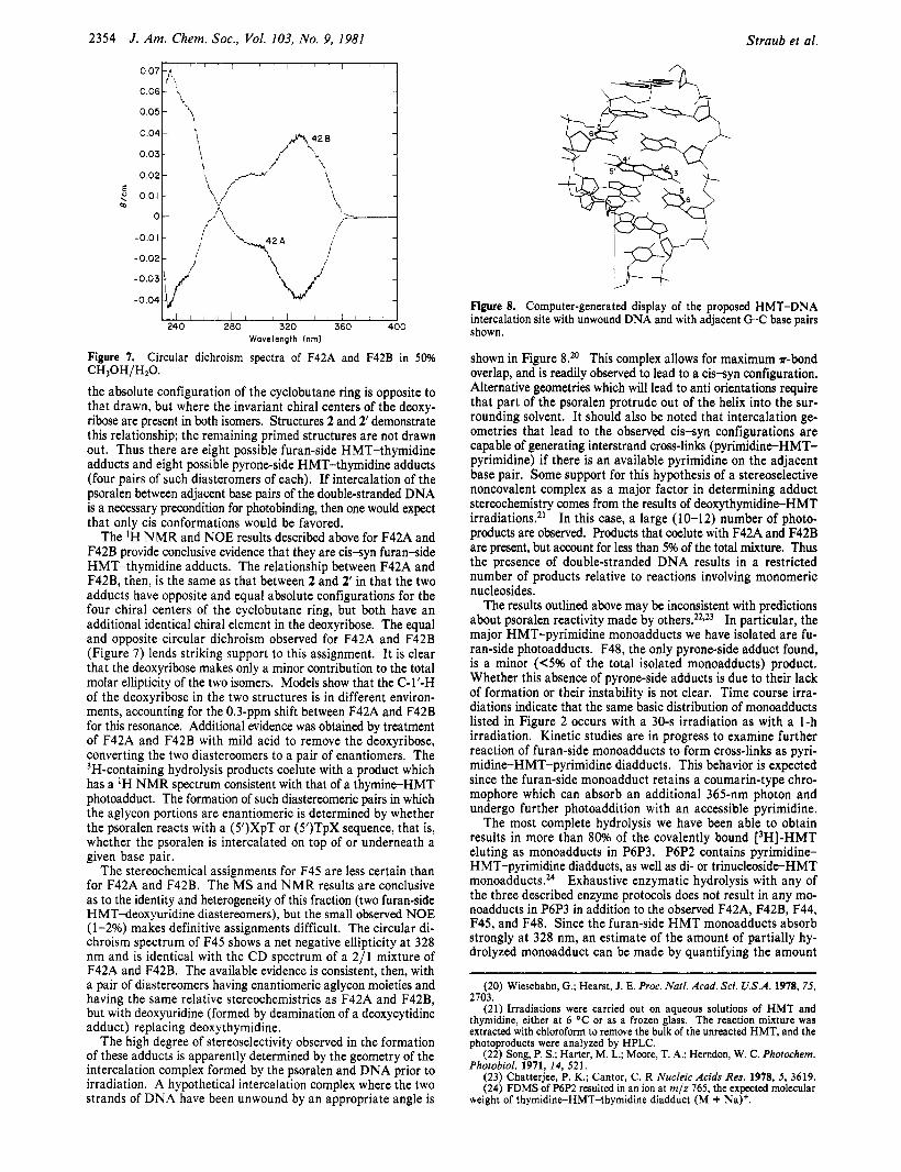
The high degree of stereoselectivity observed in the formation of these adducts is apparently determined by the geometry of the intercalation complex formed by the psoralen and DNA prior to irradiation. A hypothetical intercalation complex where the two strands of DNA have been unwound by an appropriate angle is
CH,OH/H20.
Figure 8. Computer-generated display of the proposed HMT-DNA intercalation site with unwound DNA and with adjacent G C base pairs shown.
shown in Figure 8.*O This complex allows for maximum r-bond overlap, and is readily observed to lead to a c issyn configuration. Alternative geometries which will lead to anti orientations require that part of the psoralen protrude out of the helix into the sur- rounding solvent. It should also be noted that intercalation ge- ometries that lead to the observed cis-syn configurations are capable of generating interstrand cross-links (pyrimidine-HMT- pyrimidine) if there is an available pyrimidine on the adjacent base pair. Some support for this hypothesis of a stereoselective noncovalent complex as a major factor in determining adduct stereochemistry comes from the results of deoxythymidine-HMT irradiations.21 In this case, a large (10-12) number of photo- products are observed. Products that coelute with F42A and F42B are present, but account for less than 5% of the total mixture. Thus the presence of double-stranded DNA results in a restricted number of products relative to reactions involving monomeric nucleosides.
The results outlined above may be inconsistent with predictions about psoralen reactivity made by other^.^^*^^ In particular, the major HMT-pyrimidine monoadducts we have isolated are fu- ran-side photoadducts. F48, the only pyrone-side adduct found, is a minor (6% of the total isolated monoadducts) product. Whether this absence of pyrone-side adducts is due to their lack of formation or their instability is not clear. Time course irra- diations indicate that the same basic distribution of monoadducts listed in Figure 2 occurs with a 30-s irradiation as with a 1-h irradiation. Kinetic studies are in progress to examine further reaction of furan-side monoadducts to form cross-links as pyri- midine-HMT-pyrimidine diadducts. This behavior is expected since the furan-side monoadduct retains a coumarin-type chro- mophore which can absorb an additional 365-nm photon and undergo further photoaddition with an accessible pyrimidine.
The most complete hydrolysis we have been able to obtain results in more than 80% of the covalently bound 13H]-HMT eluting as monoadducts in P6P3. P6P2 contains pyrimidine- HMT-pyrimidine diadducts, as well as di- or trinucleoside-HMT m o n o a d d ~ c t s . ~ ~ Exhaustive enzymatic hydrolysis with any of the three described enzyme protocols does not result in any mo- noadducts in P6P3 in addition to the observed F42A, F42B, F44, F45, and F48. Since the furan-side H M T monoadducts absorb strongly at 328 nm, an estimate of the amount of partially hy- drolyzed monoadduct can be made by quantifying the amount
(20) Wiesehahn, G.; Hearst, J. E. Proc. Natl. Acad. Sci. U.S.A. 1978, 75, 2703.
(21) Irradiations were carried out on aqueous solutions of HMT and thymidine, either at 6 O C or as a frozen glass. The reaction mixture was extracted with chloroform to remove the bulk of the unreacted HMT, and the photoproducts were analyzed by HPLC.
(22) Song, P. S.; Harter, M. L.; Moore, T. A,; Herndon, W. C. Photochem. Photobiol. 1971, 14, 521.
(23) Chatterjee, P. K.; Cantor, C. R Nucleic Acids Res. 1978, 5 , 3619. (24) FDMS of P6P2 resulted in an ion at m/z 765, the expected molecular
weight of thymidine-HMT-thymidine diadduct (M + Na)+.

Pyrimidine-Psoralen Photoadducts from DNA
of this absorbance in P6P2 (di- and pyrone-side adducts do not absorb at 328 nm). The results of such a measurement indicate that less than 15% of the 3H-containing material in P6P2 is present as partially hydrolyzed furan-side monoadducts. It is possible that small amounts of pyrone-side adducts are present as partially hydrolyzed oligonucleotides in this fraction.
Given the stereochemistry of the observed monoadducts and the proposed intercalation complexes, it is possible to predict the stereochemistries expected for diadducts. If only cis-syn con- figurations are allowed as in Figure 8, then two thymidine- HMT-thymidine diastereomeric adducts should be possible. The two possible dT-HMT-dT diadducts have a diastereomeric re- lationship, and removal of both deoxyriboses will result in a pair of enantiomers. Similarly, two deoxycytidine-HMT-deoxycytidine diadducts are expected (isolable as deoxyuridine adducts), and four diastereomeric heterodiadducts (dT-HMT-dC). The iso- lation and characterization of these adducts is in progress.
A final point of potential significance is the fact that F42A and F42B (and the two components of F45 as well) are apparently not formed in equal amounts. An approximate 2/ 1 ratio was found with all three enzymatic hydrolysis methods, suggesting that this is the ratio of diastereomeric adducts actually present in the DNA. Since the difference between F42A and F42B results from H M T reacting on top of or underneath the pyrimidine-purine base pair, this result could indicate a possible sequence specificity for pso- ralen-DNA reactivity. Such a specificity with respect to pyri- midine-purine or purine-pyrimidine sequencies has been reported for ethidium bromidez5 and actinomycin Dez6 Further investi- gation of this possibility is also in progress.
The analytical techniques developed in this study can be readily applied to the structural elucidation of other psoralen or small molecule DNA adducts. The ability to assign relative stereo- chemistries to these products could lead to increased understanding of the various factors that modulate the interactions of small
J. Am. Chem. SOC., Vol. 103, No. 9, 1981 2355
molecules with nucleic acids. In particular, direct questions may now be answered about the influence of noncovalent interactions such as intercalation on subsequent covalent binding. There is also considerable potential for utilizing the present methodology in designing chemical probes or drugs that would have enhanced binding affinities or unique selectivities toward certain types of primary and secondary nucleic acid structures. In summary, from the photobinding of a psoralen derivative,
4’-hydroxymethyl-4,5’,8-trimethylpsoralen (HMT), to double- stranded DNA we have isolated five nucleoside-HMT monoad- ducts. The major products result from photoaddition to the 4’,5’ double bond of the psoralen. They include two deoxythymidine adducts (F42A and F42B, structures 2 and 2’, 42 and 25% of the isolated monoadducts, respectively) and two deoxyuridine adducts (F45, c issyn stereochemistry, 28% of the isolated monoadducts). A minor deoxythymidine adduct, F48, results from photoaddition to the 3,4 double bond of the psoralen and accounts for 2% of the isolated monoadducts. We have also found evidence for the formation of a number of pyrimidine-HMT-pyrimidine diadducts. A limited number of stereoisomers are formed, and the stereo- chemistry of these isomers is apparently determined by the ge- ometry of the intercalation complex that occurs between the psoralen and D N A prior to irradiation.
Acknowledgment. This work was supported in part by the Division of Biomedical and Environmental Research, DOE, and the National Institute of General Medical Sciences, DHEW. K. Straub was supported by the Space Sciences Laboratory, Berkeley. N M R studies were carried out at the University of California, Davis, N M R facility, under the auspices of N S F Grant No. CHE79-04832. Mass spectrometry studies were carried out a t the University of California, Berkeley, Bio-organic, Biomedical Mass Spectrometry Resource supported by N I H Grant R R 007 19 Dr. A. L. Burlingame, Director. The authors thank Drs. J. Dallas and G. Matson for their assistance in obtaining and interpreting the N M R results. Computer modeling studies were carried out a t the Computer Graphics Laboratory, University of California, San Francisco, supported by NIH Grant R R 108 1.
(25) (a) Feinhardt, C. G.; Krugh, T. R Biochemistry 1978,17,4845; (b)
(26) Reinhardt, C. G.; Krugh, T. R. Biochemistry 1977, 16, 2890. Kastrup, R. V.; Young, M. A,; Krugh, T. K. Ibid. 1978, 17, 4855.