Isolation and characterization of D- Isolation and ... D- Isolation and characterization of...
10
Received on: 02-11-2013 Accepted on: 25-11-2013 Published on: 21-12-2013 Sadananda T.S* a Endophytic Natural Products Laboratory, Department of Biotechnology, Shridevi Institute of Engineering and Technology, Sira Road, Tumkur-572106, Karnataka, India Email: [email protected] Telephone: +91-9686114873 QR Code for Mobile users Isolation and characterization of D- Isolation and characterization of D-galactose, N- acetylgalactosamine, fructose, maltose specific lectin from eight different endophytic fungi of Viscum albumL. Sadananda TS a* , Govindappa M a , Ramachandra YL b a Endophytic Natural Products Laboratory, Department of Biotechnology, Shridevi Institute of Engineering and Technology, Sira Road, Tumkur-572106, Karnataka, India. b Dept. of P.G. Studies and Research in Biotechnology & Bioinformatics, Kuvempu University, Jnana Sahyadri, Shankaraghatta-577 451, Shimoga, Karnataka, India Abstract Eight different endophytic fungus was isolated and identified from the various parts of Viscum album, lectin was isolated from all the endophytic fungal sample and host Viscum album. Lectin isolated was blood group specific lectin agglutinating A +ve erythrocytes. Carbohydrate specificity of lectin was varied, Aspergillus flavus, Fusarium moniliforme, Fusarium oxysporum, Trichothecium sp, and Viscum album lectin was D-galactose and N-acetyl glucosamine specific lectin, Alternaria sp and Pencillium sp lectin was fructose specific lectin, Aspergillus niger and Cladosporium sp was Maltose and D-galactose specific lectin. Biochemical characterization of lectin showed that metal chelating agent EDTA has no effect on the hemaglutinating activity of Viscum album, Aspergillus flavus, Alternearia sp, Fusarium oxysporum, Trichothecium sp, lectin but EDTA inhibited the hemaglutinating activity of Pencillium sp, Cladosporium sp, Aspergillus niger lectin, activity was restored by adding divalent cations Mg 2+ , Mn 2+ . pH sensitive profile shows that lectin of Viscum album and its all endophytic fungal lectin retained the hemagglutinating activity with in the pH range 6-9, Viscum album and Aspergillus niger, Cladosporium sp, Fusarium moniliforme lectin was thermostable up to 60 0 C, Aspergillus flavus ,Alternearia sp, Fusarium oxysporum, Trichothecium sp lectin was stable upto 50 0 C. SDS PAGE of Aspergillus flavus, Fusarium moniliforme, Fusarium oxysporum, Trichothecium sp, D-galactose and N-acetyl glucosamine specific lectin showed the presence of 64kD protein similar to Viscum album lectin, PAS staining assay confirmed the presence of lectin in all endophytic fungal samples. Keywords: Lectin, endophytes, Viscum album, Hemagglutination, PAS staining. Cite this article as: Eglal Sadananda TS, Govindappa M, Ramachandra YL. Draz. Isolation and characterization of D- Isolation and characterization of D-galactose, N-acetylgalactosamine, fructose, maltose specific lectin from eight different endophytic fungi of Viscum albumL. Asian Journal of Biomedical and Pharmaceutical Sciences; 03 (26); 2013; 11-20.
-
Upload
phungduong -
Category
Documents
-
view
229 -
download
1
Transcript of Isolation and characterization of D- Isolation and ... D- Isolation and characterization of...

Received on: 02-11-2013 Accepted on: 25-11-2013 Published on: 21-12-2013
Sadananda T.S* aEndophytic Natural Products Laboratory, Department of Biotechnology, Shridevi Institute of Engineering and Technology, Sira Road, Tumkur-572106, Karnataka, India Email: [email protected]
Telephone: +91-9686114873
QR Code for Mobile users
Isolation and characterization of D- Isolation and characterization of D-galactose, N-
acetylgalactosamine, fructose, maltose specific lectin from eight different endophytic fungi of Viscum
albumL. Sadananda TSa*, Govindappa Ma, Ramachandra YLb
aEndophytic Natural Products Laboratory, Department of Biotechnology, Shridevi Institute of Engineering and Technology, Sira Road, Tumkur-572106, Karnataka, India.
bDept. of P.G. Studies and Research in Biotechnology & Bioinformatics, Kuvempu University, Jnana Sahyadri, Shankaraghatta-577 451, Shimoga, Karnataka, India
Abstract Eight different endophytic fungus was isolated and identified from the various parts of Viscum album, lectin was isolated from all the endophytic fungal sample and host Viscum album. Lectin isolated was blood group specific lectin agglutinating A+ve erythrocytes. Carbohydrate specificity of lectin was varied, Aspergillus flavus, Fusarium moniliforme, Fusarium oxysporum, Trichothecium sp, and Viscum album lectin was D-galactose and N-acetyl glucosamine specific lectin, Alternaria sp and Pencillium sp lectin was fructose specific lectin, Aspergillus niger and Cladosporium sp was Maltose and D-galactose specific lectin. Biochemical characterization of lectin showed that metal chelating agent EDTA has no effect on the hemaglutinating activity of Viscum album, Aspergillus flavus, Alternearia sp, Fusarium oxysporum, Trichothecium sp, lectin but EDTA inhibited the hemaglutinating activity of Pencillium sp, Cladosporium sp, Aspergillus niger lectin, activity was restored by adding divalent cations Mg2+, Mn2+. pH sensitive profile shows that lectin of Viscum album and its all endophytic fungal lectin retained the hemagglutinating activity with in the pH range 6-9, Viscum album and Aspergillus niger, Cladosporium sp, Fusarium moniliforme lectin was thermostable up to 600C, Aspergillus flavus ,Alternearia sp, Fusarium oxysporum, Trichothecium sp lectin was stable upto 500C. SDS PAGE of Aspergillus flavus, Fusarium moniliforme, Fusarium oxysporum, Trichothecium sp, D-galactose and N-acetyl glucosamine specific lectin showed the presence of 64kD protein similar to Viscum album lectin, PAS staining assay confirmed the presence of lectin in all endophytic fungal samples. Keywords: Lectin, endophytes, Viscum album, Hemagglutination, PAS staining.
Cite this article as:
Eglal Sadananda TS, Govindappa M, Ramachandra YL. Draz. Isolation and characterization of D- Isolation and characterization of D-galactose, N-acetylgalactosamine, fructose, maltose specific lectin from eight different endophytic fungi of Viscum albumL. Asian Journal of Biomedical and Pharmaceutical Sciences; 03 (26); 2013; 11-20.

Sadananda et al.: Asian Journal of Biomedical and Pharmaceutical Sciences; 3(26) 2013, 11-20.
© Asian Journal of Biomedical and Pharmaceutical Sciences, all rights reserved. Volume 3, Issue 26, 2013. 12
1. INTRODUCTIONLectins are proteins or glycoproteins from non-immune origin that specifically recognize cell surface molecule carbohydrate [1], and they found in all kinds of organism including animals, plants, fungi, bacteria and viruses [2,3]. It is their unique ability to recognize and bind reversibly to specific carbohydrate ligands without any chemical modification that distinguish lectins from other carbohydrate binding proteins, because of it makes them invaluable tools in biomedical and glycoconjugate research [4]. An important characteristic property of lectins is their ability to agglutinate erythrocytes in vitro. That is why; they frequently called agglutinins (e.g phytohemagglutinin). Lectins from different source inhibit cancer cells growth, they are able to induce apoptosis and activate the immune system by stimulating the proliferation of T-lymphocytes [5]. Cytotoxic properties of lectins like ricin (RCA) and abrin (APA) as potential therapies for human cancer treatment [6,7]. Viscum album L. (Loranthaceae) has been known since ancient time [8]. It is hemiparaitic plants, which are distributed in Korea, Europe and other Asian countries. Viscum album extracts has been traditionally used as a sedative, analgesic, anti-sapasolytic, cardiotonic and anticancer agents. Various chemical components have been identified from the extracts of Viscum album such as lectin, steroid, triterpene, sesquiterpene, flavanoid, alkaloid, organic acid, aminoacids and peptides [9]. Viscum album extracts has been used in adjuvant chemotherapy of human cancers for a long time [10]. Viscum album L agglutinin (VAA-I, II, III) are considered to be major active components in European mistletoe and have a molecular masses between 50 and 60 kDa [11]. They differ in their relative sugar binding specificities. VAA-I shows specificity to D-galactose, VAA-II, III prefentially bind to N –acetylgalactosamine [11]. The VAAs are type-2 ribosome inactivating proteins composed of two different subunits, an A- and B- chain linked by disulfide-bridge. The A-chain is capable of inactivating the 60S ribosomal subunit of eukaryotic cells resulting in inhibition of protein synthesis. The B-chain is capable of binding to cell surface glycoconjugates and thereby permits entry into the cell [12]. Viscum album agglutinin-I was recently found to induce cytotoxic effects on different tumor cells of lymphoid origin. Evidence suggests that this cytotoxicity may be mediated by induction of apoptosis, a highly conserved mechanism of cell death. Cell death associated with typicall apoptotic alterations such as cell shrinkage, chromatin condensation and inters nucleosomal DNA cleavage [13-15]. Based on the above reports of biological activity of lectin, we planned to screen similar type of lectin from the endophytes of Viscum album.
Endophytes are the microorganisms which inhabit normal tissues of host plants without causing apparent symptoms of pathogenesis, these endophytes are novel and rich sources of bioactive natural product producers which include alkaloids, amines, amides, steroids, terpenoids, isocoumarins, quinines, flavonoids, phenyl propanoids, lignans, phenols, aliphatics, etc [16]. Endophytes are recognized as potential; sources of novel natural products for exploitation in medicine, agriculture and industry with more bioactive natural products isolated from these microorganisms. In some cases endophytes can produce the same rare and important bioactive compounds as their host plant produces [17]. In this case this would not only reduce the need to harvest slow growing and possibly rare plants but also preserve the worlds ever-diminishing biodiversity and it is recognized that a microbial source of a valued product may be easier and more economical to produce, effectively reducing its market price. Due to this extensive biological activity of lectin as anticancer agent, the objective of this research was to isolate the lectins from the endophytes of Viscum album and to characterize lectin. The literature survey indicates that no reports are available from India and internationally about the endophytic lectin from Viscum album L. 2. MATERIALS AND METHODS 2.1 Collection of plant material:
Hemi parasitic plant material Viscum album L growing on Pongamia was collected from D.C Bungalow, Sira Gate, Tumkur, Karnataka, India during month August, 2012. The collected plant was authenticated from the Department of Botany, Manasa Gangotri, University of Mysore and Government Ayurvedic College, Mysore. The leaves were collected from the plant and dried under shade for 30 days at room temperature (26+20C) and then powdered with a mechanical grinder and stored in cool and dry place for further use. 2.2 Isolation of endophytic fungi: Endophytic fungi isolation was carried out under aseptic condition [18], the stem and leaves of the collected plant material was detached with a sterilized sharp blade, cleaned by washing with running tap water several times and soaked in 70% (v/v) ethanol for 10-20 min. It was then washed several times with sterilized water, dipped into 0.1% HgCl2 for 1-2 min, again washed with sterilized water 3-5 times and then put into a beaker of sterilized distilled water. The sterilized stem and leaves of collected plant material was then cut into small pieces of 1 to 1.5 cm, each piece put on a petri plate of potato dextrose agar (PDA) medium and the plate incubated at 300 C to promote fungal growth and sporulation. After 7-8 days individual hyphal tips of the fungus were then picked

Sadananda et al.: Asian Journal of Biomedical and Pharmaceutical Sciences; 3(26) 2013, 11-20.
© Asian Journal of Biomedical and Pharmaceutical Sciences, all rights reserved. Volume 3, Issue 26, 2013. 13
up from each part and inoculated onto another PDA medium plate individually and incubated at 300C for 1 week. The purified fungal isolates were numbered, transferred separately to PDA slants, and kept at 40C for further use. 2.3 Identification of endophytic fungi:
Isolated endophytic fungi was identified and characterization on the basis of morphology and microscopic studies, endophytic fungal isolates slides prepared from cultures were stained with lactophenol cotton blue stain and examined with a bright-field and phase-contrast microscope. Identification was based on morphological characteristics such as growth pattern, hyphae, colour of colony and medium, surface texture, margin character, aerial mycelium, mechanism of spore production and conidial characteristics, using standard identification manuals [19-20]. 2.4 Mass production of identified fungi:
Identified fugal species were cultured on Czapek Dox broth for large scale cultivation, which was then incubated at room temperature 300C for 5 days. 2.5 Extraction of lectin from Viscum album:
Mistletoe grown on Pongamia was collected during August, 2012 and sealed in a plastic bag and stored at -200 C until use. The leaves of the plant were grinded to powder. The powdered material was transferred to a warring blender placed in a well ventilated and ground into a fine powder which was transferred to an erlenmeyer flask. Approximately 10 volumes of 10 mM Tris-HCl (pH 8.31) containing 100 mM lactose were added and the suspension was stirred with a magnetic stirrer at 40C overnight [21]. The suspension was filtered through cheesecloth and then centrifuged at 10,000rpm for 10 min in a centrifuge. The pellet was discarded. The collected supernatant was used for further purification. 2.6 Extraction of lectin from endophytic fungus:
Identified endophytic fungus was cultured in a 500ml concial flasks containing 150 ml Czepadox broth and incubated at room temperature under stationary conditions. Isolation of lectin followed modefied method [22]. After 10 days, the mycelial mat was harvested and washed with distilled water on cheese cloth. Washed mycelia mat was homogenized in 50 ml (1 : 25 w/v) of 50mM sodium phosphate buffer (pH 7.2) containing 154mM NaCl (PBS) for 5 minutes and stirred overnight at 40C. The extract was centrifuged (9500 rpm) for 10 minutes at 40C. The resulting supernatant was used for the purification of the lectin. 2.7 Affinity chromatography: Plant and endophytic lectin extracted was purified by the method [23], the crude protein was loaded onto a lactose–agarose (Sigma) column equilibrated and eluted with extraction buffer at a flow rate of 3 ml/min, until the column effluent showed absorbance at 280 nm of less than 0.05. Bound proteins were
eluted with 100 mM lactose in equilibration buffer. Active fractions collected was adjusted to be saturated in 60 % ammonium sulfate, precipitated protein dissolved in PBS and dialyzed extensively against same buffer , aliquot, freeze-dried and stored at – 300
C until use. 2.8 Protein determination: The protein concentrations of the dialyzed protein sample from plant and endophytic sample was determined by the method of Lowry’s [24]. Bovine serum albumin was used for standard preparations. 2.9 Assay of hemagglutinating activity 2.9.1 Collection of blood samples: Blood groups of A, B, AB & O collected from healthy individuals in the Department of Biotechnology, S.I.E.T College, Tumkur, Karnataka, India. The blood samples were collected in heparinised bottles (EDTA) to prevent the blood from coagulating and kept in fridge to preserve them till the time of use [25]. The red blood cells obtained were then washed by centrifugation at 1500rpm for 5minutes at room temperature (26+20C) with 0.01M phosphate- buffered saline (pH 7.2). This was repeated twice, after which the cells were mixed with 3% formaldehyde in EDTA bottle and allowed to stir gently overnight, before it was centrifuged at 1500rpm for 5 minutes, the following day. The centrifuged red cells were then washed again as before, three times with 0.01M phosphate- buffered saline (pH 7.2) after which the cells were collected into a stopped bottle and 76.8ml of 0.01M phosphate- buffered saline was added to make the cell 4% thereafter, it was stored in the fridge 2.9.2 Assay for lectin activity: The assay of hemagglutination activity of plant and endophytic fungal lectin [26], serial twofold dilution of the lectin solution in microtiter U-plates (50µl) was mixed with 50µl of a 4% suspension of red blood cells in phosphate-buffered saline (pH 7.2) at room temperature. The results were read after about 1 hour, where the control (no lectin was added) had fully sedimented (red button). The hemagglutination titer, defined as the reciprocal of the highest dilution exhibiting hemagglutination, is reckoned as one hemagglutination unit. Specific activity is the number of hemagglutination units per mg protein. 2.10 Biochemical and biological characterization of lectin 2.10.1 Total sugar determination: The carbohydrate content of the purified lectin was determined by the Anthrone method [27], using D-glucose as standard. Lectin samples from plants and its endophytes were first hydrolyzed by keeping it in a boiling water bath for three hours with 3ml of 2.5N HCl and cool to room temperature and neutralized it with sodium carbonate until the effervescence ceases,
Sadananda et al.: Asian Journal of Biomedical and Pharmaceutical Sciences; 3(26) 2013, 11-20.
© Asian Journal of Biomedical and Pharmaceutical Sciences, all rights reserved. Volume 3, Issue 26, 2013. 14
samples were collected and centrifuged at 1500 rpm for 5min and supernatant were used for the carbohydrate analysis. 2.10.2 Assay of inhibition of haemagglutination and carbohydrate-binding specificity: To determine the sugar binding specificity of lectin was determined [28], different sugars including D-galactose, D-mannose, D-glucose, D-fructose, maltose, lactose, N-acetylgalactosamine were tested for their ability to inhibit lectin induced hemagglutination. Lectin concentration, just one step upstream the end point of hemagglutination titre was chosen for the hemagglutination inhibition assay. The above mentioned carbohydrates were employed as potential inhibitors. Serial two-fold dilutions of each carbohydrate were prepared in 10mM to 100mM range and dissolved 0.15 M NaCl solution and mixed with equal volumes of extract containing 4 units of haemagglutinating activity. Mixtures were incubated for 30 min at room temperature, after which a suspension of human A+ erythrocytes (4%) was added and the whole incubated for 1 h. The lowest carbohydrate concentration that produced complete inhibition of haemagglutination was determined. 2.10.3 Effect of pH on haemagglutinating activity: The effect of pH on the haemagglutinating activity was determined [29], by carrying out the haemagglutinating assay of the lectin at different pH by incubating the lectin samples in the following buffers varying from pH 2–10 for 18 h at 40C. Different buffers were used according to pH range as follows; 50 mM glycine–HCl buffer (pH 2.0 – 3.0), 50 mM sodium acetate buffer (pH 4.0 – 7.5), 50 mM Tris–HCl buffer (pH 8.0–8.5), and 50 mM glycine–NaOH buffer (pH 9.0–10). The pH sensitivity of the lectin was established by incubating aliquots of the purified lectin at respective hemagglutin titer for 1 h in buffers at pH values 2 -10, the haemagglutinating activity of the lectin was then measured after adjusting the pH of the assay solution to 7.0. 2.10.4 Effect of EDTA and divalent cations The effect of EDTA and divalent cations on the haemagglutinating activity of the lectin was carried [29], Two-fold serial dilutions of lectin that were prepared in 0.2M PBS alone and 0.2M PBS containing 5 mM EDTA was carried out. Human A+ erythrocytes (4%) in 0.2M PBS with 5 mM EDTA were used as the control. Equal volumes (50 μl) of 10 mM MgSO4, MnCl2 were later added to the haemagglutination assay that was performed in the presence of EDTA in order to evaluate their capacity to restore haemagglutination. 2.10.5 Effect of temperature on haemagglutinating activity The effect of temperature on the haemagglutinating activity was monitored [29]. Aliquots of lectin were incubated at different temperatures (20-900 C). The
heated solution was rapidly cooled in ice and assayed for agglutinating activity. Agglutinating activity of the control that was kept at 300 C for 30 min was used as a reference. 2.10.6 Polyacrylamide gel electrophoresis: The polyacrylamide gel electrophoresis was performed in 2 mm thick vertical slab gels [30], using 5% and 12.5% stacking and running gels, respectively. Endophytic lectin samples that were similar in carbohydrate specificity to Viscum album lectin were used for SDSPAGE, lectin were dissolved in 0.0625 M Tris-HCl pH 6.8, containing 1% SDS buffer, 0.1 CBB and 10% glycerol then incubated at 90oC for 5 min. The molecular markers employed were β-galactosidase (116 kDa), fructose-6-phosphate kinase (80 kDa), bovine serum albumin (72 kDa), ovalbumin (68 kDa), glutamate (60 kDa), carbonic anhydrase (36 kDa), myoglobin (29 kDa) (Aristogene Bioscience Ltd, Bangalore). Electrophoresis was carried out at a constant current of 50 v for 3 h. After electrophoresis, the gel was stained with Coomassie brilliant blue R-250. The molecular weight of the purified lectin was determined by comparing its electrophoresis mobility with the standard molecular weight marker proteins. 2.10.7 Demonstration of glycoprotein: Glycoslylation of lectin was demonstrated in the gel [31], SDS PAGE of lectin samples carried using 5% and 12.5% stacking and running gels. The gel after electrophoretic run was washed continuously with 2.6L of 40% methanol and 7% acetic acid overnight. The solution was changed and gel was put in 7.5% acetic acid and kept in RT for 1h. The gel was transferred to a tank containing 1% periodic acid, kept immersed for 1 h in dark at 40 C. The gel was washed in 7.5 % acetic acid for 10 min and the washing repeated 6 times. After washing the gel was incubated in Schiff's reagent at 40 C in dark for 1 h and washed in 0.5% sodium metabisulphate. Pink colored bands were observed. The gel was preserved in 7.5% acetic acid.
3. RESULTS AND DISCUSSION 3.1 Isolation and identification of endophytes:
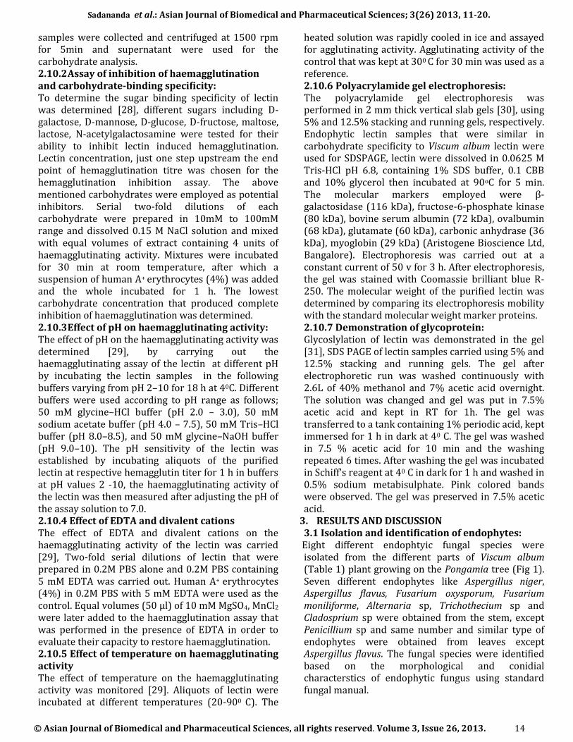
Eight different endophtyic fungal species were isolated from the different parts of Viscum album (Table 1) plant growing on the Pongamia tree (Fig 1). Seven different endophytes like Aspergillus niger, Aspergillus flavus, Fusarium oxysporum, Fusarium moniliforme, Alternaria sp, Trichothecium sp and Cladosprium sp were obtained from the stem, except Penicillium sp and same number and similar type of endophytes were obtained from leaves except Aspergillus flavus. The fungal species were identified based on the morphological and conidial characterstics of endophytic fungus using standard fungal manual.
Sadananda et al.: Asian Journal of Biomedical and Pharmaceutical Sciences; 3(26) 2013, 11-20.
© Asian Journal of Biomedical and Pharmaceutical Sciences, all rights reserved. Volume 3, Issue 26, 2013. 15
Fig 1.Viscum album on Pongamia
Endophytic fungi
Plant parts
Stem Leaves
Aspergillus niger + +
Aspergillus flavus + -
Fisarium oxysporum + +
Fusarium moniliforme + +
Alternaria sp. + +
Penicillium sp. - +
Trichothecium sp. + +
Cladosporium sp + +
Table 1. Endophytes from different parts of Viscum album plant
+: Presence,-: Absence, Repeated each experiment thrice
3.2 Isolation and purification of lectin from plant and endophytes
The identified endophytic fungus was cultured on Czapek Dox broth for mass production, lectin from the plant was isolated using 10mM Tris-HCl (pH 8.3, containing 100 mM lactose) and endophytic lectin using 50mM sodium phosphate buffer, pH 7.2, containing 154mM NaCl, crude protein was purified by affinity chromatography using Agarose-lactose column and was able to absorb lectin effectively from plant and endophytic extract. All the proteins without D-galactose specific characteristics were eluted put from the column rapidly; the bound fractions were eluted with 0.2M D-galactose buffered saline. Eluted protein was fractionated at 60 % ammonium sulfate precipitation, dialyzed against Phosphate buffered salaine (pH 7.2) and protein was estimated in the entire eluted sample using Lowry’s method, the concentration of protein from Viscum album and its endophytic was depicted in the (Fig 2)
Figure 2. Protein concentration of Viscum album and fungal
endophytic
3.3 Assay for lectin activity Purified protein extracts from the leaves of Viscum album and endophytic fungi were found to contain a hemagglutinating protein. Two-fold serial diluted purified protein was used for hemagglutination assay (22 to 210) (Table 2).
Sr.No
Endophytes
Erythrocytes
Titer
Concentration.
1 Aspergillus
niger
Human type A+
erythrocytes
26 0.025 µg/ml
2 Aspergillus
flavus
Human type A+
erythrocytes
28 0.018 µg/ml
3 Fisarium
oxysporum
Human type A+
erythrocytes
26 0.038 µg/ml
4 Fusarium
moniliforme
Human type A+
erythrocytes
26 0.084 µg/ml
5 Alternaria
sp.
Human type A+
erythrocytes
28 0.0078 µg/ml
6 Penicillium
sp.
Human type A+
erythrocytes
26 0.094 µg/ml
7 Trichotheci
um sp.
Human type A+
erythrocytes
26 0.012 µg/ml
8 Cladospori
um sp.
Human type A+
erythrocytes
26 0.098 µg/ml
9 Viscum album
L.(plant)
Human type A+
erythrocytes
28 0.32 µg/
ml
Table 2: Hemagglutination titer concentration *Each experiment was repeated thrice
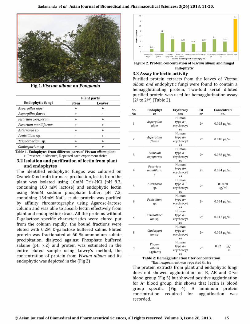
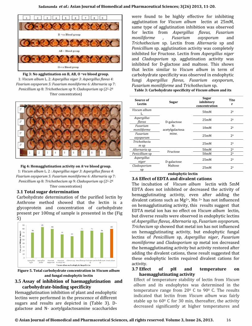
The protein extracts from plant and endophytic fungi does not showed agglutination on B, AB and O+ve blood group (Fig 3) but showed positive agglutination for A+ blood group, this shows that lectin is blood group specific (Fig 4). A minimum protein concentration required for agglutination was recorded.
Sadananda et al.: Asian Journal of Biomedical and Pharmaceutical Sciences; 3(26) 2013, 11-20.
© Asian Journal of Biomedical and Pharmaceutical Sciences, all rights reserved. Volume 3, Issue 26, 2013. 16
Fig 3: No agglutination on B, AB, O +ve blood group.
1: Viscum album L. 2: Aspergillus niger 3: Aspergillus flavus 4:
Fisarium oxysporum 5: Fusarium moniliforme 6: Alternaria sp 7:
Penicillium sp 8: Trichothecium sp 9: Cladosporium sp (22-28
Titer concentration)
Fig 4: Hemagglutination activity on A+ve blood group.
1: Viscum album L. 2 : Aspergillus niger 3: Aspergillus flavus 4:
Fisarium oxysporum 5: Fusarium moniliforme 6: Alternaria sp 7:
Penicillium sp 8: Trichothecium sp 9: Cladosporium sp (22-28
Titer concentration)
3.1 Total sugar determination Carbohydrate determination of the purified lectin by Anthrone method showed that the lectin is a glycoprotein and concentration of carbohydrate present per 100mg of sample is presented in the (Fig 5)
Figure 5. Total carbohydrate concentration in Viscum album
and fungal endophytic lectin
3.5 Assay of inhibition of haemagglutination and
carbohydrate-binding specificity Hemagglutination inhibition of plant and endophytic lectins were performed in the prescence of different sugars and results are depicted in (Table 3). D-galactose and N- acetylgalactosamine scaccharides
were found to be highly effective for inhibiting agglutination for Viscum album lectin at 25mM, same type of agglutination inhibition was observed for lectin from Aspergillus flavus, Fusarium moniliforme , Fusarium oxysporum and Trichothecium sp. Lectin from Alternaria sp and Penicillium sp. agglutination activity was completely inhibited for Fructose. Lectin from Aspergillus niger and Cladosporium sp. agglutination activity was inhibited for D-glactose and maltose. This shows that lectin similar to Viscum album in term of carbohydrate specificity was observed in endophytic fungi Aspergillus flavus, Fusarium oxysporum, Fusarium moniliforme and Trichothecium sp.
Table 3: Carbohydrate specificity of Viscum album and its
endophytic lectin
3.6 Effect of EDTA and divalent cations The incubation of Viscum album lectin with 5mM EDTA does not inhibited or decreased the activity of hemagglutinating activity, even after adding the divalent cations such as Mg2+, Mn 2+ has not influenced on hemagglutinating activity, this results suggest that EDTA metal ion has no effect on Viscum album lectin, but diverse results were observed in endophytic lectins of Aspergillus flavus, Alternaria sp, Fusarium oxysporum, Trichecium sp showed that metal ion has not influenced on hemagglutinating activity, but endophytic fungal lectins of Penicillium sp, Aspergillus niger, Fusarium moniliforme and Cladosporium sp metal ion decreased the hemagglutinating activity but activity restored after adding the divalent cations, these result suggested that these endophytic lectin required divalent cations for activity. 3.7 Effect of pH and temperature on
haemagglutinating activity Effect of temperature stability of lectin from Viscum album and its endophytes was determined in the temperature range from 200 C to 900 C. The results indicated that lectin from Viscum album was fairly stable up to 600 C for 30 min, thereafter, the activity decreased significantly at higher temperatures and
Source of Lectin
Sugar Sugar
inhibitory concentration
Titer
Viscum album L.
D-galactose N-
acetylgalactosamine.
50mM 26
Aspergillus flavus
25mM 26
Fusarium moniliforme
10mM 26
Fusarium oxysporum
25mM 24
Trichothecium sp
25mM 24
Alternaria sp Fructose
50mM 24 Penicillium sp 50mM 24
Aspergillus niger D-galactose
Maltose
25mM 24
Cladosporium sp
25mM 24
Sadananda et al.: Asian Journal of Biomedical and Pharmaceutical Sciences; 3(26) 2013, 11-20.
© Asian Journal of Biomedical and Pharmaceutical Sciences, all rights reserved. Volume 3, Issue 26, 2013. 17
was totally inactivated when incubated at 900 C for 30 min (Fig 6). But endophytic lectin extract was fairly different in Aspergillus flavus, Fusarium oxysporum, Trichothecium sp and Alternaria sp was stable up to 500 C, there after activity decreases and was totally inactivated at 800 C. Aspergillus niger, Cladosporium sp, Fusarium moniliforme lectin was fairly stable up to 600
C but decrease thereafter and completely inactivated at 800 C. The pH sensitivity profile of lectin of Viscum album and its endophytic fungal lectin showed that lectin retained hemagglutinating activity within the pH range 6.0-9.0, it was sensitive to acidic pH 3.0 and to basic 12 pH under these conditions the hemagglutinating activity was completely lost (Fig 7)
Fig 6. Effect of Temperature on hemagglutination activity of
Viscum album and endophytic fungal lectin
Fig 7. Effect of pH on the hemagglutination activity of
Viscum albums and its endophytic fungal lectin
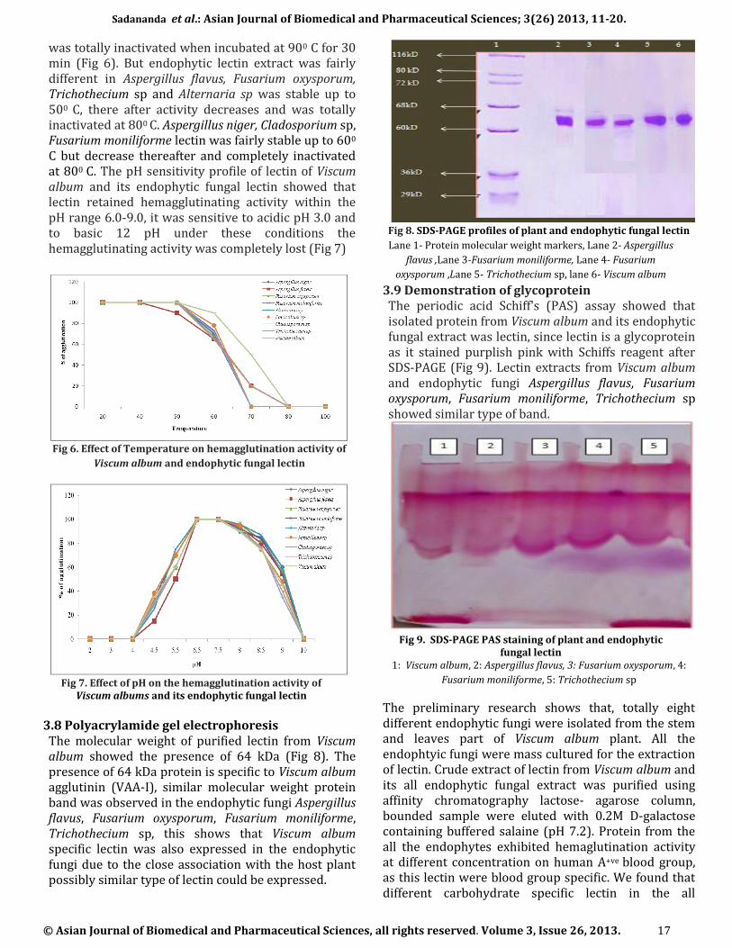
3.8 Polyacrylamide gel electrophoresis The molecular weight of purified lectin from Viscum album showed the presence of 64 kDa (Fig 8). The presence of 64 kDa protein is specific to Viscum album agglutinin (VAA-I), similar molecular weight protein band was observed in the endophytic fungi Aspergillus flavus, Fusarium oxysporum, Fusarium moniliforme, Trichothecium sp, this shows that Viscum album specific lectin was also expressed in the endophytic fungi due to the close association with the host plant possibly similar type of lectin could be expressed.
Fig 8. SDS-PAGE profiles of plant and endophytic fungal lectin
Lane 1- Protein molecular weight markers, Lane 2- Aspergillus
flavus ,Lane 3-Fusarium moniliforme, Lane 4- Fusarium
oxysporum ,Lane 5- Trichothecium sp, lane 6- Viscum album
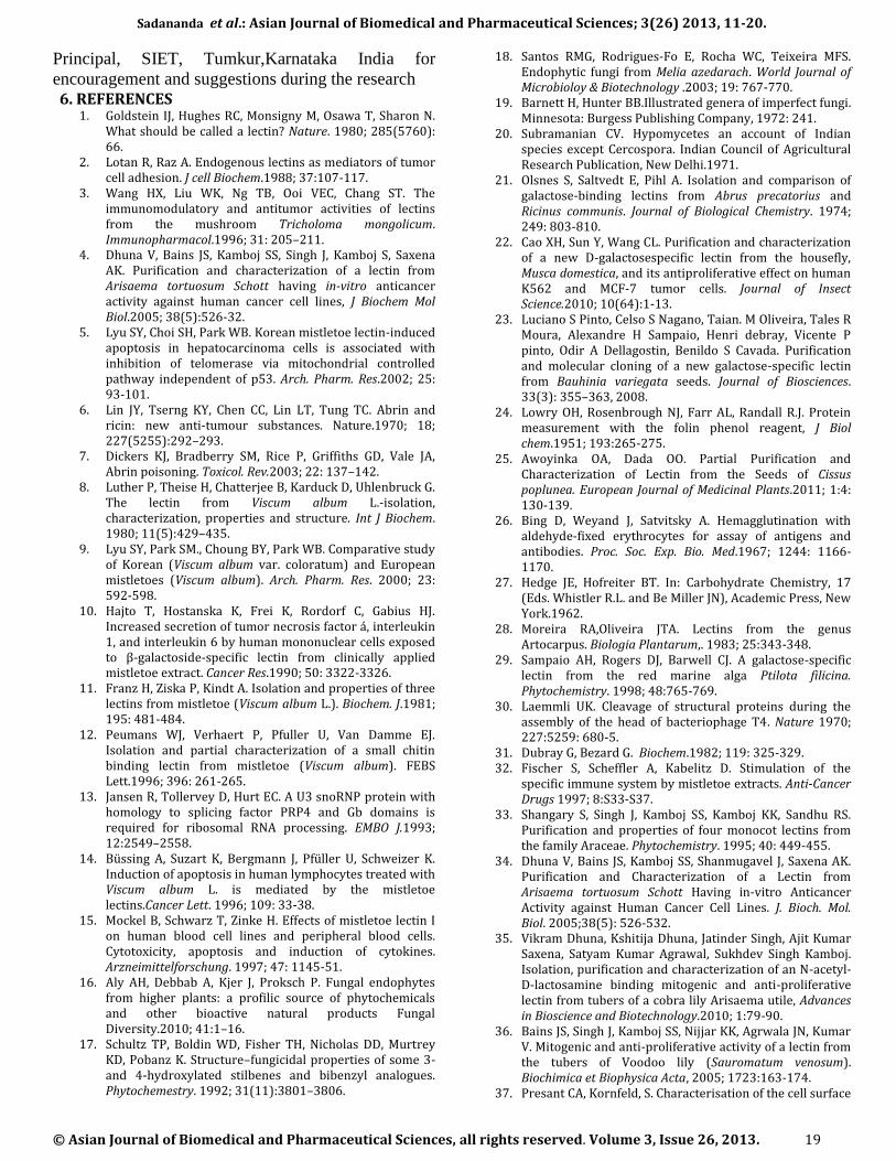
3.9 Demonstration of glycoprotein The periodic acid Schiff's (PAS) assay showed that isolated protein from Viscum album and its endophytic fungal extract was lectin, since lectin is a glycoprotein as it stained purplish pink with Schiffs reagent after SDS-PAGE (Fig 9). Lectin extracts from Viscum album and endophytic fungi Aspergillus flavus, Fusarium oxysporum, Fusarium moniliforme, Trichothecium sp showed similar type of band.
Fig 9. SDS-PAGE PAS staining of plant and endophytic
fungal lectin 1: Viscum album, 2: Aspergillus flavus, 3: Fusarium oxysporum, 4:
Fusarium moniliforme, 5: Trichothecium sp
The preliminary research shows that, totally eight different endophytic fungi were isolated from the stem and leaves part of Viscum album plant. All the endophtyic fungi were mass cultured for the extraction of lectin. Crude extract of lectin from Viscum album and its all endophytic fungal extract was purified using affinity chromatography lactose- agarose column, bounded sample were eluted with 0.2M D-galactose containing buffered salaine (pH 7.2). Protein from the all the endophytes exhibited hemaglutination activity at different concentration on human A+ve blood group, as this lectin were blood group specific. We found that different carbohydrate specific lectin in the all

Sadananda et al.: Asian Journal of Biomedical and Pharmaceutical Sciences; 3(26) 2013, 11-20.
© Asian Journal of Biomedical and Pharmaceutical Sciences, all rights reserved. Volume 3, Issue 26, 2013. 18
endophytic fungi and Viscum album. Viscum album plant and Aspergillus flavus, Fusarium moniliforme, Fusarium oxysporum, Trichothesium sp were D-galactose and N-acetylaminegalactose specific lectin. Results showed that lectin obtained from the endophytic fungi was similar in carbohydrate specificity specific to Viscum album lectin (VAA-I) that were D-galactose and N-acetylgalactoseamine specific lectin belongs to ribosomal inactivating protein [32]. Whereas Alternaria sp, Penicillium sp, were fructose specific, Aspergillus niger, Cladosporium sp were D-galactose and maltose specific. Monocot lectins have shown there inhibition by N-acetyl galactose amine [33, 34], mannose [35], D-galactose [36] and fructose [37, 38]. Based on the earlier reports, our results also indicate that agglutination of blood is always depends on different carbohydrates molecules on the surface of erythrocytes. Some of the endophytic fungal lectin from Penicillium sp, Aspergillus niger, Fusarium oxysporum, Cladosporium sp. showed metal ion dependence on hemaglutination activitiy but other endophytes Aspergillus flavus, Alternaria sp, Fusarium moniliforme, Tricothesium sp lectin has not depend on metal ion requirement. In different temperature (200 to 900C), Viscum album lectin have shown stable up to 600C and thereafter hemagglutination activity decreased, in endophytes of Aspergillus flavus, Fusarium oxysporum, Alternaria sp lectin was stable at 500 C there after hemagglutination activity decreased, Aspergillus niger, Cladosporiium sp, Fusrarium molaniforme lectin was stable up to 600 C, our results were supported with the findings of [29,39] [38-41], from different plant lectin. Similar results were observed in effect of pH on hemagglutination activity from the Viscum album and all its endophytes lectin. The maximum activity on hemagglutination at pH 7 was observed in the lectin from Viscum album and its all endophytic fungi exhibited similar results. Similar results were found in plant species viz., Kalanchoe crenata [39], Ptilota filicina [29], S.krinatum [38]. The pH change is associated with a change in the ionization state of molecule which in turn determines the binding forces between enzyme and substrate [41]. It is also possible that increase in OH- ions caused changes in ionization state of lectin there by affecting binding forces between the lectin and erythrocyte membrane that eventually led to a loss of activity. The endophytic fungi Aspergillus flavus, Fusarium moniliforme, Fusarium oxysporum, Trichothecium sp and Viscum album lectin showed similar carbohydrate specificity, in turn this lectin have showed the presence of 64 kDa protein on SDSPAGE. Previously reported that Viscum album agglutinin showed 64kDa protein [32]. Results obtained confirms the presence of similar type of lectin with same carbohydrate specificity and similar molecular weight protein from the fungal
endophytes of Viscum album, it has been reported that endophytes are capable of producing similar type of compound what the host plant is producing [42], Similar molecular weight lectin were reported 46 kDa lectin from Artocarpus integrifolia and 44 kDa from Maclura pomifera [43]. The PAS assay confirms the presence of lectin in all the endophytic fungi and plant extract with the findings of [31], they made very specific test for glycoprotein. If the extracts have glycoprotein (lectins) they will take pink colour after treating gel with Schiff reagent. All the four endophytic fungi have shown presence of lectin by agglutination and PAS staining method, this lectin shown the molecular weight of 64kD. This is the first report in India and internationally, all the eight endophytic fungi of V. album showing the presence of lectins (glycoproteins). These endophytes can be used for production of lectins at high concentration within a short period of time. In summary, all the four endophytic fungi Aspergillus flavus, Fusarium moniliforme, Fusarium oxysporum, Trichothecium sp, have D-galactose and N-acetyl galactose amine specific lectin similar to Viscum album lectin. Further studies on the cytotoxic activities of endophytic fungal lectins and identification of lectin are underway. 4. CONCLUSION Endophytes are the potential producers of novel metabolites of therapeutic value, the plant selected Viscum album (European Mistletoe) is known for toxic lectin Viscum album agglutinin it has been showed the anticancer activity, immunomodulatory, antifungal, HIV-1 reverse transcriptase inhibitory and anti-insect activities and is has been found that it can induce cytotoxic effects on different tumor cell. It is potent ribosome inactivating lectin, induces typical apoptotic alterations such as cell shrinkage, chromatin condensation and inters nucleosomal DNA cleavage. Due to its important activity in cancer treatment, we tried to isolate the similar lectin from endophytes of Viscum album growing on pongamia, the endophytic fungi Aspergillus flavus, Fusarium moniliforme, Fusarium oxysporum, Trichothecium sp showed the presence of 64kDa proteins, lectin characterization showed that similar characteristic activity comparable to host lectin Viscum album agglutinin (VAA). The preliminary research showed the presence of lectin in the endophytic fungi of Viscum album, its characterization showed that similar acitivity to plant lectin Viscum album agglutinin (VAA) and confirmed by PAS assay. Further research is going on to confirm its in vitro anticancer activity; this area of research holds great potential in anticancer drug discovery. 5. ACKNOWLEDGEMENT We thank Dr MR Hulinaykar, Managing Trustee, Sri
Shridevi Charitable Trust (R.) and Dr K. Sukumaran,
Sadananda et al.: Asian Journal of Biomedical and Pharmaceutical Sciences; 3(26) 2013, 11-20.
© Asian Journal of Biomedical and Pharmaceutical Sciences, all rights reserved. Volume 3, Issue 26, 2013. 19
Principal, SIET, Tumkur,Karnataka India for
encouragement and suggestions during the research 6. REFERENCES
1. Goldstein IJ, Hughes RC, Monsigny M, Osawa T, Sharon N. What should be called a lectin? Nature. 1980; 285(5760): 66.
2. Lotan R, Raz A. Endogenous lectins as mediators of tumor cell adhesion. J cell Biochem.1988; 37:107-117.
3. Wang HX, Liu WK, Ng TB, Ooi VEC, Chang ST. The immunomodulatory and antitumor activities of lectins from the mushroom Tricholoma mongolicum. Immunopharmacol.1996; 31: 205–211.
4. Dhuna V, Bains JS, Kamboj SS, Singh J, Kamboj S, Saxena AK. Purification and characterization of a lectin from Arisaema tortuosum Schott having in-vitro anticancer activity against human cancer cell lines, J Biochem Mol Biol.2005; 38(5):526-32.
5. Lyu SY, Choi SH, Park WB. Korean mistletoe lectin-induced apoptosis in hepatocarcinoma cells is associated with inhibition of telomerase via mitochondrial controlled pathway independent of p53. Arch. Pharm. Res.2002; 25: 93-101.
6. Lin JY, Tserng KY, Chen CC, Lin LT, Tung TC. Abrin and ricin: new anti-tumour substances. Nature.1970; 18; 227(5255):292–293.
7. Dickers KJ, Bradberry SM, Rice P, Griffiths GD, Vale JA, Abrin poisoning. Toxicol. Rev.2003; 22: 137–142.
8. Luther P, Theise H, Chatterjee B, Karduck D, Uhlenbruck G. The lectin from Viscum album L.-isolation, characterization, properties and structure. Int J Biochem. 1980; 11(5):429–435.
9. Lyu SY, Park SM., Choung BY, Park WB. Comparative study of Korean (Viscum album var. coloratum) and European mistletoes (Viscum album). Arch. Pharm. Res. 2000; 23: 592-598.
10. Hajto T, Hostanska K, Frei K, Rordorf C, Gabius HJ. Increased secretion of tumor necrosis factor á, interleukin 1, and interleukin 6 by human mononuclear cells exposed to β-galactoside-specific lectin from clinically applied mistletoe extract. Cancer Res.1990; 50: 3322-3326.
11. Franz H, Ziska P, Kindt A. Isolation and properties of three lectins from mistletoe (Viscum album L.). Biochem. J.1981; 195: 481-484.
12. Peumans WJ, Verhaert P, Pfuller U, Van Damme EJ. Isolation and partial characterization of a small chitin binding lectin from mistletoe (Viscum album). FEBS Lett.1996; 396: 261-265.
13. Jansen R, Tollervey D, Hurt EC. A U3 snoRNP protein with homology to splicing factor PRP4 and Gb domains is required for ribosomal RNA processing. EMBO J.1993; 12:2549–2558.
14. Büssing A, Suzart K, Bergmann J, Pfüller U, Schweizer K. Induction of apoptosis in human lymphocytes treated with Viscum album L. is mediated by the mistletoe lectins.Cancer Lett. 1996; 109: 33-38.
15. Mockel B, Schwarz T, Zinke H. Effects of mistletoe lectin I on human blood cell lines and peripheral blood cells. Cytotoxicity, apoptosis and induction of cytokines. Arzneimittelforschung. 1997; 47: 1145-51.
16. Aly AH, Debbab A, Kjer J, Proksch P. Fungal endophytes from higher plants: a profilic source of phytochemicals and other bioactive natural products Fungal Diversity.2010; 41:1–16.
17. Schultz TP, Boldin WD, Fisher TH, Nicholas DD, Murtrey KD, Pobanz K. Structure–fungicidal properties of some 3- and 4-hydroxylated stilbenes and bibenzyl analogues. Phytochemestry. 1992; 31(11):3801–3806.
18. Santos RMG, Rodrigues-Fo E, Rocha WC, Teixeira MFS. Endophytic fungi from Melia azedarach. World Journal of Microbioloy & Biotechnology .2003; 19: 767-770.
19. Barnett H, Hunter BB.Illustrated genera of imperfect fungi. Minnesota: Burgess Publishing Company, 1972: 241.
20. Subramanian CV. Hypomycetes an account of Indian species except Cercospora. Indian Council of Agricultural Research Publication, New Delhi.1971.
21. Olsnes S, Saltvedt E, Pihl A. Isolation and comparison of galactose-binding lectins from Abrus precatorius and Ricinus communis. Journal of Biological Chemistry. 1974; 249: 803-810.
22. Cao XH, Sun Y, Wang CL. Purification and characterization of a new D-galactosespecific lectin from the housefly, Musca domestica, and its antiproliferative effect on human K562 and MCF-7 tumor cells. Journal of Insect Science.2010; 10(64):1-13.
23. Luciano S Pinto, Celso S Nagano, Taian. M Oliveira, Tales R Moura, Alexandre H Sampaio, Henri debray, Vicente P pinto, Odir A Dellagostin, Benildo S Cavada. Purification and molecular cloning of a new galactose-specific lectin from Bauhinia variegata seeds. Journal of Biosciences. 33(3): 355–363, 2008.
24. Lowry OH, Rosenbrough NJ, Farr AL, Randall R.J. Protein measurement with the folin phenol reagent, J Biol chem.1951; 193:265-275.
25. Awoyinka OA, Dada OO. Partial Purification and Characterization of Lectin from the Seeds of Cissus poplunea. European Journal of Medicinal Plants.2011; 1:4: 130-139.
26. Bing D, Weyand J, Satvitsky A. Hemagglutination with aldehyde-fixed erythrocytes for assay of antigens and antibodies. Proc. Soc. Exp. Bio. Med.1967; 1244: 1166-1170.
27. Hedge JE, Hofreiter BT. In: Carbohydrate Chemistry, 17 (Eds. Whistler R.L. and Be Miller JN), Academic Press, New York.1962.
28. Moreira RA,Oliveira JTA. Lectins from the genus Artocarpus. Biologia Plantarum,. 1983; 25:343-348.
29. Sampaio AH, Rogers DJ, Barwell CJ. A galactose-specific lectin from the red marine alga Ptilota filicina. Phytochemistry. 1998; 48:765-769.
30. Laemmli UK. Cleavage of structural proteins during the assembly of the head of bacteriophage T4. Nature 1970; 227:5259: 680-5.
31. Dubray G, Bezard G. Biochem.1982; 119: 325-329. 32. Fischer S, Scheffler A, Kabelitz D. Stimulation of the
specific immune system by mistletoe extracts. Anti-Cancer Drugs 1997; 8:S33-S37.
33. Shangary S, Singh J, Kamboj SS, Kamboj KK, Sandhu RS. Purification and properties of four monocot lectins from the family Araceae. Phytochemistry. 1995; 40: 449-455.
34. Dhuna V, Bains JS, Kamboj SS, Shanmugavel J, Saxena AK. Purification and Characterization of a Lectin from Arisaema tortuosum Schott Having in-vitro Anticancer Activity against Human Cancer Cell Lines. J. Bioch. Mol. Biol. 2005;38(5): 526-532.
35. Vikram Dhuna, Kshitija Dhuna, Jatinder Singh, Ajit Kumar Saxena, Satyam Kumar Agrawal, Sukhdev Singh Kamboj. Isolation, purification and characterization of an N-acetyl-D-lactosamine binding mitogenic and anti-proliferative lectin from tubers of a cobra lily Arisaema utile, Advances in Bioscience and Biotechnology.2010; 1:79-90.
36. Bains JS, Singh J, Kamboj SS, Nijjar KK, Agrwala JN, Kumar V. Mitogenic and anti-proliferative activity of a lectin from the tubers of Voodoo lily (Sauromatum venosum). Biochimica et Biophysica Acta, 2005; 1723:163-174.
37. Presant CA, Kornfeld, S. Characterisation of the cell surface
Sadananda et al.: Asian Journal of Biomedical and Pharmaceutical Sciences; 3(26) 2013, 11-20.
© Asian Journal of Biomedical and Pharmaceutical Sciences, all rights reserved. Volume 3, Issue 26, 2013. 20
receptor for the Agaricus bisporus heamagglutinin. J.Biol. Chem.1972; 247: 6937-6945.
38. Souza MA, Amaˆncio-Pereira F, Cardoso CRB, Silva AG, Silva EG, Andrade LR, Pena JDO, Lanza H, Afonso-Cardoso SR. Isolation and partial characterization of a D-galactose-binding lectin from the latex of Synadenium carinatum. Brazilian Arch Biol Technol.2005; 48:705–716.
39. Adenike K, Eretan OB. Purification and partial characterization of a lectin from the fresh leaves of Kalanchoe crenata (Andr.) Haw. J.Biochem. Mol. Biol.2004; 37:2: 229-233.
40. Wittsuwannakul R, Wittsuwannakul D, Sakulborirug C. A lectin from the bark of the rubber tree (Hevea brasiliensis). Phytochem.1998; 47: 183-187.
41. Adolph L,Lorenz R. Enzyme Diagnosis in Diseases of the Heart, Liver and Pancreas, John Wiley and Sons Inc.New York, USA. 1982.
42. Gunatilaka A L. Natural products from plant-associated microorganisms: distribution, structural diversity, bioactivity, and implications of their occurrence. Journal of Natural Products. 2006; 69: 509- 506.
43. Moreira RA, Castelo-Branco CC, Monteiro ACO, Tavares RO, Beltramini LM. Isolation and partial characterization of a lectin from Artocarpus incisa L. seeds. Phytochemistry, 1998; 47(7): 1183-1188.