Who cares about Rho GTPases? Bordetella spp.Neisseria spp. Clostridium spp. Salmonella spp.
International Journal of Microbiology and Biotechnology 2019; 4(2): 29-37
http://www.sciencepublishinggroup.com/j/ijmb
doi: 10.11648/j.ijmb.20190402.11
ISSN: 2578-9678 (Print); ISSN: 2578-9686 (Online)
Isolation and Characterization of a Bacillus spp. Against Vibrio Parahaemolyticus from Shrimp Culture Ponds
Mengfan Peng1, †
, Ye Zhang1, 2, 3, †
, Zengfu Song1, 2, 3, *
1National Demonstration Center for Experimental Fisheries Science Education, Shanghai Ocean University, Shanghai, China 2Key Laboratory of Freshwater Aquatic Genetic Resources, Ministry of Aquaculture, Shanghai Ocean University, Shanghai, China 3National Pathogen Collection for Aquatic Animals, Shanghai Ocean University, Shanghai, China
Email address:
*Corresponding author
† Mengfan Peng and Ye Zhang are co-first authors.
To cite this article: Mengfan Peng, Ye Zhang, Zengfu Song. Isolation and Characterization of a Bacillus spp. Against Vibrio Parahaemolyticus from Shrimp
Culture Ponds. International Journal of Microbiology and Biotechnology. Vol. 4, No. 2, 2019, pp. 29-37. doi: 10.11648/j.ijmb.20190402.11
Received: February 23, 2019; Accepted: May 6, 2019; Published: June 4, 2019
Abstract: Pathogenic Vibrio species is one of the major factors affecting the development of aquaculture and the safety of
seafood. Using the antagonistic activity of probiotics against pathogens offers a promising alternative to fish and shrimp
aquaculture. In the present study, nine strains of bacteria were isolated from the shrimp culture ponds and screened for their
directly antimicrobial activity against pathogenic Vibrio parahaemolyticus Vp1. Strain G, showing significant antimicrobial and
non-hemolytic activity, was selected for further assays. The results of biochemical and 16S rRNA sequence analysis indicated
that strain G highly related to Bacillus licheniformis. The present study also evaluated the in vitro and in vivo antagonistic effect
of strain G against the Vibrios. Strain G exhibited significant inhibitory activity of Vibrio fluvialis FX-2, Vibrio parahaemolyticus
K, and V. parahaemolyticus Vp1 in vitro. The inhibition diameter of strain G against Vibrio spp. ranged from 16 to 20 mm on
Nutrient Agar. Under in vivo conditions, strain G was non-toxic to zebrafish and effectively protected zebrafish against V.
parahaemolyticus Vp1. The non-toxicity of strain G showed final survival rate of 100% in zebrafish at inoculation densities up to
5.6×10¹⁰
CFU/ml at 96 h postchallenge. A significant reduction in mortality (P<0.001) was found by addition of 1.5×10⁸ CFU/ml
or 1.5×10⁷ CFU/ml strain G in zebrafish against V. parahaemolyticus Vp1. In conclusion, the present study result reveals that
strain G is a promising probiotic candidate and has potential applications for controlling pathogenic Vibrios in aquaculture
practices.
Keywords: Antagonist, Aquaculture, Bacillus. spp, Vibrio parahaemolyticus, Probiotic
1. Introduction
With the increase in seafood consumption around the world,
the occurrence of seafood safety issues presents a rising trend
in recent years [1, 2]. Human pathogen Vibrio
parahaemolyticusis is widely distributed in the marine
environments and frequently isolated from a variety of raw
seafood, particularly shellfish. In the recent years, it is
recognized as a main causative agent of human gastroenteritis
associated with seafood consumption in many coastal
countries, including China, Japan, India, and the United States
[3-7]. V. parahaemolyticus infections were found in almost all
the cultured marine animals such as crustacean, mollusks, and
fish, and serious infections often led to mass mortality. The V.
parahaemolyticus infected animals, including farmed aquatic
animals, are the principal vehicle in the transmission of the
pathogenic bacteria to human [5].
Currently, the use of various antibiotics to control vibriosis
in farmed aquatic animals has a serious negative impact on
environment caused by rapid increase of antibiotic resistance
in pathogenic bacteria. Many pathogenic Vibrio strains
isolated from fish show resistance to a variety of antibiotics
[8]. In addition, the overuse of antibiotics as prophylactic
agents in feed results in high levels of drug residues in
aquaculture products, which may cause toxicity, allergic
reactions and alteration of normal microflora of consumer, and
30 Mengfan Peng et al.: Isolation and Characterization of a Bacillus spp. Against Vibrio
Parahaemolyticus from Shrimp Culture Ponds
stimulate the development of resistance in bacterial pathogen
[9-11]. Recently, biocontrol agents are used as environmental
friendly countermeasure to the diseases in aquaculture.
Bacterial antagonistic activity against Vibrio have been
reported in Carnobacterium spp. [12], lactic acid bacteria
(LAB) [13], Pseudomonas sp. PS-1 [14], and Roseobacter
strain 27-4 [15]. Hence, antagonistic activity of probiotics is
used as an alternative strategy to antibiotics for controlling the
food-borne pathogen V. parahaemolyticus in aquaculture [16].
However, there is little work to isolate an antagonistic
bacterial against a broad range of Vibrio.
Bacteria of the genus Bacillus are widely distributed in the
nature, useful in agriculture and industry, and occasionally
directly harmful to humans [17]. Bacillus has been
definitively classified by the sequence analysis of 16S rRNA
genes. Many Bacillus species have been proven safe in
humans, and used as fermentation strains for food production
or as probiotics drugs for oral consumption. The use of a few
Bacillus isolates as biological control agents also reported in
previous studies, which suggests that bacteria of the genus
Bacillus should be a potential source of probiotics [18].
However, recent studies indicated that Bacillus might contain
toxin producing genes [19]. Some screened Bacillus spp. lack
safety assessments and therefore have an adverse effect on
subsequent actual production applications in aquaculture [20].
Consequently, these results have given rise to concern about
the safety of Bacillus products. A more rigorous selection
process is thus required for Bacillus probiotic candidates.
In order to develop biological control agents against
pathogenic V. parahaemolyticus in aquaculture, we isolated
several antibiotic-producing bacterial strains from the shrimp
culture ponds. One of the isolates, strain G exhibited
significant antibacterial activity against broad range of fish
pathogenic Vibrio. In this paper we describe characters,
phylogenetic analysis based on 16S rDNA sequence, and
antagonistic activity of strain G against V. parahaemolyticus
Vp1 of pathogenic Vibrio species. We also describe the
pathogenic property of strain G to evaluate the safety for
marine aquaculture application.
2. Materials and Methods
2.1. Bacterial Strains and Media
Antibiotic-producing strain G was isolated from the water
samples collected from high-intensive shrimp culture ponds in
Cixi, Zhejiang, China. Pathogenic Vibrio species, V. fluvialis
FX-2, V. parahaemolyticus K and V. alginolyticus SH-1 used
in this study were kindly provided by National Pathogen
Collection Center for Aquatic Animals (NPCCAA), Shanghai
Ocean University. Vibrio parahaemolyticus Vp1 (MF943220)
was isolated from local sewer of aquatic products wholesale
market in Zhejiang province and conserved by NPCCAA.
The media used in this study were listed as following: (1)
2216E agar media for isolating bacteria against V.
parahaemolyticus Vp1: 5 g of peptone, 1 g of yeast extract,
0.1 g of Ferric citrate, and 15 g of agar per 1,000 ml ddH2O; (2)
Tryptic Soy Broth (TSB) for growth of Vibrio species: 15 g of
tryptone, 5 g of soy peptone, and 5 g of NaCl per 1,000 ml
ddH2O; (3) Nutrient Agar (NA) for growth of isolated strain G:
5 g of peptone, 3 g of beef extract, 5 g of NaCl, and 15 g of
agar per 1,000 ml ddH2O; (4) Luria-Bertani broth (LB) for
growth of isolated strain G: 5 g of yeast extract, 10 g of
peptone, and 10 g of NaCl per 1,000 ml ddH2O. All media
used in this study were supplied by LuQiao Co. Ltd (Beijing,
China) and adjusted to pH 7.2±0.2.
2.2. Isolation of Bacteria
The water samples collected from shrimp culture ponds
were serially diluted ten-fold (to 10-4
) with 0.85% sterile
saline solution. 100 µl of each dilution was spread-plated in
triplicate on 2216E agar plates. All plates were incubated at
28°C until the morphology of the colony could be
distinguished (24–48 h). The growth well, single-irregular,
rough surface colonies were picked out from the each sample
and plated-streaking individually. Those purified colonies
were incubated on NA medium plates at 28°C for 24 h, the
single colonies were picked up for further assay.
2.3. Antimicrobial Activity Assay
The antimicrobial activity of all isolates against Vp1 was
assayed by using a spot inoculation method [21]. The indicator
bacteria V. parahaemolyticus Vp1 were activated from
dormant status and transferred to TSB medium. Following
incubation at 28°C with vigorous shaking for 20 h, the
concentration of V. parahaemolyticus Vp1 culture was
calculated by dilution plating. V. parahaemolyticus Vp1
cultures were ultimately diluted to 1×10⁵ CFU/ml for the
direct assay. All isolates obtained in this study were spotted in
triplicate onto NA plates pre-inoculated with indicator
bacteria V. parahaemolyticus Vp1. Following incubation at
28°C for 24–48 h, the inhibition zone diameters were
measured with a Vernier caliper.
2.4. Morphological, Biochemical, and Physiological
Characterization of Strain G
The morphological, biochemical and physiological tests of
strain G were carried out based on the methods described in
Bergey’s Manual of Systematic Bacteriology. The colony
morphology was determined after 24 h incubation at 28 °C on
NA medium. The temperature range for growth was tested at
10°C, 20°C, 30°C, 40°C, 50°C and 60°C in LB medium. The
pH range for growth was examined at pH 2, 4, 6, 8, 10 and 12
in LB medium. Tolerance to sodium chloride (2, 4, 6, 8 and
10 %) was tested using LB medium. Various biochemical tests
were carried out using micro-biochemical tubes (Hangzhou
Tianhe Micro-organism Reagent, China). The results of
biochemical tests were interpreted by referring to the
identification code book of Hangzhou Tianhe Micro-organism
Reagent Co. (China). The inoculation of micro-biochemical
tubes used the protocol as described by the manufacturer and
was carried out at 28°C for 24 h.
International Journal of Microbiology and Biotechnology 2019; 4(2): 29-37 31
2.5. Molecular Identification of the Bacterial Strain G
The total genomic DNA was extracted from 24 h-broth
culture of strain G by using Bacterial genomic DNA extraction
kit (TianGen) according to the procedure described by the
manufacturer. 16S rDNA sequence was amplified with
universal primers: 16S-27F
(5’-AGAGTTTGATCCTGGCTCAG-3’) and 16S-1492R
(5’-TACGGCTAC CTTGTTACGACTT-3’). The PCR
reaction mixture (total volume 50 µl) consisted of 25 µl
2×PCR Master Mix, 1 µl DNA template, 2 µl Primer F, 2 µl
Primer R and 20 µl sterile ddH2O. PCR reactions were carried
out in a mastercycler thermocyclers (Eppendorf, Germany)
with three-step cycling programmed as following: one cycle
of 94°C for 5 min; 25-30 cycles of 94°C for 20 s, 50°C for 60 s,
and 72°C for 1.5 min; a final extension at 72°C for 10 min.
The PCR product was observed by 1% agarose gel
electrophoresis. The sequencing of 16S rRNA gene was
performed by Sangon Biotech Co. Ltd, Shanghai, China. The
sequences which shared over 98% similarity with currently
available sequences were considered to be the same species.
The multiple sequence alignment of the representative
sequences was performed by using the ClustalX 2.0.6 [22]. A
neighbour-joining analysis [23] and bootstrap analysis of
1,000 data re-samplings were performed to determine the
robustness of each topology. A phylogenetic tree was
constructed by using MEGA 5.05 [24].
2.6. Antagonistic Spectrum Activity Test of Strain G
The antagonistic spectrum of strain G were investigated
using the same method described in Antimicrobial activity
assay. After 24 h incubation at 28 °C on 5 ml LB medium,
culture of strain G was adjusted at the absorbance of 0.8
(UV/VIS Spectrophotometer, 1650-PC, Shimadzu, Japan).
Twenty microliter of the bacterial suspension was spotted in
triplicate onto NA plates pre-inoculated with different
pathogenic Vibrio, including V. alginolyticus SH-1, V.
parahaemolyticus K, or V. fluvialis FX-2. Following
incubation at 28°C for 24 h, the results were determined by
measuring the inhibition zone diameters.
2.7. Pathogenicity of Potentially Probiotic Strain G
The healthy and energetic zebrafish (~3 months old) used
throughout this study were obtained from institute of life
sciences, Chinese academy of sciences, all fishes were
maintained in recirculating aquarium systems. Fish husbandry
followed the methods of Westerfield [25]. Seventy zebrafish
were randomly divided into five experimental, one positive
control, and one negative control groups, each comprising 10
fish. Strain G and V. parahaemolyticus Vp1 was cultured in
LB and TSB respectively, incubated at 28°C for 24 h. The
negative and positive control groups were injected
intraperitoneally (i.p.) with 10 µl of 0.85% sterile saline
solution and 3.5×10⁹ CFU/ml live V. parahaemolyticus Vp1,
respectively. The five experimental groups were injected i.p.
with 10 µl of 5.6×10⁶, 5.6×10
7, 5.6×1 0
8, 5.6×10
9, or 5.6×10
10
CFU/ml live strain G. The injected fish were then kept at 28 °C
for 4 d and the mortality was recorded. The pathogenicity of
strain G to the zebra fish was evaluated using the methods
described in GB/T 13267-91 [26]. The procedures have been
approved by the Authors’ Institution’s Ethic Committee.
2.8. Virulence of Pathogenic V. parahaemolyticus Vp1
Ninety zebrafish were randomly divided into eight
experimental and one negative control groups, each
comprising 10 fish. The negative control group was injected
i.p. with 10 µl of 0.85% sterile saline solution. The eight
experimental groups were injected i.p. with 10 µl of 1.2×10⁹,
4×10⁸, 1.3×10
8, 4.4×10
7, 1.5×10
7, 4.9×10
6, 1.6×10
6, or
5.5×10⁵
CFU/ml live V. parahaemolyticus Vp1. The injected
fish were then kept at 28 °C for 4 d and the mortality were
recorded. The mean percentage mortality was plotted against
the logarithm of dose (CFU/ml), and the dose killing fifty
percent of the zerafish (LD50) was calculated using the method
described by [27].
2.9. Virulence Assays Mixing Potentially Probiotic and
Pathogenic Strains
The virulence of mixed infection with potentially probiotic
strain G and pathogenic V. parahaemolyticus Vp1 in zebrafish
was assayed using the method described by Ravi [28]. Fifty
zebrafish were randomly divided into five groups, each
comprising 10 fish. Infection experiments were carried out by
intraperitoneal co-injection of strain G and V.
parahaemolyticus Vp1. Treatments for all five groups were
listed in Table 1. The group exposed only to 0.85% sterile
saline or pathogenic strain Vp1 were also used as negative and
positive controls, respectively. The injected fish were then
kept at 28°C for 4 d and the mortality were recorded.
Table 1. The experimental groups (injection volume 20 µl /fish).
Groups Injection (i.p.)
Group 1 Vp1: 2.1×10⁸ CFU/ml (10 µl) + 0.85% sterile saline (10 µl)
Group 2 Vp1: 2.1×10⁸ CFU/ml (10 µl), strain G: 1.5×10⁹ CFU/ml (10 µl)
Group 3 Vp1: 2.1×10⁸ CFU/ml (10 µl), strain G: 1.5×10⁸ CFU/ml (10 µl)
Group 4 Vp1: 2.1×10⁸ CFU/ml (10 µl), strain G: 1.5×10⁷ CFU/ml (10 µl)
Group 5 0.85% sterile saline (20 µl)
3. Results
3.1. Antagonistic Activity of Bacterial Isolates Against V.
parahaemolyticus Vp1
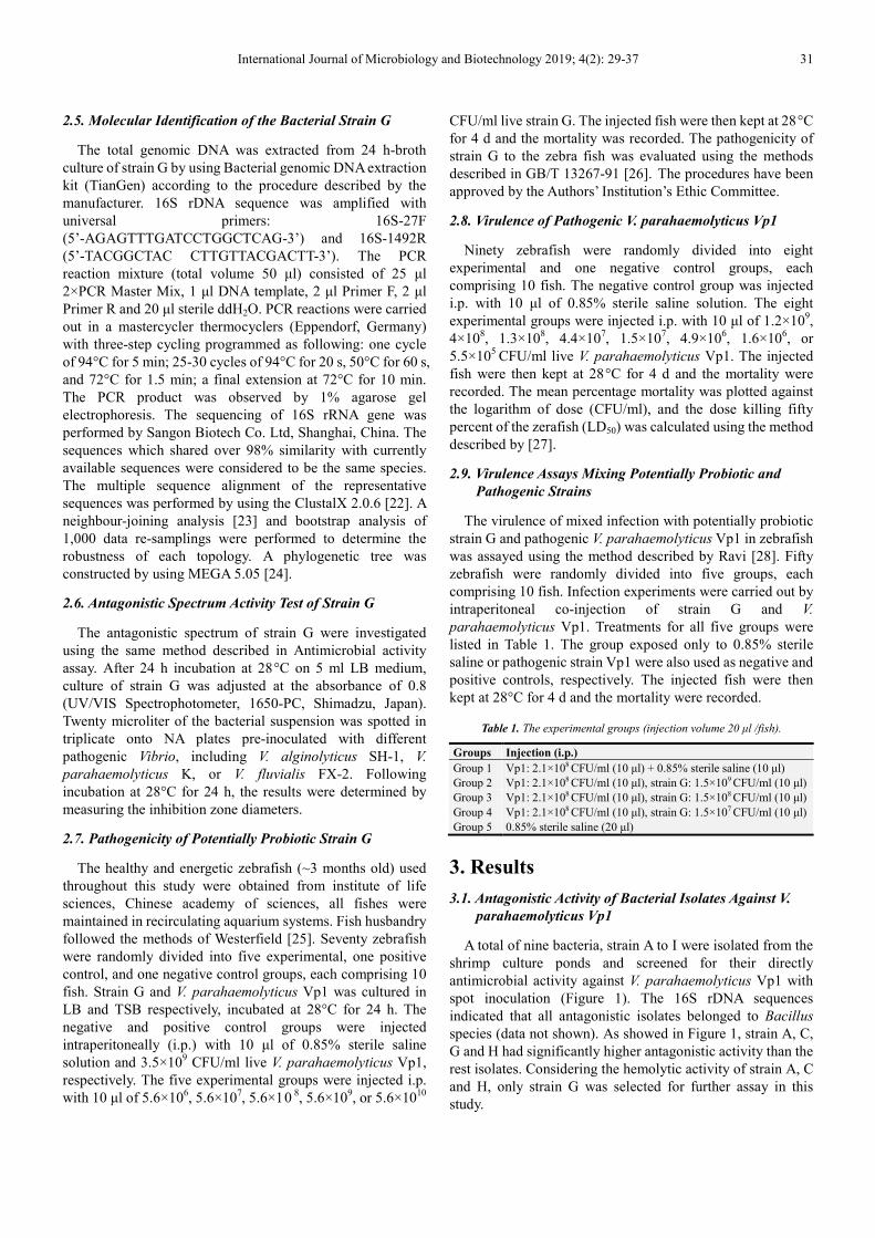
A total of nine bacteria, strain A to I were isolated from the
shrimp culture ponds and screened for their directly
antimicrobial activity against V. parahaemolyticus Vp1 with
spot inoculation (Figure 1). The 16S rDNA sequences
indicated that all antagonistic isolates belonged to Bacillus
species (data not shown). As showed in Figure 1, strain A, C,
G and H had significantly higher antagonistic activity than the
rest isolates. Considering the hemolytic activity of strain A, C
and H, only strain G was selected for further assay in this
study.
32 Mengfan Peng et al.: Isolation and Characterization of a Bacillus spp. Against Vibrio
Parahaemolyticus from Shrimp Culture Ponds
Figure 1. Antagonistic activity of potential strains against V. parahaemolyticus Vp1 in vitro.
3.2. Identification of Potential Probiotics Strain G
3.2.1. Preliminary Morphological and Physiological
Identifications
The colony morphology of strain G was (light yellow,
circular, opaque, flat, with a rough surface, and irregular edges
around the colony on Nutrient Agar). This potential probiotic
was Gram-positive, Bacillus spp. The spores of strains G were
(located in the middle of the cell, and underwent no swelling).
Strain G could not grow in the presence of sodium chloride at
concentrations above 8%. The temperature range for growth
was 20-40°C and the pH range for growth was 4.0–10.0. The
results of biochemical analyses for strain G were summarized
in Table 2. The morphological, cultural, and physiological
characteristics indicated that strains G was closely related to
Bacillus spp. which is well known for having the widely
antimicrobial activity [2, 29, 30, 31].
Table 2. Physiological characters of the isolated strain G.
Parameter Characters
Colony morphology
light yellow, circular, opaque, flat, with a rough
surface, and irregular edges around the colony
on nutrient agar
Gram strain Gram positive
Growth in temperature
10°C -
20°C +
30°C +
40°C +
50°C +
60°C -
Parameter Characters
Growth in pH
2 -
4 +
6 +
8 +
10 +
12 -
Growth in NaCl
2% +
4% +
6% +
8% +
10% -
Nitrate reduction +
Citrate utilization +
Nitrite reduction +
Amylohydrolysis +
Urea hydrolysis -
Casein hydrolysis +
Adonitol +
Arabinose +
Fructose +
Sorbitol -
Lactose -
Galactose +
Sorbitol -
V-P test +
Indole test +
Catalase test +
Gelatin test +
Lecithin test +
Methyl red test +
International Journal of Microbiology and Biotechnology 2019; 4(2): 29-37 33
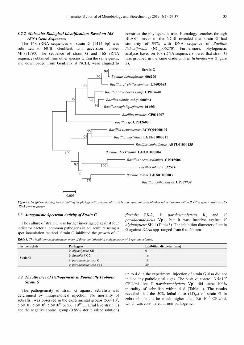
3.2.2. Molecular Biological Identifications Based on 16S
rRNA Gene Sequences
The 16S rRNA sequences of strain G (1414 bp) was
submitted to NCBI GenBank with accession number
MF871790. The sequence of strain G and 16S rRNA
sequences obtained from other species within the same genus,
and downloaded from GenBank at NCBI, were aligned to
construct the phylogenetic tree. Homology searches through
BLAST server of the NCBI revealed that strain G had
similarity of 99% with DNA sequence of Bacillus
licheniformis (NC_006270). Furthermore, phylogenetic
analysis based on 16S rDNA sequence showed that strain G
was grouped in the same clade with B. licheniformis (Figure
2).
Figure 2. Neighbour-joining tree exhibiting the phylogenetic position of strain G and representatives of other related strains within Bacillus genus based on 16S
rRNA gene sequence.
3.3. Antagonistic Spectrum Activity of Strain G
The culture of strain G was further investigated against four
indicator bacteria, common pathogens in aquaculture using a
spot inoculation method. Strain G inhibited the growth of V.
fluvialis FX-2, V. parahaemolyticus K, and V.
parahaemolyticus Vp1, but it was inactive against V.
alginolyticus SH-1 (Table 3). The inhibition diameter of strain
G against Vibrio spp. ranged from 0 to 20 mm.
Table 3. The inhibitory zone diameter (mm) of direct antimicrobial activity assay with spot inoculation.
Active isolate Pathogens Inhibition diameter (mm)
Strain G
V. alginolyticus SH-1 0
V. fluvialis FX-2 16
V. parahaemolyticus K 18
V. parahaemolyticus Vp1 20
3.4. The Absence of Pathogenicity in Potentially Probiotic
Strain G
The pathogenicity of strain G against zebrafish was
determined by intraperitoneal injection. No mortality of
zebrafish was observed in the experimental groups (5.6×10⁶,
5.6×10⁷, 5.6×10
8, 5.6×10
9, or 5.6×10
10 CFU/ml live strain G)
and the negative control group (0.85% sterile saline solution)
up to 4 d in the experiment. Injection of strain G also did not
induce any pathological signs. The positive control, 3.5×10⁹
CFU/ml live V. parahaemolyticus Vp1 did cause 100%
mortality of zebrafish within 4 d (Table 4). The results
revealed that the 50% lethal dose (LD50) of strain G in
zebrafish should be much higher than 5.6×10¹⁰
CFU/ml,
which was considered as non-pathogenic.
34 Mengfan Peng et al.: Isolation and Characterization of a Bacillus spp. Against Vibrio
Parahaemolyticus from Shrimp Culture Ponds
Table 4. Acute toxicity of the strain G on Zebra fish.
Group Dosage
mg/l
Concentration
CFU/ml
Death number Mortality
(%) 1h 2h 4h 6h 8h 24h 48h 72h 96h
Control
group
normal
saline 0.85%
0 0 0 0 0 0 0 0 0
0 0 0 0 0 0 0 0 0 0
0 0 0 0 0 0 0 0 0
1 0.233 5.6×10⁶
0 0 0 0 0 0 0 0 0
0 0 0 0 0 0 0 0 0 0
0 0 0 0 0 0 0 0 0
2 2.33 5.6×10⁷
0 0 0 0 0 0 0 0 0
0 0 0 0 0 0 0 0 0 0
0 0 0 0 0 0 0 0 0
3 23.3 5.6×10⁸
0 0 0 0 0 0 0 0 0
0 0 0 0 0 0 0 0 0 0
0 0 0 0 0 0 0 0 0
4 233 5.6×10⁹
0 0 0 0 0 0 0 0 0
0 0 0 0 0 0 0 0 0 0
0 0 0 0 0 0 0 0 0
5 2330 5.6×10¹⁰ 0 0 0 0 0 0 0 0 0 0
0 0 0 0 0 0 0 0 0
Vp1 23.3 3.5×10⁹
0 0 0 0 0 0 0 0 0
100% 0 0 0 0 0 3 6 8 9
0 0 0 0 0 4 5 7 10
0 0 0 1 3 4 7 8 10
* 10 zebra fish of each group
3.5. Virulence of Pathogenic V. parahaemolyticus Vp1
The virulence of pathogenic strain V. parahaemolyticus
Vp1 against zebrafish was determined on the basis of LD50.
According to the mortality caused by various concentrations
(5.5×10⁵-1.2×10
9 CFU/ml), Vp1 strain had an LD50 of 4.4×10
7
CFU/ml. The Vp1 strain did cause 100% mortality in the
groups challenged with 1.2×10⁹
or 4×10⁸
CFU/ml per fish
within 4 d. No mortality was observed in the negative control
group challenged with 0.85% sterile saline solution up to 4 d.
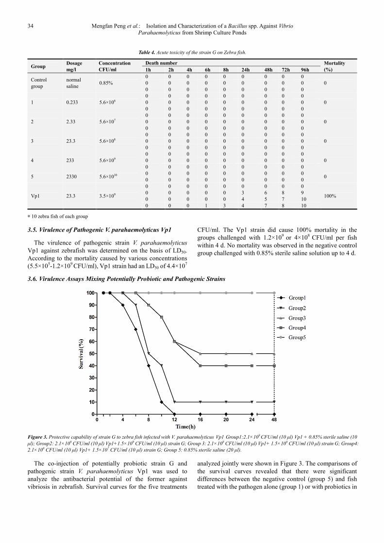
3.6. Virulence Assays Mixing Potentially Probiotic and Pathogenic Strains
Figure 3. Protective capability of strain G to zebra fish infected with V. parahaemolyticus Vp1 Group1:2.1×10⁸ CFU/ml (10 µl) Vp1 + 0.85% sterile saline (10
µl); Group2: 2.1×10⁸ CFU/ml (10 µl) Vp1+1.5×10⁹ CFU/ml (10 µl) strain G; Group 3: 2.1×10⁸ CFU/ml (10 µl) Vp1+ 1.5×10⁸ CFU/ml (10 µl) strain G; Group4:
2.1×10⁸ CFU/ml (10 µl) Vp1+ 1.5×10⁷ CFU/ml (10 µl) strain G; Group 5: 0.85% sterile saline (20 µl).
The co-injection of potentially probiotic strain G and
pathogenic strain V. parahaemolyticus Vp1 was used to
analyze the antibacterial potential of the former against
vibriosis in zebrafish. Survival curves for the five treatments
analyzed jointly were shown in Figure 3. The comparisons of
the survival curves revealed that there were significant
differences between the negative control (group 5) and fish
treated with the pathogen alone (group 1) or with probiotics in
International Journal of Microbiology and Biotechnology 2019; 4(2): 29-37 35
the mixed challenges (group 2-4). Negative control treated
with 0.85% sterile saline solution (group 5) showed survival
rate of 100% within 48 h after treatment. Fish infected with V.
parahaemolyticus Vp1 alone (group 1) showed survival rate
of 0 at 12 h postchallenge. The analyses also showed that the
survival of zebrafish was significantly increased by the
addition of potentially probiotic strain G. The final survival
rate in the presence of strain G was up to 50% (group 3) at 16 h
postchallenge. Significant differences between fish treated
with varied dose of probiotics in the mixed challenges were
found as well. The final survival rates in the presence of
1.5×10⁹
CFU/ml, 1.5×10⁸
CFU/ml, or 1.5×10⁷
CFU/ml strain
G were 10% (group 2), 50% (group 3), or 40% (group 4),
respectively. The results of virulence assays indicated that
strain G provided significant protection against the pathogenic
action of V. parahaemolyticus Vp1 under the given
experimental conditions.
4. Discussion
The concept of using probiotics as biological control
against pathogens has received widespread attention during
the last few decades [32]. In this study, we demonstrated that
the growth of fish pathogenic Vibrio was controlled by
non-pathogenic strain G isolated from the highly intensive
shrimp culture ponds, in vitro and in vivo conditions (Figure
1-3 & Table 1-3). Besides of the morphological and
physiological characters, the homology search based on 16s
rDNA sequence showed that strain G was the number of
Bacillus spp. and highly related to Bacillus licheniformis with
99% sequence similarity. In vivo examination carried out in
this study confirmed that the Bacillus isolate strain G was
non-hemolytic activity and nonpathogenic bacteria for
zebrafish, suggesting that strain G could be a good candidate
for probiotics in aquaculture. In addition, recent studies
indicated that Bacillus might contain toxin producing genes
[19], might conduct to the insecurity for food and environment
and limits it’s clinical application. Hence, the strain G might
have the potential possibility to be used as a safe probiotic in
aquaculture.
In the present study, zero survival was observed in zebrafish
treated with V. parahaemolyticus Vp1 alone, which confirmed
that the Vibrio strain Vp1 is highly pathogenic. When the fish
were exposed to the mixture of pathogenic strain Vp1 and
potential probiotic strain G, their survival rate was
significantly (P<0.001) increased (Figure 3). In vivo
antagonistic activity results were well correlated with our in
vitro observations (Table 2). This result finds support of other
workers. Kennedy [33] had recorded that the inoculation of a
probiotic B. subtilis isolate into the rearing water resulted in
the apparent elimination of Vibrio spp. from the snook larvae.
A similar effect has been observed with another probiotic B.
cereus isolate at 105 CFU/ml that protects shrimp larvae of P.
monodon against V. harveyi with 104 CFU/ml by increasing
the survival rate to 60% [28]. Co-injection experiments
showed that the final survival rate of Vibrio infected zebrafish
increased from 40% to 50% with increasing concentration of
strain G (antagonist) from 1.5×10⁷ to 1.5×10
8 CFU/ml. To act
as probiotics, strain G must be present at significantly high
levels and the degree of probiotic protect increased with the
level of strain G. However, the final survival rate dropped to
10% when the Vibrio infected zebrafish was treated with
higher concentration of strain G (1.5×10⁹ CFU/ml). This result
suggested that the optimal concentration for strain G to exhibit
the best probiotic effect against Vibrio infection in zebrafish
should be close to 1.5×10⁸ CFU/ml.
Several species are known to be antibiotic-producing
bacteria, such as Carnobacterium spp. [12], lactic acid
bacteria (LAB) [13], Pseudomonas sp. [14], and Roseobacter
[15], and Bacillus spp. [17, 35, 36]. Especially Pseudomonas
S2V2 inhibited V. alginolyticus, V. anguillarum, V. fluvialis, V.
harveyi, V. metschnikovii, V. splendidus, V. ordalii, V.
parahaemolyticus, and V. vulnificus but inactive against V.
campbellii [34], Susceptibility variations to strain S2V2 were
exhibited among species and among strains in the same
species of Vibrio sp. However, there is a lack of
broad-spectrum antibacterial activity against Vibrio in
Bacillus such as B. pumilus B16, Bacillus mojavensis J7 [17],
Bacillus licheniformis DAHB1 [35], Bacillus subtilisb [36],
but for strain G, it exhibited significant inhibitory activity of V.
fluvialis FX-2, V. parahaemolyticus K, and V.
parahaemolyticus Vp1 in vitro. The inhibition diameter of
strain G against Vibrio spp. ranged from 16 to 20 mm on
Nutrient Agar. The spectrum activity implies that the nature of
antibiotic produced by strain G is different to antibiotics
produced by other Baccillus species reported before.
The mechanism of antagonistic effects could be the growth
inhibition of bacterial pathogens due to the bioactive
compounds produced by probiotics. Previous studies also
showed that Bacillus species produce various bioactive
compounds such as subtilin [37], subtilomycin [38], cerecidin
[39], and haloduracin [40], which display inhibitory activity
against a broad spectrum of bacteria, including
Staphylococcus, Listeria, Aeromonas, Vibrio, Pseudomonas,
and Alteromonas species, etc [41]. In the present study, strain
G exhibited zone of clearance against pathogenic Vibrios on
Nutrient Agar plates (Figure 1 & Table 3). It could be
considered that the growth of pathogenic Vibrios is inhibited
by the diffusion of antibacterial compounds produced by
strain G. Thus, purification and characterization of the
antibacterial compounds produced by strain G may contribute
to a better understanding of the mechanism of the antagonistic
effect.
5. Conclusion
From this study, it can be concluded that the Bacillus isolate,
strain G is non-toxic to zebrafish and can effectively inhibit
the growth of pathogenic Vibrios on Nutrient Agar plates and
protect zebrafish against V. parahaemolyticus Vp1 by
significantly (P<0.001) increasing the survival rate in culture
systems. Therefore, the ability of strain G to suppress
pathogen growth in vitro and in vivo conditions suggests that it
is a promising probiotic candidate that may be a good
36 Mengfan Peng et al.: Isolation and Characterization of a Bacillus spp. Against Vibrio
Parahaemolyticus from Shrimp Culture Ponds
alternative to antibiotics in aquaculture. Further study is need
to provide valuable insight into the exact mode of action of
observed probiotic effects and the possibilities and limitations
of bacterial disease control in situations directly relevant to
aquaculture conditions.
Acknowledgements
This work was supported by a grant from Shanghai aquatic
collaborative innovation center of Breeding and Genetics
(ZF1206), Doctoral Fund from Shanghai Ocean University
(A2-0203-00-100319), and Excellent Young Teachers
Program of Shanghai Municipal Commission of education
(A1-2039-17-0006).
References
[1] Fernandez-Piquer J, Bowman JP, Ross T, Tamplin ML (2011) Predictive models for the effect of storage temperature on Vibrio parahaemolyticus viability and counts of total viable bacteria in Pacific oysters (Crassostreagigas). Appl Environ Microbiol. 77:8687–8695.
[2] Xu HM, Rong YJ, Zhao MX, Song B, Chi ZM (2014) Antibacterial activity of the lipopetides produced by Bacillus amyloliquefaciens M1 against multidrug-resistant Vibrio spp. isolated from diseased marine animals. Appl Microbiol Biotechnol. 98:127–136.
[3] Aranda CP, Valenzuela C, Barrientos J, Paredes J, Leal P, Maldonado M, Godoy FA, Osorio CG (2012) Bacteriostatic anti-Vibrio parahaemolyticus activity of Pseudoalteromonas sp. Strains DIT09, DIT44 and DIT46 isolated from Southern Chilean intertidal Perumytiluspurpuratus. World J Microbiol Biotechnol. 28:2365–2374.
[4] Touraki M, Karamanlidou G, Karavida P, Chrysi K (2012) Evaluation of the probiotics Bacillus subtilis and Lactobacillus plantarumbioencapsulated in Artemianauplii against vibriosis in European sea bass larvae (Dicentrarchuslabrax, L.). World J Microbiol Biotechnol. 28:2425–2433.
[5] Zarei M, Borujeni MP, Jamnejad A, Khezrzadeh M (2012) Seasonal prevalence of Vibrio species in retail shrimps with an emphasis on Vibrio parahaemolyticus. Food Control. 25:107–109.
[6] Yu WT, Jong KJ, Lin YR, Tsai SE, Tey YH, Wong HC (2013) Prevalence of Vibrio parahaemolyticusin oyster and clam culturing environments in Taiwan. Int J Food Microbiol. 160:185–192.
[7] Wu YN, Wen J, Ma Y, Ma XC, Chen Y (2014) Epidemiology of foodborne disease outbreaks caused by Vibrio parahaemolyticus, China, 2003–2008. Food Control. 46:197–202.
[8] Nithya C, Aravindraja C, Pandian SK (2010) Bacillus pumilus of Palk Bay origin inhibits quorum-sensing-mediated virulence factors in Gram-negative bacteria. Res Microbiol. 161:293–304.
[9] Sinhaseni P, Limpoka M, Samatiwat O (2000) Human health aspects of the use of chemicals in aquaculture, with special emphasis on food safety and regulations. In: Arthur JR, Lavilla-Pitogo CR, Subasinghe RP (eds) Use of chemicals in
aquaculture in Asia. SEAFDEC, Iloilo, pp 55–60.
[10] Cabello FC (2006) Heavy use of prophylactic antibiotics in aquaculture: a growing problem for human and animal health and for the environment. Environ Microbiol. 8:1137–1144.
[11] Esposito A, Fabrizi L, Lucchetti D, Marvasi L, Coni E, Guandalini E (2007) Orally administered erythromycin in rainbow trout (Oncorhynchus mykiss): residues in edible tissues and withdrawal time. Antimicrob Agents Chemother. 51:1043–1047.
[12] Bacon CW, Hinton DM, Mitchell TR, Snook ME, Olubajo B (2012) Characterization of endophytic strains of Bacillus mojavensis and their production of surfactin isomers. Biol Control. 62:1–9.
[13] Chahad OB, El Bour M, Calo-Mata P, Boudabous A, Barros Vela `zquez J (2012) Discovery of novel biopreservation agents with inhibitory effects on growth of food-borne pathogens and their application to seafood products. Res Microbiol. 163:44–54.
[14] Makino K, Oshima K, Kurokawa K (2003) Genome sequence of Vibrio parahaemolyticus: a pathogenic mechanism distinct from that of V cholera. The Lancet. 9359: 743-749.
[15] Planas M, Pe ´rez-Lorenzo M, Hjelm M, Gram L, Fiksdal IU, Bergh Ø, Pintado J. (2006) Probiotic effect in vivo of Roseobacter strain 27-4 against Vibrio (Listonella) anguillarum infections in turbot (Scophthalmusmaximus L.) larvae. Aquaculture. 255:323–333.
[16] Zokaeifar H, Balca ´zar JL, Saad CR, Kamarudin MS, Sijam K, Arshad A, Nejat N (2012) Effects of Bacillus subtilis on the growth performance, digestive enzymes, immune gene expression and disease resistance of white shrimp, Litopenaeusvannamei. Fish Shellfish Immunol. 33:683–689.
[17] Robertson PAW, Dowd CO, Burrells C, Williams P, Austin B (2000) Use of Carnobacterium sp. as a probiotic for Atlantic salmon_Salmosalar L. and rainbow trout, Oncorhynchusmykiss, Walbaum. Aquaculture. 185:235–243.
[18] Villamil L, Figueras A, Planas M, Novoa B (2003) Control of Vibrio alginolyticus in Artemia culture by treatment with bacterial robiotics. Aquaculture. 219:43–56.
[19] Duc, H. L., Hong, H. A., Barbosa, T. M., Henriques, A. O., & Cutting, S. M. (2004). Characterization of Bacillus probiotics available for human use. Appl Environ Microbiol, 70(4), 2161-2171.
[20] Liu, X. F, Li, Y, Li, J. R, Cai, L. Y, Li, X. X, & Chen, J. R. (2015). Isolation and characterisation of Bacillus spp. antagonistic to Vibrio parahaemolyticus for use as probiotics in aquaculture. World J Microb Biot, 31(5), 795-803.
[21] Kamiso HN, Isnansetyo A, Triyanto, Istiqomah I, Murdjani M (2005) Isolation, identification and characterization of pathogenic Vibrio spp. causative agents of vibriosis in grouper at brackishwater Aquaculture Development Centre, Situbondo. J Fish Sci VII: 80–94.
[22] Thompson JD, Gibson TJ, Plewniak F, Jeanmougin F, Higgins DG (1997) The ClustalX windows interface: flexible strategies for multiple sequence alignment aided by quality analysis tools. Nucleic Acids Res. 25:4876–4882.
[23] Saitou N, Nei M (1987) The neighbour-joining method: a new method for reconstructing phylogenic trees. Mol Biol Evol. 4:406–425.
International Journal of Microbiology and Biotechnology 2019; 4(2): 29-37 37
[24] Tamura K, Peterson D, Peterson N, Stecher G, Nei M, Kumar S (2011) MEGA5: molecular evolutionary genetics analysis using maximum likelihood, evolutionary distance, and maximum parsimony methods. Mol Biol Evol. 10:2731–2739.
[25] M. Westerfield (2000) The Zebrafish Book: a Guide for the Laboratory Use of Zebrafish (Danio rerio), fourth ed., Oregon, OR.
[26] Xie FJ, Chen HZ, Zhao YY (1992) Water quality-Determination of the acute toxiclty of substance to freshwater fish (Brachydanio rerio Hamilton-Buchanan). National Standard of the People’s Republic of China, GB/T13267-91.
[27] Reed LJ and Munench H (1938) A simple method of estimating fifty percent endpoints. Am J Hyg. 27: 493–497.
[28] Ravi A. V, Musthafa K. S, Jegathammbal G, Kathiresan K, Pandian S. K (2007) Screening and evaluation of probiotics as a biocontrol agent against pathogenic Vibrios in marine aquaculture. Lett Appl Microbiol. 45:219–223.
[29] Aunpad R, Na-Bangchang K (2007) Pumilicin 4, a novel bacteriocin with anti-MRSA and anti-VRE activity produced by newly isolated bacteria Bacillus pumilus strain WAPB4. Curr Micobiol. 55:308–313.
[30] Aunpad R, Panbangred W (2012) Evidence for two putative holin-like peptides encoding genes of Bacillus pumilus strain WAPB4. Curr Microbiol. 64:343–348.
[31] Li GG, Liu BS, Shang YJ, Yu ZQ, Zhang RJ (2012) Novel activity evaluation and subsequent partial purification of antimicrobial peptides produced by Bacillus subtilis LFB112. Ann Microbiol. 62:667–674.
[32] Sugita H, Ohta K, Kuruma A, Sagesaka T (2008) An antibacterial effect of Lactococcus lactis isolated from the intestinal tract of the Amur catfish, Silurus asotus Linnaeus. Aquac Res. 38:1002–1004.
[33] Kennedy, S. B, Tucker, J. W, Neidig, C. L, Vermeer, G. K, Cooper, V. R, Jarrell, J. L. and Sennett, D. G. (1998) Bacterial management strategies for stock enhancement of warm water marine fish: a case study with common snook (Centropomus undecimalis). Bulletin of Marine Sciences. 62, 573–588.
[34] Isnansetyo, A, Istiqomah, I, Muhtadi, Sinansari, S, Hernawan, R. K, & Triyanto. (2009). A potential bacterial biocontrol agent, strain S2V2 against pathogenic marinevibrioin aquaculture. World J Microb Biot, 25(6), 1103-1113.
[35] Vinoj, G, Vaseeharan, B, Thomas, S, Spiers, A. J, & Shanthi, S. (2014). Quorum-quenching activity of the AHL-lactonase from Bacillus licheniformis DAHB1 inhibits Vibrio biofilm formation in vitro and reduces shrimp intestinal colonisation and mortality. Mar Biotechnology, 16(6), 707.
[36] Xu D, Wang Y, Sun L. Inhibitory activity of a novel antibacterial peptide AMPNT-6 from Bacillus subtilis against Vibrio parahaemolyticus in shrimp. Food Control, 2013, 30(1):58-61.
[37] Chan, W. C, Bycroft, B. W, Leyland, M. L, Lian, L.-Y, and Roberts, G. C. K (1993) A novel post-translational modification of the peptide antibiotic subtilin: isolation and characterization of a natural variant from Bacillus subtilis A. T. C. C. 6633. Biochem. J. 291:23−27.
[38] Phelan, R. W, Barret, M, Cotter, P. D, O’Connor, P. M, Chen, R., Morrissey, J. P, Dobson, A. D. W, O’Gara, F, and Barbosa, T. M. (2013) Subtilomycin: a new lantibiotic from Bacillus subtilis strain MMA7 isolated from the marine sponge Haliclona simulans. Mar. Drugs. 11:1878−1898.
[39] Wang, J, Zhang, L, Teng, K, Sun, S, Sun, Z, and Zhong, J (2014) Cerecidins, novel lantibiotics from Bacillus cereus with potent antimicrobial activity. Appl. Environ. Microbiol. 80: 2633-2643.
[40] Lawton, E. M, Cotter, P. D, Hill, C, and Ross, R. P (2007) Identification of a novel two-peptide lantibiotic, haloduracin, produced by the alkaliphile Bacillus halodurans C-125. FEMS Microbiol. Lett. 267: 64−71.
[41] Barbosa, J, Caetano, T, and Mendo, S. J (2015) Class I and Class II Lanthipeptides Produced by Bacillus spp. Nat. Prod. 78: 2850–2866.


![Enhanced FAD Production in Eremothecium ashbyi with ...article.ijmicrobio.org/pdf/10.11648.j.ijmb.20200501.12.pdf · Amongst the different types of stress tested [10], oxidative stress](https://static.fdocuments.net/doc/165x107/5f3a456edbfa997a22309c52/enhanced-fad-production-in-eremothecium-ashbyi-with-amongst-the-different-types.jpg)