Isolated Pituitary Neurosarcoidosis: A Case Report and ...
4
196 Isolated Pituitary Neurosarcoidosis: A Case Report and Review of the Literature İzole Pituiter Nörosarkoidoz: Olgu Sunumu ve Literatürün Gözden Geçirilmesi Özet Sarkoidoz genellikle genç erişkinlerde görülen ve sıklıkla bilateral hilar len- fadenopati ve pulmoner infiltrasyonla presente olan, birçok sistemi tutan granülamatoz bir hastalıktır. Santral sinir sistemi (SSS) tutulumu oldukça nadirdir. Sarkoidozun nedeni bilinmemektedir. Sarkoidoz tanısı etkilenen organlarda histopatolojik incelemede gözlenen kazeifiye olmayan granu- lomların klinik ve radyolojik bulgularla uyumlu olması ile konur. Burada, kli- niğimize amenore ve poliüri ile başvuran 30 yaşında bir kadın hastayı sun- maktayız. Pituiter stalkı tutan lezyonun radyolojik görünümü sarkoidoz, sifi- lis, tüberküloz ve yabancı cisim granülamatozu için patognomik olan infla- matuar infiltrasyon görüntüsündeydi. Laboratuar testleri ile sifilis ve tüber- küloz tanıları ekarte edildi. Olası tanı sarkoidozdu. Diğer sistemleri araştır- dığımızda akciğerler, lenf nodları, deri ve gözlerde sarkoidoz tutulumu göz- lenmedi. Transkranyal insizyonel biyopsi histopatolojik incelemesi makro- faj, epiteloid hücreler, sitokin salgılayan multinükleer dev hücrelerden olu- şan kazeifiye olmayan granülom göstermekteydi. Santral korda CD4 ve CD8 lenfositler, B lenfositler, plazma hücreleri ve fibroblastlar gözlendi. Tanı nörosarkoidozdu. Bu olguyu pituiter lezyonların ayırıcı tanısında izole nöro- sarkoidozun da yeri olduğunu hatırlatmak ve bu durumun tanı ve tedavi- sinde son bilgileri gözden geçirmek için sunmaktayız. (Marmara Üniversi- tesi Tıp Fakültesi Dergisi 2011;24:196-9) Anahtar Kelimeler: Pituiter lezyonlar, Sarkoidoz, Nörosarkoidoz, Kazeifiye olmayan granülomlar Abstract Sarcoidosis is a multisystem granulomatous disorder, commonly affecting young adults and usually presenting with bilateral hilar lymphadenopathy and pulmonary infiltration. Central nervous system (CNS) involvement is extremely rare. The cause of sarcoidosis is unknown. The diagnosis of sarcoidosis is firmly established when histopathological evidence of non-caseating granulomas in affected organs supports compatible clinicoradiographic findings. Here, we present a case of a 30-year-old woman referred to our clinic with amenorrhea and polyuria. The radiological appearance of a lesion involving the pituitary stalk was an image of inflammatory infiltration, which is pathognomic for sarcoidosis, syphilis, tuberculosis and foreign body granulomatosis. Laboratory tests were done to rule out syphilis and tuberculosis. A possible diagnosis was sarcoidosis. When we searched other systems for the involvement of sarcoidosis, lungs, lymph nodes, skin and eyes were not involved by the disease. Histopathological examination of a transcranial incisional biopsy revealed a non-caseating granuloma, consisting of macrophages, epithelioid cells, and multinucleated giant cells that secrete cytokines. Around this central core, CD4 and CD8 lymphocytes, B lymphocytes, plasma cells, and fibroblasts were detected. The diagnosis was neurosarcoidosis. We present this case to draw attention to the possibility of isolated neurosarcoidosis as the differential diagnosis of pituitary lesions and review recent advances in the investigation, diagnosis and treatment of this condition.. (Marmara Medical Journal 2011;24:196-9) Key Words: Pituitary lesions, Sarcoidosis, Neurosarcoidosis, Non-caseating granulomas. Case Report / Olgu Sunumu Akin AKAKİN 1 , Deniz KONYA 1 , Dilek AKAKİN 2 , Turker KİLIÇ 1 1 Marmara University, School of Medicine, Department of Neurosurgery, Istanbul, Turkey 2 Marmara University, School of Medicine, Department of Histology and Embryology, Istanbul, Turkey Correspondence to/İletişim: Akın Akakın M.D., Marmara University, School of Medicine, Department of Neurosurgery, İstanbul, Turkey. E-mail: [email protected] Submitted/Başvuru Tarihi: 31.07.2011 Accepted/Kabul Tarihi: 16.09.2011 © Marmara Medical Journal, Published by Galenos Publishing. / © Marmara Üniversitesi Tıp Fakültesi Dergisi, Galenos Yayınevi tarafından basılmıştır. DOI: 10.5472/MMJ.2011.02028.1 Introduction Sarcoidosis is a multisystem granulomatous disorder, with an unknown etiology, commonly affecting young adults and usually presenting with bilateral hilar lymphadenopathy, pulmonary infiltration and skin or eye lesions 1 . Although involvement of the central nervous system (CNS) is rare (5%), the disease can lead to severe neurological problems 2 . Patients with sarcoidosis rarely present with sellar mass 3 .
Transcript of Isolated Pituitary Neurosarcoidosis: A Case Report and ...

196
Isolated Pituitary Neurosarcoidosis: A Case Report andReview of the Literatureİzole Pituiter Nörosarkoidoz: Olgu Sunumu ve Literatürün Gözden Geçirilmesi
Özet
Sarkoidoz genellikle genç erişkinlerde görülen ve sıklıkla bilateral hilar len-fadenopati ve pulmoner infiltrasyonla presente olan, birçok sistemi tutangranülamatoz bir hastalıktır. Santral sinir sistemi (SSS) tutulumu oldukçanadirdir. Sarkoidozun nedeni bilinmemektedir. Sarkoidoz tanısı etkilenenorganlarda histopatolojik incelemede gözlenen kazeifiye olmayan granu-lomların klinik ve radyolojik bulgularla uyumlu olması ile konur. Burada, kli-niğimize amenore ve poliüri ile başvuran 30 yaşında bir kadın hastayı sun-maktayız. Pituiter stalkı tutan lezyonun radyolojik görünümü sarkoidoz, sifi-lis, tüberküloz ve yabancı cisim granülamatozu için patognomik olan infla-matuar infiltrasyon görüntüsündeydi. Laboratuar testleri ile sifilis ve tüber-küloz tanıları ekarte edildi. Olası tanı sarkoidozdu. Diğer sistemleri araştır-dığımızda akciğerler, lenf nodları, deri ve gözlerde sarkoidoz tutulumu göz-lenmedi. Transkranyal insizyonel biyopsi histopatolojik incelemesi makro-faj, epiteloid hücreler, sitokin salgılayan multinükleer dev hücrelerden olu-şan kazeifiye olmayan granülom göstermekteydi. Santral korda CD4 veCD8 lenfositler, B lenfositler, plazma hücreleri ve fibroblastlar gözlendi. Tanınörosarkoidozdu. Bu olguyu pituiter lezyonların ayırıcı tanısında izole nöro-sarkoidozun da yeri olduğunu hatırlatmak ve bu durumun tanı ve tedavi-sinde son bilgileri gözden geçirmek için sunmaktayız. (Marmara Üniversi-tesi Tıp Fakültesi Dergisi 2011;24:196-9)Anah tar Ke li me ler: Pituiter lezyonlar, Sarkoidoz, Nörosarkoidoz, Kazeifiyeolmayan granülomlar
Abstract
Sarcoidosis is a multisystem granulomatous disorder, commonly affecting youngadults and usually presenting with bilateral hilar lymphadenopathy and pulmonary infiltration. Central nervous system (CNS) involvement is extremelyrare. The cause of sarcoidosis is unknown. The diagnosis of sarcoidosis is firmlyestablished when histopathological evidence of non-caseating granulomas inaffected organs supports compatible clinicoradiographic findings. Here, we present a case of a 30-year-old woman referred to our clinic with amenorrheaand polyuria. The radiological appearance of a lesion involving the pituitary stalkwas an image of inflammatory infiltration, which is pathognomic for sarcoidosis,syphilis, tuberculosis and foreign body granulomatosis. Laboratory tests weredone to rule out syphilis and tuberculosis. A possible diagnosis was sarcoidosis.When we searched other systems for the involvement of sarcoidosis, lungs,lymph nodes, skin and eyes were not involved by the disease. Histopathologicalexamination of a transcranial incisional biopsy revealed a non-caseating granuloma, consisting of macrophages, epithelioid cells, and multinucleatedgiant cells that secrete cytokines. Around this central core, CD4 and CD8 lymphocytes, B lymphocytes, plasma cells, and fibroblasts were detected. Thediagnosis was neurosarcoidosis. We present this case to draw attention to thepossibility of isolated neurosarcoidosis as the differential diagnosis of pituitarylesions and review recent advances in the investigation, diagnosis and treatmentof this condition.. (Marmara Medical Journal 2011;24:196-9)Key Words: Pituitary lesions, Sarcoidosis, Neurosarcoidosis, Non-caseatinggranulomas.
Case Report / Olgu Sunumu
Akin AKAKİN1, Deniz KONYA1, Dilek AKAKİN2, Turker KİLIÇ1
1Marmara University, School of Medicine, Department of Neurosurgery, Istanbul, Turkey2Marmara University, School of Medicine, Department of Histology and Embryology, Istanbul, Turkey
Correspondence to/İletişim: Akın Akakın M.D., Marmara University, School of Medicine, Department of Neurosurgery, İstanbul, Turkey.E-mail: [email protected]
Submitted/Başvuru Tarihi: 31.07.2011 Ac cep ted/Ka bul Ta ri hi: 16.09.2011© Marmara Medical Journal, Pub lis hed by Ga le nos Pub lis hing. / © Marmara Üniversitesi Tıp Fakültesi Der gi si, Ga le nos Ya yı ne vi ta ra fın dan ba sıl mış tır.
DO I: 10.5472/MMJ.2011.02028.1
Introduction
Sarcoidosis is a multisystem granulomatous disorder, with an unknown etiology, commonly affecting young adults and usually
presenting with bilateral hilar lymphadenopathy, pulmonary infiltrationand skin or eye lesions1. Although involvement of the central nervoussystem (CNS) is rare (5%), the disease can lead to severe neurologicalproblems2. Patients with sarcoidosis rarely present with sellar mass3.
Intracranial masses as a manifestation of neurosarcoidosis are occasionally seen. In this article, we present a case of neurosarcoidosis,pituitary sarcaidosis and discuss the relevant literature.
Case Report
A 30-year-old woman was referred to our clinic with an intracranial lesion, that was presented with amenorrhea and polyuria. Laboratory blood analysis for complete blood count, urea,serum electrolytes, liver function, thyroid function, immunoglobulinelectrophoresis, serum angiotensin converting enzyme (ACE), prothrombin time, partial thromboplastin time, autoimmune profi-le including anti-neutrophil cytoplasmic antibodies, cardiolipin andphospholipid antibodies were within the normal range. The onlyabnormality was hyponatremia (116mEq/L –normal range 137-143 mEq/L-) due to diabetes insipidus. A cerebrospinal fluid (CSF)cell count was normal, but the protein level was elevated to 0.89g/dl, and there were identical oligoclonal bands in the CSF and inthe blood pointing to a systemic disorder with CNS involvementrather than a pure CNS disorder. The chest x-ray, lung functiontests and neurological examination were within normal ranges.
Magnetic resonance imaging (MRI) showed a heterogeneouslyenhanced intrasellar and suprasellar dumbbell shaped mass andthickening of the pituitary stalk. After injection of a contrast medium, we detected dynamic images at the coronal plane atspin echo T1 (Figure 1). In both T1 and T2 sequences gray matter signal voiding was equal to hypophysis. A sagittal T1-weighted image of the pituitary shows a large low intensity massin the sellar and suprasellar area with irregular thickening of thewall and extension into the infundibulum (Figure 2). The
diaphragma sellae was more convex than normal. A pituitaryenlargement with thickening of the pituitary stalk was detected byMRI with gadolinium enhancement and attenuation in the intensity of the pituitary. The lesion involving the pituitary stalkshowed inflammatory infiltration that is pathognomic for sarcoidosis, syphilis, tuberculosis and foreign body granulomatosis.In order to rule out syphilis and tuberculosis, venereal disease research laboratory studies, fluorescent treponemal antibody-absorption and tuberculin tests were done, and all were negative.The case was evaluated as "probable" neurosarcoidosis dependingon the clinical picture, laboratory investigations such as cerebrospinal fluid lymphocyte sub-populations and MRI findings.
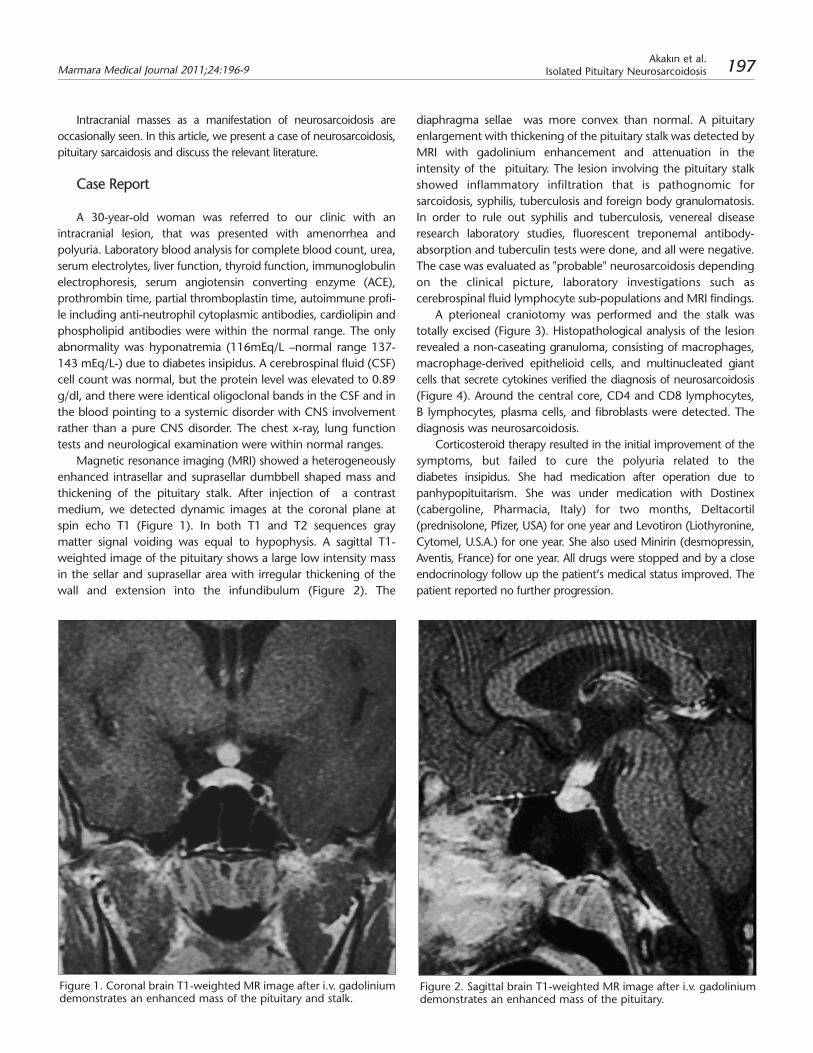
A pterioneal craniotomy was performed and the stalk wastotally excised (Figure 3). Histopathological analysis of the lesionrevealed a non-caseating granuloma, consisting of macrophages,macrophage-derived epithelioid cells, and multinucleated giantcells that secrete cytokines verified the diagnosis of neurosarcoidosis(Figure 4). Around the central core, CD4 and CD8 lymphocytes,B lymphocytes, plasma cells, and fibroblasts were detected. Thediagnosis was neurosarcoidosis.
Corticosteroid therapy resulted in the initial improvement of thesymptoms, but failed to cure the polyuria related to the diabetes insipidus. She had medication after operation due to panhypopituitarism. She was under medication with Dostinex(cabergoline, Pharmacia, Italy) for two months, Deltacortil (prednisolone, Pfizer, USA) for one year and Levotiron (Liothyronine,Cytomel, U.S.A.) for one year. She also used Minirin (desmopressin,Aventis, France) for one year. All drugs were stopped and by a closeendocrinology follow up the patient’s medical status improved. Thepatient reported no further progression.
Figure 1. Coronal brain T1-weighted MR image after i.v. gadoliniumdemonstrates an enhanced mass of the pituitary and stalk.
Figure 2. Sagittal brain T1-weighted MR image after i.v. gadoliniumdemonstrates an enhanced mass of the pituitary.
Marmara Medical Journal 2011;24:196-9Akakın et al.
Isolated Pituitary Neurosarcoidosis 197

Discussion
The incidence of sarcoidosis varies from 1 to 40 cases per100,000 population with a peak in the 3rd and 4th decades of life2,4.Although the cause is unknown, a defective immune system, environmental factors such as heavy metals, organic/inorganicdusts, inherited/genetic factors and infections caused by variousmicroorganisms are thought to be the possible causative agents4. Insymptomatic patients, sarcoidosis can involve one or more organsystems and present with a wide variety of signs and symptomswhich can be constitutional: fatigue, weight loss, fever or malaise;generalized, or focused on a single organ. The onset of the diseaseis usually insidious, but can be acute also. Respiratory symptoms aremost common and include cough and chest discomfort, anddyspnea. Following the lung, lymph nodes, skin and the eyes aremost often the organ involved. Despite the fact that there are granulomas on histological examination of many organs in themajority of patients, these much less often produce signs andsymptoms. Primary CNS involvement alone is a rare condition. Inmost of the cases with primary CNS involvement the disease occursin the hypothalamus alone or in both the hypothalamus and pituitary, but rarely in the pituitary alone1. This case is a good example of extreme neurosarcoidosis presentation.
The clinical presentation of neurosarcoidosis is widely variable, itcan be manifested in a maltitude of ways including cranial neuropathy, aseptic meningitis, encephalopathy, vasculopathy, seizures, psychiatric manifestations, hydrocephalus, hypothalamicpituitary disorders, myelopathy and peripheral neuropathy1,4-7. The disease has a predilection to spread from the leptomeninges to the Virchow-Robin spaces leading to invasionand thrombosis of associated blood vessels resulting in granulomatous angitis. In our case, the patient presented with achief complaint of dysmenorrhea.
It must be kept in mind that the Kveim test has a low sensitivity in neurosarcoidosis and thus is of limited use. Galliumuptake may demonstrate an extracranial granuloma available forbiopsy8. A whole-body gallium scan shows increased uptake rela-ted to CNS disease in less than 5% of patients with this condition but may give evidence of the presence of systemicdisease in 45% of patients with CNS involvement9. However, inselected cases of isolated CNS disorders, a meningeal or cerebralbiopsy may be required if standard investigations are not conclu-sive in order to exclude other causes such as tumor metastasis,lymphoma, vasculitis and remaining granulomatous disorders.ACE levels in the serum and cerebrospinal fluid may be increased,decreased or normal2. Serum or CSF ACE levels are found to beelevated in approximately 70-80% of patients with sarcoidosis,hypercalcemia may be found in 2-15% of the patients due toenhanced sensitivity to vitamin D, however the diagnosis of sarcoidosis is confirmed by histopathological examination5,10,11. In ourcase the CSF cell count was normal but the protein was slightly ele-vated to 0.89 gm/dL and there were identical oligoclonal bandsin CSF and in blood pointing to a systemic disorder with CNSinvolvement rather than a pure CNS disorder.
Figure 3. Coronal section T1-weighted MR postoperative image after i.vgadolinium demonstrates no enhancing mass or pituitary stalk.
Figure 4. Granulomas in typical position within central collections ofepithelioid cells and the encircling rim of lymphocytes.
Marmara Medical Journal 2011;24:196-9Akakın et al.Isolated Pituitary Neurosarcoidosis198

Both computed tomography and MRI scans are helpful in disease evaluation; however MRI scan is the modality of choice5,12.MRIs show a wide range of CNS abnormalities including hypothalamic-pituitary infiltrating lesions, cerebral parenchyme masses, leptomeningeal lesions, and focal white-matter lesions 5,13.The use of gadolinium improves the sensitivity of detecting leptome-ningeal lesions. Additional MRI findings include white matter andperiventricular hyperintensity mimicking multiple sclerosis, hydro-cephalus, atrophy, periventricular enhancement, chiasmal edema,extra-axial masses, and parenchymal or spinal cord masses7.Neurosarcoidosis is usually a diagnosis of exclusion. However, theradiographic features are suggestive. There are classically two radiographic patterns described for neurosarcoidosis: 1) Chronic basilar leptomeningitis with involvement of the hypothalamus, pituitary stalk, optic nerve, and chiasm; 2) Parenchymal sarcoidnodules, which occasionally calcify10,11. Differential diagnosis ofcentraldiabetes insipidus should be considered by an endocrinologist. Thedisease is associated with intrathoracic lesions in about 70% of cases;therefore, an intensive search for enlarged pulmonary lymph nodesshould be performed14. MRI is the modality of choice in neurosarcoidosis evaluation5,12 and although any technique can beused to diagnose suprasellar lesions, tissue biopsy taken from the lesi-on is required for definitive diagnosis and to exclude other cerebral pathologies.
Corticosteroids are the mainstay of neurosarcoidosis treatment, alleviating symptoms and potentially slowing diseaseprogression; however there is no known cure. Aggressive diseaseor frequent recurrence may require other immunosuppressivedrugs such as methotrexate or cyclophosphamide. Approximatelytwo thirds of patients with neurosarcoidosis have a self-limited illness, while the remainder have a chronic remitting and relapsingcourse15. Neurological deficits have been reported to respond tocorticosteroids in contrast to hormonal abnormalities that gene-rally persist despite therapy16. However, in our case the patient was totally cured in her close endocrinology follow-upeven after the medication was stopped one year later. The prognosis of chronic neurosarcoidosis is poor. The mortality rateof sarcoidosis is 1-6%15. Severe involvement of lung parenchymeleading to pulmonary fibrosis and respiratory failure and myocardial involvement leading to arhythmias and cardiac failureare the most common causes of death in sarcoidosis15.
In conclusion, sarcoidosis is associated with diverse neurologicalmanifestations and neuroimaging findings. The diagnosis of neurosarcoidosis can reasonably be supported in many patients byMRI findings although the definite diagnosis of isolated CNS sarcoidosis requires a biopsy to exclude neoplasms and other granulomatous diseases. The optimum management of patients withneurosarcoidosis relies on the ability of clinicians to recognize thebroad spectrum of clinical and neuroimaging manifestations of thedisorder and the final neuropathological confirmation. This diseaseneeds multidiciplinary treatment due to systemic involvement.
References
1. Stern BJ, Krumholz A, Johns C, Scott P. Nissim J. Sarcoidosis and its neurological manifestations. Arch Neurol 1985;42:909-17.
2. Sharma OP. Neurosarcoidosis: a personal perspective based on the studyof 37 patients. Chest 1997;112:220-8. doi: 10.1378/chest.112.1.220
3. Sato N, Sze G, Kim JH. Cystic pituitary mass in neurosarcoidosis. AJNR1997;18:1182-5.
4. Pentland B, Mitchell JD, Cull RE, Ford MJ. Central nervous system sarcoidosis. Q J Med 1985;56:457-65.
5. Fels C, Riegel A, Javaheripour-Otto K, Obenauer S. Neurosarcoidosis findings in MRI. Clin Imaging 2004;28:166-9. doi: 10.1007/s11060-008-9687-1
6. Wiederholt WC, Siekert RG. Neurological manifestations of sarcoidosis.Neurology 1965;15:1147-54.
7. Younger DS, Hayo AP, Brust JC, Rawland LP. Granulomatous angiitis of thebrain. An inflammatory reaction of diverse etiology. Arch Neurol1988;45:514-8.
8. Pickuth D, Heywang-Kobrunner SH. Neurosarcoidosis: evaluation withMRI. J Neuroradiol 2000;27:185-8.
9. Zajicek JP, Scolding NJ, Foster O, et al. Central nervous system sarcoidosis:diagnosis and management. Q J Med 1999;92:103-17.
10. Christoforidis GA, Spickler EM, Recio MV, Mehta BM. MR of CNS sarcoidosis: correlation of imaging features to clinical symptoms and response to treatment. AJNR Am J Neuroradiol 1999;20:655-69.
11. Osborn AG, Blaser SI, Salzman KL, et al. Diagnostic Imaging: Brain. 1stedition. Altona:Amirsys Inc., 2004: II 4:52-5.
12. Smith JK, Matheus MG, Castillo M. Imaging manifestations of neurosarcoidosis. AJR Am J Roentgenol 2004;182:289-95.
13. Wolfsberger S, Ba-Ssalamah A, Pinker K, et al. Application of three-teslamagnetic resonance imaging for diagnosis and surgery of sellar lesions. JNeurosurg 2004;100:278-86. doi: 10.3171/jns.200.100.2.0278
14. Chapelon C, Ziza JM, Piette JC, et al. Neurosarcoidosis: signs, course andtreatment in 35 confirmed cases. Medicine 1990;69:261-76.
15. Luke RA, Stern BJ, Krumholz A, Johns CJ. Neurosarcoidosis: The long termclinical course. Neurology 1987;37:461-3.
16. Freda PU, Silverberg SJ, Post KD, Wardlaw SL. Hypothalamic-pituitary sarcoidosis. Trends Endocrinol Metab 1992;3:321-5. doi:10.1016/1043-2760(92)90110-M
Marmara Medical Journal 2011;24:196-9Akakın et al.
Isolated Pituitary Neurosarcoidosis 199