Isolated liver mass for web - BC · PDF file• Radiologic interventions: RFA, TACE, SIR ....
24
Hypervascular liver metastasis from pancreatic neuroendocrine tumor. arterial PV arterial
Transcript of Isolated liver mass for web - BC · PDF file• Radiologic interventions: RFA, TACE, SIR ....

Hypervascular liver metastasis from pancreatic
neuroendocrine tumor.
arterial PV
arterial

Hypervascular hepatic metastases from
neuroendocrine pancreatic primary
T2
arterial PV arterial
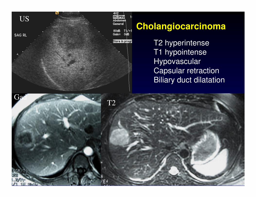
US
GadT2
Cholangiocarcinoma
T2 hyperintenseT1 hypointense
Hypovascular
Capsular retractionBiliary duct dilatation

CholangiocarcinomaCT
Gad T2
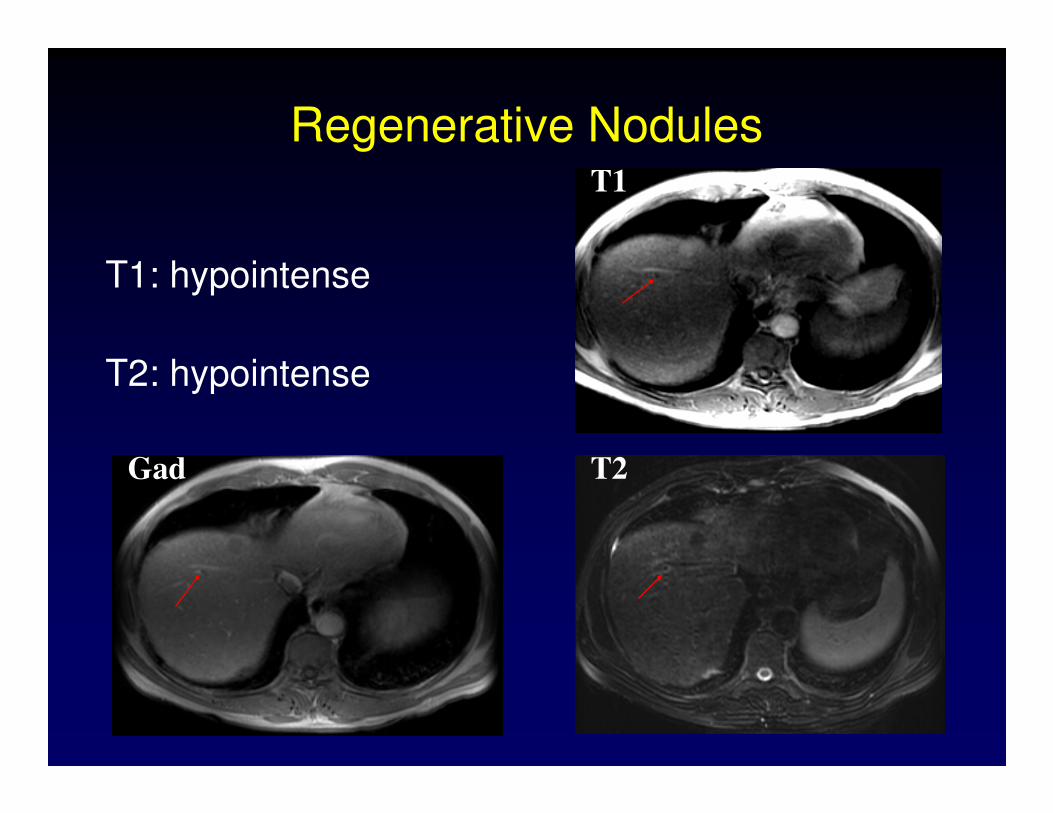
Regenerative Nodules
T1: hypointense
T2: hypointense
T1
T2Gad

Dysplastic Nodules
T1
T2 Gad
•Pathologically show abnormal tissue development but lack
definite histopathologic findings of
malignancy•Classified as low grade or high
grade•T1: hyperintense
•T2: hypointense•Post gad: hypovascular

T2
T1 arterial PV
HCC•T2 hyperintense•T1 hypointense
•Hypervascular with washout

HCC with microscopic fat(signal drop out on out of
phase)
T2
In phase Out of phase

HCC – ablated & new lesion
T2 LAVA pre-gad Arterial phase
PV phase substraction Rt PV thrombosis

Diffusion Weighted Imaging• Diffusion Weighted Imaging (DWI):
Random motion of water molecules (Brownian motion) within extracellular, within extracellular,
intracellular and intravascular spaces.intracellular and intravascular spaces.

DWI• Restricted diffusion:
• Malignancy (increased number of cells )
• Ischemia (cytotoxic edema)
• Abscess (increased viscosity)
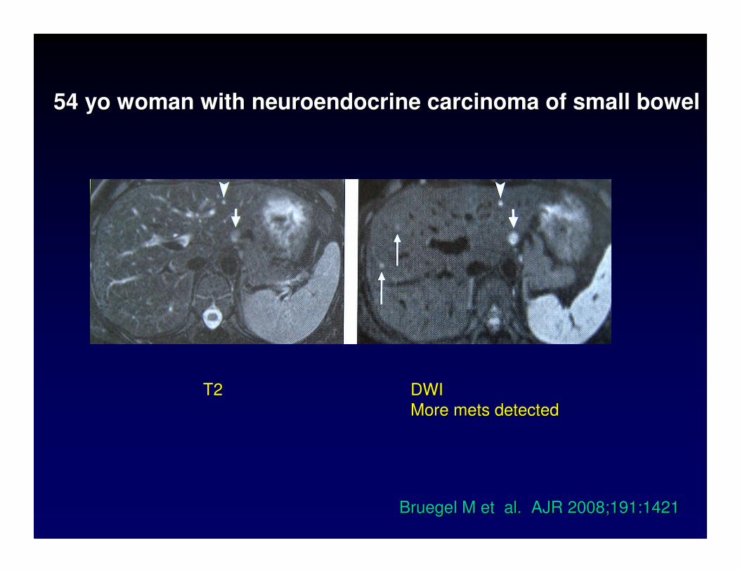
DWIDWI
More More metsmets detecteddetectedT2 T2
BruegelBruegel M et al. AJR 2008;191:1421M et al. AJR 2008;191:1421
54 54 yoyo woman with woman with neuroendocrineneuroendocrine carcinoma of small bowelcarcinoma of small bowel

Focal Hepatic Lesions
Hypervasc, washout, +/- fat, edema, vascular invasion
HCC
bright on T1, dark on T2, not vascularDN/well diff HCC
dark on all sequencesRN
Heterogeneous/peripheral enhancement, may show delayed enhancement, capsular retraction, peripheral duct dilatation
CholangioCA
Heterogeneous/peripheral enhancementMets
Similar to FNH unless contains hemorrhage, fat
Central scar does not enhance
Adenoma
Homogeneous, hypervascular -> isointense. Central scar T2 bright & delayed enhancement.
FNH
T2 bright, peripheral enhancement with fill inHemangioma

Clinical Features•• Asymptomatic/symptomaticAsymptomatic/symptomatic
•• AgeAge
•• GenderGender
•• Oral contraceptives, anabolic steroids, glycogen storage diseaseOral contraceptives, anabolic steroids, glycogen storage disease
•• Risk factors for chronic liver diseaseRisk factors for chronic liver disease
•• History of primary malignancyHistory of primary malignancy
•• Travel historyTravel history
•• Lab tests, including tumor markersLab tests, including tumor markers
•• Imaging studiesImaging studies
•• Majority of lesions characterized without biopsy.Majority of lesions characterized without biopsy.
•• 156/160 (98%) correct pre156/160 (98%) correct pre--op diagnosis.op diagnosis.
Torzilli et al. Hepatology 199;30:889

Fine Needle Aspiration Biopsy
•• ““Think first, then donThink first, then don’’t do itt do it””
•• Commonly nonCommonly non--diagnostic for hepatic diagnostic for hepatic
adenomas and focal nodular hyperplasiaadenomas and focal nodular hyperplasia
•• HCC > 1cm diagnosed with imagingHCC > 1cm diagnosed with imaging+
•• Risks: Risks:
1.1. Bleeding: Bleeding: hemangiomashemangiomas and adenomasand adenomas
2.2. Seeding: metaSeeding: meta--analysis analysis --> 2.7 % risk for > 2.7 % risk for
HCC*HCC*+AASLD July 2010 update
*Silva et al. Gut 2008;57:1592

Fine Needle Aspiration Biopsy
•• UnresectableUnresectable lesionlesion
•• Problematic caseProblematic case
•• USUS--guided biopsy preferredguided biopsy preferred
•• CTCT--guided biopsy, if US not feasibleguided biopsy, if US not feasible
•• ContrastContrast--enhanced US, if availableenhanced US, if available
FNA or Core Liver Biopsy
•• INR INR < < 1.51.5
• PTT < 50
• Platelets > 50

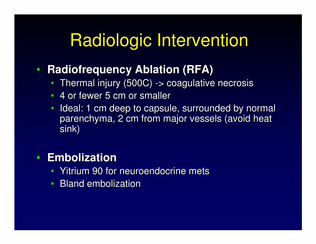
Radiologic Intervention
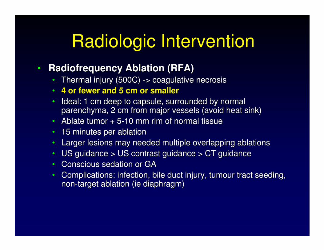
• Radiofrequency Ablation (RFA)
• Transcatheter Arterial Chemoembolization (TACE)
• Selective Internal Radiation (SIR)
Radiologic Intervention
•• Radiofrequency Ablation (RFA)Radiofrequency Ablation (RFA)•• Thermal injury (500C) Thermal injury (500C) --> > coagulativecoagulative necrosisnecrosis
•• 4 or fewer 5 cm or smaller4 or fewer 5 cm or smaller
•• Ideal: 1 cm deep to capsule, surrounded by normal Ideal: 1 cm deep to capsule, surrounded by normal parenchyma, 2 cm from major vessels (avoid heat parenchyma, 2 cm from major vessels (avoid heat sink)sink)
•• EmbolizationEmbolization•• YitriumYitrium 90 for 90 for neuroendocrineneuroendocrine metsmets
•• Bland Bland embolizationembolization
Radiologic Intervention
•• Radiofrequency Ablation (RFA)Radiofrequency Ablation (RFA)•• Thermal injury (500C) Thermal injury (500C) --> > coagulativecoagulative necrosisnecrosis
•• 4 or fewer and 5 cm or smaller4 or fewer and 5 cm or smaller
•• Ideal: 1 cm deep to capsule, surrounded by normal Ideal: 1 cm deep to capsule, surrounded by normal parenchyma, 2 cm from major vessels (avoid heat sink)parenchyma, 2 cm from major vessels (avoid heat sink)
•• Ablate tumor + 5Ablate tumor + 5--10 mm rim of normal tissue10 mm rim of normal tissue
•• 15 minutes per ablation15 minutes per ablation
•• Larger lesions may needed multiple overlapping ablationsLarger lesions may needed multiple overlapping ablations
•• US guidance > US contrast guidance > CT guidanceUS guidance > US contrast guidance > CT guidance
•• Conscious sedation or GAConscious sedation or GA
•• Complications: infection, bile duct injury, Complications: infection, bile duct injury, tumourtumour tract seeding, tract seeding, nonnon--target ablation (target ablation (ieie diaphragm)diaphragm)

Radiologic Intervention
• Transcatheter Arterial Chemoembolization (TACE)
•• Bland Bland embolizationembolization
•• GelfoamGelfoam: temporary, : temporary, recanalizationrecanalization in 2in 2--6 wks6 wks
•• Polyvinyl alcohol: permanentPolyvinyl alcohol: permanent
•• LipiodolLipiodol: oily contrast with affinity for HCC (drug vehicle): oily contrast with affinity for HCC (drug vehicle)
•• Chemotherapeutic agentsChemotherapeutic agents
•• fluorodeoxyuridinefluorodeoxyuridine, doxorubicin, , doxorubicin, cisplatincisplatin, , mitomycinmitomycin
• Chemoembolization• ischemia and prolonged contact of the chemotherapeutic agent with the tumor
• dramatically increase the local concentration of the chemotherapeutic agent
Radiologic Intervention
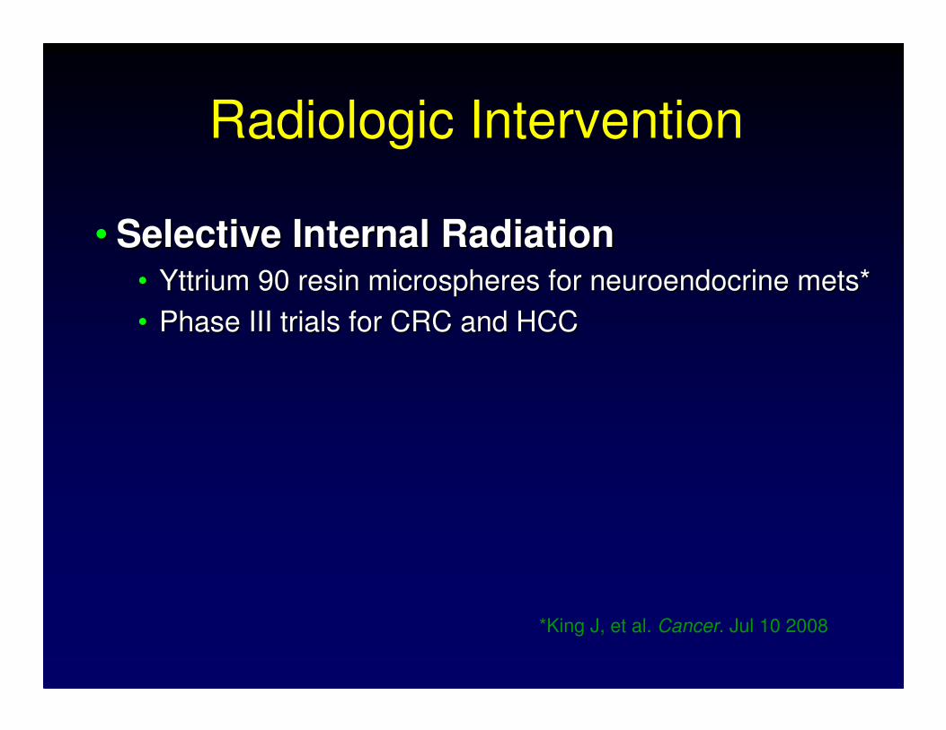
•• Selective Internal RadiationSelective Internal Radiation•• Yttrium 90 resin microspheres for Yttrium 90 resin microspheres for neuroendocrineneuroendocrine metsmets**
•• Phase III trials for CRC and HCCPhase III trials for CRC and HCC
*King J, et al. Cancer. Jul 10 2008
Conclusion
• Most solitary liver lesions can be characterized with CT and /or MR imaging
• Role of biopsy has decreased
• Imaging work-up depends on local expertise and
resources
• Radiologic interventions: RFA, TACE, SIR

Thank You