Isolated dilatation of the inferior vena cavakjim.org/upload/kjim-29-2-15_241-245.pdfDilatation of...
5
Korean J Intern Med 2014;29:241-245 http://dx.doi.org/10.3904/kjim.2014.29.2.241 Copyright © 2014 The Korean Association of Internal Medicine This is an Open Access article distributed under the terms of the Creative Commons Attribution Non-Commercial License (http://creativecommons.org/licenses/ by-nc/3.0/) which permits unrestricted noncommercial use, distribution, and reproduction in any medium, provided the original work is properly cited. pISSN 1226-3303 eISSN 2005-6648 http://www.kjim.org CASE REPORT Isolated dilatation of the inferior vena cava Jae-Joon Kim 1 , Kyoung-Im Cho 2 , Ji-Hoon Kang 1 , Ja-Jun Goo 1 , Kyoung-Nyoun Kim 1 , Ja-Young Lee 1 , and Seong-Man Kim 1 1 Department of Internal Medicine, Maryknoll Medical Center, Busan; 2 Department of Internal Medicine, Kosin University School of Medicine, Busan, Korea Received : August 22, 2012 Revised : October 23, 2012 Accepted: November 9, 2012 Correspondence to Kyoung-Im Cho, M.D. Division of Cardiology, Depart- ment of Internal Medicine, Kosin University School of Medicine, 262 Gamcheon-ro, Seo-gu, Busan 602-702, Korea Tel: +82-51-990-6105 Fax: +82-51-990-3005 E-mail: [email protected] The diameter and collapsibility of the inferior vena cava (IVC) should be inter- preted in consideration with other clinical and echocardiographic parameters be- fore drawing definitive diagnostic conclusions. We report a case of a 46-year-old female with isolated IVC dilation and diminished inspiratory collapse without other abnormalities, and provide a brief review of the literature. Keywords: Vena cava, inferior; Echocardiography; Cardiac catheterization INTRODUCTION The diameter of the inferior vena cava (IVC) and degree of inspiratory collapse are used as indices in the echo- cardiographic estimation of right atrial (RA) pressure. Under normal RA pressure, the maximum IVC diame- ter is less than 20 mm, and the inspiratory collapse is more than 50%. Under high RA pressure, the IVC is di- lated (more than 20 mm) and the inspiratory collapse of IVC is diminished. We report a case of a female patient with dilated IVC with normal RA pressure. This condi- tion is rare [1], and to our knowledge, this is the first case of isolated dilatation of the IVC reported in Korea. CASE REPORT A 46-year-old female underwent abdominal computed tomography (CT) imaging at a private clinic due to mild right upper quadrant abdominal discomfort and back pain during the previous 2 weeks. She was re- ferred to our hospital to evaluate the cause of IVC dila- tation shown by abdominal CT (Fig. 1). The patient had no history of hypertension or diabetes mellitus and no family history of aortic, collagen, vascular or congeni- tal heart disease. She had latent hepatitis B virus infec- tion and her mother died of hepatocellular carcinoma. Ultrasonography of the liver showed hepatic vein dila- tation without obstruction or thrombus in either the hepatic vein or IVC. The abdominal CT showed promi- nent dilatation of the IVC and hepatic vein with no evi- dence of liver disease such as cirrhosis, hepatocellular carcinoma or Budd–Chiari syndrome. Her vital signs included blood pressure of 107/64 mmHg, pulse of 60 beats per minute, respiration of 20 breaths per minute, and body temperature of 36.5°C. During the physical examination, cardiac auscultation revealed no definite murmurs and her electrocardiography demonstrated
Transcript of Isolated dilatation of the inferior vena cavakjim.org/upload/kjim-29-2-15_241-245.pdfDilatation of...

Korean J Intern Med 2014;29:241-245http://dx.doi.org/10.3904/kjim.2014.29.2.241
Copyright © 2014 The Korean Association of Internal MedicineThis is an Open Access article distributed under the terms of the Creative Commons Attribution Non-Commercial License (http://creativecommons.org/licenses/by-nc/3.0/) which permits unrestricted noncommercial use, distribution, and reproduction in any medium, provided the original work is properly cited.
pISSN 1226-3303eISSN 2005-6648
http://www.kjim.org
CASE REPORT
Isolated dilatation of the inferior vena cava Jae-Joon Kim1, Kyoung-Im Cho2, Ji-Hoon Kang1, Ja-Jun Goo1, Kyoung-Nyoun Kim1, Ja-Young Lee1, and Seong-Man Kim1
1Department of Internal Medicine, Maryknoll Medical Center, Busan; 2Department of Internal Medicine, Kosin University School of Medicine, Busan, Korea
Received : August 22, 2012Revised : October 23, 2012Accepted: November 9, 2012
Correspondence toKyoung-Im Cho, M.D.Division of Cardiology, Depart-ment of Internal Medicine, Kosin University School of Medicine, 262 Gamcheon-ro, Seo-gu, Busan 602-702, KoreaTel: +82-51-990-6105Fax: +82-51-990-3005E-mail: [email protected]
The diameter and collapsibility of the inferior vena cava (IVC) should be inter-preted in consideration with other clinical and echocardiographic parameters be-fore drawing definitive diagnostic conclusions. We report a case of a 46-year-old female with isolated IVC dilation and diminished inspiratory collapse without other abnormalities, and provide a brief review of the literature.
Keywords: Vena cava, inferior; Echocardiography; Cardiac catheterization
INTRODUCTION
The diameter of the inferior vena cava (IVC) and degree of inspiratory collapse are used as indices in the echo-cardiographic estimation of right atrial (RA) pressure. Under normal RA pressure, the maximum IVC diame-ter is less than 20 mm, and the inspiratory collapse is more than 50%. Under high RA pressure, the IVC is di-lated (more than 20 mm) and the inspiratory collapse of IVC is diminished. We report a case of a female patient with dilated IVC with normal RA pressure. This condi-tion is rare [1], and to our knowledge, this is the first case of isolated dilatation of the IVC reported in Korea.
CASE REPORT
A 46-year-old female underwent abdominal computed tomography (CT) imaging at a private clinic due to
mild right upper quadrant abdominal discomfort and back pain during the previous 2 weeks. She was re-ferred to our hospital to evaluate the cause of IVC dila-tation shown by abdominal CT (Fig. 1). The patient had no history of hypertension or diabetes mellitus and no family history of aortic, collagen, vascular or congeni-tal heart disease. She had latent hepatitis B virus infec-tion and her mother died of hepatocellular carcinoma. Ultrasonography of the liver showed hepatic vein dila-tation without obstruction or thrombus in either the hepatic vein or IVC. The abdominal CT showed promi-nent dilatation of the IVC and hepatic vein with no evi-dence of liver disease such as cirrhosis, hepatocellular carcinoma or Budd–Chiari syndrome. Her vital signs included blood pressure of 107/64 mmHg, pulse of 60 beats per minute, respiration of 20 breaths per minute, and body temperature of 36.5°C. During the physical examination, cardiac auscultation revealed no definite murmurs and her electrocardiography demonstrated
242 www.kjim.org http://dx.doi.org/10.3904/kjim.2014.29.2.241
The Korean Journal of Internal Medicine Vol. 29, No. 2, March 2014
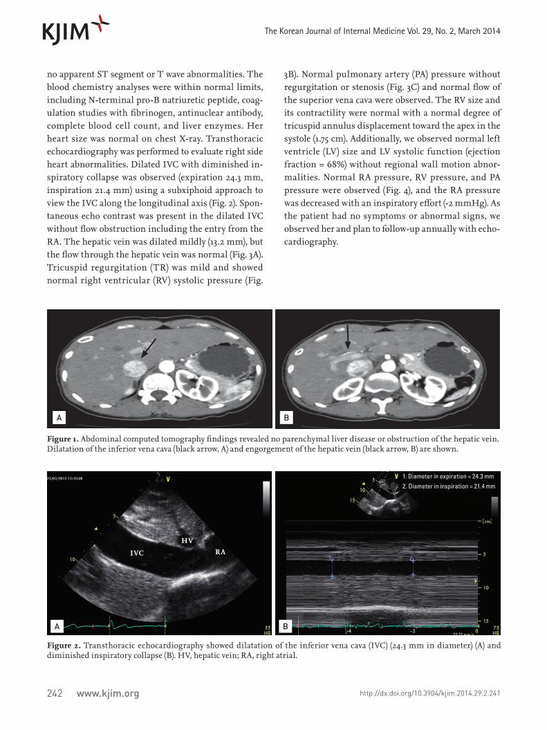
no apparent ST segment or T wave abnormalities. The blood chemistry analyses were within normal limits, including N-terminal pro-B natriuretic peptide, coag-ulation studies with fibrinogen, antinuclear antibody, complete blood cell count, and liver enzymes. Her heart size was normal on chest X-ray. Transthoracic echocardiography was performed to evaluate right side heart abnormalities. Dilated IVC with diminished in-spiratory collapse was observed (expiration 24.3 mm, inspiration 21.4 mm) using a subxiphoid approach to view the IVC along the longitudinal axis (Fig. 2). Spon-taneous echo contrast was present in the dilated IVC without flow obstruction including the entry from the RA. The hepatic vein was dilated mildly (13.2 mm), but the flow through the hepatic vein was normal (Fig. 3A). Tricuspid regurgitation (TR) was mild and showed normal right ventricular (RV) systolic pressure (Fig.
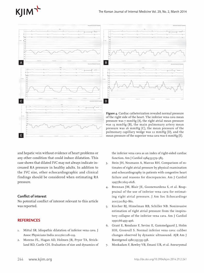
3B). Normal pulmonary artery (PA) pressure without regurgitation or stenosis (Fig. 3C) and normal flow of the superior vena cava were observed. The RV size and its contractility were normal with a normal degree of tricuspid annulus displacement toward the apex in the systole (1.75 cm). Additionally, we observed normal left ventricle (LV) size and LV systolic function (ejection fraction = 68%) without regional wall motion abnor-malities. Normal RA pressure, RV pressure, and PA pressure were observed (Fig. 4), and the RA pressure was decreased with an inspiratory effort (-2 mmHg). As the patient had no symptoms or abnormal signs, we observed her and plan to follow-up annually with echo-cardiography.
Figure 1. Abdominal computed tomography findings revealed no parenchymal liver disease or obstruction of the hepatic vein. Dilatation of the inferior vena cava (black arrow, A) and engorgement of the hepatic vein (black arrow, B) are shown.
Figure 2. Transthoracic echocardiography showed dilatation of the inferior vena cava (IVC) (24.3 mm in diameter) (A) and diminished inspiratory collapse (B). HV, hepatic vein; RA, right atrial.
A B
B
1. Diameter in expiration = 24.3 mm2. Diameter in inspiration = 21.4 mm
A
IVC
HVRA

243www.kjim.orghttp://dx.doi.org/10.3904/kjim.2014.29.2.241
Kim JJ, et al. Dilatation of IVC
DISCUSSION
The IVC is a highly collapsible major vein, and its di-ameter correlates closely with right side cardiac func-tions [2,3]. The IVC diameter is altered with volume sta-tus and respiration, with higher IVC diameter during expiration than inspiration. An IVC diameter greater than 20 mm is commonly regarded as an upper limit of normal, which is a noninvasive indication of increased RA pressure in patients with cardiac or renal disease [4]. The RA pressure is correlated with the diameter and collapsibility of the IVC [5], and under normal RA pressure, the IVC diameter is decreased during inspi-ration [6]. In this case, the patient had increased diame-ter of the IVC (24.3 mm) with diminished collapsibility; however, there was no evidence of high RA pressure. IVC aneurysm can be seen in association with elevated right heart pressure, RV dysfunction, significant TR [7], none of which were present in this case. Moreover, the morphology of the IVC did not resemble that of an
aneurysm. Ultrasonography of the liver and abdominal CT findings showed no liver disease and no mass-like leiomyoma. The possibility of Budd–Chiari syndrome [8] was ruled out as there was no thrombus or compres-sion of the IVC. Additionally, normal hemoglobin and blood clot tests, and normal f low of the hepatic vein further excluding Budd–Chiari syndrome. We ob-served normal renal function including the serum glo-merular filtration rate and normal kidney findings us-ing the aforementioned imaging tests. Another study reported dilated IVCs in many competitive young ath-letes without heart disease [9]. However, our patient was a normal middle-aged housewife without regular exercise. With few reports in the literature of isolated dilatation of the IVC, the prognosis is unknown. A re-cent study suggested that dilated IVC in healthy sub-jects (without volume overload, pericardial disease and right heart abnormalities) might be a marker of de-creased abdominal venous tone and/or increased com-pliance [10]. In conclusion, this patient had dilated IVC
Figure 3. Transthoracic echocardiography. (A) The hepatic vein f low was normal. (B) No regurgitation or stenosis of the pulmonary artery were observed. (C) Mild grade tricuspid regurgitation flow was observed with normal right ventricular systolic pressure. VR, ventricular reversal; AR, atrial reversal; D, diastolic forward flow; S, systolic forward flow.
A
C
B
244 www.kjim.org http://dx.doi.org/10.3904/kjim.2014.29.2.241
The Korean Journal of Internal Medicine Vol. 29, No. 2, March 2014
and hepatic vein without evidence of heart problems or any other condition that could induce dilatation. This case shows that dilated IVC may not always indicate in-creased RA pressure in healthy adults. In addition to the IVC size, other echocardiographic and clinical f indings should be considered when estimating RA pressure.
Conflict of interestNo potential conflict of interest relevant to this article was reported.
REFERENCES
1. Mittal SR. Idiopathic dilatation of inferior vena cava. J Assoc Physicians India 2012;60:118-119.
2. Moreno FL, Hagan AD, Holmen JR, Pryor TA, Strick-land RD, Castle CH. Evaluation of size and dynamics of
the inferior vena cava as an index of right-sided cardiac function. Am J Cardiol 1984;53:579-585.
3. Stein JH, Neumann A, Marcus RH. Comparison of es-timates of right atrial pressure by physical examination and echocardiography in patients with congestive heart failure and reasons for discrepancies. Am J Cardiol 1997;80:1615-1618.
4. Brennan JM, Blair JE, Goonewardena S, et al. Reap-praisal of the use of inferior vena cava for estimat-ing right atrial pressure. J Am Soc Echocardiogr 2007;20:857-861.
5. Kircher BJ, Himelman RB, Schiller NB. Noninvasive estimation of right atrial pressure from the inspira-tory collapse of the inferior vena cava. Am J Cardiol 1990;66:493-496.
6. Grant E, Rendano F, Sevinc E, Gammelgaard J, Holm HH, Gronvall S. Normal inferior vena cava: caliber changes observed by dynamic ultrasound. AJR Am J Roentgenol 1980;135:335-338.
7. Mookadam F, Rowley VB, Emani UR, et al. Aneurysmal
Figure 4. Cardiac catheterization revealed normal pressure of the right side of the heart. The inferior vena cava mean pressure was 7 mmHg (A), the right atrial mean pressure was 13 mmHg (B), the main pulmonary artery mean pressure was 16 mmHg (C), the mean pressure of the pulmonary capillary wedge was 12 mmHg (D), and the mean pressure of the superior vena cava was 6 mmHg (E).
A
C
E
B
D

245www.kjim.orghttp://dx.doi.org/10.3904/kjim.2014.29.2.241
Kim JJ, et al. Dilatation of IVC
dilatation of the inferior vena cava. Echocardiography 2011;28:833-842.
8. Menon KV, Shah V, Kamath PS. The Budd-Chiari syn-drome. N Engl J Med 2004;350:578-585.
9. Goldhammer E, Mesnick N, Abinader EG, Sagiv M. Di-lated inferior vena cava: a common echocardiographic
finding in highly trained elite athletes. J Am Soc Echo-cardiogr 1999;12:988-993.
10. Styczynski G, Jaltuszewska M, Kosiorowska N, Kostrze-wska M, Szmigielski C. Dilated inferior vena cava in young adults with vasovagal syncope. Arch Intern Med 2009;169:1634-1635.