Is the development of cirrhosis in hereditary hemorrhagic telangiectasia incidental?
1
April 1995 AASLD Al183 • PORTAL HYPERTENSION REDUCES EXPRESSION OF bFGF AND ITS RECEPTOR GENES IN ESOPHAGEAL MUCOSA: IMPORTANT FACTOR CONTRIBUTING TO VARICEAL RUPTURE? K. Tanoue, A. Tarnawski, IJ. Sarfeh, AM. Santos, F.L. Irwin, Jr, J.J. Lee, K.J. Wahlstrom, and K. Sugimachi. DVA Med Cent, Long Beach (CA), Univ of Calif, Irvine, USA and Kyushu Univ, Fukuoka, Japan. In the portal hypertensive {PHT) state, varices are prone to rupture in the lower esophagus, but the mechanism is unknown. Since basic fibrublast growth factor (bFGF) is important in maintaining tissue integrity, we determined expression ofmRNAs and proteins for bFGF and its receptors (FGFR-1 and-2) in PHT and normal esophageal mucosa. METHODS: In 15 PHT rats and 15 sham operated (SO) rats, 2 weeks postoperatively ihe lower esophagus was removed and frozen in liquid nitrogen or fixed in 4% paraformaldehyde. STUDIES: 1) Reverse transcfiption-polymerase chain reaction (RT/PCR) for bFGF and FGFR-I, 2 and 4 mRNAs with specific primers; 2) Quantitative histology; 3) Immunostaining with specific antibodies against bFGF and FGFR-I and-2, and quantitative assessment of fluorescent signal with Image 1, video image analysis system. RESULTS: Portal pressures in PHT and control rats were 28.3:t:2.6 and 13.54-1.8 cm saline, respectively (p<0.01). The esophageal muscularis mucosae and epithelium in the areas overlying dilated submucosal veins was significantlythinner in PHT rats than in controls (27.7:t:5.3 vs 36.6+6.0 lam, p<0.01, 40.4i6.0 vs 5254-9.8, p<0.05 respectively). Basic FGF and FGFR-2 mRNAS expression in PHT esophageal wall were significantlyreduced vs controls by 30.8% and 30.3%, respectively, (p<0.01). Intensity ofbFGF and FGFR-2 specific fluorescence of the muscularis mucosae was significantly reduced in PHT rats (42.14-2.3 and 71.3:~6.5 units vs controls 49.54-5.6 and 78.64-5.7 units, respectively, both p<0.05). CONCLUSIONS: 1) Muscularis mueosae and epithelium in the lower esophageal mucosa of PHT rats are thinner than in controls. 2) At this site, expression of bFGF, FGFR proteins and mRNAs are reduced, which will weaken structural integrity, defense and reparative ability. 3) Since bFGF is important for muscle growth, reduction of bFGF may be the cause of muscularis mucusae thinning. Combined, these findings suggest a mechanism for predilection of varices to rupture in the lower esophagus. IS THE DEVELOPrlENT OF CIRRIIOSIS IN HEREDITARY HEIIORRHAGIC TELANGIECTASIA INCIDENTAL? H. Tanyol, Departments of Hedi- cine and Physiology, Thomas Jefferson University, Phila- delphia, PA. Current Affiliation: Germantmm IIospital and IIedica] Center, Philadelphia, PA. The involvement of the liver in hereditary hemorrhagic telangiectasia (HHT) has long been the subject of dehate and controversy. However, the passage of time has produced studies which have provided histological findings of the liver in adequate numbers to allow objective examination of the subject. By using the Index riedicus, we studied 79 re- pocks that included autopsy and/or blopsy findings published between 1901 (the publication date of the first autopsied case by Osler) and 1994. In 39 cases (49%) the authors made the histologicai diagnosis of cirrhosis (C). dolliffe and Jellinek (1941), based on a careful appraisal of 2,045 autopsy findings, estimated that 8% of alcoholics ultimately developed C. ~lhen these figures (8% versus 49%) are com- pared, one may conclude that HHT is significantly more cir- rhogenic than alcohol. (Probability: very close to zero.) See the figure. An analysis of the 79 reports shm'fs that trans- fusion has no effect on the development of C. On the basis of observations by me and others (Tanyo]:1960,1961,1963; Factor:1976;Rubin:1979), I drev~ attention to the anqiofibro- proliferative effects of ethanol. The findings on the alco- holic anf]iopathy are extensions of the earlier ohservations on the vascular origin of Laehnec's C. (Tanyol:Llien kl llschr 1944;57:2(l~,,1964,1%5A). An arterio- venous shunt mechanism ~lhich seems to 491 undei~lie the alcoholic angiopathy and HHT generates in both conditions a 4C ," .,, ," vascular spectrum comprised of arterial ~ • • • dilation, varicose veins, angioma, fi- --~3G . " , " . " brosis and vascular bleeding. The ~ , ° , * , " Study of the natural history of C in ~20 • " • " • " both alcoholic angiopathy and tlilT re- ~. • * • veals the presence of an identical I0 " • " vascular prelude to the cirrhotic • 8,~ • • • process. These observations are incom- • , , , • , patible with the prevailing view of O | I ..... hepatocellular origin of C. ALC()HOLI HNT I O INTERFERON DOWN-REGULATES CYP 3A GENES IN WELL- DIFFERENTIATED RAT AND HUMAN HEPATOCYTES IN PRIMARY CULTURE. M. Tapner, B. Goodwin, C. Liddle, M. Murray, J. George and FarrelL Storr Liver Unit, University of Sydney at Westmead Hospital, Westmead, NSW 2145 Background and aims: We have previously shown that interfemns down- regulate expression of the important constitutive hepatic cytochrome P450 (CYP) 3A2 in male rats at a pretranslational level (Mol Pharm 38:313-8,1990). The analogous CYP 3A4 is quantitatively the most important component of total P450 in human liver. Using the 14C-erythromycin breath test as an in vivo probe for CYPs 3A in human liver, we have demonstrated that interferon decreases the activity of these enzymes (Hepatol 17:230-5,1993). However, because the interferons release other cytokines in vivo, it is unclear whether interferons modulate CYP expression directly. The present studies addressed (1) whether interferon directly down-regulates CYP 3A genes in cultured rat hepatocytes, and (2) whether similar changes in CYP 3A were elicited at a pretranslational level in cultured human hepatocytes. Methods: Hepatocytes were isolated from adult male rat liver or normal human liver (segments of adult liver cut-down for pediatric recipients) using collagenase perfusion methods. Hepatocytes were cultured in William's E medium on matrigel, conditions which provided phenotypically well-differentiated hepatocytes. Experiments were performed on day 4 (human) or day 5 (rat) after isolation when levels of 3A mRNA species were found to be maximal, mRNA was quantified by solution hybridization using 35S-labeled gene-specific probes. Total 3A protein was immunoquantitated using anti-rat 3A1 or anti-human 3A4 antibodies, and 3A catalytic activity was measured as testosterone 6g- hydroxylase (T-61~OH). Results: In cultured rat hepatocytes, expression of 3A2 fell rapidly, but stable levels of 3A2 mRNA (30% of freshly isolated cells) were attained at day 5. Addition of 2mM phenobarbital (PB) increased levels of 3A2 mRNA, immunoquantifiable 3A protein and T-613OH activity to between 60% and 100% of freshly-isolated cells by day 5; dexamethasone was without effect on 3A2 mRNA. In phenobarbital-exposed rat hepatocytes, rat interferon-7 (100 U/culture plate - 3x10⁶ cells) added at day 1 decreased mRNA levels to 1.5% of PB-induced control at day 5. Levels of rat 3A protein were also decreased by interferon but to a lesser extent (75% of PB control); this may reflect the relative preservation of other inducible 3A proteins. In human hepatocytes, levels of 3A4 mRNA fell during isolation of ceils but essentially recovered (without addition of exogenous chemicals) after 4 days in primary culture. Addition of human interferon-o.2b (100,000 U/3xl06 cells) on day 1 lowered 3A4 mRNA levels to 21% of control by day 4. Conclusions: Experimentalconditions have been identified that allow down-regulation of CYP 3A genes to be studied in hepatocyte cultures. In both rat and human hepatocytes, interferon decreased concentrations of mRNA species corresponding to major 3A proteins. Thus interferon acts directly on hepatocytes to down-regulate CYPs 3A at a pretranslational level. O IS CHRONIC C HEPATITIS POSSIBLE IN ANTIHCVNEGATIVE PATIENTS? G.TARANTINO, L.MORELLI,N.MORLANDO, *M.L.GOBBO,*R.SORRENTiNO, *S.GAROFALO. Institute of Internal Medicine and Dismetabolic Diseases; *mipartimento di Biologia e Patologia Cellulare e Molecolare. School of Medicine "Federico II", Naples, ITALY. Aim of the present study was to detect HCV replication by reverse transcriptase polymerase chain reaction (RT-PCR) in the sera of pts with hypertransaminasemia, who were antiHCV negative and previously submitted to liver biopsy. By using a nested RT-PCR assay for the 5' untranslated region (5'UTR) of the HCV-RNA (Bioline Diagnostics, Italy) we found positivity for HCV RNA in 17 samples out of 21 pts. All the positive pts, having a median age of 45+/-18 yrs had a history of fluctuant hypertransaminasemia and serological, instrumental and clinical findings suggestive of CH. We excluded, as the cause of the hepatitis, autoimmune diseases, metabolic disorders, drug addictions or a toxic state. All pts, instead, had a history suggestive of viral exposure: in fact, one or more members of the family of some pts had suffered from CH (NANB) (70% of the pts), while some of the other pts (30%) had undergone major or minor surgery. Furthermore, we have evaluated these pts for a period of 2 yrs, initially by an antiHCV 2nd generation EIA and RIBA II test and, subsequently, by RIBA III test. All of these tests were constantly negative, even though the pts showed no signs of an immunodeficiency state. All positive pts were subjected to a liver biopsy and the histological sections of the biopsies showed the presence of prominent microvescicular fatty changes with sporadic lobular inflammatory infiltrates suggestive of minimal chronic hepatitis or steatohepatitis. Although our pts did not fulfill all international criteria, they were diagnosed as having chronic C hepatitis. We are at the present determining the genotypes of the HCV while in HCV pts an interferon therapy cycle (3 MU three times weekly for 12 months was started from October 1994) has been initiated for the treatment of all 17 pts. These findings could be explained by assuming that there is a small proportion of the pts with chronic C hepatitis who had a major cell damage and a low response by host's ir~nune system. In any case, the histological findings of steatosis or steatohepatitis should have carefully evaluated by hepatologist.
Transcript of Is the development of cirrhosis in hereditary hemorrhagic telangiectasia incidental?
April 1995 AASLD A l 1 8 3
• PORTAL HYPERTENSION REDUCES EXPRESSION OF bFGF AND ITS RECEPTOR GENES IN ESOPHAGEAL MUCOSA: IMPORTANT FACTOR CONTRIBUTING TO VARICEAL RUPTURE? K. Tanoue, A. Tarnawski, IJ. Sarfeh, AM. Santos, F.L. Irwin, Jr, J.J. Lee, K.J. Wahlstrom, and K. Sugimachi. DVA Med Cent, Long Beach (CA), Univ of Calif, Irvine, USA and Kyushu Univ, Fukuoka, Japan.
In the portal hypertensive {PHT) state, varices are prone to rupture in the lower esophagus, but the mechanism is unknown. Since basic fibrublast growth factor (bFGF) is important in maintaining tissue integrity, we determined expression ofmRNAs and proteins for bFGF and its receptors (FGFR-1 and-2) in PHT and normal esophageal mucosa. METHODS: In 15 PHT rats and 15 sham operated (SO) rats, 2 weeks postoperatively ihe lower esophagus was removed and frozen in liquid nitrogen or fixed in 4% paraformaldehyde. STUDIES: 1) Reverse transcfiption-polymerase chain reaction (RT/PCR) for bFGF and FGFR-I, 2 and 4 mRNAs with specific primers; 2) Quantitative histology; 3) Immunostaining with specific antibodies against bFGF and FGFR-I and-2, and quantitative assessment of fluorescent signal with Image 1, video image analysis system. RESULTS: Portal pressures in PHT and control rats were 28.3:t:2.6 and 13.54-1.8 cm saline, respectively (p<0.01). The esophageal muscularis mucosae and epithelium in the areas overlying dilated submucosal veins was significantly thinner in PHT rats than in controls (27.7:t:5.3 vs 36.6+6.0 lam, p<0.01, 40.4i6.0 vs 5254-9.8, p<0.05 respectively). Basic FGF and FGFR-2 mRNAS expression in PHT esophageal wall were significantly reduced vs controls by 30.8% and 30.3%, respectively, (p<0.01). Intensity ofbFGF and FGFR-2 specific fluorescence of the muscularis mucosae was significantly reduced in PHT rats (42.14-2.3 and 71.3:~6.5 units vs controls 49.54-5.6 and 78.64-5.7 units, respectively, both p<0.05). CONCLUSIONS: 1) Muscularis mueosae and epithelium in the lower esophageal mucosa of PHT rats are thinner than in controls. 2) At this site, expression of bFGF, FGFR proteins and mRNAs are reduced, which will weaken structural integrity, defense and reparative ability. 3) Since bFGF is important for muscle growth, reduction of bFGF may be the cause of muscularis mucusae thinning. Combined, these findings suggest a mechanism for predilection of varices to rupture in the lower esophagus.
IS THE DEVELOPrlENT OF CIRRIIOSIS IN HEREDITARY HEIIORRHAGIC TELANGIECTASIA INCIDENTAL? H. Tanyol, Departments of Hedi- cine and Physiology, Thomas Jefferson University, Phila- delphia, PA. Current A f f i l i a t ion : Germantmm IIospital and IIedica] Center, Philadelphia, PA.
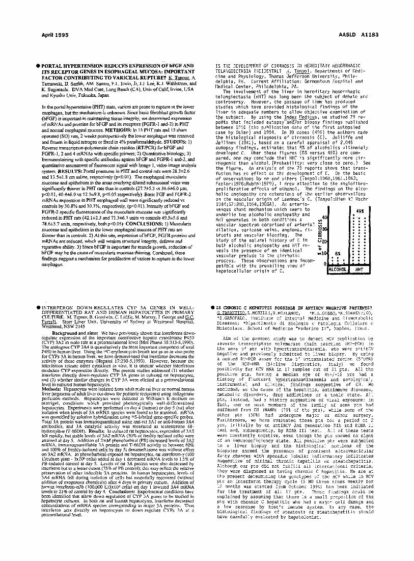
The involvement of the l i ver in hereditary hemorrhagic telangiectasia (HHT) has long been the subject of dehate and controversy. However, the passage of time has produced studies which have provided histological findings of the l iver in adequate numbers to allow objective examination of the subject. By using the Index riedicus, we studied 79 re- pocks that included autopsy and/or blopsy findings published between 1901 (the publication date of the f i r s t autopsied case by Osler) and 1994. In 39 cases (49%) the authors made the histologicai diagnosis of cirrhosis (C). do l l i f fe and Jellinek (1941), based on a careful appraisal of 2,045 autopsy findings, estimated that 8% of alcoholics ultimately developed C. ~lhen these figures (8% versus 49%) are com- pared, one may conclude that HHT is s igni f icant ly more c i r - rhogenic than alcohol. (Probability: very close to zero.) See the figure. An analysis of the 79 reports shm'fs that trans- fusion has no effect on the development of C. On the basis of observations by me and others (Tanyo]:1960,1961,1963; Factor:1976;Rubin:1979), I drev~ attention to the anqiofibro- prol i ferat ive effects of ethanol. The findings on the alco- holic anf]iopathy are extensions of the earl ier ohservations on the vascular origin of Laehnec's C. (Tanyol:Llien kl llschr 1944;57:2(l~,,1964,1%5A). An arterio- venous shunt mechanism ~lhich seems to 491 undei~lie the alcoholic angiopathy and HHT generates in both cond i t i ons a 4 C ," .,, ," vascu la r spectrum comprised o f a r t e r i a l ~ • • • di lat ion, varicose veins, angioma, f i - --~3G . " , " . " brosis and vascular bleeding. The ~ , ° , * , " Study of the natural history of C in ~20 • " • " • " both a l c o h o l i c ang iopathy and tlilT re - ~. • * • veals the presence of an identical I0 " • " vascular prelude to the cirrhotic • 8,~ • • • process. These observations are incom- • , , , • , patible with the prevailing view of O | I . . . . . hepatocellular origin of C. ALC()HOLI HNT I
O INTERFERON DOWN-REGULATES CYP 3A GENES IN WELL- DIFFERENTIATED RAT AND HUMAN HEPATOCYTES IN PRIMARY CULTURE. M. Tapner, B. Goodwin, C. Liddle, M. Murray, J. George and FarrelL Storr Liver Unit, University of Sydney at Westmead Hospital, Westmead, NSW 2145
Background and aims: We have previously shown that interfemns down- regulate expression of the important constitutive hepatic cytochrome P450 (CYP) 3A2 in male rats at a pretranslational level (Mol Pharm 38:313-8,1990). The analogous CYP 3A4 is quantitatively the most important component of total P450 in human liver. Using the 14C-erythromycin breath test as an in vivo probe for CYPs 3A in human liver, we have demonstrated that interferon decreases the activity of these enzymes (Hepatol 17:230-5,1993). However, because the interferons release other cytokines in vivo, it is unclear whether interferons modulate C Y P expression directly. The present studies addressed (1) whether interferon directly down-regulates CYP 3A genes in cultured rat hepatocytes, and (2) whether similar changes in CYP 3A were elicited at a pretranslational level in cultured human hepatocytes. Methods: Hepatocytes were isolated from adult male rat liver or normal human liver (segments of adult liver cut-down for pediatric recipients) using collagenase perfusion methods. Hepatocytes were cultured in William's E medium on matrigel, conditions which provided phenotypically well-differentiated hepatocytes. Experiments were performed on day 4 (human) or day 5 (rat) after isolation when levels of 3A mRNA species were found to be maximal, mRNA was quantified by solution hybridization using 35S-labeled gene-specific probes. Total 3A protein was immunoquantitated using anti-rat 3A1 or anti-human 3A4 antibodies, and 3A catalytic activity was measured as testosterone 6g- hydroxylase (T-61~OH). Results: In cultured rat hepatocytes, expression of 3A2 fell rapidly, but stable levels of 3A2 mRNA (30% of freshly isolated cells) were attained at day 5. Addition of 2mM phenobarbital (PB) increased levels of 3A2 mRNA, immunoquantifiable 3A protein and T-613OH activity to between 60% and 100% of freshly-isolated cells by day 5; dexamethasone was without effect on 3A2 mRNA. In phenobarbital-exposed rat hepatocytes, rat interferon-7 (100 U/culture plate - 3x10⁶ cells) added at day 1 decreased mRNA levels to 1.5% of PB-induced control at day 5. Levels of rat 3A protein were also decreased by interferon but to a lesser extent (75% of PB control); this may reflect the relative preservation of other inducible 3A proteins. In human hepatocytes, levels of 3A4 mRNA fell during isolation of ceils but essentially recovered (without addition of exogenous chemicals) after 4 days in primary culture. Addition of human interferon-o.2b (100,000 U/3xl06 cells) on day 1 lowered 3A4 mRNA levels to 21% of control by day 4. Conclusions: Experimentalconditions have been identified that allow down-regulation of CYP 3A genes to be studied in hepatocyte cultures. In both rat and human hepatocytes, interferon decreased concentrations of mRNA species corresponding to major 3A proteins. Thus interferon acts directly on hepatocytes to down-regulate CYPs 3A at a pretranslational level.
O IS CHRONIC C HEPATITIS POSSIBLE IN ANTIHCVNEGATIVE PATIENTS? G.TARANTINO, L.MORELLI,N.MORLANDO, *M.L.GOBBO,*R.SORRENTiNO, *S.GAROFALO. Institute of Internal Medicine and Dismetabolic Diseases; *mipartimento di Biologia e Patologia Cellulare e Molecolare. School of Medicine "Federico II", Naples, ITALY.
Aim of the present study was to detect HCV replication by reverse transcriptase polymerase chain reaction (RT-PCR) in the sera of pts with hypertransaminasemia, who were antiHCV negative and previously submitted to liver biopsy. By using a nested RT-PCR assay for the 5' untranslated region (5'UTR) of the HCV-RNA (Bioline Diagnostics, Italy) we found positivity for HCV RNA in 17 samples out of 21 pts. All the positive pts, having a median age of 45+/-18 yrs had a history of fluctuant hypertransaminasemia and serological, instrumental and clinical findings suggestive of CH. We excluded, as the cause of the hepatitis, autoimmune diseases, metabolic disorders, drug addictions or a toxic state. All pts, instead, had a history suggestive of viral exposure: in fact, one or more members of the family of some pts had suffered from CH (NANB) (70% of the pts), while some of the other pts (30%) had undergone major or minor surgery. Furthermore, we have evaluated these pts for a period of 2 yrs, initially by an antiHCV 2nd generation EIA and RIBA II test and, subsequently, by RIBA III test. All of these tests were constantly negative, even though the pts showed no signs of an immunodeficiency state. All positive pts were subjected to a liver biopsy and the histological sections of the biopsies showed the presence of prominent microvescicular fatty changes with sporadic lobular inflammatory infiltrates suggestive of minimal chronic hepatitis or steatohepatitis. Although our pts did not fulfill all international criteria, they were diagnosed as having chronic C hepatitis. We are at the present determining the genotypes of the HCV while in HCV pts an interferon therapy cycle (3 MU three times weekly for 12 months was started from October 1994) has been initiated for the treatment of all 17 pts. These findings could be explained by assuming that there is a small proportion of the pts with chronic C hepatitis who had a major cell damage and a low response by host's ir~nune system. In any case, the histological findings of steatosis or steatohepatitis should have carefully evaluated by hepatologist.