IRON DEFICIENCY ANEMIA -...
31
SECTION 6 IRON DEFICIENCY ANEMIA
Transcript of IRON DEFICIENCY ANEMIA -...
SECTION 6
IRON DEFICIENCY ANEMIA


Table of Content
6.0 Iron Deficiency Anemia 6.0.1 Introduction 6.0.2 Purpose 6.0.3 Objectives
6.1 The Role of Iron in the Body 6.1.1 Reutilization and Loss of Iron 6.1.2 Causes of Iron Deficiency Anemia 6.1.3 Laboratory Screening for Iron-Deficiency Anemia
6.2 Recommendations for Iron Intake 6.2.1 Iron Requirements for Infants 6.2.2 Iron Requirements for Pregnancy
6.3 Food Sources of Iron
6.4 Factors Influencing Iron Absorption 6.4.1 Inhibition of Iron Absorption 6.4.2 Enhancement of Iron Absorption
6.5 Prevention of Iron Deficiency Anemia 6.5.1 (0 – 12 Months) and Preschool Children (1-5 Years of Age) 6.5.2 Pregnant Women 6.5.3 Postpartum Women
6.6 Other Dietary Anemias 6.6.1 Megaloblastic (Pernicious) (Folate Deficiency) Anemia 6.6.2 Vitamin B12 Deficiency 6.6.3 Gastrointestinal Disorders 6.6.4 Vegetarian Diets
6.7 Sickle Cell Anemia 6.7.1 Sickle Cell Trait Versus Anemia 6.7.2 Signs and Symptoms of Sickle Cell Anemia 6.7.3 Treatment for Sickle Cell Anemia
6.8 Anemia Counseling Tips
6.9 Self-Test Questions 6.10 Case Study 6.11 References
6.0 IRON DEFICIENCY ANEMIA
6.0.1 Introduction
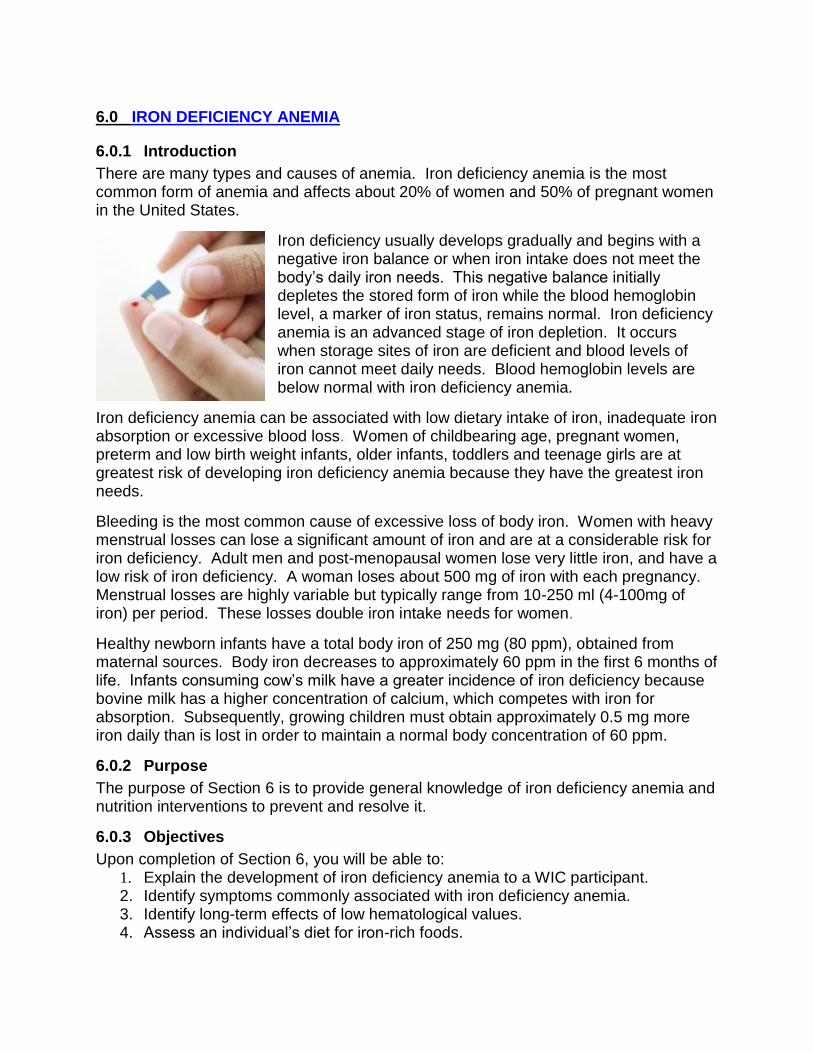
There are many types and causes of anemia. Iron deficiency anemia is the most common form of anemia and affects about 20% of women and 50% of pregnant women in the United States.
Iron deficiency usually develops gradually and begins with a negative iron balance or when iron intake does not meet the body’s daily iron needs. This negative balance initially depletes the stored form of iron while the blood hemoglobin level, a marker of iron status, remains normal. Iron deficiency anemia is an advanced stage of iron depletion. It occurs when storage sites of iron are deficient and blood levels of iron cannot meet daily needs. Blood hemoglobin levels are below normal with iron deficiency anemia.
Iron deficiency anemia can be associated with low dietary intake of iron, inadequate iron absorption or excessive blood loss. Women of childbearing age, pregnant women, preterm and low birth weight infants, older infants, toddlers and teenage girls are at greatest risk of developing iron deficiency anemia because they have the greatest iron needs.
Bleeding is the most common cause of excessive loss of body iron. Women with heavy menstrual losses can lose a significant amount of iron and are at a considerable risk for iron deficiency. Adult men and post-menopausal women lose very little iron, and have a low risk of iron deficiency. A woman loses about 500 mg of iron with each pregnancy. Menstrual losses are highly variable but typically range from 10-250 ml (4-100mg of iron) per period. These losses double iron intake needs for women.
Healthy newborn infants have a total body iron of 250 mg (80 ppm), obtained from maternal sources. Body iron decreases to approximately 60 ppm in the first 6 months of life. Infants consuming cow’s milk have a greater incidence of iron deficiency because bovine milk has a higher concentration of calcium, which competes with iron for absorption. Subsequently, growing children must obtain approximately 0.5 mg more iron daily than is lost in order to maintain a normal body concentration of 60 ppm.
6.0.2 Purpose
The purpose of Section 6 is to provide general knowledge of iron deficiency anemia and nutrition interventions to prevent and resolve it.
6.0.3 Objectives
Upon completion of Section 6, you will be able to: 1. Explain the development of iron deficiency anemia to a WIC participant. 2. Identify symptoms commonly associated with iron deficiency anemia. 3. Identify long-term effects of low hematological values. 4. Assess an individual’s diet for iron-rich foods.
5. Recommend dietary changes to increase the absorption of iron from the diet. 6. Identify probable diet-related cause(s) of iron deficiency. 7. Be familiar with other types of anemia.
6.1 THE ROLE OF IRON IN THE BODY
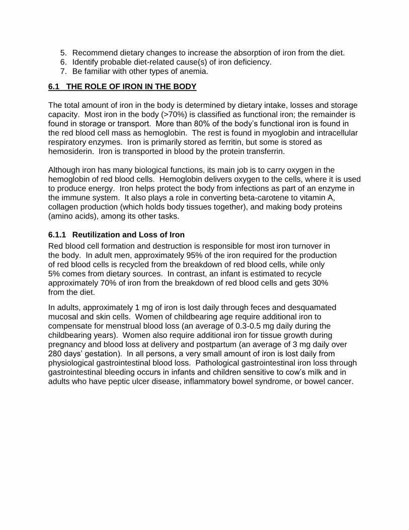
The total amount of iron in the body is determined by dietary intake, losses and storage capacity. Most iron in the body (>70%) is classified as functional iron; the remainder is found in storage or transport. More than 80% of the body’s functional iron is found in the red blood cell mass as hemoglobin. The rest is found in myoglobin and intracellular respiratory enzymes. Iron is primarily stored as ferritin, but some is stored as hemosiderin. Iron is transported in blood by the protein transferrin.
Although iron has many biological functions, its main job is to carry oxygen in the hemoglobin of red blood cells. Hemoglobin delivers oxygen to the cells, where it is used to produce energy. Iron helps protect the body from infections as part of an enzyme in the immune system. It also plays a role in converting beta-carotene to vitamin A, collagen production (which holds body tissues together), and making body proteins (amino acids), among its other tasks.
6.1.1 Reutilization and Loss of Iron
Red blood cell formation and destruction is responsible for most iron turnover in the body. In adult men, approximately 95% of the iron required for the production of red blood cells is recycled from the breakdown of red blood cells, while only 5% comes from dietary sources. In contrast, an infant is estimated to recycle approximately 70% of iron from the breakdown of red blood cells and gets 30% from the diet.
In adults, approximately 1 mg of iron is lost daily through feces and desquamated mucosal and skin cells. Women of childbearing age require additional iron to compensate for menstrual blood loss (an average of 0.3-0.5 mg daily during the childbearing years). Women also require additional iron for tissue growth during pregnancy and blood loss at delivery and postpartum (an average of 3 mg daily over 280 days’ gestation). In all persons, a very small amount of iron is lost daily from physiological gastrointestinal blood loss. Pathological gastrointestinal iron loss through gastrointestinal bleeding occurs in infants and children sensitive to cow’s milk and in adults who have peptic ulcer disease, inflammatory bowel syndrome, or bowel cancer.
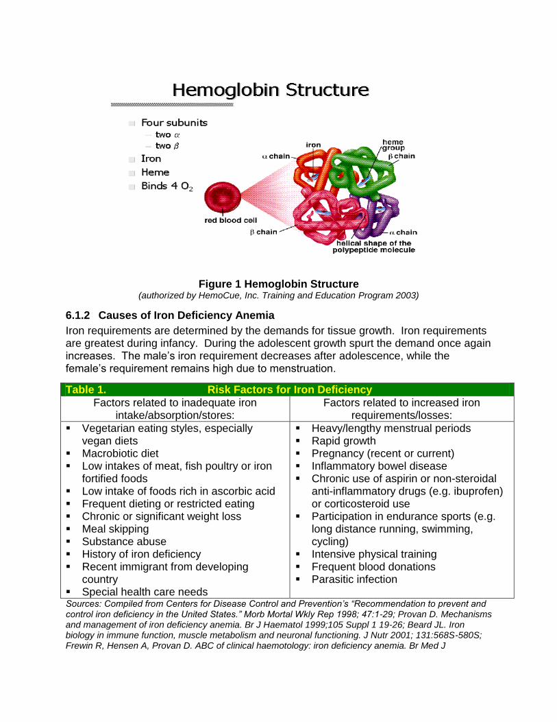
Figure 1 Hemoglobin Structure
(authorized by HemoCue, Inc. Training and Education Program 2003)
6.1.2 Causes of Iron Deficiency Anemia
Iron requirements are determined by the demands for tissue growth. Iron requirements are greatest during infancy. During the adolescent growth spurt the demand once again increases. The male’s iron requirement decreases after adolescence, while the female’s requirement remains high due to menstruation.
Table 1. Risk Factors for Iron Deficiency
Factors related to inadequate iron intake/absorption/stores:
Factors related to increased iron requirements/losses:
Vegetarian eating styles, especially vegan diets
Macrobiotic diet Low intakes of meat, fish poultry or iron
fortified foods Low intake of foods rich in ascorbic acid Frequent dieting or restricted eating Chronic or significant weight loss Meal skipping Substance abuse History of iron deficiency Recent immigrant from developing
country Special health care needs
Heavy/lengthy menstrual periods Rapid growth Pregnancy (recent or current) Inflammatory bowel disease Chronic use of aspirin or non-steroidal
anti-inflammatory drugs (e.g. ibuprofen) or corticosteroid use
Participation in endurance sports (e.g. long distance running, swimming, cycling)
Intensive physical training Frequent blood donations Parasitic infection
Sources: Compiled from Centers for Disease Control and Prevention’s “Recommendation to prevent and control iron deficiency in the United States.” Morb Mortal Wkly Rep 1998; 47:1-29; Provan D. Mechanisms and management of iron deficiency anemia. Br J Haematol 1999;105 Suppl 1 19-26; Beard JL. Iron biology in immune function, muscle metabolism and neuronal functioning. J Nutr 2001; 131:568S-580S; Frewin R, Hensen A, Provan D. ABC of clinical haemotology: iron deficiency anemia. Br Med J
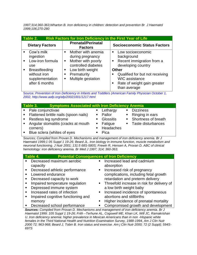
1997;314;360-363;Wharton B. Iron deficiency in children: detection and prevention Br J Haematol 1999;106;270-280
Table 2. Risk Factors for Iron Deficiency in the First Year of Life
Dietary Factors Prenatal/Perinatal
Factors Socioeconomic Status Factors
Cow’s milk ingestion
Low-iron formula use
Breastfeeding without iron supplementation after 6 months
Mother with anemia during pregnancy
Mother with poorly controlled diabetes
Low birth weight Prematurity Multiple gestation
Low socioeconomic background
Recent immigration from a developing country
Other Qualified for but not receiving
WIC assistance Rate of weight gain greater
than average
Source: Prevention of Iron Deficiency in Infants and Toddlers (American Family Physician October 1, 2002, http://www.aafp.org/afp/20021001/1217.html
Table 3. Symptoms Associated with Iron Deficiency Anemia
Pale conjunctivae Flattened brittle nails (spoon nails) Restless leg syndrome Angular stomatitis (cracks at mouth
corners) Blue sclera (whites of eyes
Lethargy Pallor Glossitis Fatigue Headaches Pica
Dizziness Ringing in ears Shortness of breath Taste disturbances
Sources; Compiled from Provan D. Mechanisms and management of iron deficiency anemia. Br J Haematol 1999;1 05 Suppl 1 19-26; Beard JL. Iron biology in immune function, muscle metabolism and neuronal functioning. J Nutr 2001; 131:5 68S-580S; Frewin R, Hensen A, Provan D. ABC of clinical hemotology: iron deficiency anemia. Br Med J 1997; 314; 360-363.
Table 4. Potential Consequences of Iron Deficiency
Decreased maximum aerobic capacity
Decreased athletic performance Lowered endurance Decreased capacity to work Impaired temperature regulation Depressed immune system Increased rates of infection Impaired cognitive functioning and
memory Decreased school performance
Increased lead and cadmium absorption
Increased risk of pregnancy complications, including fetal growth retardation and preterm delivery
Threefold increase in risk for delivery of a low birth weight baby
Increased incidence of spontaneous abortions and stillbirths
Higher incidence of prenatal mortality Compromised growth and development
Sources: Compiled from Provan D. Mechanisms and management of iron deficiency anemia. Br J Haematol 1999; 105 Suppl 1 19-26; Frith –Terhune AL, Cogswell ME, Khan LK, Will JC, Ramakrishan U. Iron deficiency anemia: higher prevalence in Mexican Americans than in non -Hispanic white females in the Third National Health and Nutrition Examination Survey, 1988-1994, Am J Clin Nutr 2000 72; 963-968; Beard J, Tobin B. Iron status and exercise. Am j Clin Nutr 2000; 72 (2 Suppl); 594S-697S

6.1.3 Laboratory Screening for Iron-Deficiency Anemia
The WIC Program uses two laboratory tests to screen for iron level, Hemoglobin (Hgb) and Hematocrit (Hct).
HEMOGLOBIN Hemoglobin is the iron-containing protein in red blood cells that carries oxygen to the body tissues and transports some carbon dioxide away from the tissues. It is responsible for the red color of blood. In Missouri, the HemoCue machine is used to measure the hemoglobin level. The hemoglobin value WIC uses to screen for iron deficiency anemia is based on a participant’s status as a smoker, prenatal trimester (if pregnant) and age.
HEMATOCRIT Hematocrit is a measure of how much of the blood is red cells (RBCs). To measure hematocrit, a sample of blood is spun at a high speed in a centrifuge. The spinning packs the RBCs together. The packed RBCs are then measured. The hematocrit is not considered as sensitive of an anemia indicator as hemoglobin is. The hematocrit value WIC uses to screen for iron deficiency anemia is based on a participant’s status as a smoker, prenatal trimester (if pregnant) and age.
6.2 RECOMMENDATIONS FOR IRON INTAKE
Recommendations for iron are provided in the Dietary Reference Intakes (DRI) developed by the Institute of Medicine (IOM) of the National Academy of Sciences. Iron absorption is the amount of dietary iron the body obtains and uses from food. Healthy adults absorb about 10% to 15% of dietary iron, but individual absorption is influenced by several factors.
In general the DRI for iron can be met if an individual consumes at least two iron-rich foods each day. In addition, the individual must consume a well-balanced diet based on recommended foods.
Table 5. Dietary Reference Intakes for Iron for Infants (7-12 months), Children and Adults
Age Males (mg/day)
Females (mg/day)
Pregnancy (mg/day)
Lactation (mg/day)
0-6 months 0.27* 0.27* N/A N/A
7 to 12 months 11 11 N/A N/A
1 to 3 years 7 7 N/A N/A
4 to 8 years 10 10 N/A N/A
9 to 13 years 8 8 N/A N/A
14 to 18 years 11 15 27 10
19 to 50 years 8 18 27 9
51+ years 8 8 N/A N/A * Adequate Intake Source http://ods.od.nih.gov/factsheets/iron.asp Institute of Medicine. Food and Nutrition Board. Dietary Reference Intakes for Vitamin A, Vitamin K, Arsenic, Boron, Chromium, Copper, Iodine, Iron, Manganese, Molybdenum, Nickel, Silicon, Vanadium and Zinc. Washington, DC: National Academy Press, 2001
6.2.1 Iron Requirements for Infants
Healthy full term infants are born with a supply of iron that lasts for 4 to 6 months. There is not enough evidence available to establish an RDA for iron for infants from
birth through 6 months of age. Recommended iron intake for this age group is based on an Adequate Intake (AI) that reflects the average iron intake of healthy infants fed breast milk.
Iron in human breast milk is well absorbed by infants. It is estimated that infants can use more than 50% of the iron in breast milk compared to less than 12% of the iron provided in infant formula. The amount of iron in cow’s milk is low and poorly absorbed. Feeding cow’s milk to infants may also result in gastrointestinal bleeding. For these reasons, cow’s milk should not be fed to infants until they are at least one year of age. The American Academy of Pediatrics (AAP) recommends that infants be exclusively breastfed for the first six months of life. Gradual introduction of iron-enriched solid foods should complement breast milk from 6 to 12 months of age. Infants weaned from breast milk before 12 months of age should receive iron-fortified infant formula. Infant formulas that contain from 4 to 12 milligrams of iron per liter are considered iron-fortified.
6.2.2 Iron Requirements for Pregnancy
Table 6. Iron Requirements for Pregnancy
Iron requirements (mg)
Iron requirements during pregnancy 300
Fetus placenta 50
Expansion of maternal erythrocyte mass 450
Basal iron losses 240
Total iron requirement 1040
Net iron balance after delivery
Contraction of maternal erythrocyte mass +450
Maternal blood loss -250
Pregnancy Net Iron Balance +200
Net iron requirements for pregnancy if sufficient maternal iron stores are present (1040 - 200 = 840)
840
Source: Report of a joint FAO/WHO expert consultation, Bangkok, ThailandFAO & WHO Series title: Training materials for agricultural planning -Non-series title 2002 Y2809/ Chapter 13
http://www.fao.org/DOCREP/004/Y2809E/y2809e0j.htm
Early in pregnancy there are marked hormonal, hemodynamic and hematological changes. There is a very early increase in the plasma volume, which has been used to explain the physiologic anemia of pregnancy observed even in iron-replete women. The average blood loss during the birth process results in about 250 mg iron loss. At the same time, however, the hemoglobin mass of the mother is gradually normalized, which implies that about 200 mg of iron from the expanded hemoglobin mass (150-250 mg) is returned to the mother. A further 300 mg of iron must be accumulated in iron stores in order for the woman to start her next pregnancy with about 500 mg of stored iron. Such restitution is not possible with current diets. There is an association between low hemoglobin values and prematurity. An extensive study showed that a woman with a hematocrit of 37 percent had double the risk of having a premature birth, as a woman with a hematocrit between 41 percent and 44 percent. The iron requirement during
lactation is 1.1 mg/day which comes from a 0.3 mg daily iron loss in milk and basal iron losses of 0.8 mg.
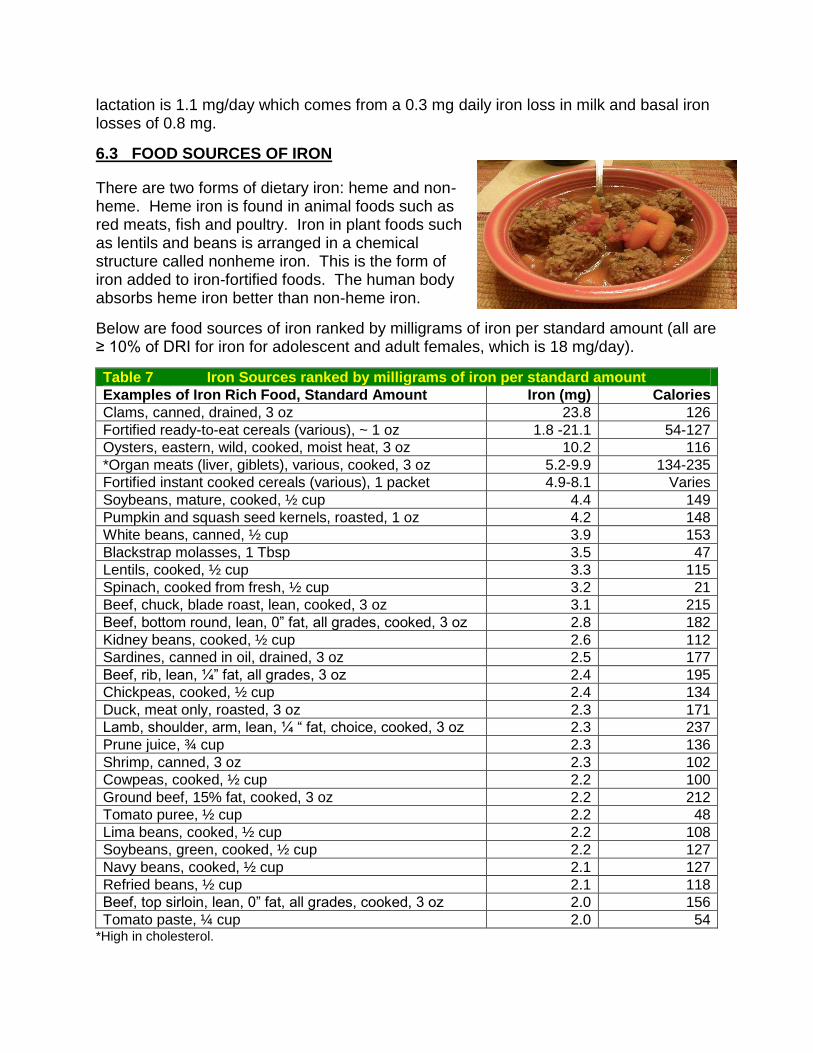
6.3 FOOD SOURCES OF IRON
There are two forms of dietary iron: heme and non-heme. Heme iron is found in animal foods such as red meats, fish and poultry. Iron in plant foods such as lentils and beans is arranged in a chemical structure called nonheme iron. This is the form of iron added to iron-fortified foods. The human body absorbs heme iron better than non-heme iron.
Below are food sources of iron ranked by milligrams of iron per standard amount (all are ≥ 10% of DRI for iron for adolescent and adult females, which is 18 mg/day).
Table 7 Iron Sources ranked by milligrams of iron per standard amount
Examples of Iron Rich Food, Standard Amount Iron (mg) Calories
Clams, canned, drained, 3 oz 23.8 126
Fortified ready-to-eat cereals (various), ~ 1 oz 1.8 -21.1 54-127
Oysters, eastern, wild, cooked, moist heat, 3 oz 10.2 116
*Organ meats (liver, giblets), various, cooked, 3 oz 5.2-9.9 134-235
Fortified instant cooked cereals (various), 1 packet 4.9-8.1 Varies
Soybeans, mature, cooked, ½ cup 4.4 149
Pumpkin and squash seed kernels, roasted, 1 oz 4.2 148
White beans, canned, ½ cup 3.9 153
Blackstrap molasses, 1 Tbsp 3.5 47
Lentils, cooked, ½ cup 3.3 115
Spinach, cooked from fresh, ½ cup 3.2 21
Beef, chuck, blade roast, lean, cooked, 3 oz 3.1 215
Beef, bottom round, lean, 0‖ fat, all grades, cooked, 3 oz 2.8 182
Kidney beans, cooked, ½ cup 2.6 112
Sardines, canned in oil, drained, 3 oz 2.5 177
Beef, rib, lean, ¼‖ fat, all grades, 3 oz 2.4 195
Chickpeas, cooked, ½ cup 2.4 134
Duck, meat only, roasted, 3 oz 2.3 171
Lamb, shoulder, arm, lean, ¼ ― fat, choice, cooked, 3 oz 2.3 237
Prune juice, ¾ cup 2.3 136
Shrimp, canned, 3 oz 2.3 102
Cowpeas, cooked, ½ cup 2.2 100
Ground beef, 15% fat, cooked, 3 oz 2.2 212
Tomato puree, ½ cup 2.2 48
Lima beans, cooked, ½ cup 2.2 108
Soybeans, green, cooked, ½ cup 2.2 127
Navy beans, cooked, ½ cup 2.1 127
Refried beans, ½ cup 2.1 118
Beef, top sirloin, lean, 0‖ fat, all grades, cooked, 3 oz 2.0 156
Tomato paste, ¼ cup 2.0 54 *High in cholesterol.
Source: Nutrient values from Agricultural Research Service (ARS) Nutrient Database for Standard Reference, Release 17. Foods are from ARS single nutrient reports, sorted in descending order by nutrient content in terms of common household measures. Food items and weights in the single nutrient reports are adapted from those in 2002 revision of USDA Home and Garden Bulletin No. 72, Nutritive Value of Foods. Mixed dishes and multiple preparations of the same food item have been omitted from this table.
6.4 FACTORS INFLUENCING IRON ABSORPTION
Iron absorption can be influenced both positively and negatively by additional components consumed with the iron source. This deserves important consideration when counseling on inclusion of iron-rich foods in the diet to prevent iron deficiency anemia. The following sections discuss factors that influence and inhibit iron absorption. This information is summarized in Table 8.
6.4.1 Inhibition of Iron Absorption
Several dietary factors can interfere with the body’s absorption of iron. These include phytates, phenolic compounds and calcium.
PHYTATES Phytates, found in all kinds of grains, seeds, nuts, vegetables and roots (e.g. potatoes) can inhibit iron absorption. Phytates, chemically inositol hexaphosphate salts, are a storage form of phosphates and minerals. In North American and European diets, about 90 percent of phytates originate from cereals. Bran has a high phytate content. Whole wheat flour has a much higher content of phytates than white wheat flour. By contrast, non-phytate-containing dietary fiber components have almost no influence on iron absorption.
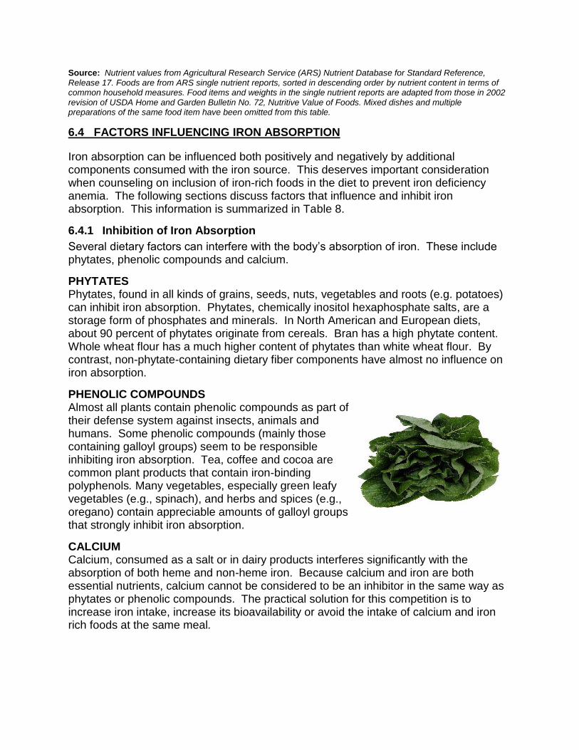
PHENOLIC COMPOUNDS Almost all plants contain phenolic compounds as part of their defense system against insects, animals and humans. Some phenolic compounds (mainly those containing galloyl groups) seem to be responsible inhibiting iron absorption. Tea, coffee and cocoa are common plant products that contain iron-binding polyphenols. Many vegetables, especially green leafy vegetables (e.g., spinach), and herbs and spices (e.g., oregano) contain appreciable amounts of galloyl groups that strongly inhibit iron absorption.
CALCIUM Calcium, consumed as a salt or in dairy products interferes significantly with the absorption of both heme and non-heme iron. Because calcium and iron are both essential nutrients, calcium cannot be considered to be an inhibitor in the same way as phytates or phenolic compounds. The practical solution for this competition is to increase iron intake, increase its bioavailability or avoid the intake of calcium and iron rich foods at the same meal.
Table 8 Factors Influencing Dietary Iron Absorption
Heme iron absorption depends on:
Iron status Amount of heme iron consumed at one time Food preparation methods (time and temperature)
Non-Heme iron absorption depends on:
Current iron status Amount of potentially available non-heme iron (adjustment for
fortification iron and contamination iron) Balance between enhancing and inhibiting factors
Enhancing Factors
Ascorbic acid (e.g., certain fruit juices, fruits, potatoes and certain vegetables)
Presence of heme iron sources such as meat, chicken, fish and other seafood
Presence of fermented vegetables (e.g., sauerkraut) or fermented soy sauces, etc.
Inhibiting Factors
Phytates and other inositol phosphates (e.g., bran products, bread made from high-extraction flour, breakfast cereals, oats, rice [especially unpolished rice], pasta, cocoa, nuts, soya beans and peas)
Iron-binding phenolic compounds (e.g., tea, coffee, cocoa, certain spices, certain vegetables and most red wines)
Calcium (e.g., milk, cheese) Soy proteins
Source: http://www.fao.org/DOCREP/004/Y2809E/y2809e0j.htm
6.4.2 Enhancement of Iron Absorption
Ascorbic acid found naturally in fruits, vegetables and juices and synthetic vitamin C enhance non-heme iron absorption. At least 25 mg of ascorbic acid is needed to enhance iron absorption. More may be needed if the meal contains inhibitors of iron absorption. When establishing vitamin C requirements to prevent vitamin C deficiency (especially scurvy) a requirement for ascorbic acid intake should be considered to assist in iron absorption.
Meat intake promotes iron status in two ways; first by augmenting the absorption of both heme and non-heme iron and second, by supplying a source of the well-absorbed heme iron. Meat, poultry, fish and seafood all promote the absorption of non-heme iron although the mechanism for this has not been determined. Meat enhances the heme iron absorption to about the same extent. Organic acids, such as citric acid, have been found to enhance the absorption of non-heme iron in some studies. This effect is not observed as consistently as is the effect of ascorbic acid. Sauerkraut and other fermented vegetables and even some fermented soy sauces enhance iron absorption although the nature of this enhancement has not yet been determined.
6.5 PREVENTION OF IRON DEFICIENCY ANEMIA
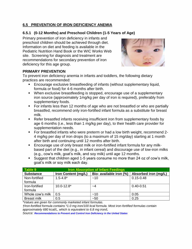
6.5.1 (0-12 Months) and Preschool Children (1-5 Years of Age)
Primary prevention of iron deficiency in infants and preschool children should be achieved through diet. Information on diet and feeding is available in the Pediatric Nutrition Hand Book or the WIC Works Web site. Screening for diagnosis and treatment are recommendations for secondary prevention of iron deficiency for this age group.
PRIMARY PREVENTION To prevent iron deficiency anemia in infants and toddlers, the following dietary practices are recommended:
Encourage exclusive breastfeeding of infants (without supplementary liquid, formula or food) for 4-6 months after birth.
When exclusive breastfeeding is stopped, encourage use of a supplementary iron source (approximately 1mg/kg per day of iron is required), preferably from supplementary foods.
For infants less than 12 months of age who are not breastfed or who are partially breastfed, recommend only iron-fortified infant formula as a substitute for breast milk.
Refer breastfed infants receiving insufficient iron from supplementary foods by age 6 months (i.e., less than 1 mg/kg per day), to their health care provider for supplementation needs.
For breastfed infants who were preterm or had a low birth weight, recommend 2-4 mg/kg per day of iron drops (to a maximum of 15 mg/day) starting at 1 month after birth and continuing until 12 months after birth.
Encourage use of only breast milk or iron-fortified infant formula for any milk-based part of the diet (e.g., in infant cereal) and discourage use of low-iron milks (e.g., cow’s milk, goat’s milk, and soy milk) until age 12 months.
Suggest that children aged 1-5 years consume no more than 24 oz of cow’s milk, goat’s milk or soy milk each day.
Table 9 Iron Absorption of Infant Feedings
Substance Iron Content (mg/L) Bio- available iron (%) Absorbed iron (mg/L)
Non-fortified formula
1.5-4.8* ~10 0.15-0.48
Iron-fortified formula
10.0-12.8* ~4 0.40-0.51
Whole cow’s milk 0.5 ~10 0.05
Breast milk 0.5 ~50 0.25 *Values are given for commonly marketed infant formulas. †Iron-fortified formula contains ³1.0 mg iron/100 kcal formula. Most iron-fortified formulas contain approximately 680 kcal/L, which is equivalent to 6.8 mg iron/L Source: Recommendations to Prevent and Control Iron Deficiency in the United States
RECOMMENDATIONS FOR SOLID FOODS At 6 months of age or when the extrusion reflex disappears, recommend that
infants be introduced to plain, iron-fortified infant cereal. By approximately age 6 months, encourage caregivers to offer vitamin C-rich
foods (e.g., fruits and vegetables) daily to improve iron absorption, preferably with meals.
Suggest introducing plain, pureed meats after age 6 months or when the infant is developmentally ready to consume such food.
SCREENING FOR IRON DEFICIENCY ANEMIA In infant and preschool populations at high risk for iron-deficiency anemia (e.g., children from low-income families, WIC-eligible children), all children should be screened for anemia between ages 9 and 12 months, 15 and 18 months and annually from ages 2 to 5 years.
Additionally, the following children should be screened: Preterm or low-birth weight infants Infants fed a diet of non-iron-fortified infant formula for greater than 2 months Infants introduced to cow’s milk before age 12 months Breastfed infants who do not consume a diet adequate in iron after age 6 months
(i.e., who receive insufficient iron from supplementary foods) Children who consume greater than 24 oz daily of cow’s milk Children with special health care needs (e.g., children who use medications that
interfere with iron absorption and children who have chronic infection, inflammatory disorders, restricted diets, or extensive blood loss from a wound, accident, or surgery)
6.5.2 Pregnant Women
Primary prevention of iron deficiency during pregnancy includes adequate dietary iron intake and iron supplementation. Pregnant women enrolled in WIC should be screened at the earliest opportunity during the pregnancy.
Specific recommendations preventing iron deficiency in pregnant women include: Start oral, low-dose (30 mg/day) iron supplements at the first prenatal visit.
Iron supplements should be taken with water or juice and not milk. Encourage pregnant women to eat iron-rich foods and foods that enhance
iron absorption. Pregnant women whose diets are low in iron
are at additional risk for iron deficiency anemia; guide these women in optimizing their dietary iron intake.
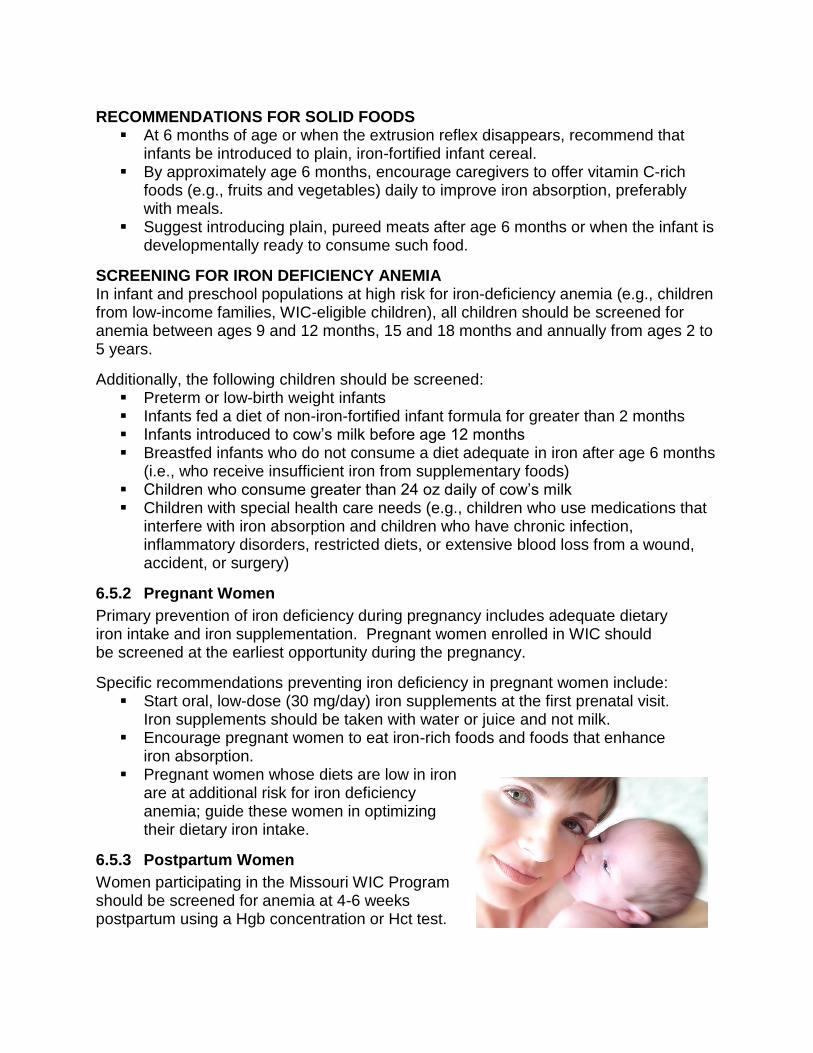
6.5.3 Postpartum Women
Women participating in the Missouri WIC Program should be screened for anemia at 4-6 weeks postpartum using a Hgb concentration or Hct test.
Risk factors for postpartum iron deficiency include: Anemia continued through the third trimester Excessive blood loss during delivery Multifetal birth
Treatment and follow-up for iron-deficiency anemia in postpartum women is the same as for nonpregnant women. If no risk factors for anemia are present, supplemental iron should be stopped at delivery.
6.6 OTHER DIETARY ANEMIAS
Iron deficiency anemia is only one type of anemia. Other dietary anemias include those caused by dietary deficiency of folic acid, vitamin B12, copper or vitamin C and lack of Intrinsic Factor (IF) in the stomach.
6.6.1 Megaloblastic (Pernicious) (Folate Deficiency) Anemia
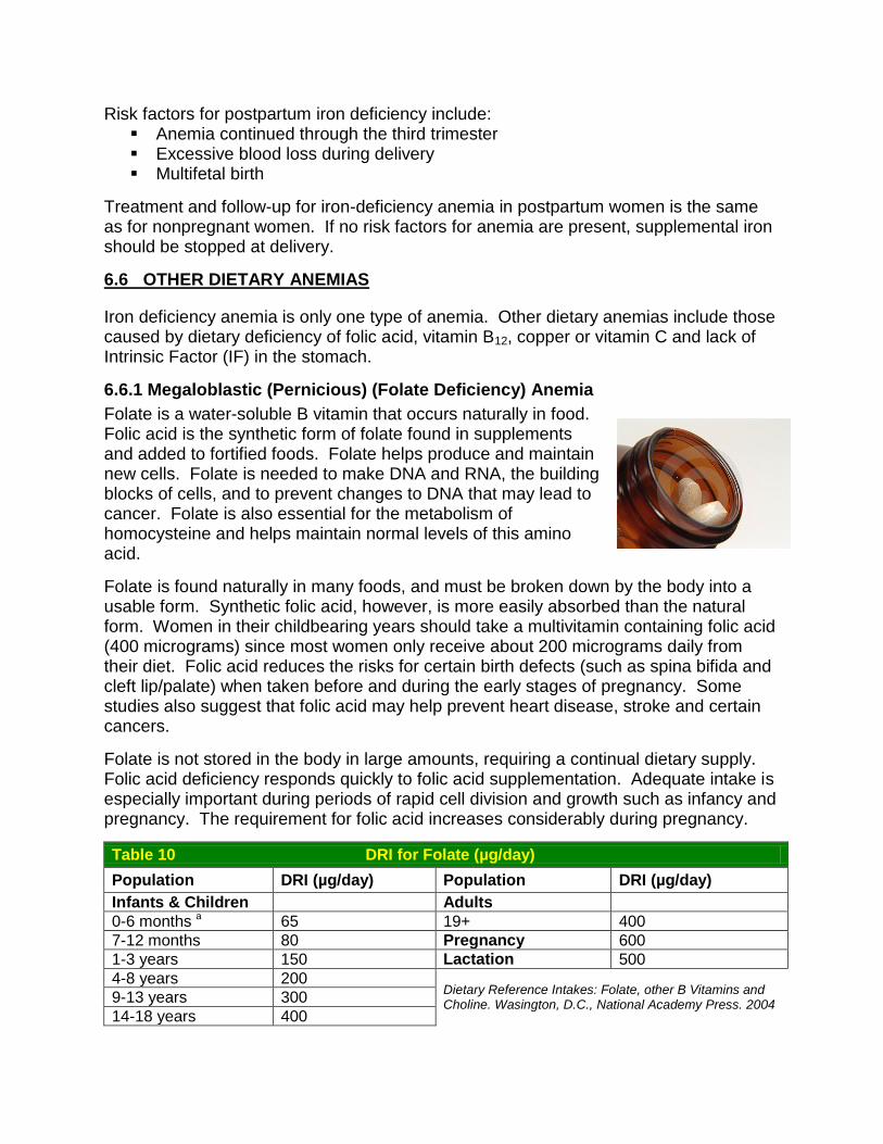
Folate is a water-soluble B vitamin that occurs naturally in food. Folic acid is the synthetic form of folate found in supplements and added to fortified foods. Folate helps produce and maintain new cells. Folate is needed to make DNA and RNA, the building blocks of cells, and to prevent changes to DNA that may lead to cancer. Folate is also essential for the metabolism of homocysteine and helps maintain normal levels of this amino acid.
Folate is found naturally in many foods, and must be broken down by the body into a usable form. Synthetic folic acid, however, is more easily absorbed than the natural form. Women in their childbearing years should take a multivitamin containing folic acid (400 micrograms) since most women only receive about 200 micrograms daily from their diet. Folic acid reduces the risks for certain birth defects (such as spina bifida and cleft lip/palate) when taken before and during the early stages of pregnancy. Some studies also suggest that folic acid may help prevent heart disease, stroke and certain cancers.
Folate is not stored in the body in large amounts, requiring a continual dietary supply. Folic acid deficiency responds quickly to folic acid supplementation. Adequate intake is especially important during periods of rapid cell division and growth such as infancy and pregnancy. The requirement for folic acid increases considerably during pregnancy.
Table 10 DRI for Folate (µg/day)
Population DRI (µg/day) Population DRI (µg/day)
Infants & Children Adults
0-6 months a 65 19+ 400
7-12 months 80 Pregnancy 600
1-3 years 150 Lactation 500
4-8 years 200 Dietary Reference Intakes: Folate, other B Vitamins and Choline. Wasington, D.C., National Academy Press. 2004 9-13 years 300
14-18 years 400
Lack of folate can cause megaloblastic (pernicious) anemia. Folic acid is required for the production of normal red blood cells. Megaloblastic anemia is characterized by very large red blood cells with underdeveloped cell parts. This malformation causes the bone marrow to produce fewer cells, which are oval instead of being round or disc-shaped. Sometimes the cells die before their 120-day life expectancy.
Risk factors for folic acid deficiency include: Poor diet (seen frequently in the poor and elderly, and with inadequate fresh fruit
or vegetable intake) Overcooking food Chronic alcoholism History of malabsorption diseases (celiac disease) Pregnancy Use of certain medications (specifically those that prevent seizures, such as
phenytoin, primidone, and Phenobarbital) that inhibit folic acid absorption
The inability to absorb folic acid may also be inherited. Poor folic acid absorption can lead to megaloblastic anemia, which requires early intensive treatment to prevent long-term problems such as mental retardation. Folic acid deficiency has an incidence rate of 4 out of 100,000 people.
SOURCES OF FOLIC ACID
Table 11 Selected Food Sources of Folate and Folic Acid
Food Micrograms (μg) % DV^
*Breakfast cereals fortified with 100% of the DV, ¾ cup 400 100
Beef liver, cooked, braised, 3 ounces 185 45
Cowpeas (black eye peas), immature, cooked, boiled, ½ cup 105 25
*Breakfast cereals, fortified with 25% of the DV, ¾ cup 100 25
Spinach, frozen, cooked, boiled, ½ cup 100 25
Great Northern beans, boiled, ½ cup 90 20
Asparagus, boiled, 4 spears 85 20
*Rice, white, long-grain, parboiled, enriched, cooked, ½ cup 65 15
Vegetarian baked beans, canned, 1 cup 60 15
Spinach, raw, 1 cup 60 15
Green peas, frozen, boiled, ½ cup 50 15
Broccoli, chopped, frozen, cooked, ½ cup 50 15
*Egg noodles, cooked, enriched, ½ cup 50 15
Broccoli, raw, 2 spears (each 5 inches long) 45 10
Avocado, raw, all varieties, sliced, ½ cup sliced 45 10
Peanuts, all types, dry roasted, 1 ounce 40 10
Lettuce, Romaine, shredded, ½ cup 40 10
Wheat germ, crude, 2 Tablespoons 40 10
Tomato Juice, canned, 6 ounces 35 10
Orange juice, chilled, includes concentrate, ¾ cup 35 10
Turnip greens, frozen, cooked, boiled, ½ cup 30 8
Orange, all commercial varieties, fresh, 1 small 30 8
*Bread, white, 1 slice 25 6
*Bread, whole wheat, 1 slice 25 6
Egg, whole, raw, fresh, 1 large 25 6
Cantaloupe, raw, ¼ medium 25 6
Papaya, raw, ½ cup cubes 25 6
Banana, raw, 1 medium 20 6
Source; Dietary Supplement Fact Sheet: Folate. Items marked with an asterisk (*) are fortified with folic acid as part of the Folate Fortification Program. ^ DV = Daily Value. DVs are reference numbers developed by the Food and Drug Administration (FDA) to help consumers determine if a food contains a lot or a little of a specific nutrient. The DV for folate is 400 micrograms (μg). Most food labels do not list a food’s magnesium content. The percent DV (%DV) listed on the table indicates the percentage of the DV provided in one serving. A food providing 5% of the DV or less is a low source while a food that provides 10-19% of the DV is a good source. A food that provides 20% or more of the DV is high in that nutrient. It is important to remember that foods that provide lower percentages of the DV also contribute to a healthful diet. For foods not listed in this table, please refer to the U.S. Department of Agriculture’s Nutrient Database Web site.
SIGNS AND SYMPTOMS OF FOLATE DEFICIENCY AND MEGALOBLASTIC ANEMIA
Common signs and symptoms of folate deficiency include: Folate deficient women who become pregnant are at greater risk of giving birth to
low birth weight, premature and/or infants with neural tube defects. Slow overall growth rate in children and infants Digestive disorders such as diarrhea, loss of appetite and weight loss Weakness Sore tongue Headaches Heart palpitations Irritability Forgetfulness Behavioral disorders Elevated blood homocysteine levels
The following are the most common symptoms of megaloblastic anemia, however each individual may experience symptoms differently.
Abnormal paleness or lack of color in the skin Decreased appetite Lack of energy or tiring easily (fatigue) Irritability Smooth and tender tongue Diarrhea
Many of these subtle symptoms are general and can also result from a variety of medical conditions other than folate deficiency. It is important to have a physician evaluate these symptoms so that appropriate medical care can be given.
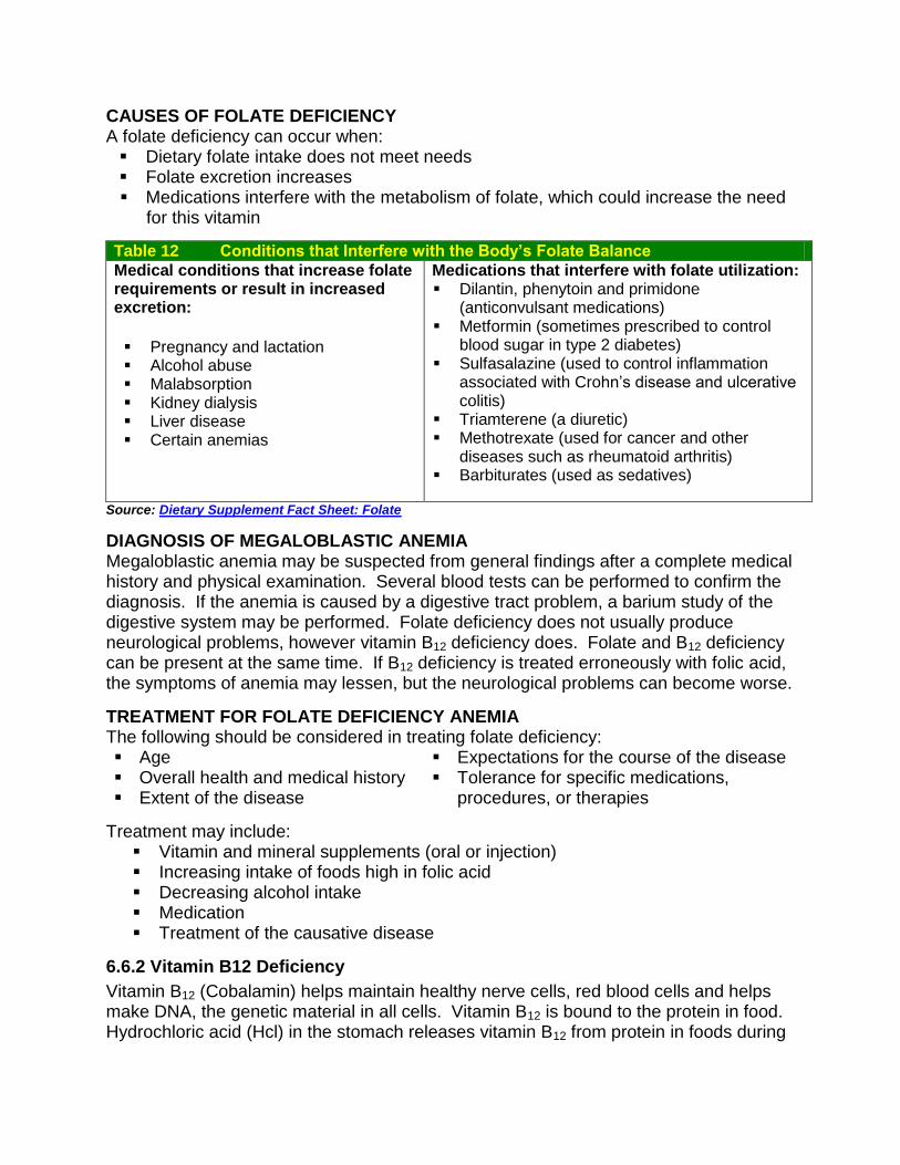
CAUSES OF FOLATE DEFICIENCY A folate deficiency can occur when: Dietary folate intake does not meet needs Folate excretion increases Medications interfere with the metabolism of folate, which could increase the need
for this vitamin
Table 12 Conditions that Interfere with the Body’s Folate Balance
Medical conditions that increase folate requirements or result in increased excretion:
Pregnancy and lactation Alcohol abuse Malabsorption Kidney dialysis Liver disease Certain anemias
Medications that interfere with folate utilization: Dilantin, phenytoin and primidone
(anticonvulsant medications) Metformin (sometimes prescribed to control
blood sugar in type 2 diabetes) Sulfasalazine (used to control inflammation
associated with Crohn’s disease and ulcerative colitis)
Triamterene (a diuretic) Methotrexate (used for cancer and other
diseases such as rheumatoid arthritis) Barbiturates (used as sedatives)
Source: Dietary Supplement Fact Sheet: Folate
DIAGNOSIS OF MEGALOBLASTIC ANEMIA Megaloblastic anemia may be suspected from general findings after a complete medical history and physical examination. Several blood tests can be performed to confirm the diagnosis. If the anemia is caused by a digestive tract problem, a barium study of the digestive system may be performed. Folate deficiency does not usually produce neurological problems, however vitamin B12 deficiency does. Folate and B12 deficiency can be present at the same time. If B12 deficiency is treated erroneously with folic acid, the symptoms of anemia may lessen, but the neurological problems can become worse.
TREATMENT FOR FOLATE DEFICIENCY ANEMIA The following should be considered in treating folate deficiency: Age Overall health and medical history Extent of the disease
Expectations for the course of the disease Tolerance for specific medications,
procedures, or therapies
Treatment may include: Vitamin and mineral supplements (oral or injection) Increasing intake of foods high in folic acid Decreasing alcohol intake Medication Treatment of the causative disease
6.6.2 Vitamin B12 Deficiency
Vitamin B12 (Cobalamin) helps maintain healthy nerve cells, red blood cells and helps make DNA, the genetic material in all cells. Vitamin B12 is bound to the protein in food. Hydrochloric acid (Hcl) in the stomach releases vitamin B12 from protein in foods during
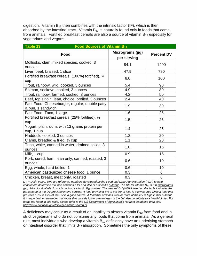
digestion. Vitamin B12 then combines with the intrinsic factor (IF), which is then absorbed by the intestinal tract. Vitamin B12 is naturally found only in foods that come from animals. Fortified breakfast cereals are also a source of vitamin B12 especially for vegetarians and vegans.
Table 13 Food Sources of Vitamin B12
Food Micrograms ( g)
per serving Percent DV
Mollusks, clam, mixed species, cooked, 3 ounces
84.1 1400
Liver, beef, braised, 1 slice 47.9 780
Fortified breakfast cereals, (100%) fortified), ¾ cup
6.0 100
Trout, rainbow, wild, cooked, 3 ounces 5.4 90
Salmon, sockeye, cooked, 3 ounces 4.9 80
Trout, rainbow, farmed, cooked, 3 ounces 4.2 50
Beef, top sirloin, lean, choice, broiled, 3 ounces 2.4 40
Fast Food, Cheeseburger, regular, double patty & bun, 1 sandwich
1.9 30
Fast Food, Taco, 1 large 1.6 25
Fortified breakfast cereals (25% fortified), ¾ cup
1.5 25
Yogurt, plain, skim, with 13 grams protein per cup, 1 cup
1.4 25
Haddock, cooked, 3 ounces 1.2 20
Clams, breaded & fried, ¾ cup 1.1 20
Tuna, white, canned in water, drained solids, 3 ounces
1.0 15
Milk, 1 cup 0.9 15
Pork, cured, ham, lean only, canned, roasted, 3 ounces
0.6 10
Egg, whole, hard boiled, 1 0.6 10
American pasteurized cheese food, 1 ounce 0.3 6
Chicken, breast, meat only, roasted 0.3 6 DV = Daily Value. DVs are reference numbers developed by the Food and Drug Administration (FDA) to help consumers determine if a food contains a lot or a little of a specific nutrient. The DV for vitamin B12 is 6.0 micrograms (μg). Most food labels do not list a food’s vitamin B12 content. The percent DV (%DV) listed on the table indicates the percentage of the DV provided in one serving. A food providing 5% of the DV or less is a low source while a food that provides 10% to 19% of the DV is a good source. A food that provides 20% or more of the DV is high in that nutrient. It is important to remember that foods that provide lower percentages of the DV also contribute to a healthful diet. For foods not listed in this table, please refer to the US Department of Agriculture’s Nutrient Database Web site: http://www.nal.usda.gov/fnic/cgi-bin/nut_search.pl.
A deficiency may occur as a result of an inability to absorb vitamin B12 from food and in strict vegetarians who do not consume any foods that come from animals. As a general rule, most individuals who develop a vitamin B12 deficiency have an underlying stomach or intestinal disorder that limits B12 absorption. Sometimes the only symptoms of these
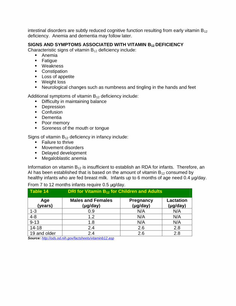
intestinal disorders are subtly reduced cognitive function resulting from early vitamin B12 deficiency. Anemia and dementia may follow later.
SIGNS AND SYMPTOMS ASSOCIATED WITH VITAMIN B12 DEFICIENCY Characteristic signs of vitamin B12 deficiency include:
Anemia Fatigue Weakness Constipation Loss of appetite Weight loss Neurological changes such as numbness and tingling in the hands and feet
Additional symptoms of vitamin B12 deficiency include: Difficulty in maintaining balance Depression Confusion Dementia Poor memory Soreness of the mouth or tongue
Signs of vitamin B12 deficiency in infancy include: Failure to thrive Movement disorders Delayed development Megaloblastic anemia
Information on vitamin B12 is insufficient to establish an RDA for infants. Therefore, an AI has been established that is based on the amount of vitamin B12 consumed by healthy infants who are fed breast milk. Infants up to 6 months of age need 0.4 μg/day.
From 7 to 12 months infants require 0.5 g/day.
Table 14 DRI for Vitamin B12 for Children and Adults
Age (years)
Males and Females (μg/day)
Pregnancy (μg/day)
Lactation (μg/day)
1-3 0.9 N/A N/A
4-8 1.2 N/A N/A
9-13 1.8 N/A N/A
14-18 2.4 2.6 2.8
19 and older 2.4 2.6 2.8 Source: http://ods.od.nih.gov/factsheets/vitaminb12.asp
PREVENTION OF B12 DEFICIENCY Vitamin B12 must bind with intrinsic factor in the stomach before it can be absorbed and used by the body. An absence of intrinsic factor prevents normal absorption of vitamin B12 and results in pernicious anemia. Severe gastric atrophy, a condition that prevents gastric cells from secreting intrinsic factor can result in pernicious anemia.
Most individuals with pernicious anemia need parenteral (deep subcutaneous) injections of vitamin B12 as initial therapy to replenish depleted body stores of vitamin B12. Vitamin B12 body stores can then be maintained through a daily oral B12 supplement. A health care provider will manage the treatment required to maintain vitamin B12 status of individuals with pernicious anemia.
Individuals with gastrointestinal disorders or pernicious anemia may benefit from or require a vitamin B12 supplement. A decrease in stomach acid and thus the ability to absorb B12, increases the risk for B12 deficiency in older adults. Chronic use of medications that decrease vitamin B12 absorption may also result in a need for additional vitamin B12.
Vitamin B12, like other nutrients, is transferred across the placenta during pregnancy. Breastfed infants receive their nutrition, including vitamin B12, through breast milk. Vitamin B12 deficiency in infants is rare but can occur as a result of maternal deficiency. Breastfed infants of women who follow strict vegetarian diets have very limited reserves of vitamin B12 and can develop a vitamin B12 deficiency within months of birth. This is of particular concern because undetected and untreated vitamin B12 deficiency in infants can result in permanent neurological damage. Mothers following a strict vegetarian diet should consult with a pediatrician regarding appropriate use of vitamin B12 supplementation for themselves and their infants and children.
6.6.3 Gastrointestinal Disorders
Individuals with stomach and small intestine disorders may be unable to absorb enough vitamin B12 from food to maintain healthy body stores. Intestinal disorders that may result in malabsorption of vitamin B12 include:
Celiac disease (CD), a genetic disorder. People with CD are intolerant to the wheat protein gluten, which can trigger damage to the small intestine, where most nutrient absorption occurs. Affected individuals experience nutrient malabsorption and must follow a gluten-free diet.
Crohn’s disease, which is an inflammatory bowel disease that affects the small intestine. People with Crohn’s disease often experience diarrhea and nutrient malabsorption.
Surgical procedures in the gastrointestinal tract. Surgery to remove all or part of the stomach often results in the loss of cells that secrete hydrochloric acid and the intrinsic factor. Surgical removal of the distal ileum, a section of the intestine, can also result in the inability to absorb vitamin B12. Anyone who has had either of these surgeries usually requires lifelong vitamin B12 supplementation to prevent deficiency and routine follow up with their physician.
6.6.4 Vegetarian Diets
Table 15 Types of Vegetarianism
Foods Consumed: Meat including fish and poultry
Eggs Dairy Honey
Lacto-Ovo-Vegetarian No Yes Yes Yes
Lacto-Vegetarian No No Yes Yes
Ovo-Vegetarian No Yes No Yes
Vegan No No No No
There has been a surge in popularity of vegetarian diets. The term vegetarianism is subject to a wide range of interpretations as seen above. The American Dietetic Association (ADA) states that ―Vegetarian diets offer a number of nutritional benefits, including lower levels of saturated fat, cholesterol and animal protein as well as higher levels of carbohydrates, fiber, magnesium, potassium, folate and antioxidants such as vitamins C and E and phytochemicals.‖
A vegetarian is someone who does not eat meat, fish, or fowl or products containing these foods. Common reasons for choosing a vegetarian diet include health considerations, concern for the environment, and animal welfare factors. The eating patterns of vegetarians can vary considerably. Individual assessment is important to determine the diet’s nutritional adequacy when someone reports they consume a vegetarian diet.
Strict vegetarians and vegans are at greater risk of developing vitamin B12 deficiency than lacto-ovo-vegetarians and non-vegetarians because natural food sources of B12 are limited to animal food sources. Sources of vitamin B12 for vegetarians include milk products, eggs, and foods that have been fortified with vitamin B12. These include breakfast cereals, soy-based beverages, veggie burgers, and nutritional yeast. Fortified cereals are an important source of vitamin B12 for strict vegetarians and vegans who should discuss the need for vitamin B12 supplements with their physician.
Types of vegetarianism include: Semivegetarian. Semivegetarians are not true vegetarians. They may describe
themselves as vegetarian however as they choose to limit intake of meat in certain ways, such as not eating certain types of meat, such as beef and pork, but eating fish or fowl. A semivegetarian may also choose to eat meat once or twice a week.
Lacto-ovo-vegetarian. A vegetarian who does not eat any meat, fish or fowl or products containing these foods, but does eat eggs, dairy products, nuts, legumes, seeds, grains, fruits and vegetables is considered a lacto-ovo-vegetarian. Without careful planning, this type of diet may be low in foods sources rich in iron and zinc. Food sources of iron compatible with a lacto-ovo-vegetarian diet include whole grain enriched breads, deep green leafy vegetables, legumes, eggs and dried fruit.
Vegan. Vegans do not eat any animal products, including eggs or dairy products. They only eat foods from plant sources such as vegetables, fruits,
grains, legumes, nuts and seeds. A vegan diet may be completely lacking in vitamin B12 since this vitamin is only found in animal products. Sources of vitamin B12 that vegans may use to meet their vitamin B12 requirement include fortified foods (such as some brands of soy milk, breakfast cereals and nutritional yeast) or supplements. Vegan diets should include reliable sources of B12 daily. Vitamin D status may also be a concern for someone following a vegan diet. If there is limited synthesis because of limited sunlight exposure, skin tone, season or sunscreen use, a vitamin D supplement or fortified foods should be considered. Vegan diets may also be low in calcium, iron and zinc. Because vegan diets utilize only protein from plant sources, which may have lower digestibility than protein from animal sources, it is important that vegans consume a variety of protein sources such as grains, nuts, legumes and seeds to get all essential amino acids. Dietary quality of plant proteins varies with the source.
6.7 SICKLE CELL ANEMIA
Sickle cell disease affects millions of people worldwide. It is most common among people whose ancestors come from Africa; Mediterranean countries such as Greece, Turkey, and Italy; the Arabian Peninsula; India; and Spanish-speaking regions of South and Central America and parts of the Caribbean.
Sickle cell disease is the most common inherited blood disorder in the United States, affecting 70,000 to 80,000 Americans. The disease is estimated to occur in 1 in 500 African American births and 1 in 36,000 Hispanic American births. About 2 million Americans have the sickle cell trait, while about 1 in 12 African Americans have the sickle cell trait. In Missouri, the disease occurs in about one in 400 African American births.
Normal red blood cells are disc-shaped, like doughnuts without holes in the center. They move easily through the blood vessels. Sickle cell anemia is a disease in which the body makes sickle-shaped or ―C-shaped‖ red blood cells. Abnormal hemoglobin causes the cells to have a sickle shape. Sickle-shaped cells are stiff and sticky and tend to form clumps and get stuck in the blood vessels. This causes blocked blood vessels, which can cause pain, serious infections and organ damage.
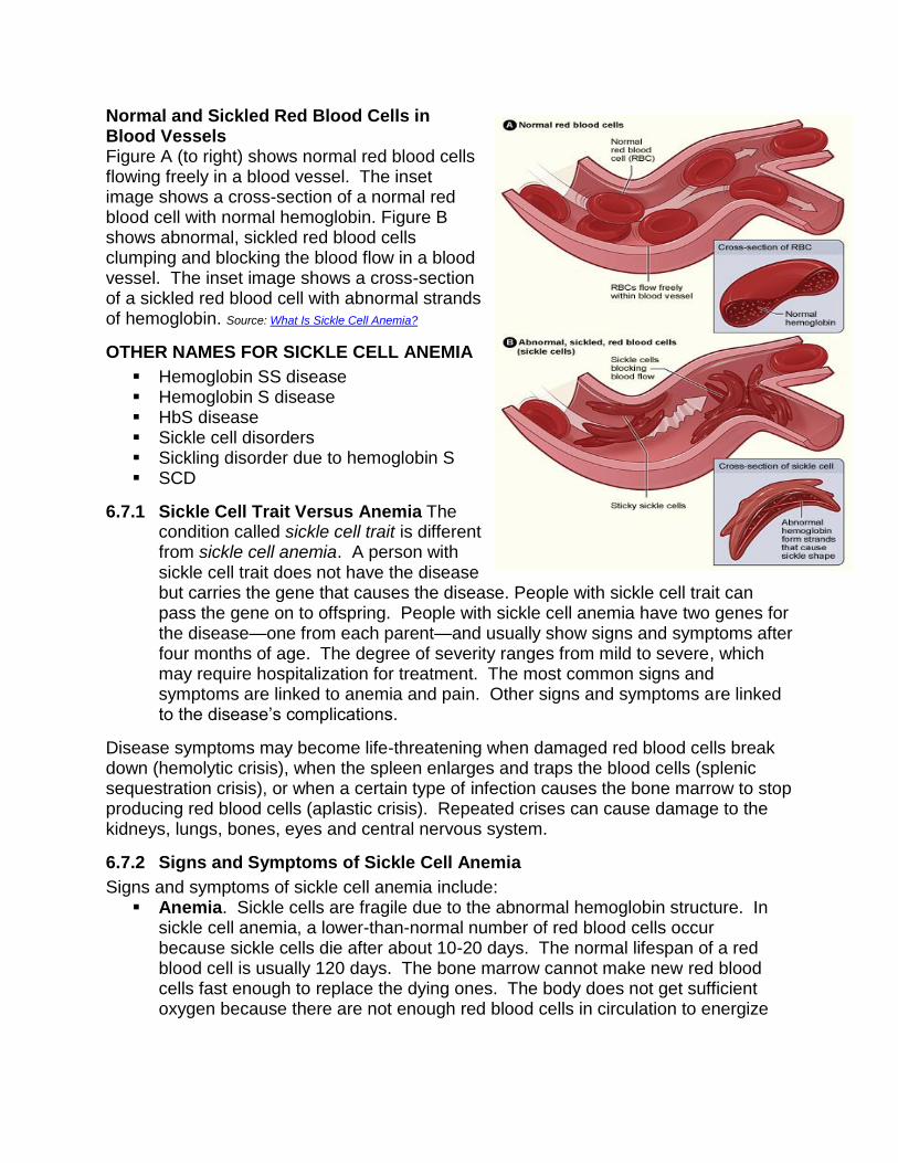
Normal and Sickled Red Blood Cells in Blood Vessels Figure A (to right) shows normal red blood cells flowing freely in a blood vessel. The inset image shows a cross-section of a normal red blood cell with normal hemoglobin. Figure B shows abnormal, sickled red blood cells clumping and blocking the blood flow in a blood vessel. The inset image shows a cross-section of a sickled red blood cell with abnormal strands of hemoglobin. Source: What Is Sickle Cell Anemia?
OTHER NAMES FOR SICKLE CELL ANEMIA
Hemoglobin SS disease Hemoglobin S disease HbS disease Sickle cell disorders Sickling disorder due to hemoglobin S SCD
6.7.1 Sickle Cell Trait Versus Anemia The condition called sickle cell trait is different from sickle cell anemia. A person with sickle cell trait does not have the disease but carries the gene that causes the disease. People with sickle cell trait can pass the gene on to offspring. People with sickle cell anemia have two genes for the disease—one from each parent—and usually show signs and symptoms after four months of age. The degree of severity ranges from mild to severe, which may require hospitalization for treatment. The most common signs and symptoms are linked to anemia and pain. Other signs and symptoms are linked to the disease’s complications.
Disease symptoms may become life-threatening when damaged red blood cells break down (hemolytic crisis), when the spleen enlarges and traps the blood cells (splenic sequestration crisis), or when a certain type of infection causes the bone marrow to stop producing red blood cells (aplastic crisis). Repeated crises can cause damage to the kidneys, lungs, bones, eyes and central nervous system.
6.7.2 Signs and Symptoms of Sickle Cell Anemia
Signs and symptoms of sickle cell anemia include: Anemia. Sickle cells are fragile due to the abnormal hemoglobin structure. In
sickle cell anemia, a lower-than-normal number of red blood cells occur because sickle cells die after about 10-20 days. The normal lifespan of a red blood cell is usually 120 days. The bone marrow cannot make new red blood cells fast enough to replace the dying ones. The body does not get sufficient oxygen because there are not enough red blood cells in circulation to energize

the body. Signs of anemia include shortness of breath, dizziness, headaches, coldness in hands and feet, pale skin and chest pain.
Episodes of pain. Periodic episodes of sudden pain, called ―sickle cell crisis‖, are a major symptom of sickle cell anemia. Pain develops when sickled cells block blood flow through tiny blood vessels to the chest, abdomen and joints. Pain can also occur in the bones and can cause organ damage. The pain may vary in intensity and can last for a few hours to a few weeks. Some people experience only a few episodes of pain, while others experience a dozen or more crises a year. A severe crisis may need hospitalization so that painkillers can be injected intravenously.
Hand-and-foot syndrome. Swollen hands and feet are often the first signs of sickle cell anemia in babies. The swelling is caused by sickled red blood cells blocking blood flow out of the hands and feet.
Jaundice. Jaundice is a yellowing of the skin and eyes that occurs because of liver damage or dysfunction. Occasionally, people who have sickle cell anemia have some degree of jaundice because the liver, which filters harmful substances from the blood, is overwhelmed by the rapid breakdown of red blood cells. In people with dark skin, jaundice is visible mostly as yellowing of the whites of their eyes.
Frequent infections. Sickled cells can damage the spleen, which helps fight infections. Physicians commonly give infants and children with sickle cell anemia antibiotics to prevent potentially life-threatening infections, such as pneumonia.
Stunted growth. Red blood cells provide the body with oxygen and nutrients for growth. A shortage of healthy red blood cells slows growth in infants and children and delays puberty in teenagers.
Vision problems. Some people with sickle cell anemia experience vision problems. Blood vessels that supply the eyes may become plugged with sickle cells, which may result in damage to the retina.
6.7.3 Treatment for Sickle Cell Anemia
Effective treatments exist for the symptoms and complications of sickle cell anemia, but there is no cure. People with sickle cell anemia need regular medical care. Some doctors and clinics specialize in treating people with the disease.
The goals of treating sickle cell anemia are to relieve pain, prevent infections and control complications as they occur. Medications, such as pain relievers are prescribed to manage complications. Hydroxyurea is another medication that has shown positive effects in reducing sickle cell crises. Blood transfusions are commonly used to treat worsening anemia and sickle cell complications, such as enlargement of the spleen. Some, but not all, patients need transfusions to prevent life-threatening events such as stroke or pneumonia. Many patients are prescribed folic acid (folate) to help prevent some of the complications of sickle cell anemia.
Additional treatment revolves around specific treatment for complications.

PREVENTING INFECTIONS Infection is a major complication of sickle cell anemia. In fact, pneumonia is the leading cause of death in children with the disease. Other infections common in people with the disease include meningitis, influenza and hepatitis.
PREVENTING EYE DAMAGE Children may develop damage to the blood vessels in the back of their eyes. Parents should ask the child’s doctor about regular checkups with an eye doctor who specializes in diseases of the retina.
NEW TREATMENTS Research on sickle cell anemia is looking at new medicines, bone marrow transplant and gene therapy. The hope is that these studies will provide new treatments and find a cure for sickle cell anemia. Researchers are also looking for a way to predict the severity of the disease.
BONE MARROW TRANSPLANT Bone marrow is responsible for making new red blood cells to replace old ones. Bone marrow transplant can be a very effective treatment for sickle cell anemia, but there are risks which make it unsuitable for certain patients. Some researchers believe that bone marrow transplantation may offer a cure in a small percentage of cases. Bone marrow used for a transplant must come from a closely matched donor, usually a close family member who does not have the disease.
GENE THERAPY Researchers are investigating if a normal gene can be implanted in the bone marrow of a person with sickle cell anemia, and thus cause the body to produce normal red blood cells. They are also studying the possibility of treatment to ―turn off‖ the sickle cell gene or ―turn on‖ a gene that makes red blood cells behave normally.
NEW MEDICINES Butyric acid. This is a food additive that may increase normal hemoglobin in the
blood. Clotrimazole. This is used to treat fungus infections. This medicine helps
prevent the loss of water from a red blood cell and can keep the cell from turning into a sickle cell.
Nitric oxide. This may make sickle cells less sticky and keep blood vessels open. People with sickle cell anemia have low levels of nitric acid in their blood.
Decitadine. This medicine increases hemoglobin F levels (this type of hemoglobin carries more oxygen).
6.8 ANEMIA COUNSELING TIPS
The following is a list of recommendations for counseling when a WIC participant is screened as having a low hgb or hct.
Identify the probable cause of the low hematological values. Review the participant’s dietary intake and food habits. Take a thorough health history and information on the participant’s socioeconomic background.
Explain the consequences of iron-deficiency anemia to the participant.
Recommend dietary changes based on the probable cause of the low hemoglobin. These recommendations should be individualized to meet the participant’s needs.
Help the participant set a food behavior objective, which will help improve the hemoglobin level at future screenings.
Give the participant a WIC pamphlet on anemia. Review the pamphlet with the participant and highlight the most important points.
Encourage the participant to read the pamphlet thoroughly at home and bring back any questions he/she might have.
Always provide the participant positive reinforcement when appropriate, especially when the participant is making progress in meeting his/her food behavior objective.
Document necessary referrals according to the high-risk care plan.
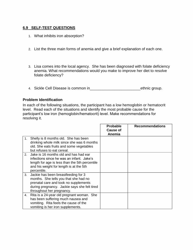
6.9 SELF-TEST QUESTIONS
1. What inhibits iron absorption?
2. List the three main forms of anemia and give a brief explanation of each one.
3. Lisa comes into the local agency. She has been diagnosed with folate deficiency anemia. What recommendations would you make to improve her diet to resolve folate deficiency?
4. Sickle Cell Disease is common in________________________ethnic group.
Problem Identification
In each of the following situations, the participant has a low hemoglobin or hematocrit level. Read each of the situations and identify the most probable cause for the participant’s low iron (hemoglobin/hematocrit) level. Make recommendations for resolving it.
Probable Cause of Anemia
Recommendations
1. Shelly is 8 months old. She has been drinking whole milk since she was 6 months old. She eats fruits and some vegetables but refuses to eat cereal.
2. Jake is 16 months old and has had ear infections since he was an infant. Jake’s length for age is less than the 5th percentile and his weight for length is at the 5th percentile.
3. Jackie has been breastfeeding for 3 months. She tells you that she had no prenatal care and took no supplements during pregnancy. Jackie says she felt tired throughout her pregnancy.
4. Rita is a 24-year old pregnant woman. She has been suffering much nausea and vomiting. Rita feels the cause of the vomiting is her iron supplements.
6.10 CASE STUDY
Lactating Woman
Bonnie is a WIC participant and is one week postpartum. She is breastfeeding her baby, and has hemoglobin of 9.0mg/dl. Bonnie’s diet is as follows:
Breakfast Snack Lunch Snack Dinner Snack
½ c. WIC cereal
½ c. peaches
Vegetable salad (broccoli, greens, zucchini, onion, cucumber)
1 apple 2 oz. chicken 12 oz. milk
12 oz. milk
1 oz. cheese 6 crackers ½ c. rice 1 slice whole wheat toast
1 banana 1 slice whole wheat bread
lettuce salad w/dressing
1 t. marg.
1 slice whole wheat toast
1 t. margarine 12 oz. milk
1 t. margarine
12 oz. milk
A. Bonnie may exhibit the following symptoms because of low hemoglobin
a.
b.
c.
d.
B. What is the most probable cause of Bonnie’s low hemoglobin?
C. Give specific examples (based on Bonnie’s diet) of how Bonnie could increase the amount of iron absorbed from her diet.
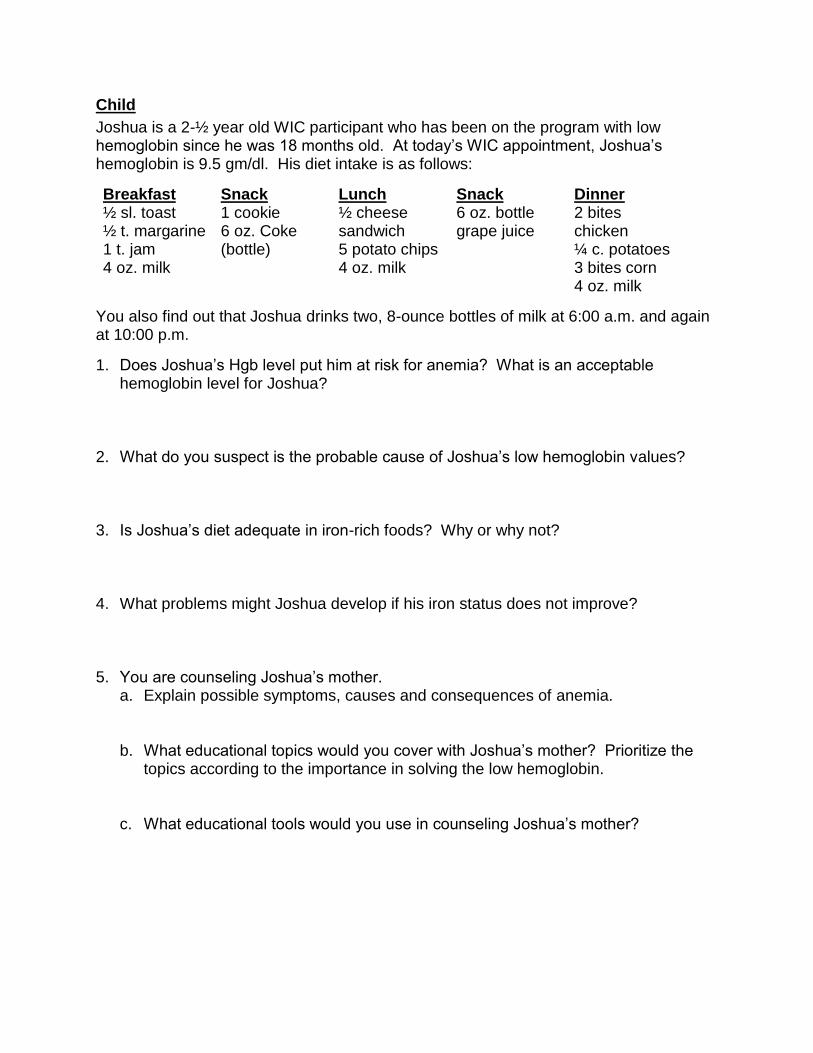
Child
Joshua is a 2-½ year old WIC participant who has been on the program with low hemoglobin since he was 18 months old. At today’s WIC appointment, Joshua’s hemoglobin is 9.5 gm/dl. His diet intake is as follows:
Breakfast ½ sl. toast ½ t. margarine 1 t. jam 4 oz. milk
Snack 1 cookie 6 oz. Coke (bottle)
Lunch ½ cheese sandwich 5 potato chips 4 oz. milk
Snack 6 oz. bottle grape juice
Dinner 2 bites chicken ¼ c. potatoes 3 bites corn 4 oz. milk
You also find out that Joshua drinks two, 8-ounce bottles of milk at 6:00 a.m. and again at 10:00 p.m.
1. Does Joshua’s Hgb level put him at risk for anemia? What is an acceptable hemoglobin level for Joshua?
2. What do you suspect is the probable cause of Joshua’s low hemoglobin values? 3. Is Joshua’s diet adequate in iron-rich foods? Why or why not? 4. What problems might Joshua develop if his iron status does not improve? 5. You are counseling Joshua’s mother.
a. Explain possible symptoms, causes and consequences of anemia.
b. What educational topics would you cover with Joshua’s mother? Prioritize the topics according to the importance in solving the low hemoglobin.
c. What educational tools would you use in counseling Joshua’s mother?
6.11 REFERENCES
http://www.mayoclinic.com/health/anemia/DS00321/DSECTION=3). http://www.emedicine.com/med/topic1188.htm#target6 Recommendations to Prevent and Control Iron Deficiency in the United States MMWR 47(RR-3);1-36 1998 http://wonder.cdc.gov/wonder/prevguid/m0051880/m0051880.asp Source: Prevention of Iron Deficiency in Infants and Toddlers (American Family Physician October 1, 2002, http://www.aafp.org/afp/20021001/1217.html http://www.fao.org/DOCREP/004/Y2809E/y2809e0j.htm Report of a joint FAO/WHO expert consultation, Bangkok, Thailand FAO & WHO Series title: Training materials for agricultural planning - Non-series title 2002 Y2809/ Chapter 13 http://www.fao.org/DOCREP/004/Y2809E/y2809e0j.htm Factors influencing dietary iron absorption http://www.fao.org/DOCREP/004/Y2809E/y2809e0j.htm http://www.health.gov/dietaryguidelines/dga2005/document/html/appendixB.htm Dietary Supplement Fact Sheet: Folate http://www.nal.usda.gov/fnic/foodcomp/search/index.html http://ods.od.nih.gov/factsheets/vitaminb12.asp MyPyramid.gov - United States Department of Agriculture - Home http://ghr.nlm.nih.gov/condition=sicklecelldisease What Is Sickle Cell Anemia?