Iron-Deficiency Anemia - iacld.irIron-Deficiency Anemia A Global Health Problem Iron deficiency...
12
The new england journal of medicine n engl j med 372;19 nejm.org May 7, 2015 1832 Review Article I ron deficiency and iron-deficiency anemia are global health problems and common medical conditions seen in everyday clinical practice. Although the prevalence of iron-deficiency anemia has recently declined some- what, iron deficiency continues to be the top-ranking cause of anemia worldwide, and iron-deficiency anemia has a substantial effect on the lives of young children and premenopausal women in both low-income and developed countries. 1 The diagnosis and treatment of this condition could clearly be improved. Iron is crucial to biologic functions, including respiration, energy production, DNA synthesis, and cell proliferation. 2 The human body has evolved to conserve iron in several ways, including the recycling of iron after the breakdown of red cells and the retention of iron in the absence of an excretion mechanism. How- ever, since excess levels of iron can be toxic, its absorption is limited to 1 to 2 mg daily, and most of the iron needed daily (about 25 mg per day) is provided through recycling by macrophages that phagocytose senescent erythrocytes. The latter two mechanisms are controlled by the hormone hepcidin, which maintains total-body iron within normal ranges, avoiding both iron deficiency and excess. Iron deficiency refers to the reduction of iron stores that precedes overt iron- deficiency anemia or persists without progression. Iron-deficiency anemia is a more severe condition in which low levels of iron are associated with anemia and the presence of microcytic hypochromic red cells. Iron-restricted erythropoiesis indicates that the delivery of iron to erythroid precursors is impaired, no matter how replete the stores. 3,4 Stores may be normal or even increased because of iron sequestration in cases of anemia of chronic in- flammation, which is observed in patients with autoimmune disorders, cancer, infections, and chronic kidney diseases. 3,4 The presence of both iron deficiency and anemia of chronic disorders is common and may be seen in elderly patients 5 and patients with chronic kidney disease. 6 However, a substantial fraction of the ane- mia that is typical in elderly patients occurs in the absence of iron deficiency or elevated hepcidin levels. 7 Functional iron deficiency is a state of iron-poor erythropoiesis 8 in which there is insufficient mobilization of iron from stores in the presence of increased de- mands, as is observed after treatment with erythropoiesis-stimulating agents. 9 (See the Glossary for definitions of terms related to iron-deficiency anemia.) This review reconsiders iron deficiency and its anemia in light of advances in the understanding of systemic iron homeostasis and examines causes, patho- physiological features, and treatment options in adults. Readers are referred else- where for information on the presentation, symptoms, and diagnosis of iron-defi- ciency anemia through laboratory tests and on issues that are specific to children or pregnancy. 10-13 From Vita Salute University and San Raf- faele Scientific Institute, Milan. Address reprint requests to Dr. Camaschella at Vita Salute University, San Raffaele Sci- entific Institute, Via Olgettina, 58, 20132 Milan, Italy, or at [email protected]. N Engl J Med 2015;372:1832-43. DOI: 10.1056/NEJMra1401038 Copyright © 2015 Massachusetts Medical Society. Dan L. Longo, M.D., Editor Iron-Deficiency Anemia Clara Camaschella, M.D. The New England Journal of Medicine Downloaded from nejm.org on May 15, 2015. For personal use only. No other uses without permission. Copyright © 2015 Massachusetts Medical Society. All rights reserved.
Transcript of Iron-Deficiency Anemia - iacld.irIron-Deficiency Anemia A Global Health Problem Iron deficiency...
T h e n e w e ngl a nd j o u r na l o f m e dic i n e
n engl j med 372;19 nejm.org May 7, 20151832
Review Article
Iron deficiency and iron-deficiency anemia are global health problems and common medical conditions seen in everyday clinical practice. Although the prevalence of iron-deficiency anemia has recently declined some-
what, iron deficiency continues to be the top-ranking cause of anemia worldwide, and iron-deficiency anemia has a substantial effect on the lives of young children and premenopausal women in both low-income and developed countries.1 The diagnosis and treatment of this condition could clearly be improved.
Iron is crucial to biologic functions, including respiration, energy production, DNA synthesis, and cell proliferation.2 The human body has evolved to conserve iron in several ways, including the recycling of iron after the breakdown of red cells and the retention of iron in the absence of an excretion mechanism. How-ever, since excess levels of iron can be toxic, its absorption is limited to 1 to 2 mg daily, and most of the iron needed daily (about 25 mg per day) is provided through recycling by macrophages that phagocytose senescent erythrocytes. The latter two mechanisms are controlled by the hormone hepcidin, which maintains total-body iron within normal ranges, avoiding both iron deficiency and excess.
Iron deficiency refers to the reduction of iron stores that precedes overt iron-deficiency anemia or persists without progression. Iron-deficiency anemia is a more severe condition in which low levels of iron are associated with anemia and the presence of microcytic hypochromic red cells.
Iron-restricted erythropoiesis indicates that the delivery of iron to erythroid precursors is impaired, no matter how replete the stores.3,4 Stores may be normal or even increased because of iron sequestration in cases of anemia of chronic in-flammation, which is observed in patients with autoimmune disorders, cancer, infections, and chronic kidney diseases.3,4 The presence of both iron deficiency and anemia of chronic disorders is common and may be seen in elderly patients5 and patients with chronic kidney disease.6 However, a substantial fraction of the ane-mia that is typical in elderly patients occurs in the absence of iron deficiency or elevated hepcidin levels.7
Functional iron deficiency is a state of iron-poor erythropoiesis8 in which there is insufficient mobilization of iron from stores in the presence of increased de-mands, as is observed after treatment with erythropoiesis-stimulating agents.9 (See the Glossary for definitions of terms related to iron-deficiency anemia.)
This review reconsiders iron deficiency and its anemia in light of advances in the understanding of systemic iron homeostasis and examines causes, patho-physiological features, and treatment options in adults. Readers are referred else-where for information on the presentation, symptoms, and diagnosis of iron-defi-ciency anemia through laboratory tests and on issues that are specific to children or pregnancy.10-13
From Vita Salute University and San Raf-faele Scientific Institute, Milan. Address reprint requests to Dr. Camaschella at Vita Salute University, San Raffaele Sci-entific Institute, Via Olgettina, 58, 20132 Milan, Italy, or at camaschella . clara@ hsr . it.
N Engl J Med 2015;372:1832-43.DOI: 10.1056/NEJMra1401038Copyright © 2015 Massachusetts Medical Society.
Dan L. Longo, M.D., Editor
Iron-Deficiency AnemiaClara Camaschella, M.D.
The New England Journal of Medicine Downloaded from nejm.org on May 15, 2015. For personal use only. No other uses without permission.
Copyright © 2015 Massachusetts Medical Society. All rights reserved.
n engl j med 372;19 nejm.org May 7, 2015 1833
Iron-Deficiency Anemia
A Gl ob a l He a lth Problem
Iron deficiency affects more than 2 billion people worldwide,1 and iron-deficiency anemia remains the top cause of anemia, as confirmed by the analysis of a large number of reports on the burden of disease in 187 countries between 1990 and 201014 and by a survey on the burden of ane-mia in persons at risk, such as preschool chil-dren and young women.15 Prevention programs have decreased rates of iron-deficiency anemia globally; the prevalence is now highest in Cen-tral and West Africa and South Asia.14,15 The esti-mated prevalence of iron deficiency worldwide is twice as high as that of iron-deficiency anemia.
The reported prevalence of iron deficiency in the absence of dietary fortification is approxi-mately 40% in preschool children, 30% in men-struating girls and women, and 38% in pregnant women.14-16 These rates reflect the increased physiological need for dietary iron during spe-cific life stages and according to sex. The growth spurt of adolescence is another critical period. For patients in any of these categories, pathologic causes of iron-deficiency anemia are often absent and extensive diagnostic workups are not advised. However, as discussed below, when the response to treatment is unsatisfacto-ry, multiple causes should be considered, even in patients in these high-risk groups.
In developing countries, iron deficiency and iron-deficiency anemia typically result from in-sufficient dietary intake, loss of blood due to intestinal worm colonization, or both. In high-income countries, certain eating habits (e.g., a
vegetarian diet or no intake of red meat) and pathologic conditions (e.g., chronic blood loss or malabsorption) are the most common causes. Paradoxically, it appears to be more difficult to reduce the prevalence of iron-deficiency anemia in high-income countries than in lower-income countries. One reason for this seeming paradox is the high rate of iron deficiency in aging popu-lations.14
Modific ations of Iron Homeos ta sis in Iron Deficienc y
The mechanisms of iron acquisition are tightly regulated by hepcidin-based homeostatic con-trols.2 Hepcidin is a peptide hormone that is synthesized primarily in the liver. It functions as an acute-phase reactant that adjusts fluctuations in plasma iron levels caused by absorptive entero-cytes and macrophages in the spleen by binding to and inducing the degradation of ferroportin, which exports iron from cells.17 Hepcidin expres-sion increases in response to high circulating and tissue levels of iron and in persons with systemic inflammation or infection. Its produc-tion is inhibited by the expansion of erythropoie-sis, iron deficiency, and tissue hypoxia in response to signals originating in the bone marrow, the liver, and probably muscle tissue and adipo-cytes.2,18 Increases in hepcidin levels that are induced by inflammatory cytokines, especially interleukin-6, explain the iron sequestration and reduced supply of erythropoietic iron that occurs in the anemia of chronic disease.
In the general population, hepcidin levels are
Anemia of chronic disorders or anemia of inflammation: Multifactorial anemia associated with increased cytokine pro-duction, up-regulation of hepcidin, and abnormal iron homeostasis.
Functional iron deficiency: Insufficient mobilization of erythroid iron in the presence of increased requests, as occurs after treatment with erythropoiesis-stimulating agents.
Iron deficiency: Depressed levels of total body iron, especially iron stores, with preservation of levels of erythroid iron.
Iron-deficiency anemia: Depressed levels of total body iron in the presence of anemia.
Iron-restricted erythropoiesis: A reduced supply of iron for the purpose of erythropoiesis, regardless of the level of iron stores, which are usually replete.
Iron-refractory iron-deficiency anemia (IRIDA): Iron-deficiency anemia that is unresponsive to oral iron treatment, in most cases referring to the genetic disease caused by a mutation in TMPRSS6, the gene encoding transmembrane protease, serine 6, also known as matriptase-2.
Glossary
The New England Journal of Medicine Downloaded from nejm.org on May 15, 2015. For personal use only. No other uses without permission.
Copyright © 2015 Massachusetts Medical Society. All rights reserved.

n engl j med 372;19 nejm.org May 7, 20151834
T h e n e w e ngl a nd j o u r na l o f m e dic i n e
low in girls and young women and higher — similar to levels in men — in postmenopausal women; fluctuations in hepcidin levels have a
strong direct correlation with serum levels of ferritin.19,20 In iron deficiency, the transcription of hepcidin is suppressed. This adaptive mecha-
Figure 1. The Iron Cycle — Mechanisms of Adaptation to Iron Deficiency.
The mechanisms of adaptation to iron deficiency are centered on the suppression of the hepatic hormone hepcidin and the tissue hypoxia that develops consequent to anemia. The production of erythropoietin (EPO) by the kidney increases in response to enhanced levels of hypoxia-inducible factor 2α (HIF-2α). As a consequence of the stimula-tion of erythropoietin, erythropoiesis is increased and hypochromic microcytic red cells are produced owing to the low availability of iron. Senescent red cells are destroyed by macrophages, and their iron is recycled. The increase in erythropoiesis suppresses the production of hepcidin. In mice, this function is mediated by erythroferrone (ERFE), which is secreted by the erythroblasts21 to maintain adequate iron absorption and efficiency in erythropoiesis. HIF-2α increases the expression of the duodenal divalent metal transporter 1 (DMT1)22 on the apical surface of enterocytes to increase the transfer of dietary iron from the lumen to enterocytes. Hepcidin levels are depressed in response to a reduction in the physiologic signals that maintain its production (e.g., increases in levels of iron-bound transferrin and in the iron content of the liver),2,18 to the increased activity of the inhibitor transmembrane protease, serine 6 (TMPRSS6),23 to the reduction in levels of the activator bone morphogenetic protein 6 (BMP6), and to increased in-hibition from erythropoietin-stimulated erythropoiesis. Ferroportin (FPN), which is no longer being degraded because of the low levels of hepcidin, exports the available iron across the enterocyte basal membrane and from macrophage stores17 to the circulation. Once stores are exhausted, levels of circulating iron decrease, even if absorption from the lumen is increased. Reduced levels of iron in the liver trigger increases in the synthesis of the iron carrier transferrin (referred to as apotransferrin when not bound to iron), further decreasing levels of iron-bound transferrin, the ligand of the transferrin receptor. Consequently, the uptake of iron from transferrin receptors by all cells and organs (e.g., skeletal muscles and the heart) is reduced.
Hepatocyte
Enterocyte
FPN
FPN
DMT1
Heart
Skeletalmuscle
↓Fe↓Fe
+
↑HIF-2α
↑HIF-2α
EPO
↓Fe
↓Fe
↑Fe
Fe
Fe
↑Fe
↑TMPRSS6
ERFE?
Erythropoiesis
Erythroblasts
Spleenmacrophage
Kidney
Red cells
BMP6BMP6
Fe
Hepcidin
ERFE?
HepcidinHepcidin
TMPRSS6
↓HepcidinHepcidinHepcidinHepcidin
↓Fe
↓Fe
↓FeApotransferrin
Fe
↓Fe
ApotransferrinApotransferrin
Allorgans
↓organsorgans
↓FeFe ↓↓↓↓↓↓↓↓
The New England Journal of Medicine Downloaded from nejm.org on May 15, 2015. For personal use only. No other uses without permission.
Copyright © 2015 Massachusetts Medical Society. All rights reserved.
n engl j med 372;19 nejm.org May 7, 2015 1835
Iron-Deficiency Anemia
nism facilitates the absorption of iron (Fig. 1) and the release of iron from body stores. Intes-tinal iron uptake from the gut lumen through divalent metal transporter 1 (DMT1) is increased by the activation of hypoxia-inducible factor 2α.22 The degree of store repletion determines the rapidity with which iron deficiency develops in cases of blood loss or a drastic reduction in iron absorption. Hepatocytes appear to be a long-term reservoir for iron and release it more slowly than macrophages.
C auses of Iron-Deficienc y A nemi a
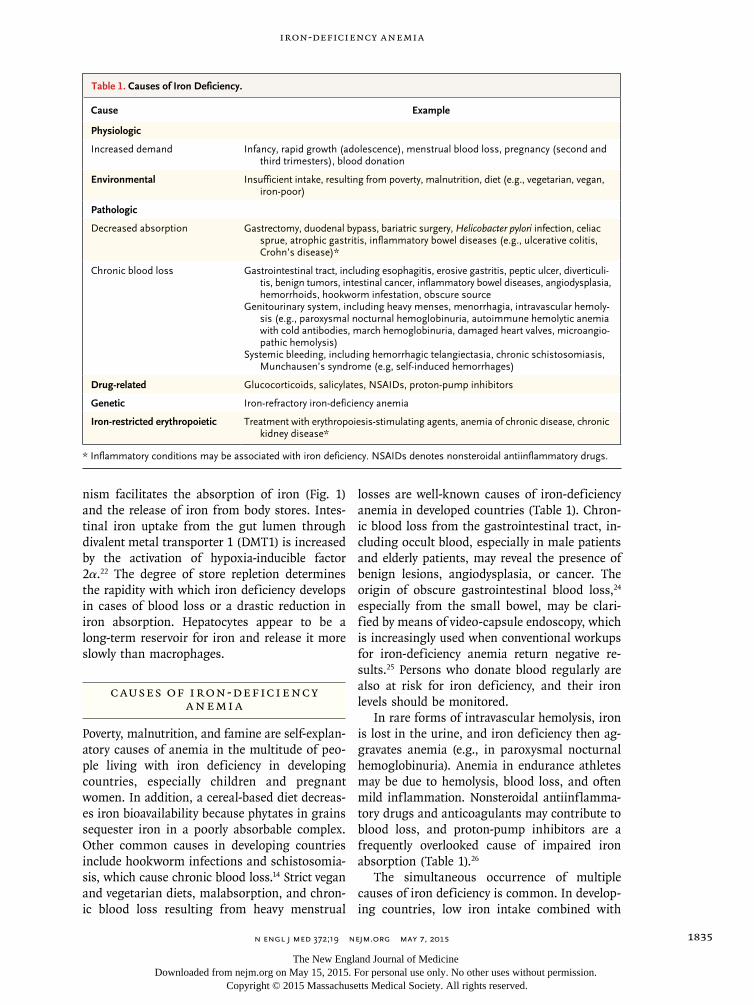
Poverty, malnutrition, and famine are self-explan-atory causes of anemia in the multitude of peo-ple living with iron deficiency in developing countries, especially children and pregnant women. In addition, a cereal-based diet decreas-es iron bioavailability because phytates in grains sequester iron in a poorly absorbable complex. Other common causes in developing countries include hookworm infections and schistosomia-sis, which cause chronic blood loss.14 Strict vegan and vegetarian diets, malabsorption, and chron-ic blood loss resulting from heavy menstrual
losses are well-known causes of iron-deficiency anemia in developed countries (Table 1). Chron-ic blood loss from the gastrointestinal tract, in-cluding occult blood, especially in male patients and elderly patients, may reveal the presence of benign lesions, angiodysplasia, or cancer. The origin of obscure gastrointestinal blood loss,24 especially from the small bowel, may be clari-fied by means of video-capsule endoscopy, which is increasingly used when conventional workups for iron-deficiency anemia return negative re-sults.25 Persons who donate blood regularly are also at risk for iron deficiency, and their iron levels should be monitored.
In rare forms of intravascular hemolysis, iron is lost in the urine, and iron deficiency then ag-gravates anemia (e.g., in paroxysmal nocturnal hemoglobinuria). Anemia in endurance athletes may be due to hemolysis, blood loss, and often mild inflammation. Nonsteroidal antiinflamma-tory drugs and anticoagulants may contribute to blood loss, and proton-pump inhibitors are a frequently overlooked cause of impaired iron absorption (Table 1).26
The simultaneous occurrence of multiple causes of iron deficiency is common. In develop-ing countries, low iron intake combined with
Cause Example
Physiologic
Increased demand Infancy, rapid growth (adolescence), menstrual blood loss, pregnancy (second and third trimesters), blood donation
Environmental Insufficient intake, resulting from poverty, malnutrition, diet (e.g., vegetarian, vegan, iron-poor)
Pathologic
Decreased absorption Gastrectomy, duodenal bypass, bariatric surgery, Helicobacter pylori infection, celiac sprue, atrophic gastritis, inflammatory bowel diseases (e.g., ulcerative colitis, Crohn’s disease)*
Chronic blood loss Gastrointestinal tract, including esophagitis, erosive gastritis, peptic ulcer, diverticuli-tis, benign tumors, intestinal cancer, inflammatory bowel diseases, angiodysplasia, hemorrhoids, hookworm infestation, obscure source
Genitourinary system, including heavy menses, menorrhagia, intravascular hemoly-sis (e.g., paroxysmal nocturnal hemoglobinuria, autoimmune hemolytic anemia with cold antibodies, march hemoglobinuria, damaged heart valves, microangio-pathic hemolysis)
Systemic bleeding, including hemorrhagic telangiectasia, chronic schistosomiasis, Munchausen’s syndrome (e.g, self-induced hemorrhages)
Drug-related Glucocorticoids, salicylates, NSAIDs, proton-pump inhibitors
Genetic Iron-refractory iron-deficiency anemia
Iron-restricted erythropoietic Treatment with erythropoiesis-stimulating agents, anemia of chronic disease, chronic kidney disease*
* Inflammatory conditions may be associated with iron deficiency. NSAIDs denotes nonsteroidal antiinflammatory drugs.
Table 1. Causes of Iron Deficiency.
The New England Journal of Medicine Downloaded from nejm.org on May 15, 2015. For personal use only. No other uses without permission.
Copyright © 2015 Massachusetts Medical Society. All rights reserved.
n engl j med 372;19 nejm.org May 7, 20151836
T h e n e w e ngl a nd j o u r na l o f m e dic i n e
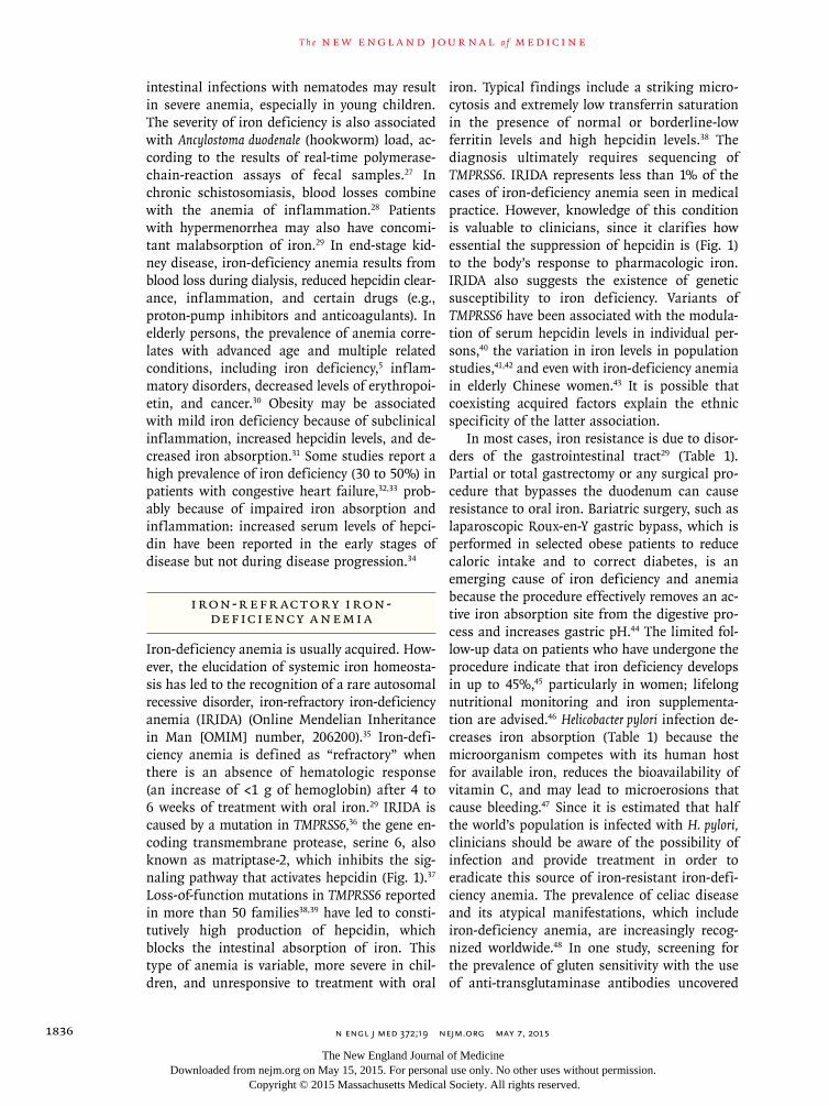
intestinal infections with nematodes may result in severe anemia, especially in young children. The severity of iron deficiency is also associated with Ancylostoma duodenale (hookworm) load, ac-cording to the results of real-time polymerase-chain-reaction assays of fecal samples.27 In chronic schistosomiasis, blood losses combine with the anemia of inflammation.28 Patients with hypermenorrhea may also have concomi-tant malabsorption of iron.29 In end-stage kid-ney disease, iron-deficiency anemia results from blood loss during dialysis, reduced hepcidin clear-ance, inflammation, and certain drugs (e.g., proton-pump inhibitors and anticoagulants). In elderly persons, the prevalence of anemia corre-lates with advanced age and multiple related conditions, including iron deficiency,5 inflam-matory disorders, decreased levels of erythropoi-etin, and cancer.30 Obesity may be associated with mild iron deficiency because of subclinical inflammation, increased hepcidin levels, and de-creased iron absorption.31 Some studies report a high prevalence of iron deficiency (30 to 50%) in patients with congestive heart failure,32,33 prob-ably because of impaired iron absorption and inflammation: increased serum levels of hepci-din have been reported in the early stages of disease but not during disease progression.34
Iron-R efr ac t or y Iron-Deficienc y A nemi a
Iron-deficiency anemia is usually acquired. How-ever, the elucidation of systemic iron homeosta-sis has led to the recognition of a rare autosomal recessive disorder, iron-refractory iron-deficiency anemia (IRIDA) (Online Mendelian Inheritance in Man [OMIM] number, 206200).35 Iron-defi-ciency anemia is defined as “refractory” when there is an absence of hematologic response (an increase of <1 g of hemoglobin) after 4 to 6 weeks of treatment with oral iron.29 IRIDA is caused by a mutation in TMPRSS6,36 the gene en-coding transmembrane protease, serine 6, also known as matriptase-2, which inhibits the sig-naling pathway that activates hepcidin (Fig. 1).37 Loss-of-function mutations in TMPRSS6 reported in more than 50 families38,39 have led to consti-tutively high production of hepcidin, which blocks the intestinal absorption of iron. This type of anemia is variable, more severe in chil-dren, and unresponsive to treatment with oral
iron. Typical findings include a striking micro-cytosis and extremely low transferrin saturation in the presence of normal or borderline-low ferritin levels and high hepcidin levels.38 The diagnosis ultimately requires sequencing of TMPRSS6. IRIDA represents less than 1% of the cases of iron-deficiency anemia seen in medical practice. However, knowledge of this condition is valuable to clinicians, since it clarifies how essential the suppression of hepcidin is (Fig. 1) to the body’s response to pharmacologic iron. IRIDA also suggests the existence of genetic susceptibility to iron deficiency. Variants of TMPRSS6 have been associated with the modula-tion of serum hepcidin levels in individual per-sons,40 the variation in iron levels in population studies,41,42 and even with iron-deficiency anemia in elderly Chinese women.43 It is possible that coexisting acquired factors explain the ethnic specificity of the latter association.
In most cases, iron resistance is due to disor-ders of the gastrointestinal tract29 (Table 1). Partial or total gastrectomy or any surgical pro-cedure that bypasses the duodenum can cause resistance to oral iron. Bariatric surgery, such as laparoscopic Roux-en-Y gastric bypass, which is performed in selected obese patients to reduce caloric intake and to correct diabetes, is an emerging cause of iron deficiency and anemia because the procedure effectively removes an ac-tive iron absorption site from the digestive pro-cess and increases gastric pH.44 The limited fol-low-up data on patients who have undergone the procedure indicate that iron deficiency develops in up to 45%,45 particularly in women; lifelong nutritional monitoring and iron supplementa-tion are advised.46 Helicobacter pylori infection de-creases iron absorption (Table 1) because the microorganism competes with its human host for available iron, reduces the bioavailability of vitamin C, and may lead to microerosions that cause bleeding.47 Since it is estimated that half the world’s population is infected with H. pylori, clinicians should be aware of the possibility of infection and provide treatment in order to eradicate this source of iron-resistant iron-defi-ciency anemia. The prevalence of celiac disease and its atypical manifestations, which include iron-deficiency anemia, are increasingly recog-nized worldwide.48 In one study, screening for the prevalence of gluten sensitivity with the use of anti-transglutaminase antibodies uncovered
The New England Journal of Medicine Downloaded from nejm.org on May 15, 2015. For personal use only. No other uses without permission.
Copyright © 2015 Massachusetts Medical Society. All rights reserved.
n engl j med 372;19 nejm.org May 7, 2015 1837
Iron-Deficiency Anemia
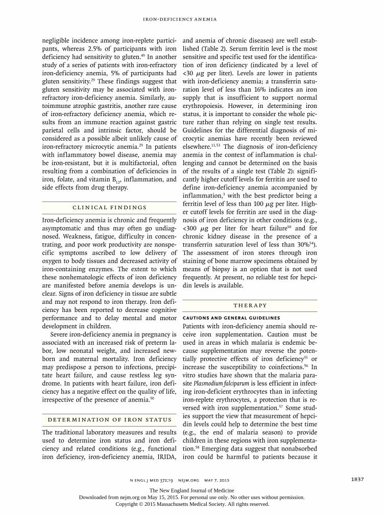
negligible incidence among iron-replete partici-pants, whereas 2.5% of participants with iron deficiency had sensitivity to gluten.49 In another study of a series of patients with iron-refractory iron-deficiency anemia, 5% of participants had gluten sensitivity.29 These findings suggest that gluten sensitivity may be associated with iron-refractory iron-deficiency anemia. Similarly, au-toimmune atrophic gastritis, another rare cause of iron-refractory deficiency anemia, which re-sults from an immune reaction against gastric parietal cells and intrinsic factor, should be considered as a possible albeit unlikely cause of iron-refractory microcytic anemia.29 In patients with inflammatory bowel disease, anemia may be iron-resistant, but it is multifactorial, often resulting from a combination of deficiencies in iron, folate, and vitamin B12, inflammation, and side effects from drug therapy.
Clinic a l Findings
Iron-deficiency anemia is chronic and frequently asymptomatic and thus may often go undiag-nosed. Weakness, fatigue, difficulty in concen-trating, and poor work productivity are nonspe-cific symptoms ascribed to low delivery of oxygen to body tissues and decreased activity of iron-containing enzymes. The extent to which these nonhematologic effects of iron deficiency are manifested before anemia develops is un-clear. Signs of iron deficiency in tissue are subtle and may not respond to iron therapy. Iron defi-ciency has been reported to decrease cognitive performance and to delay mental and motor development in children.
Severe iron-deficiency anemia in pregnancy is associated with an increased risk of preterm la-bor, low neonatal weight, and increased new-born and maternal mortality. Iron deficiency may predispose a person to infections, precipi-tate heart failure, and cause restless leg syn-drome. In patients with heart failure, iron defi-ciency has a negative effect on the quality of life, irrespective of the presence of anemia.50
De ter mination of Iron S tat us
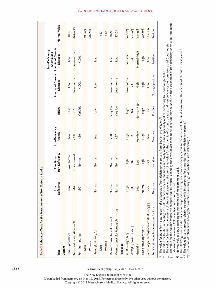
The traditional laboratory measures and results used to determine iron status and iron defi-ciency and related conditions (e.g., functional iron deficiency, iron-deficiency anemia, IRIDA,
and anemia of chronic diseases) are well estab-lished (Table 2). Serum ferritin level is the most sensitive and specific test used for the identifica-tion of iron deficiency (indicated by a level of <30 μg per liter). Levels are lower in patients with iron-deficiency anemia; a transferrin satu-ration level of less than 16% indicates an iron supply that is insufficient to support normal erythropoiesis. However, in determining iron status, it is important to consider the whole pic-ture rather than relying on single test results. Guidelines for the differential diagnosis of mi-crocytic anemias have recently been reviewed elsewhere.11,53 The diagnosis of iron-deficiency anemia in the context of inflammation is chal-lenging and cannot be determined on the basis of the results of a single test (Table 2): signifi-cantly higher cutoff levels for ferritin are used to define iron-deficiency anemia accompanied by inflammation,3 with the best predictor being a ferritin level of less than 100 μg per liter. High-er cutoff levels for ferritin are used in the diag-nosis of iron deficiency in other conditions (e.g., <300 μg per liter for heart failure50 and for chronic kidney disease in the presence of a transferrin saturation level of less than 30%54). The assessment of iron stores through iron staining of bone marrow specimens obtained by means of biopsy is an option that is not used frequently. At present, no reliable test for hepci-din levels is available.
Ther a py
Cautions and General Guidelines
Patients with iron-deficiency anemia should re-ceive iron supplementation. Caution must be used in areas in which malaria is endemic be-cause supplementation may reverse the poten-tially protective effects of iron deficiency55 or increase the susceptibility to coinfections.56 In vitro studies have shown that the malaria para-site Plasmodium falciparum is less efficient in infect-ing iron-deficient erythrocytes than in infecting iron-replete erythrocytes, a protection that is re-versed with iron supplementation.57 Some stud-ies support the view that measurement of hepci-din levels could help to determine the best time (e.g., the end of malaria season) to provide children in these regions with iron supplementa-tion.58 Emerging data suggest that nonabsorbed iron could be harmful to patients because it
The New England Journal of Medicine Downloaded from nejm.org on May 15, 2015. For personal use only. No other uses without permission.
Copyright © 2015 Massachusetts Medical Society. All rights reserved.
n engl j med 372;19 nejm.org May 7, 20151838
T h e n e w e ngl a nd j o u r na l o f m e dic i n e
Tabl
e 2.
Lab
orat
ory
Test
s fo
r th
e M
easu
rem
ent o
f Iro
n St
atus
in A
dults
.
Test
Iron
D
efic
ienc
yFu
nctio
nal
Iron
Def
icie
ncy
Iron
-Def
icie
ncy
Ane
mia
IRID
AA
nem
ia o
f Chr
onic
D
isea
ses
Iron
-Def
icie
ncy
Ane
mia
and
A
nem
ia o
f Chr
onic
D
isea
ses
Nor
mal
Val
ue
Cur
rent
Iron
— μ
mol
/lite
rLo
wLo
w–n
orm
alLo
wLo
wLo
wLo
w10
–30
Tran
sfer
rin
satu
ratio
n —
%≥1
6Lo
w–
norm
al<1
6*<1
0Lo
w–n
orm
alLo
w–n
orm
al>1
6 to
<45
Ferr
itin
— μ
g/lit
er<3
0†N
orm
al<1
0V
aria
ble
>100
‡<1
00‡
Men
40–3
00
Wom
en20
–200
Hem
oglo
bin
— g
/dl
Nor
mal
Nor
mal
Low
Low
Low
Low
Men
>13
Wom
en>1
2
Mea
n co
rpus
cula
r vo
lum
e —
flN
orm
alN
orm
al<8
0V
ery
low
Low
–nor
mal
Low
80–9
5
Mea
n co
rpus
cula
r he
mog
lobi
n —
pg
Nor
mal
Nor
mal
<27
Ver
y lo
wLo
w–n
orm
alLo
w27
–34
Prop
osed
sTFR
— m
g/lit
er§
Hig
hH
igh
Hig
hH
igh
Low
–nor
mal
Var
iabl
eV
arie
s¶
sTFR
/log
ferr
itin
inde
x‖N
AN
A>2
NA
<1>2
Var
ies¶
Hep
cidi
nLo
wLo
wV
ery
low
Nor
mal
–hig
hH
igh
Nor
mal
–hig
hV
arie
s¶
Zin
c pr
otop
orph
yrin
**N
orm
alH
igh
Hig
hH
igh
Hig
hH
igh
Var
ies¶
Ret
icul
ocyt
e he
mog
lobi
n co
nten
t — p
g††
<25
<29
Low
Low
Low
Low
31.2
±1.6
Perl
’s s
tain
ing
of b
one
mar
row
for
iron
Neg
ativ
eV
aria
ble
Neg
ativ
ePo
sitiv
eSt
rong
ly p
ositi
vePo
sitiv
ePo
sitiv
e
* Th
e va
lue
for
tran
sfer
rin
satu
ratio
n in
the
dia
gnos
is o
f iro
n-de
ficie
ncy
anem
ia is
from
Beu
tler
and
Waa
len.
51
†
The
valu
e fo
r fe
rriti
n in
the
dia
gnos
is o
f iro
n-de
ficie
ncy
anem
ia h
as a
sen
sitiv
ity o
f 92%
and
a s
peci
ficity
of 8
3% a
ccor
ding
to
Goo
dnou
gh e
t al
.4
‡
The
valu
e fo
r fe
rriti
n in
the
ane
mia
of c
hron
ic d
isea
se a
nd t
he c
ombi
ned
valu
e fo
r th
e an
emia
of c
hron
ic d
isea
se a
nd ir
on d
efic
ienc
y ar
e fr
om W
eiss
and
Goo
dnou
gh.3
§ Th
e va
lue
for
the
solu
ble
tran
sfer
rin
rece
ptor
(sT
FR),
whi
ch is
she
d by
the
ery
thro
blas
t m
embr
ane
in s
erum
, may
be
usef
ul in
the
ass
essm
ent
of ir
on-d
efic
ienc
y an
emia
, but
the
met
h-od
s us
ed t
o m
easu
re s
TFR
hav
e be
en n
ot s
tand
ardi
zed.
3
¶
Nor
mal
val
ues
vary
acc
ordi
ng t
o th
e m
etho
d of
mea
sure
men
t us
ed.
‖ Th
e sT
FR/l
og fe
rriti
n in
dex
has
been
pro
pose
d to
dis
tingu
ish
iron
-def
icie
ncy
anem
ia in
the
ane
mia
of c
hron
ic d
isea
se fr
om t
he a
nem
ia o
f chr
onic
dis
ease
alo
ne.3
** T
he v
alue
s fo
r zi
nc p
roto
porp
hyri
n ar
e us
ed o
nly
in s
cree
ning
for
or m
onito
ring
iron
-def
icie
ncy
anem
ia..9
††
Red
uctio
n of
ret
icul
ocyt
e he
mog
lobi
n co
nten
t is
an
earl
y si
gn o
f fun
ctio
nal i
ron
defic
ienc
y.52
The New England Journal of Medicine Downloaded from nejm.org on May 15, 2015. For personal use only. No other uses without permission.
Copyright © 2015 Massachusetts Medical Society. All rights reserved.
n engl j med 372;19 nejm.org May 7, 2015 1839
Iron-Deficiency Anemia
might modify the gut microbiota, increasing the concentration of intestinal pathogens.59
The benefit of treating iron deficiency before the development of anemia remains uncertain. A few small studies show that the administration of intravenous iron improves fatigue in women without anemia whose ferritin levels are in the iron-deficient range.60 Some studies have also suggested that oral iron supplementation bene-fits physical performance in women of repro-ductive age,61 but such studies have included a limited number of participants and are strik-ingly heterogeneous.
Patients with severe iron-deficiency anemia that causes cardiovascular symptoms, such as heart failure or angina, should receive red-cell transfusions. This approach rapidly corrects not only hypoxia but also iron deficiency, since one unit of packed red cells provides approximately 200 mg of iron.
Oral Iron Therapy
The administration of oral iron is a convenient, inexpensive, and effective means of treating stable patients. Among the myriad preparations on the market, iron sulfate is the most frequent-ly used; gluconate and fumarate are also effec-tive iron salts. The recommended daily dose for adults with iron deficiency is 100 to 200 mg of elementary iron and that for children is 3 to 6 mg per kilogram of body weight of a liquid preparation; for both groups the supplement should be administered in divided doses without food. The addition of vitamin C may improve absorption. The low hepcidin levels in patients with iron-deficiency anemia ensure effective iron absorption and the rapid recovery of hemo-globin levels; however, 3 to 6 months of treat-ment are required for the repletion of iron stores and the normalization of serum ferritin levels. Long-term use of oral iron is limited by side ef-fects, including nausea, vomiting, constipation, and metallic taste; these side effects are frequent and, although not severe, are often worrisome to patients. Although oral iron may cause dark stools, it does not produce false positive results on tests for occult blood. If treatment with oral iron fails, the reasons may include premature termination of treatment, lack of compliance with the regimen or discontinuation by the pa-tient, or a truly refractory response to treatment. In the latter case, other, specific treatments,
such as the eradication of infection with H. py-lori or the introduction of a gluten-free diet in patients with celiac disease, may restore the ca-pacity for iron absorption and eliminate the need for supplementation in some patients.29 There are no known markers that can be used to predict which patients will or will not have a response to oral iron therapy. The oral iron chal-lenge test (in which 60 mg of oral iron is admin-istered and serum iron levels are measured 1 to 2 hours afterward) is rarely used since it has not been extensively validated. A pilot study showed that measurement of serum hepcidin levels could help to identify patients in whom a re-sponse to oral iron is probable (those with low hepcidin levels) and those in whom it is not probable (those with normal or elevated hepci-din levels).62 However, hepcidin tests are not routinely available for clinical use. Assessment of an early response to oral iron might also be useful in the treatment of iron-deficiency ane-mia in patients with anemia of chronic disease. One study in patients with rheumatologic dis-
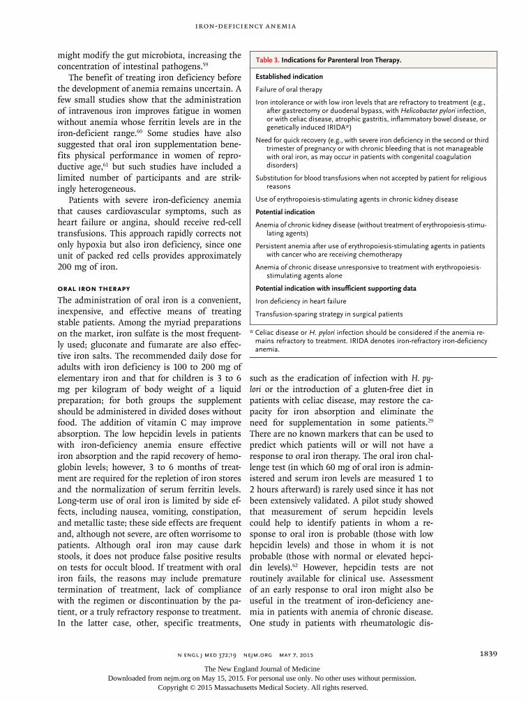
Established indication
Failure of oral therapy
Iron intolerance or with low iron levels that are refractory to treatment (e.g., after gastrectomy or duodenal bypass, with Helicobacter pylori infection, or with celiac disease, atrophic gastritis, inflammatory bowel disease, or genetically induced IRIDA*)
Need for quick recovery (e.g., with severe iron deficiency in the second or third trimester of pregnancy or with chronic bleeding that is not manageable with oral iron, as may occur in patients with congenital coagulation disorders)
Substitution for blood transfusions when not accepted by patient for religious reasons
Use of erythropoiesis-stimulating agents in chronic kidney disease
Potential indication
Anemia of chronic kidney disease (without treatment of erythropoiesis-stimu-lating agents)
Persistent anemia after use of erythropoiesis-stimulating agents in patients with cancer who are receiving chemotherapy
Anemia of chronic disease unresponsive to treatment with erythropoiesis-stimulating agents alone
Potential indication with insufficient supporting data
Iron deficiency in heart failure
Transfusion-sparing strategy in surgical patients
* Celiac disease or H. pylori infection should be considered if the anemia re-mains refractory to treatment. IRIDA denotes iron-refractory iron-deficiency anemia.
Table 3. Indications for Parenteral Iron Therapy.
The New England Journal of Medicine Downloaded from nejm.org on May 15, 2015. For personal use only. No other uses without permission.
Copyright © 2015 Massachusetts Medical Society. All rights reserved.
n engl j med 372;19 nejm.org May 7, 20151840
T h e n e w e ngl a nd j o u r na l o f m e dic i n e
ease and iron-deficiency anemia showed that a change in the hemoglobin content of reticulo-cytes (and in serum levels of iron and transferrin saturation) may predict the response to the admin-istration of oral iron after 1 week of therapy.63
Parenteral Iron Therapy
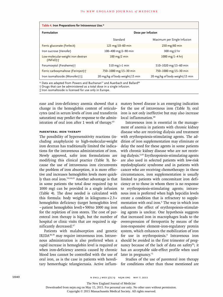
The possibility of hypersensitivity reactions (in-cluding anaphylaxis) to high-molecular-weight iron dextran has traditionally limited the indica-tions for the intravenous administration of iron. Newly approved, safer iron formulations are modifying this clinical practice (Table 3). Be-cause the use of intravenous iron circumvents the problem of iron absorption, it is more effec-tive and increases hemoglobin levels more quick-ly than oral iron.54,64,65 Another advantage is that in some patients the total dose required (up to 1000 mg) can be provided in a single infusion (Table 4). The dose needed is calculated with this formula: body weight in kilograms × 2.3 × hemoglobin deficiency (target hemoglobin level − patient hemoglobin level) + 500 to 1000 mg iron for the repletion of iron stores. The cost of par-enteral iron therapy is high, but the number of hospital or clinic visits that are required is sig-nificantly decreased.67
Patients with malabsorption and genetic IRIDA38,39 may require intravenous iron. Intrave-nous administration is also preferred when a rapid increase in hemoglobin level is required or when iron-deficiency anemia caused by chronic blood loss cannot be controlled with the use of oral iron, as is the case in patients with heredi-tary hemorrhagic telangiectasia. Active inflam-
matory bowel disease is an emerging indication for the use of intravenous iron (Table 3); oral iron is not only ineffective but may also increase local inflammation.68
Intravenous iron is essential in the manage-ment of anemia in patients with chronic kidney disease who are receiving dialysis and treatment with erythropoiesis-stimulating agents. The ad-dition of iron supplementation may eliminate or delay the need for these agents in some patients with chronic kidney disease who are not receiv-ing dialysis.6,69 Erythropoiesis-stimulating agents are also used in selected patients with low-risk myelodysplastic syndrome and in patients with cancer who are receiving chemotherapy: in these circumstances, iron supplementation is usually limited to patients with concomitant iron defi-ciency or to those in whom there is no response to erythropoiesis-stimulating agents; intrave-nous iron is preferred when high hepcidin levels create a condition that is refractory to supple-mentation with oral iron.6 The way in which iron enhances the effect of erythropoiesis-stimulat-ing agents is unclear. One hypothesis suggests that increased iron in macrophages leads to the overexpression of ferroportin by means of the iron-responsive element–iron-regulatory protein system, which enhances the mobilization of iron for use in erythropoiesis.4 Intravenous iron should be avoided in the first trimester of preg-nancy because of the lack of data on safety70; it has an acceptable side-effect profile when used later in pregnancy.71
Studies of the use of parenteral iron therapy for conditions other than those mentioned are
Formulation Dose per Infusion
Standard Maximum per Single Infusion
Ferric gluconate (Ferlecit) 125 mg/10–60 min 250 mg/60 min
Iron sucrose (Venofer) 100–400 mg/2–90 min 300 mg/2 hr
Low-molecular-weight iron dextran (INFeD)†
100 mg/2 min 1000 mg/1–4 hr)
Ferumoxytol (Feraheme)† 510 mg/>1 min 510–1020 mg/15–60 min
Ferric carboxymaltose (Ferinject)† 750–1000 mg/15–30 min 750–1000 mg/15–30 min
Iron isomaltoside (Monofer)†‡ 20 mg/kg of body weight/15 min 20 mg/kg of body weight/15 min
* Data are adapted from Powers and Buchanan13 and Auerbach and Ballard66
† Drugs that can be administered as a total dose in a single infusion.‡ Iron isomaltoside is licensed for use only in Europe.
Table 4. Iron Preparations for Intravenous Use.*
The New England Journal of Medicine Downloaded from nejm.org on May 15, 2015. For personal use only. No other uses without permission.
Copyright © 2015 Massachusetts Medical Society. All rights reserved.
n engl j med 372;19 nejm.org May 7, 2015 1841
Iron-Deficiency Anemia
either limited or not controlled. A multicenter European trial of patients with iron deficiency and chronic heart failure showed that the use of intravenous iron supplementation led to im-provements in physical performance, New York Heart Association functional class,50 and quality of life independently from the correction of ane-mia72; more recently, 1 year of treatment was associated with a reduced risk of hospitaliza-tion.73 However, since these results were based largely on subjective evaluation, larger and lon-ger-term studies are required to assess the real benefit of administering iron to patients with heart failure.
The transient side effects of intravenous iron supplementation include nausea, vomiting, pru-ritus, headache, and flushing; myalgia, arthral-gia, and back and chest pain usually resolve within 48 hours, even after total dose adminis-tration.74 Hypersensitivity reactions are rare,66,74 as are severe or life-threatening reactions75; the pathophysiological features of these reactions are uncertain and might be exacerbated by re-leased free iron,70,76 a phenomenon that does not occur with currently used formulations. Predis-posing conditions are rapid infusions, a history
of atopy, and drug allergy. Practical recommen-dations for minimizing risk70 include a slow in-fusion rate, careful patient observation, and ad-ministration by trained health care personnel in an environment with access to resuscitation fa-cilities.75 The test dose may provide false reas-surance; premedication with antihistamine is no longer advised because it may cause hypotension and tachycardia.70,75,76
Clinical trials are reassuring with regard to the efficacy and side-effect profile of intrave-nous iron. Some concern persists with regard to the long-term biologic effects of iron and its effects on the generation of oxygen radicals, patient susceptibility to infections,54,66 and the potential such treatment would have to worsen conditions such as type 2 diabetes and other chronic metabolic disorders.77 Well-designed, randomized, controlled trials are needed to ver-ify the long-term effects of intravenous iron supplementation.78 In the interim, intravenous iron should be used only when the benefits out-weigh the risks.
No potential conflict of interest relevant to this article was reported.
Disclosure forms provided by the author are available with the full text of this article at NEJM.org.
References1. McLean E, Cogswell M, Egli I, Woj-dyla D, de Benoist B. Worldwide preva-lence of anaemia, WHO Vitamin and Mineral Nutrition Information System, 1993-2005. Public Health Nutr 2009; 12: 444-54.2. Hentze MW, Muckenthaler MU, Galy B, Camaschella C. Two to tango: regula-tion of mammalian iron metabolism. Cell 2010; 142: 24-38.3. Weiss G, Goodnough LT. Anemia of chronic disease. N Engl J Med 2005; 352: 1011-23.4. Goodnough LT, Nemeth E, Ganz T. Detection, evaluation, and management of iron-restricted erythropoiesis. Blood 2010; 116: 4754-61.5. Goodnough LT, Schrier SL. Evaluation and management of anemia in the elderly. Am J Hematol 2014; 89: 88-96.6. Macdougall IC. Iron supplementation in nephrology and oncology: what do we have in common? Oncologist 2011; 16: Suppl 3: 25-34.7. Ferrucci L, Semba RD, Guralnik JM, et al. Proinflammatory state, hepcidin, and anemia in older persons. Blood 2010; 115: 3810-6.8. Marković M, Majkić-Singh N, Ignjato-vić S, Singh S. Reticulocyte haemoglobin content vs. soluble transferrin receptor
and ferritin index in iron deficiency anae-mia accompanied with inflammation. Int J Lab Hematol 2007; 29: 341-6.9. Thomas DW, Hinchliffe RF, Briggs C, Macdougall IC, Littlewood T, Cavill I. Guideline for the laboratory diagnosis of functional iron deficiency. Br J Haematol 2013; 161: 639-48.10. Schrier SL. Causes and diagnosis of iron deficiency anemia in the adults. 2014 (http://www .uptodate .com/ contents/ causes-and-diagnosis-of-iron-deficiency -anemia-in-the-adult).11. DeLoughery TG. Microcytic anemia. N Engl J Med 2014; 371: 1324-31.12. Pavord S, Myers B, Robinson S, Allard S, Strong J, Oppenheimer C. UK guide-lines on the management of iron defi-ciency in pregnancy. Br J Haematol 2012; 156: 588-600.13. Powers JM, Buchanan GR. Diagnosis and management of iron deficiency ane-mia. Hematol Oncol Clin North Am 2014; 28: 729-45.14. Kassebaum NJ, Jasrasaria R, Naghavi M, et al. A systematic analysis of global anemia burden from 1990 to 2010. Blood 2014; 123: 615-24.15. Stevens GA, Finucane MM, De-Regil LM, et al. Global, regional, and national trends in haemoglobin concentration and
prevalence of total and severe anaemia in children and pregnant and non-pregnant women for 1995-2011: a systematic analy-sis of population-representative data. Lan-cet Glob Health 2013; 1(1): e16-e25.16. Pasricha SR, Drakesmith H, Black J, Hipgrave D, Biggs BA. Control of iron de-ficiency anemia in low- and middle- income countries. Blood 2013; 121: 2607-17.17. Nemeth E, Tuttle MS, Powelson J, et al. Hepcidin regulates cellular iron efflux by binding to ferroportin and inducing its internalization. Science 2004; 306: 2090-3.18. Camaschella C. Iron and hepcidin: a story of recycling and balance. Hematol-ogy Am Soc Hematol Educ Program 2013; 2013: 1-8.19. Galesloot TE, Vermeulen SH, Geurts-Moespot AJ, et al. Serum hepcidin: refer-ence ranges and biochemical correlates in the general population. Blood 2011; 117(25): e218-e225.20. Traglia M, Girelli D, Biino G, et al. Association of HFE and TMPRSS6 genetic variants with iron and erythrocyte param-eters is only in part dependent on serum hepcidin concentrations. J Med Genet 2011; 48: 629-34.21. Kautz L, Jung G, Valore EV, Rivella S, Nemeth E, Ganz T. Identification of erythroferrone as an erythroid regulator
The New England Journal of Medicine Downloaded from nejm.org on May 15, 2015. For personal use only. No other uses without permission.
Copyright © 2015 Massachusetts Medical Society. All rights reserved.
n engl j med 372;19 nejm.org May 7, 20151842
T h e n e w e ngl a nd j o u r na l o f m e dic i n e
of iron metabolism. Nat Genet 2014; 46: 678-84.22. Mastrogiannaki MMP, Matak P, Peys-sonnaux C. The gut in iron homeostasis: role of HIF-2 under normal and patho-logical conditions. Blood 2013; 122: 885-92.23. Zhang AS, Anderson SA, Wang J, et al. Suppression of hepatic hepcidin expres-sion in response to acute iron deprivation is associated with an increase of matrip-tase-2 protein. Blood 2011; 117: 1687-99.24. Goenka MK, Majumder S, Goenka U. Capsule endoscopy: present status and future expectation. World J Gastroenterol 2014; 20: 10024-37.25. Mustafa BF, Samaan M, Langmead L, Khasraw M. Small bowel video capsule endoscopy: an overview. Expert Rev Gas-troenterol Hepatol 2013; 7: 323-9.26. Heidelbaugh JJ. Proton pump inhibi-tors and risk of vitamin and mineral defi-ciency: evidence and clinical implications. Ther Adv Drug Saf 2013; 4: 125-33.27. Jonker FA, Calis JC, Phiri K, et al. Re-al-time PCR demonstrates Ancylostoma duodenale is a key factor in the etiology of severe anemia and iron deficiency in Ma-lawian pre-school children. PLoS Negl Trop Dis 2012; 6(3): e1555.28. Friedman JF, Kanzaria HK, McGarvey ST. Human schistosomiasis and anemia: the relationship and potential mecha-nisms. Trends Parasitol 2005; 21: 386-92.29. Hershko C, Camaschella C. How I treat unexplained refractory iron deficiency anemia. Blood 2014; 123: 326-33.30. Bach V, Schruckmayer G, Sam I, Kem-mler G, Stauder R. Prevalence and possi-ble causes of anemia in the elderly: a cross-sectional analysis of a large European university hospital cohort. Clin Interv Ag-ing 2014; 9: 1187-96.31. Aigner E, Feldman A, Datz C. Obesity as an emerging risk factor for iron defi-ciency. Nutrients 2014; 6: 3587-600.32. van Veldhuisen DJ, Anker SD, Poni-kowski P, Macdougall IC. Anemia and iron deficiency in heart failure: mecha-nisms and therapeutic approaches. Nat Rev Cardiol 2011; 8: 485-93.33. Cohen-Solal A, Damy T, Terbah M, et al. High prevalence of iron deficiency in patients with acute decompensated heart failure. Eur J Heart Fail 2014; 16: 984-91.34. Jankowska EA, Malyszko J, Ardehali H, et al. Iron status in patients with chronic heart failure. Eur Heart J 2013; 34: 827-34.35. Finberg KE, Heeney MM, Campagna DR, et al. Mutations in TMPRSS6 cause iron-refractory iron deficiency anemia (IRIDA). Nat Genet 2008; 40: 569-71.36. Du X, She E, Gelbart T, et al. The serine protease TMPRSS6 is required to sense iron deficiency. Science 2008; 320: 1088-92.37. Silvestri L, Pagani A, Nai A, De Do-menico I, Kaplan J, Camaschella C. The serine protease matriptase-2 (TMPRSS6)
inhibits hepcidin activation by cleaving membrane hemojuvelin. Cell Metab 2008; 8: 502-11.38. De Falco L, Sanchez M, Silvestri L, et al. Iron refractory iron deficiency anemia. Haematologica 2013; 98: 845-53.39. Heeney MM, Finberg KE. Iron-refrac-tory iron deficiency anemia (IRIDA). Hema-tol Oncol Clin North Am 2014; 28: 637-52.40. Nai A, Pagani A, Silvestri L, et al. TMPRSS6 rs855791 modulates hepcidin transcription in vitro and serum hepcidin levels in normal individuals. Blood 2011; 118: 4459-62.41. Benyamin B, Ferreira MA, Willemsen G, et al. Common variants in TMPRSS6 are associated with iron status and eryth-rocyte volume. Nat Genet 2009; 41: 1173-5.42. Tanaka T, Roy CN, Yao W, et al. A ge-nome-wide association analysis of serum iron concentrations. Blood 2010; 115: 94-6.43. An P, Wu Q, Wang H, et al. TMPRSS6, but not TF, TFR2 or BMP2 variants are as-sociated with increased risk of iron-defi-ciency anemia. Hum Mol Genet 2012; 21: 2124-31.44. Warsch S, Byrnes J. Emerging causes of iron deficiency anemia refractory to oral iron supplementation. World J Gastroin-test Pharmacol Ther 2013; 4: 49-53.45. Stein J, Stier C, Raab H, Weiner R. Re-view article: the nutritional and pharma-cological consequences of obesity surgery. Aliment Pharmacol Ther 2014; 40: 582-609.46. Obinwanne KM, Fredrickson KA, Mathiason MA, Kallies KJ, Farnen JP, Ko-thari SN. Incidence, treatment, and out-comes of iron deficiency after laparoscopic Roux-en-Y gastric bypass: a 10-year analy-sis. J Am Coll Surg 2014; 218: 246-52.47. Franceschi F, Zuccalà G, Roccarina D, Gasbarrini A. Clinical effects of Helico-bacter pylori outside the stomach. Nat Rev Gastroenterol Hepatol 2014; 11: 234-42.48. Rubio-Tapia A, Hill ID, Kelly CP, Calderwood AH, Murray JA. ACG clinical guidelines: diagnosis and management of celiac disease. Am J Gastroenterol 2013; 108: 656-76.49. Murray JA, McLachlan S, Adams PC, et al. Association between celiac disease and iron deficiency in Caucasians, but not non-Caucasians. Clin Gastroenterol Hepa-tol 2013; 11: 808-14.50. Anker SD, Comin Colet J, Filippatos G, et al. Ferric carboxymaltose in patients with heart failure and iron deficiency. N Engl J Med 2009; 361: 2436-48.51. Beutler E, Waalen J. The definition of anemia: what is the lower limit of normal of the blood hemoglobin concentration? Blood 2006; 107: 1747-50.52. Brugnara C, Mohandas N. Red cell indices in classification and treatment of anemias: from M.M. Wintrobes’s original 1934 classification to the third millenni-um. Curr Opin Hematol 2013; 20: 222-30.53. Camaschella C. How I manage pa-
tients with atypical microcytic anaemia. Br J Haematol 2013; 160: 12-24.54. Macdougall IC, Bock AH, Carrera F, et al. FIND-CKD: a randomized trial of in-travenous ferric carboxymaltose versus oral iron in patients with chronic kidney disease and iron deficiency anaemia. Nephrol Dial Transplant 2014; 29: 2075-84.55. Sazawal S, Black RE, Ramsan M, et al. Effects of routine prophylactic supple-mentation with iron and folic acid on admission to hospital and mortality in preschool children in a high malaria transmission setting: community-based, randomised, placebo-controlled trial. Lan-cet 2006; 367: 133-43.56. Drakesmith H, Prentice AM. Hepcidin and the iron-infection axis. Science 2012; 338: 768-72.57. Clark MA, Goheen MM, Fulford A, et al. Host iron status and iron supplemen-tation mediate susceptibility to erythro-cytic stage Plasmodium falciparum. Nat Commun 2014; 5: 4446.58. Atkinson SH, Armitage AE, Khand-wala S, et al. Combinatorial effects of malaria season, iron deficiency, and in-f lammation determine plasma hepcidin concentration in African children. Blood 2014; 123: 3221-9.59. Kortman GA, Raffatellu M, Swinkels DW, Tjalsma H. Nutritional iron turned inside out: intestinal stress from a gut mi-crobial perspective. FEMS Microbiol Rev 2014; 38: 1202-34.60. Krayenbuehl PA, Battegay E, Breymann C, Furrer J, Schulthess G. Intravenous iron for the treatment of fatigue in non-anemic, premenopausal women with low serum ferritin concentration. Blood 2011; 118: 3222-7.61. Pasricha SR Sr, Low M, Thompson J, Farrell A, De-Regil LM. Iron supplemen-tation benefits physical performance in women of reproductive age: a systematic review and meta-analysis. J Nutr 2014; 144: 906-14.62. Bregman DB, Goodnough LT. Expe-rience with intravenous ferric carboxy-maltose in patients with iron deficiency anemia. Ther Adv Hematol 2014; 5: 48-60.63. van Santen S, de Mast Q, Oosting JD, van Ede A, Swinkels DW, van der Ven AJ. Hematologic parameters predicting a re-sponse to oral iron therapy in chronic in-flammation. Haematologica 2014; 99(9): e171-e173.64. Onken JE, Bregman DB, Harrington RA, et al. A multicenter, randomized, active-controlled study to investigate the efficacy and safety of intravenous ferric carboxymaltose in patients with iron defi-ciency anemia. Transfusion 2014; 54: 306-15.65. Vadhan-Raj S, Strauss W, Ford D, et al. Efficacy and safety of IV ferumoxytol for adults with iron deficiency anemia previ-ously unresponsive to or unable to toler-ate oral iron. Am J Hematol 2014; 89: 7-12.
The New England Journal of Medicine Downloaded from nejm.org on May 15, 2015. For personal use only. No other uses without permission.
Copyright © 2015 Massachusetts Medical Society. All rights reserved.
n engl j med 372;19 nejm.org May 7, 2015 1843
Iron-Deficiency Anemia
66. Auerbach M, Ballard H. Clinical use of intravenous iron: administration, effi-cacy, and safety. Hematology Am Soc He-matol Educ Program 2010; 2010: 338-47.67. Bager P, Dahlerup JF. The health care cost of intravenous iron treatment in IBD patients depends on the economic evalua-tion perspective. J Crohns Colitis 2010; 4: 427-30.68. Reinisch W, Chowers Y, Danese S, et al. The management of iron deficiency in inflammatory bowel disease — an online tool developed by the RAND/UCLA appro-priateness method. Aliment Pharmacol Ther 2013; 38: 1109-18.69. Bailie GR, Larkina M, Goodkin DA, et al. Variation in intravenous iron use internationally and over time: the Dialysis Outcomes and Practice Patterns Study (DOPPS). Nephrol Dial Transplant 2013; 28: 2570-9.70. Rampton DFJ, Folkersen J, Fishbane S, et al. Hypersensitivity reactions to in-
travenous iron: guidance for risk minimi-zation and management. Haematologica 2014; 99: 1671-6.71. Gupta A, Manaktala U, Rathore AM. A randomised controlled trial to compare intravenous iron sucrose and oral iron in treatment of iron deficiency anemia in pregnancy. Indian J Hematol Blood Trans-fus 2014; 30: 120-5.72. Comin-Colet J, Lainscak M, Dickstein K, et al. The effect of intravenous ferric carboxymaltose on health-related quality of life in patients with chronic heart fail-ure and iron deficiency: a subanalysis of the FAIR-HF study. Eur Heart J 2013; 34: 30-8.73. Ponikowski P, van Veldhuisen DJ, Co-min-Colet J, et al. Beneficial effects of long-term intravenous iron therapy with ferric carboxymaltose in patients with symptomatic heart failure and iron defi-ciency. Eur Heart J 2015; 36: 657-68.74. Auerbach M, Strauss W, Auerbach S,
Rineer S, Bahrain H. Safety and efficacy of total dose infusion of 1,020 mg of feru-moxytol administered over 15 min. Am J Hematol 2013; 88: 944-7.75. New recommendations to manage risk of allergic reactions with intravenous iron-containing medicines. London: Euro-pean Medicines Agency, 2013 (http://www .ema .europa .eu/ docs/ en_GB/ document_library/ Press_release/ 2013/ 06/ WC500144874 .pdf).76. Bircher AJAM, Auerbach M. Hyper-sensitivity from intravenous iron prod-ucts. Immunol Allergy Clin North Am 2014; 34: 707-23.77. Fernández-Real JM, Manco M. Effects of iron overload on chronic metabolic dis-eases. Lancet Diabetes Endocrinol 2014; 2: 513-26.78. Fishbane S, Hazzan AD. Anaemia: FIND-CKD: intravenous iron in predialy-sis CKD. Nat Rev Nephrol 2014; 10: 488-9.Copyright © 2015 Massachusetts Medical Society.
The New England Journal of Medicine Downloaded from nejm.org on May 15, 2015. For personal use only. No other uses without permission.
Copyright © 2015 Massachusetts Medical Society. All rights reserved.