Iridoid-specificGlucosyltransferasefrom Gardenia … · Gardenia jasminoides (Rubiaceae) fruits...
10
Iridoid-specific Glucosyltransferase from Gardenia jasminoides * □ S Received for publication, March 23, 2011, and in revised form, July 4, 2011 Published, JBC Papers in Press, July 28, 2011, DOI 10.1074/jbc.M111.242586 Mai Nagatoshi ‡ , Kazuyoshi Terasaka ‡ , Akito Nagatsu § , and Hajime Mizukami ‡1 From the ‡ Graduate School of Pharmaceutical Sciences, Nagoya City University, Nagoya 467-8603 and the § School of Pharmacy, Kinjo Gakuin University, Nagoya 463-8521, Japan Iridoids are one of the most widely distributed secondary metabolites in higher plants. They are pharmacologically active principles in various medicinal plants and key intermediates in the biosynthesis of monoterpenoid indole alkaloids as well as quinoline alkaloids. Although most iridoids are present as 1-O- glucosides, the glucosylation step in the biosynthetic pathway has remained obscure. We isolated a cDNA coding for UDP- glucose:iridoid glucosyltransferase (UGT85A24) from Garde- nia jasminoides. UGT85A24 preferentially glucosylated the 1-O-hydroxyl group of 7-deoxyloganetin and genipin but exhib- ited only weak activity toward loganetin and no activity toward 7-deoxyloganetic acid. This suggests that, in the biosynthetic pathway of geniposide, a major iridoid compound in G. jasmi- noides, glucosylation occurs after methylation of 7-deoxy- loganetic acid. UGT85A24 showed negligible activity toward any acceptor substrates other than iridoid aglycones. Thus, UGT85A24 has a remarkable specificity for iridoid aglycones. The mRNA level of UGT85A24 overlaps with the marked increase in genipin glucosylation activity in the methyl jasmo- nate-treated cell cultures of G. jasminoides and is related to iridoid accumulation in G. jasminoides fruits. Iridoids are cyclopenta[c]pyran monoterpenoids distributed in numerous plant families, usually as glucosides (1). In plants, iridoids function as important defense compounds in plant- herbivore interactions (2). They play an important biogenetic role linking terpenes and alkaloids (3). Iridoids exhibit a wide range of pharmacological activities, such as cardiovascular, anti-hepatotoxic, choleretic, anti-inflammatory, immuno- modulatory, and purgative activities, and are present in various folk medicinal plants as bioactive principles (4, 5). In vivo tracer studies have shown that the iridane skeleton is formed by the cyclization of 10-oxogeranial (10-oxoneral) to yield iridodial, which is subsequently oxidized to iridotrial. Iridotrial is converted to a series of iridoid compounds through various reactions, including oxidation, methylation, and glyco- sylation as shown in Fig. 1 (6, 7). However, few studies have investigated the biochemical and molecular biological charac- terization of the iridoid biosynthetic pathway. In particular, the 1-O-glucosylation step in the iridoid biosynthetic pathway has been poorly characterized, although most iridoids are present as 1-O-glucosides in nature. Glucosyltransferase activity toward loganetin, 7-deoxyloganetin, and 7-deoxyloganetic acid was detected in the crude extract of Lonicera japonica (8). How- ever, genes for enzymes relevant to the glucosylation step in the iridoid biosynthetic pathway have yet to be characterized. Glucosylation steps in plant secondary metabolism are cata- lyzed by family 1 plant secondary product glycosyltransferases (PSPGs) 2 that attach a sugar molecule from a UDP-sugar to a low-molecular-weight acceptor substrate (9 –11). PSPGs are defined by the presence of a 44-amino acid C-terminal signa- ture motif designated the PSPG box (12). Recent investigations on the three-dimensional structures of PSPGs by x-ray crystal- lography revealed that the PSPG box functions as a sugar donor-binding pocket (13–15). Homology-based cloning using the conserved amino acid sequence within the PSPG box has been shown to be an efficient tool for isolating cDNAs encoding PSPGs (16, 17). Gardenia jasminoides (Rubiaceae) fruits accumulate iridoid compounds, such as geniposide and gardenoside (18), and the dried fruits have been used as a crude drug in traditional Chi- nese medicine. Cultured G. jasminoides cells have been used to investigate iridoid biosynthesis because they produce small amounts of iridoids even after dedifferentiation (19 –21). Thus, suspension cultures of G. jasminoides are suitable as sources for cDNA isolation and the molecular characterization of glucosyl- transferases that catalyze the 1-O-glucosylation step in the iridoid biosynthetic pathway. Geniposide is a major iridoid compound present in G. jasminoides fruits, and genipin, an aglycone of geniposide, is the only iridoid aglycone commer- cially available in relatively large amounts. Homology-based cloning using the conserved amino acid sequence within the PSPG box combined with screening of the glucosylation activ- ity of recombinant enzymes from candidate cDNA clones with genipin as a glucose-accepting substrate, instead of the intrinsic substrates such as 7-deoxyloganetic acid and 7-deoxyloganetin, which are difficult to obtain, led to the isolation of a cDNA encoding an iridoid-specific glucosyltransferase for the first time. Here, we describe the molecular cloning and charac- * This work was supported by a grant-in-aid for scientific research and a grant-in-aid for young scientists from the Ministry of Education, Culture, Sports, Science, and Technology of Japan. □ S The on-line version of this article (available at http://www.jbc.org) contains supplemental Figs. S1 and S2 and Tables S1–S3. The nucleotide sequence(s) reported in this paper has been submitted to the DDBJ/ GenBank TM /EBI Data Bank with accession number(s)AB555731–AB555743. 1 To whom correspondence should be addressed: Graduate School of Phar- maceutical Sciences, Nagoya City University, 3-1 Tanabe-dori, Mizuho-ku, Nagoya 467-8603, Japan. Fax: 81-52-836-3415; E-mail: hajimem@phar. nagoya-cu.ac.jp. 2 The abbreviations used are: PSPG, plant secondary product glycosyltrans- ferase; RACE, rapid amplification of cDNA ends; Gj, G. jasminoides. THE JOURNAL OF BIOLOGICAL CHEMISTRY VOL. 286, NO. 37, pp. 32866 –32874, September 16, 2011 © 2011 by The American Society for Biochemistry and Molecular Biology, Inc. Printed in the U.S.A. 32866 JOURNAL OF BIOLOGICAL CHEMISTRY VOLUME 286 • NUMBER 37 • SEPTEMBER 16, 2011 by guest on June 17, 2018 http://www.jbc.org/ Downloaded from
Transcript of Iridoid-specificGlucosyltransferasefrom Gardenia … · Gardenia jasminoides (Rubiaceae) fruits...

Iridoid-specific Glucosyltransferase from Gardeniajasminoides*□S
Received for publication, March 23, 2011, and in revised form, July 4, 2011 Published, JBC Papers in Press, July 28, 2011, DOI 10.1074/jbc.M111.242586
Mai Nagatoshi‡, Kazuyoshi Terasaka‡, Akito Nagatsu§, and Hajime Mizukami‡1
From the ‡Graduate School of Pharmaceutical Sciences, Nagoya City University, Nagoya 467-8603 and the §School of Pharmacy,Kinjo Gakuin University, Nagoya 463-8521, Japan
Iridoids are one of the most widely distributed secondarymetabolites in higher plants. They are pharmacologically activeprinciples in various medicinal plants and key intermediates inthe biosynthesis of monoterpenoid indole alkaloids as well asquinoline alkaloids. Although most iridoids are present as 1-O-glucosides, the glucosylation step in the biosynthetic pathwayhas remained obscure. We isolated a cDNA coding for UDP-glucose:iridoid glucosyltransferase (UGT85A24) from Garde-nia jasminoides. UGT85A24 preferentially glucosylated the1-O-hydroxyl group of 7-deoxyloganetin and genipin but exhib-ited only weak activity toward loganetin and no activity toward7-deoxyloganetic acid. This suggests that, in the biosyntheticpathway of geniposide, a major iridoid compound in G. jasmi-noides, glucosylation occurs after methylation of 7-deoxy-loganetic acid. UGT85A24 showed negligible activity towardany acceptor substrates other than iridoid aglycones. Thus,UGT85A24 has a remarkable specificity for iridoid aglycones.The mRNA level of UGT85A24 overlaps with the markedincrease in genipin glucosylation activity in the methyl jasmo-nate-treated cell cultures of G. jasminoides and is related toiridoid accumulation in G. jasminoides fruits.
Iridoids are cyclopenta[c]pyran monoterpenoids distributedin numerous plant families, usually as glucosides (1). In plants,iridoids function as important defense compounds in plant-herbivore interactions (2). They play an important biogeneticrole linking terpenes and alkaloids (3). Iridoids exhibit a widerange of pharmacological activities, such as cardiovascular,anti-hepatotoxic, choleretic, anti-inflammatory, immuno-modulatory, and purgative activities, and are present in variousfolk medicinal plants as bioactive principles (4, 5).In vivo tracer studies have shown that the iridane skeleton is
formed by the cyclization of 10-oxogeranial (10-oxoneral) toyield iridodial, which is subsequently oxidized to iridotrial.Iridotrial is converted to a series of iridoid compounds throughvarious reactions, including oxidation, methylation, and glyco-
sylation as shown in Fig. 1 (6, 7). However, few studies haveinvestigated the biochemical and molecular biological charac-terization of the iridoid biosynthetic pathway. In particular, the1-O-glucosylation step in the iridoid biosynthetic pathway hasbeen poorly characterized, although most iridoids are presentas 1-O-glucosides in nature. Glucosyltransferase activitytoward loganetin, 7-deoxyloganetin, and 7-deoxyloganetic acidwas detected in the crude extract ofLonicera japonica (8).How-ever, genes for enzymes relevant to the glucosylation step in theiridoid biosynthetic pathway have yet to be characterized.Glucosylation steps in plant secondary metabolism are cata-
lyzed by family 1 plant secondary product glycosyltransferases(PSPGs)2 that attach a sugar molecule from a UDP-sugar to alow-molecular-weight acceptor substrate (9–11). PSPGs aredefined by the presence of a 44-amino acid C-terminal signa-ture motif designated the PSPG box (12). Recent investigationson the three-dimensional structures of PSPGs by x-ray crystal-lography revealed that the PSPG box functions as a sugardonor-binding pocket (13–15). Homology-based cloning usingthe conserved amino acid sequence within the PSPG box hasbeen shown to be an efficient tool for isolating cDNAs encodingPSPGs (16, 17).Gardenia jasminoides (Rubiaceae) fruits accumulate iridoid
compounds, such as geniposide and gardenoside (18), and thedried fruits have been used as a crude drug in traditional Chi-nese medicine. CulturedG. jasminoides cells have been used toinvestigate iridoid biosynthesis because they produce smallamounts of iridoids even after dedifferentiation (19–21). Thus,suspension cultures ofG. jasminoides are suitable as sources forcDNA isolation and themolecular characterization of glucosyl-transferases that catalyze the 1-O-glucosylation step in theiridoid biosynthetic pathway. Geniposide is a major iridoidcompound present in G. jasminoides fruits, and genipin, anaglycone of geniposide, is the only iridoid aglycone commer-cially available in relatively large amounts. Homology-basedcloning using the conserved amino acid sequence within thePSPG box combined with screening of the glucosylation activ-ity of recombinant enzymes from candidate cDNA clones withgenipin as a glucose-accepting substrate, instead of the intrinsicsubstrates such as 7-deoxyloganetic acid and 7-deoxyloganetin,which are difficult to obtain, led to the isolation of a cDNAencoding an iridoid-specific glucosyltransferase for the firsttime. Here, we describe the molecular cloning and charac-
* This work was supported by a grant-in-aid for scientific research and agrant-in-aid for young scientists from the Ministry of Education, Culture,Sports, Science, and Technology of Japan.
□S The on-line version of this article (available at http://www.jbc.org) containssupplemental Figs. S1 and S2 and Tables S1–S3.
The nucleotide sequence(s) reported in this paper has been submitted to the DDBJ/GenBankTM/EBI Data Bank with accession number(s)AB555731–AB555743.
1 To whom correspondence should be addressed: Graduate School of Phar-maceutical Sciences, Nagoya City University, 3-1 Tanabe-dori, Mizuho-ku,Nagoya 467-8603, Japan. Fax: 81-52-836-3415; E-mail: [email protected].
2 The abbreviations used are: PSPG, plant secondary product glycosyltrans-ferase; RACE, rapid amplification of cDNA ends; Gj, G. jasminoides.
THE JOURNAL OF BIOLOGICAL CHEMISTRY VOL. 286, NO. 37, pp. 32866 –32874, September 16, 2011© 2011 by The American Society for Biochemistry and Molecular Biology, Inc. Printed in the U.S.A.
32866 JOURNAL OF BIOLOGICAL CHEMISTRY VOLUME 286 • NUMBER 37 • SEPTEMBER 16, 2011
by guest on June 17, 2018http://w
ww
.jbc.org/D
ownloaded from

terization of a UDP-glucose:iridoid glucosyltransferase(UGT85A24) from G. jasminoides in culture.
EXPERIMENTAL PROCEDURES
Plant Materials—Cell suspension cultures of G. jasminoidesand Catharanthus roseus were originally obtained from seed-lings of each plant and maintained in LS medium (22) supple-mented with 3% sucrose, 1 �M 2,4-dichlorophenoxyacetic acid,and 1 �M kinetin. The cells were cultured on a rotary shaker(100 rpm) at 25 °C in the dark and subcultured at 2-week inter-vals. Methyl jasmonate was dissolved in dimethyl sulfoxide and
aseptically added to the cultures at 3 days after cell inoculationthrough membrane filters at a final concentration of 250 �M.Chemicals—The reagent 7-deoxyloganin tetraacetate was
kindly provided by Professor K. Inoue (Yokohama College ofPharmacy). 7-Deoxyloganin and 7-deoxyloganic acid were pre-pared from 7-deoxyloganin tetraacetate according to themethod described previously (23). Loganetin, 7-deoxylogane-tin, and 7-deoxyloganetic acid were enzymatically preparedfrom loganin, 7-deoxyloganin, and 7-deoxyloganic acid, respec-tively, as follows. A 20-mg aliquot of each glucoside was dis-solved in 10 ml of 50 mM phosphate-citrate buffer (pH 4.8)containing 50 units/ml almond �-glucosidase (Sigma) andincubated for 3 h at 37 °C. After centrifugation, the reactionmixture was extracted three times with ethyl acetate. The ethylacetate fraction was concentrated under reduced pressure toyield each aglycone. The purity of each aglycone thus obtainedwas estimated by quantitative 1H NMR analysis (24). Genipo-side, genipin, and loganin were obtained from Wako PureChemicals (Osaka, Japan). Gardenoside was a generous giftfrom Tsumura & Co. (Tokyo, Japan). All other chemicals werecommercial reagent-grade quality.Biotransformation of Genipin—Genipin (25 �mol) was dis-
solved inMe2SO and added to the cultures throughmembranefilters. The cells were collected by vacuum filtration at anappropriate time, immediately frozen in liquid nitrogen, andstored at �70 °C until used.The frozen cells (�0.5 g) were extracted with 1 ml of meth-
anol by sonication, and themethanol extracts were subjected toHPLC analysis on a reversed-phase column (COSMOSIL5C18-AR-II, 4.6� 150mm;Nacalai Tesque, Kyoto, Japan). Theeluate was monitored using a photodiode array detector. Thefollowing gradient elution programwas used at a flow rate of 1.0ml/min: 0–15 min, 15–35% methanol; 15–17 min, 35–100%methanol; and 17–20 min, 100% methanol. The amounts ofproducts formed were calculated on the basis of calibrationcurves prepared using the respective genipin glucosides.Isolation and Identification of Glucosylation Products of
Genipin—To identify the glucosylation products, themethanolextract was concentrated to dryness in vacuo. The residue wassuspended in water and extracted first with ethyl acetate andthen with water-saturated butanol. The butanol extract wasapplied to a silica gel TLC plate, which was developed withchloroform/methanol (3:1). The bands corresponding to twoglycosylation products were scraped and extracted with meth-anol. The products thus obtained were analyzed by 1H and 13CNMR spectroscopies using a JNM �-500 spectrometer (JEOL,Tokyo).PCR Cloning of Glycosyltransferase cDNAs—Total RNA was
isolated from G. jasminoides cells using an RNeasy plant mini-kit (Qiagen, Hilden, Germany). RT-PCRwas performed using aCapFishing full-length cDNA premix kit (Seegene, Rockville,MD). Two degenerate primers, UGT2mFw (TT(T/C)(T/C/G)TI(A/T)(G/C)ICA(T/C)TG(T/C)GGITGGAA) and PSPG2Fw(TG(T/C)GGITGGAA(T/C)TCI(A/G)(T/C)I(T/C)TIGA),were designed on the basis of amino acid sequences F(L/V)(T/S)HCGWN and CGWNS(T/V)LE, respectively, in the con-served PSPG box of plant glucosyltransferases (12, 16). A 5-�laliquot of the cDNA was used as a template for PCR amplifica-
FIGURE 1. A, iridoid biosynthetic pathway. B, proposed biosynthetic pathwayfrom iridotrial to geniposide based on the present results. G10H, geraniol10-hydroxylase; ADH, acyclic monoterpene primary alcohol dehydrogenase;MC, monoterpene cyclase; MIAs, monoterpenoid indole alkaloids.
Iridoid Glucosyltransferase from Gardenia
SEPTEMBER 16, 2011 • VOLUME 286 • NUMBER 37 JOURNAL OF BIOLOGICAL CHEMISTRY 32867
by guest on June 17, 2018http://w
ww
.jbc.org/D
ownloaded from

tion in a 50-�l reaction mixture containing 1 �M primerUGT2mFw, 0.2 �M 3�-RACE primer from the CapFishing full-length cDNA premix kit, and 25 �l SeeAmp TaqPlus MasterMix (Seegene). A portion of the first PCR product was used asthe template for nested PCR using the PSPG2Fw and 3�-RACEprimers. The first and nested PCRs were performed under thefollowing conditions: denaturation at 94 °C for 3 min; 35 cyclesat 94 °C for 30 s, 45 °C for 1min, and 72 °C for 1min; and a finalextension step at 72 °C for 5 min. Amplified fragments of �500bp were recovered from an agarose gel and subcloned into thepT7Blue-2 (Novagen, Madison, WI) or pBluescript II SK(�)(Stratagene, La Jolla, CA) vector. Randomly selected cloned in-serts were sequenced using a BigDye terminator cycle sequenc-ing kit (Applied Biosystems, Foster City, CA) with an ABIPRISM 3100 genetic analyzer (Applied Biosystems).The 5�-ends of these cDNAs were obtained using the gene-
specific primers shown in supplemental Table S1 and the5�-RACEprimer from theCapFishing full-length cDNApremixkit. The full-length cDNAcloneswere amplified and sequencedusing the 5�- and 3�-sequences as specific primers (supplemen-tal Table S2).Heterologous Expression and Purification of Recombinant
Proteins—The full-length cDNAs were amplified by PCR withKOD FX DNA polymerase (Toyobo, Tokyo). The nucleotidesequences of the PCR primers containing appropriate restric-tion sites are shown in supplemental Table S3. The PCR prod-uctswere subcloned into the pT7Blue-2 or pBluescript II SK(�)vector to confirm the sequence and then into the pQE-30 vector(Qiagen) after digestion with appropriate restriction enzymesto createN-terminal fusion proteinswith aHis6 tag.Escherichiacoli strain JM109 was used as the host for expression. Trans-formed cells were cultured at 37 °C until the absorbance at 600nm reached 0.6. After incubation overnight at 18 °C, the cellswere harvested. The recombinant protein was affinity-purifiedon a nickel-nitrilotriacetic acid-agarose matrix (Qiagen)according to the manufacturer’s instructions. Protein contentin the enzyme preparations was estimated using the Bradfordmethod (25).Recombinant EnzymeAssay—The standard reactionmixture
(total volume of 50 �l) contained 50 mM Tris-HCl (pH 8.0), 5mM UDP-sugar (UDP-glucose, UDP-galactose, or UDP-glucu-ronic acid), 1 mM acceptor substrates, and the enzyme prepara-tion. The reaction was performed at 30 °C and then terminatedby the addition of 100 �l of methanol. After centrifugation at12,000 � g for 5 min, the reaction products were analyzed byHPLC, and elution was monitored using the photodiode arraydetector.To separate the substrates and their glucosides, the following
gradient elution programs were used: for iridoid glucosides,30–60% methanol for 0–16 min and 60% methanol for 16–19min; for flavonoids, coumarins, and various phenolic gluco-sides, 15–52% acetonitrile for 0–26 min, 52–100% acetonitrilefor 26–29 min, and 100% acetonitrile for 29–33 min; and forcrocetin derivatives, 20–40% methanol for 0–20 min,40–100% methanol for 20–25 min, and 100% methanol for25–27.5 min. For trans-zeatin and its glucosides, the HPLCconditions were as described by Hou and co-workers (26).
To determine the kinetic parameters, enzyme assays wereperformed in triplicate at each substrate concentration with10–25 �g of the purified enzyme at 30 °C for 10 min. The sub-strate concentrations used were 0.1–10 mM sugar acceptorswith UDP-glucose at 5 mM for acceptor kinetics and 0.05–2.5mM UDP-glucose with 7-deoxyloganetin at 0.5 mM for donorkinetics. The initial velocity data were visualized by Lin-eweaver-Burk plots, and kinetic parameters were calculatedbased on linear regression analysis using Excel 2007 (MicrosoftJapan, Tokyo).Determination of Genipin Glucosyltransferase Activity in
Cultured Cells—G. jasminoides cells frozen in liquid nitrogenwere ground to a fine powder using a mortar and pestle. Thepowdered cells were homogenized in 3 volumes of extractionbuffer (50 mM Tris-HCl (pH 7.5), 5 mM EDTA, 0.5 mM phenyl-methylsulfonyl fluoride, and 5% (w/v) polyvinylpolypyrroli-done). The homogenate was filtered through two layers ofMiracloth (Merck) and centrifuged at 12,000 � g for 20 min at4 °C. The supernatant was desalted on a PD-10 column (GEHealthcare), which was pre-equilibrated with 50 mM Tris-HCl(pH 7.5). To determine enzymatic activity, a 100-�l reactionmixture containing 5 mM UDP-glucose, 1 mM genipin, and theenzyme preparation was used. The reaction was performed at30 °C for 30 min and then terminated in liquid nitrogen. Aftercentrifugation at 12,000 � g for 5 min, the reaction productswere analyzed by HPLC as described above.Analysis of Gene Expression by RNA Gel Blot Analysis and
RT-PCR—Total RNA was prepared from cultured cells andorgans of G. jasminoides using the RNeasy plant minikit and aFruit-mate (Takara, Shiga, Japan) for RNA purification. ForRNAgel blot analysis, RNA (7�g)was separated by formamide-containing 1% agarose gel electrophoresis and then blottedonto a nylon membrane (Hybond N�, GE Healthcare). Themembrane was hybridized with a digoxigenin-labeled probe offull-lengthG. jasminoides (Gj)UGT2 cDNAand detected usinga chemiluminescent reagent. The specificity of the probe forGjUGT2 cDNA was confirmed by Southern blotting of theprobe with the cDNA inserts of GjUGT1–GjUGT13 (supple-mental Fig. S2).First-strand cDNAs for RT-PCR were synthesized from 0.5
�g of total RNA using SuperScript III RNase H� reverse tran-scriptase (Invitrogen). The specific primers GjUGT2_Fw3(GATGCCCTCAATGGCTTGCCT) andGjUGT2_Rv3 (TCT-TGGCTCTCTCAGTCTCTTGC) were used for GjUGT2amplification using AmpliTaq Gold PCRMaster Mix (AppliedBiosystems). The specific annealing of the primer set toGjUGT2 cDNA was confirmed by PCR using cDNA clones ofGjUGT1–GjUGT13 as templates. The 60 S acidic ribosomalprotein P0 (RPP0C) primer pair was used as an internal control.PCR was performed under the following conditions: precyclingat 94 °C for 7 min, followed by 32 cycles of 30 s at 94 °C, 30 s at60 °C, and 30 s at 72 °C and then 5 min at 72 °C. The PCRproducts were separated on a 1.5% agarose gel and stained withethidium bromide, followed by fluorescent signal detection.Analysis of signal intensity was performed using ImageJ soft-ware. The RT-PCR products were extracted from the gel usingaWizard SV gel and PCR clean-up system (Promega, Madison,WI), subcloned into T-Vector pMD20 (Takara Biotechnology,
Iridoid Glucosyltransferase from Gardenia
32868 JOURNAL OF BIOLOGICAL CHEMISTRY VOLUME 286 • NUMBER 37 • SEPTEMBER 16, 2011
by guest on June 17, 2018http://w
ww
.jbc.org/D
ownloaded from

Otsu, Japan), and sequenced using a BigDye terminator cyclesequencing kit with an ABI PRISM 3100 genetic analyzer.Quantitative Determination of Genipin Glucosides—Plant
tissues of G. jasminoides were ground in liquid nitrogen to afine powder using a mortar and pestle, and the powdered tis-sues (0.5 g) were extracted with 1 ml of 70% (v/v) methanol bysonication. The methanol extracts were subjected to HPLCseparation. The following gradient elution program was used:0–40 min, 10–50%methanol; 40–42 min, 50–80%methanol;and 42–47 min, 80%methanol. The amounts of genipin gluco-sides formed were calculated on the basis of a calibration curveprepared using their respective standards.
RESULTS
Genipin Glucosylation by Cultured G. jasminoides Cells—Genipinwas added to cell suspension cultures ofG. jasminoidesat a final concentration of 0.25 mM at 7 days after cell inocula-tion. The cells were collected at 4, 8, 24, 48, and 72 h after theaddition of genipin. Two potentially glycosylated products (1and 2) were detected when the methanol extract of G. jasmi-noides cells supplemented with genipin was analyzed by HPLCas shown in Fig. 2. These products were isolated by preparativeTLC and identified as geniposide (1) and gardenoside (2) on thebasis of their 1H and 13C NMR data (27, 28).
Time course changes in genipin glucosylation in G. jasmi-noides cell cultures were examined (Fig. 3A). Immediately afteraddition, genipin was rapidly converted to geniposide. Thegeniposide content reached a maximum level at 8 h after theaddition of genipin and then gradually decreased. In contrast,gardenoside formation gradually increased until 72 h after theaddition of genipin. About 70% of the exogenously added geni-pin was transformed into its glucosides after 3 days. The gluco-sylation activity of cultured G. jasminoides cells was examinedduring the growth cycle of the cell suspension cultures (Fig. 3B).
Cultures at 3, 6, 9, 12, and 15 days after cell inoculation wereincubated with genipin for 4 h before cell collection. Theamount of genipin glucosides (geniposide plus gardenoside) pergram of cells (fresh weight) was highest when genipin wasadded to the cultures on day 3 (during the early growth phase)and then decreased during the later stages of the growth cycle.The addition of genipin did not affect the growth G. jasmi-noides cell suspension cultures.Cloning of Glycosyltransferase cDNAs from Cultured G. jas-
minoides Cells—On the basis of the results of our biotransfor-mation experiments, we used G. jasminoides cells collectedduring the early growth phase for isolating cDNAs encodingiridoid glucosyltransferases. Total RNA was prepared from thecells and used as the template for RT-PCRdesigned on the basis
FIGURE 2. HPLC profiles of the biotransformation products of genipin bycell suspension cultures of G. jasminoides. Genipin was added to cell sus-pension cultures of G. jasminoides and incubated for 4 h (A) and 72 h (B). Themethanol extracts of the collected cells were subjected to HPLC analysis.C, HPLC profile of a mixture comprising standards for genipin, geniposide,and gardenoside.
FIGURE 3. Genipin glucosylation by cell suspension cultures of G. jasmi-noides. A, time course changes in the accumulation of glucosylation productsin cultured G. jasminoides cells. G. jasminoides cell cultures were supple-mented with genipin (25 �mol/flask). The cells were collected at 4, 8, 24, 48,and 72 h after the addition of genipin, and the amounts of the glucosidesformed from genipin were estimated by HPLC. B, glucosylation activity ofG. jasminoides cell cultures. Genipin (25 �mol/flask) was added to the cellcultures at 3, 6, 9, 12, and 15 days after cell inoculation, and glucoside forma-tion was estimated after the cells were cultured for 1 additional day. F,biomass fresh weight per flask at 3, 6, 9, 12, and 15 days after cell inocula-tion. Each circle and bar represent an average value with S.D. from tripli-cate measurements.
Iridoid Glucosyltransferase from Gardenia
SEPTEMBER 16, 2011 • VOLUME 286 • NUMBER 37 JOURNAL OF BIOLOGICAL CHEMISTRY 32869
by guest on June 17, 2018http://w
ww
.jbc.org/D
ownloaded from
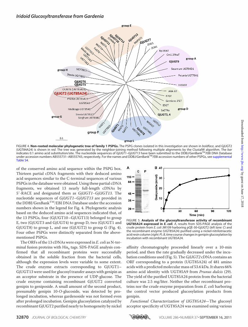
of the conserved amino acid sequence within the PSPG box.Thirteen partial cDNA fragments with their deduced aminoacid sequences similar to the C-terminal sequences of variousPSPGs in the databasewere obtained.Using these partial cDNAfragments, we obtained 13 nearly full-length cDNAs by5�-RACE and designated them as GjUGT1–GjUGT13. Thenucleotide sequences of GjUGT1–GjUGT13 are provided intheDDBJ/GenBankTM/EBIDNADatabase under the accessionnumbers shown in the legend for Fig. 4. Phylogenetic analysisbased on the deduced amino acid sequences indicated that, ofthe 13 PSPGs, four (GjUGT10–GjUGT13) belonged to groupE, two (GjUGT3 and GjUGT4) to group D, two (GjUGT1 andGjUGT8) to group L, and one (GjUGT2) to group G (Fig. 4).Four other PSPGs were distinctly separated from the above-mentioned PSPGs.TheORFs of the 13 cDNAswere expressed inE. coli asN-ter-
minal fusion proteins with His6 tags. SDS-PAGE analysis con-firmed that all recombinant proteins were successfullyobtained in the soluble fraction from the bacterial cells,although the expression levels were variable to some extent.The crude enzyme extracts corresponding to GjUGT1–GjUGT13were used for glucosyl transfer assays with genipin asan acceptor substrate in the presence of UDP-glucose. Thecrude enzyme containing recombinant GjUGT2 convertedgenipin to geniposide. A small amount of the second product,presumably genipin 10-O-glucoside, was detected by pro-longed incubation, whereas gardenoside was not formed evenafter prolonged incubation. Genipin glucosylation catalyzed byrecombinantGjUGT2 purified nearly to homogeneity by nickel
affinity chromatography proceeded linearly over a 10-minperiod, and then the rate gradually decreased under the incu-bation conditions used (Fig. 5). TheGjUGT2 cDNAcontains anORF corresponding to a protein (UGT85A24) of 481 aminoacidswith a predictedmolecularmass of 53.6 kDa. It shares 66%amino acid identity with UGT85A9 from Prunus dulcis (29).The yield of the purified UGT85A24 protein from the bacterialculture was 2.5 mg/liter. Neither the other recombinant pro-teins nor the crude enzyme preparation from E. coli harboringthe control vector produced glucosylation products fromgenipin.Functional Characterization of UGT85A24—The glucosyl
acceptor specificity of UGT85A24 was examined using various
FIGURE 4. Non-rooted molecular phylogenetic tree of family 1 PSPGs. The PSPG clones isolated in this investigation are shown in boldface, and GjUGT2(UGT84A24) is shown in red. The tree was generated by the neighbor-joining method following multiple alignments by the ClustalW algorithm. The barindicates 0.1 amino acid substitution/site. The nucleotide sequences of GjUGT1–GjUGT13 have been submitted to the DDBJ/GenBankTM/EBI DNA Databaseunder accession numbers AB555731–AB555743, respectively. For the names and DDBJ/GenBankTM/EBI accession numbers of other PSPGs, see supplementalTable S4.
FIGURE 5. Analysis of the glucosyltransferase activity of recombinantUGT85A24 expressed in E. coli. A, results from SDS-PAGE analysis of thecrude protein from E. coli JM109 harboring pQE-30-GjUGT2 (left lane: C) andthe recombinant enzyme (UGT85A24) purified using a nickel-nitrilotriaceticacid resin column (right: P). B, time course changes in genipin glucosylation byincubation with recombinant UGT85A24.
Iridoid Glucosyltransferase from Gardenia
32870 JOURNAL OF BIOLOGICAL CHEMISTRY VOLUME 286 • NUMBER 37 • SEPTEMBER 16, 2011
by guest on June 17, 2018http://w
ww
.jbc.org/D
ownloaded from

substrates. The chemical structures of the compounds used areshown in supplemental Fig. S1. HPLC analysis revealed the for-mation of a product with a retention time and UV spectrumidentical to those of 7-deoxyloganin (7-deoxyloganetin 1-O-glucoside, 3) when 7-deoxyloganetin (4) was used as a substrate(Fig. 6A). Loganin (loganetin 1-O-glucoside, 5) was also pro-duced from loganetin (6), but the yield was extremely low com-pared with that from 7-deoxyloganin (Fig. 6B). No product wasformed from 7-deoxyloganetic acid. UGT85A24 exhibited littleglucosyltransferase activity toward several phenolic com-pounds, including 4-nitrophenol, curcumin, flavonoids, cou-marins, crocetin, and phytohormones such as trans-zeatinand indoleacetic acid. As for the glycosyl donor specificity,UGT85A24 exhibited no activity toward either UDP-galactoseor UDP-glucuronic acid.The kinetic parameters of UGT85A24 for iridoid aglycones
using UDP-glucose as the sugar donor substrate were deter-mined using the affinity-purified protein (Table 1). The appar-ent Km values for genipin and 7-deoxyloganetin were 8.8 and0.61 mM, respectively. The kcat/Km ratio for 7-deoxyloganetinwas 2-fold higher than that for genipin. The kinetic parametersfor UDP-glucose were also determined using 7-deoxyloganetinas the sugar acceptor (Table 1).
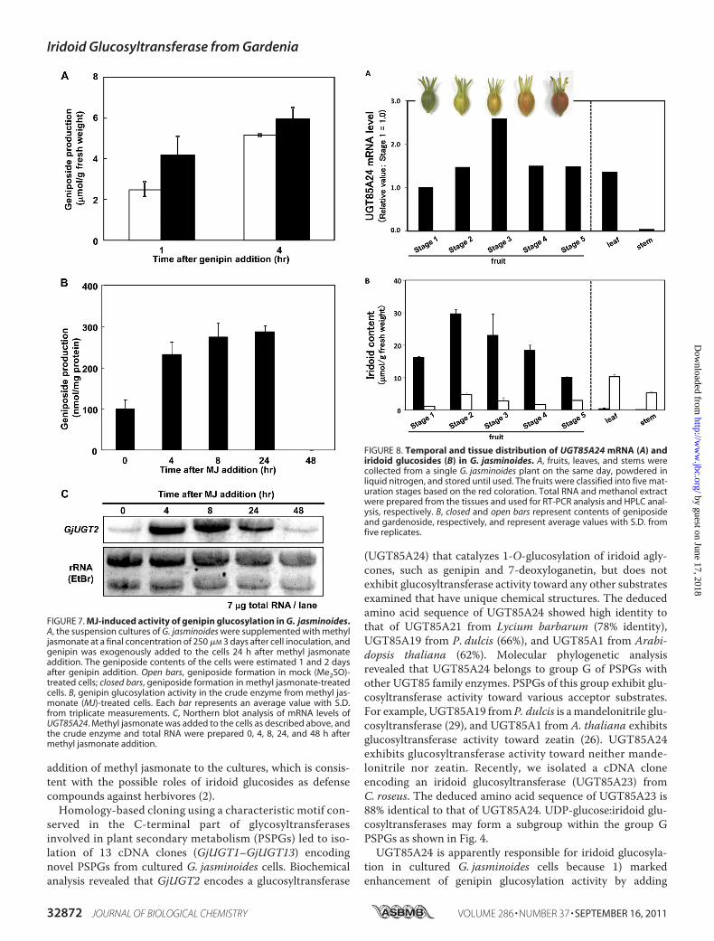
Expression of the UGT85A24 Gene in G. jasminoides—Therelationship between the enzymatic activity of genipin gluco-syltransferase and the mRNA level of UGT85A24 in culturedG. jasminoides cells was examined. The genipin glucosylationactivity of the cultured cells (Fig. 7A) and that of the crudeenzyme extract from the cultured cells (Fig. 7B) were markedlyincreased by adding methyl jasmonate to the cell cultures. ThemRNA level ofUGT85A24 in the cells was also strongly up-reg-ulated, and the pattern of up-regulation correlatedwell with thepattern of increased enzymatic activity (Fig. 7C).The expression levels ofUGT85A24mRNA in variousG. jas-
minoides organs were examined by RT-PCR (Fig. 8A). Toexamine the specificity of the PCR primers forUGT85A24, thePCR products were subcloned into T-Vector pMD20. Twentyclones were randomly selected and sequenced. All of the clonesexhibited the same sequence with UGT85A24 cDNA, indicat-ing that the PCR primers annealed specifically to UGT85A24cDNA. UGT85A24 mRNA was detected at similar levels in allorgans examined except the stems, in which the expressionlevel was very low. The mRNA level gradually increased duringthe early stage of fruit ripening, whereas it remained low in themature fruits. Geniposide and gardenoside contents in G. jas-minoides organs were quantitatively determined (Fig. 8B).Geniposide was found to be a predominant iridoid inG. jasmi-noides fruits, whereas gardenoside was found to be a majoriridoid compound in other organs. Iridoid contents in the fruitsgradually increased in the early stages of development andgradually decreased afterward. Iridoid content was somewhatlower in the vegetative tissues than in the flowers and fruits. Astrict correlation between the mRNA level and iridoid contentin the plant organs was not exhibited, except that both themRNA level ofUGT85A24 and the iridoid content were low inthe stems compared with those in the other organs.
DISCUSSION
Cultured G. jasminoides cells efficiently glucosylated exoge-nously added genipin to geniposide and converted geniposideto gardenoside. We also examined genipin glucosylation usingcell suspension cultures of C. roseus, which produces no geni-poside in planta. Only �3% of the initially supplemented geni-pin was converted to glucoside in the cell suspension cultures,and no further transformation of geniposide to gardenosidewas observed. Therefore, the cellular activity of genipin gluco-sylation is specific to G. jasminoides, which has biosyntheticactivity for geniposide-related iridoid glucosides. The gluco-sylation activity in theG. jasminoides cells was increased by the
FIGURE 6. A and B, glucosylation of 7-deoxyloganetin and loganetin, respec-tively, by UGT85A24. Each iridoid aglycone was incubated with UGT85A24 for60 min, and the assay mixture was subjected to HPLC analysis. C, HPLC profileof a mixture comprising standards. 3, 7-deoxyloganin; 4, 7-deoxyloganetin; 5,loganin; 6, loganetin.
TABLE 1Kinetic parameters of recombinant UGT85A24 for iridoid aglyconesand UDP-glucoseData represent means � S.D. from triplicate measurements. NA, no activity; ND,cannot be determined because only trace amounts of the product were formed(detection limit, 0.1 nmol of product/50 �l of reaction mixture).
Km kcat kcat/Km
mM s�1 mM�1 s�1
Genipin 8.82 � 0.09 1.04 � 0.04 0.12 � 0.0047-Deoxyloganetin 0.61 � 0.08 0.13 � 0.04 0.21 � 0.0337-Deoxyloganetic acid NA NA NALoganetin ND ND NDUDP-glucose 0.17 � 0.03 0.16 � 0.02 1.00 � 0.21
Iridoid Glucosyltransferase from Gardenia
SEPTEMBER 16, 2011 • VOLUME 286 • NUMBER 37 JOURNAL OF BIOLOGICAL CHEMISTRY 32871
by guest on June 17, 2018http://w
ww
.jbc.org/D
ownloaded from
addition of methyl jasmonate to the cultures, which is consis-tent with the possible roles of iridoid glucosides as defensecompounds against herbivores (2).Homology-based cloning using a characteristic motif con-
served in the C-terminal part of glycosyltransferasesinvolved in plant secondary metabolism (PSPGs) led to iso-lation of 13 cDNA clones (GjUGT1–GjUGT13) encodingnovel PSPGs from cultured G. jasminoides cells. Biochemicalanalysis revealed that GjUGT2 encodes a glucosyltransferase
(UGT85A24) that catalyzes 1-O-glucosylation of iridoid agly-cones, such as genipin and 7-deoxyloganetin, but does notexhibit glucosyltransferase activity toward any other substratesexamined that have unique chemical structures. The deducedamino acid sequence of UGT85A24 showed high identity tothat of UGT85A21 from Lycium barbarum (78% identity),UGT85A19 from P. dulcis (66%), and UGT85A1 from Arabi-dopsis thaliana (62%). Molecular phylogenetic analysisrevealed that UGT85A24 belongs to group G of PSPGs withother UGT85 family enzymes. PSPGs of this group exhibit glu-cosyltransferase activity toward various acceptor substrates.For example, UGT85A19 from P. dulcis is amandelonitrile glu-cosyltransferase (29), and UGT85A1 from A. thaliana exhibitsglucosyltransferase activity toward zeatin (26). UGT85A24exhibits glucosyltransferase activity toward neither mande-lonitrile nor zeatin. Recently, we isolated a cDNA cloneencoding an iridoid glucosyltransferase (UGT85A23) fromC. roseus. The deduced amino acid sequence of UGT85A23 is88% identical to that of UGT85A24. UDP-glucose:iridoid glu-cosyltransferases may form a subgroup within the group GPSPGs as shown in Fig. 4.UGT85A24 is apparently responsible for iridoid glucosyla-
tion in cultured G. jasminoides cells because 1) markedenhancement of genipin glucosylation activity by adding
FIGURE 7. MJ-induced activity of genipin glucosylation in G. jasminoides.A, the suspension cultures of G. jasminoides were supplemented with methyljasmonate at a final concentration of 250 �M 3 days after cell inoculation, andgenipin was exogenously added to the cells 24 h after methyl jasmonateaddition. The geniposide contents of the cells were estimated 1 and 2 daysafter genipin addition. Open bars, geniposide formation in mock (Me2SO)-treated cells; closed bars, geniposide formation in methyl jasmonate-treatedcells. B, genipin glucosylation activity in the crude enzyme from methyl jas-monate (MJ)-treated cells. Each bar represents an average value with S.D.from triplicate measurements. C, Northern blot analysis of mRNA levels ofUGT85A24. Methyl jasmonate was added to the cells as described above, andthe crude enzyme and total RNA were prepared 0, 4, 8, 24, and 48 h aftermethyl jasmonate addition.
FIGURE 8. Temporal and tissue distribution of UGT85A24 mRNA (A) andiridoid glucosides (B) in G. jasminoides. A, fruits, leaves, and stems werecollected from a single G. jasminoides plant on the same day, powdered inliquid nitrogen, and stored until used. The fruits were classified into five mat-uration stages based on the red coloration. Total RNA and methanol extractwere prepared from the tissues and used for RT-PCR analysis and HPLC anal-ysis, respectively. B, closed and open bars represent contents of geniposideand gardenoside, respectively, and represent average values with S.D. fromfive replicates.
Iridoid Glucosyltransferase from Gardenia
32872 JOURNAL OF BIOLOGICAL CHEMISTRY VOLUME 286 • NUMBER 37 • SEPTEMBER 16, 2011
by guest on June 17, 2018http://w
ww
.jbc.org/D
ownloaded from
methyl jasmonate toG. jasminoides cell cultures was consistentwith the strong up-regulation of UGT85A24 mRNA expres-sion, and 2) G. jasminoides cells efficiently glucosylatedexogenously added 7-deoxyloganetin to glucoside but poorlyglucosylated 7-deoxyloganetic acid (data not shown),which is consistent with the substrate preferentiality ofUGT85A24. UGT85A24 mRNA expression was alsodetected in various G. jasminoides organs. The gradualincrease in the mRNA level during the early phase of fruitripening correlates with an increase in iridoid accumulationduring the fruit development. Furthermore, the relativelylow level of iridoid accumulation (geniposide plus gardeno-side) in the vegetative tissues is consistent with the distribu-tion pattern of UGT85A24mRNA in the plant organs. Thus,UGT85A24 may at least partially limit the iridoid accumula-tion in planta.Iridoid biosynthesis has been extensively studied using cell
cultures ofC. roseus and L. japonica because the biosynthesis ofsecologanin, a major iridoid metabolite in these species, hasbeen considered a bottleneck for monoterpenoid indole alka-loid production in C. roseus cell cultures (7). Tracer experi-ments and enzymatic studies revealed that the iridane skeletonis formed by the cyclization of 10-oxogeranial biosynthesizedfrom geraniol through 10-hydroxygeraniol. Conversion of thecyclization product iridodial to loganin and then to secologanininvolves several steps, including 1-O-glucosylation of theiridane skeleton (Fig. 1). However, the glucosylation step hasremained elusive so far. Kinetic analysis of UGT85A24 indi-cated that the Km value for 7-deoxyloganetin is �13-fold lowerthan that for genipin, whereas the kcat value for genipin is �8times higher than that for 7-deoxyloganetin. Thus, the catalyticefficiency (kcat/Km) is�2-fold higher for 7-deoxyloganetin thanfor genipin. This result suggests that 7-deoxyloganetin is thepreferential sugar acceptor for UGT85A24 and probably anendogenous substrate of the glucosylation step in the biosyn-thetic pathways of geniposide and gardenoside in G. jasmi-noides, although genipin is a good substrate because of its struc-tural similarity to 7-deoxyloganetin. In G. jasminoides,7-deoxyloganetic acid is converted to 7-deoxyloganetin, whichis then glucosylated to 7-deoxyloganin. Although the Km valueof UGT85A24 for 7-deoxyloganetin (�0.6 mM) seems to behigher compared with the values of, for example, flavonoid glu-cosyltransferases (30, 31), it is in the same range of the Kmvalues for terpenoid glucosyltransferases so far reported (32,33).Hydroxylation of 7-deoxyloganin at C-10 results in the for-
mation of geniposide as shown in Fig. 1B. Yamamoto and co-workers (8) examined the kinetic parameters of the partiallypurified glucosyltransferase preparation from L. japonica cellsand noted that the preparation had the highest affinity for 7-de-oxyloganetic acid comparedwith 7-deoxyloganetin and logane-tin, although the specific activity was the highest for loganetin.On the basis of their result, they proposed that 7-deoxy-loganetic acid is a sugar-accepting substrate in the iridoid bio-synthetic pathway leading to loganin. Glucosylation of iridoidprecursors may occur in different steps between loganin bio-synthesis in L. japonica and geniposide biosynthesis in G. jas-minoides. However, the partially purified enzyme preparation
from L. japonica cells may be amixture of glucosyltransferases,which makes the estimated the kinetic parameters difficult todiscern.In conclusion, we isolated a novel UDP-glucose:7-deoxy-
loganetin glucosyltransferase, UGT85A24 from G. jasmi-noides. Our results shed light not only on the glucosylation stepin iridoid biosynthesis in G. jasminoides but also on the isola-tion of various iridoid-specific glucosyltransferases fromdiverse iridoid- and indole alkaloid-producing plant species,leading to the engineering of biosynthetic pathways producingiridoids and/or monoterpenoid indole alkaloids.
Acknowledgment—We thank Professor K. Inoue for the generous giftof 7-deoxyloganin tetraacetate.
REFERENCES1. Dinda, B., Debnath, S., and Harigaya, Y. (2007) Chem. Pharm. Bull. 55,
159–2222. Harvey, J. A., van Nouhuys, S., and Biere, A. (2005) J. Chem. Ecol. 31,
287–3023. El-Sayed, M., and Verpoorte, R. (2007) Phytochem. Rev. 6, 277–3054. Ghisalberti, E. L. (1998) Phytomedicine 5, 147–1635. Dinda, B., Debnath, S., and Harigaya, Y. (2007) Chem. Pharm. Bull. 55,
689–7286. Meijer, A. H., Verpoorte, R., and Hoge, J. H. (1993) J. Plant Res. 3,
145–1647. Oudin, A., Courtois, M., Rideau, M, and Clastre, M. (2007) Phytochem.
Rev. 6, 259–2768. Yamamoto, H., Sha, M., Kitamura, K., Yamaguchi, Y., Katano, N., and
Inoue, K. (2002) Plant Biotechnol. 19, 295–3019. Vogt, T., and Jones, P. (2000) Trends Plant Sci. 5, 380–38610. Lim, E. K., and Bowles, D. J. (2004) EMBO J. 23, 2915–292211. Gachon, C. M., Langlois-Meurinne, M., and Saindrenan, P. (2005) Trends
Plant Sci. 10, 542–54912. Hughes, J., and Hughes, M. A. (1994) DNA Seq. 5, 41–4913. Offen, W., Martinez-Fleites, C., Yang, M., Kiat-Lim, E., Davis, B. G., Tar-
ling, C. A., Ford, C. M., Bowles, D. J., and Davies, G. J. (2006) EMBO J. 25,1396–1405
14. Li, L., Modolo, L. V., Escamilla-Trevino, L. L., Achnine, L., Dixon, R. A.,and Wang, X. (2007) J. Mol. Biol. 370, 951–963
15. Brazier-Hicks, M., Offen, W. A., Gershater, M. C., Revett, T. J., Lim, E. K.,Bowles, D. J., Davies, G. J., and Edwards, R. (2007) Proc. Natl. Acad. Sci.U.S.A. 104, 20238–20243
16. Noguchi, A., Fukui, Y., Iuchi-Okada, A., Kakutani, S., Satake, H., Iwashita,T., Nakao, M., Umezawa, T., and Ono, E. (2008) Plant J. 54, 415–427
17. Masada, S., Terasaka, K., Oguchi, Y., Okazaki, S., Mizushima, T., andMizukami, H. (2009) Plant Cell Physiol. 50, 1401–1415
18. Yu, Y., Xie, Z. L., Gao, H., Ma, W. W., Dai, Y., Wang, Y., Zhong, Y., andYao, X. S. (2009) J. Nat. Prod. 72, 1459–1464
19. Ueda, S., Kobayashi, K.,Muramatsu, T., and Inouye,H. (1981)PlantaMed.41, 186–191
20. Uesato, S., Ueda, S., Kobayashi, K., and Inouye, H. (1983) Chem. Pharm.Bull. 31, 4185–4188
21. Uesato, S., Ueda, S., Kobayashi, K., Miyauchi, M., and Inouye, H. (1984)Tetrahedron Lett. 25, 573–576
22. Linsmaier, E. M., and Skoog, F. (1965) Physiol. Plant. 18, 100–12723. Inoue, K., Ono,M., Nakajima, H., Fujie, I., Inouye, H., and Fujita, T. (1992)
Heterocycles 33, 673–69524. Hasada, K., Yoshida, T., Yamazaki, T., Sugimoto, N., Nishimura, T., Na-
gatsu, A., and Mizukami, H. (2010) J. Nat. Med. 64, 161–16625. Bradford, M. M. (1976) Anal. Biochem. 72, 248–25426. Hou, B., Lim, E. K., Higgins, G. S., and Bowles, D. J. (2004) J. Biol. Chem.
279, 47822–4783227. Chaudhuri, R. K., Afifi-Yazar, F. U., and Sticher,O. (1980)Tetrahedron 36,
Iridoid Glucosyltransferase from Gardenia
SEPTEMBER 16, 2011 • VOLUME 286 • NUMBER 37 JOURNAL OF BIOLOGICAL CHEMISTRY 32873
by guest on June 17, 2018http://w
ww
.jbc.org/D
ownloaded from

2317–232628. Ono, M., Ueno, M., Masuoka, C., Ikeda, T., and Nohara, T. (2005) Chem.
Pharm. Bull. 53, 1342–134429. Franks, T. K., Yadollahi, A.,Wirthensohn,M.G.,Guerin, J. R., Kaiser, B. K.,
Sedgley, M., and Ford, C. M. (2008) Funct. Plant Biol. 35, 236–24630. Nakatsuka, T., Sato, K., Takahashi, H., Yamamura, S., and Nishihara, M.
(2008) J. Exp. Bot. 59, 1241–1252
31. Noguchi, A., Horikawa, M., Fukui, Y., Fukuchi-Mizutani, M., Iuchi-Okada, A., Ishiguro, M., Kiso, Y., Nakayama, T., and Ono, E. (2009) PlantCell 21, 1556–1572
32. Achnine, L., Huhman, D. V., Farag, M. A., Sumner, L. W., Blount, J. W.,and Dixon, R. A. (2005) Plant J. 41, 875–887
33. Naoumkina,M.A.,Modolo, L. V., Huhman,D. V., Urbanczyk-Wochniak, E.,Tang, Y., Sumner, L.W., and Dixon, R. A. (2010) Plant Cell 22, 850–866
Iridoid Glucosyltransferase from Gardenia
32874 JOURNAL OF BIOLOGICAL CHEMISTRY VOLUME 286 • NUMBER 37 • SEPTEMBER 16, 2011
by guest on June 17, 2018http://w
ww
.jbc.org/D
ownloaded from

Mai Nagatoshi, Kazuyoshi Terasaka, Akito Nagatsu and Hajime MizukamiGardenia jasminoidesIridoid-specific Glucosyltransferase from
doi: 10.1074/jbc.M111.242586 originally published online July 28, 20112011, 286:32866-32874.J. Biol. Chem.
10.1074/jbc.M111.242586Access the most updated version of this article at doi:
Alerts:
When a correction for this article is posted•
When this article is cited•
to choose from all of JBC's e-mail alertsClick here
Supplemental material:
http://www.jbc.org/content/suppl/2011/07/28/M111.242586.DC1
http://www.jbc.org/content/286/37/32866.full.html#ref-list-1
This article cites 33 references, 5 of which can be accessed free at
by guest on June 17, 2018http://w
ww
.jbc.org/D
ownloaded from