
Iranian Journal of Basic Medical...
5
Iranian Journal of Basic Medical Sciences ijbms.mums.ac.ir Histopathological study of erythropoietin protective effect on carbon monoxide-induced cardiotoxicity in rat Mitra Asgharian Rezaee 1, 2, 3 , Seyed Adel Moallem 4, 5, 6 , Amir Hooshang Mohammadpour 1, 5 , Mahmoud Mahmoudi 7 , Mojtaba Sankian 8 , Mehdi Farzadnia 9 , Hassan Alavi 10, 11 , Mohsen Imenshahidi 5, 1 * 1 Department of Pharmacodynamics and Toxicology, School of Pharmacy, Mashhad University of Medical Sciences, Mashhad, Iran 2 Department of Toxicology and Pharmacology, Faculty of Pharmacy, Kerman University of Medical Sciences, Kerman, Iran 3 Pharmaceutical Research Center, Institute of Neuropharmacology , Kerman University of Medical Sciences, Kerman, Iran 4 Department of Pharmacology and toxicology, Faculty of Pharmacy, University of Ahl Al Bayt, Karbala, Iraq 5 Pharmaceutical Research Center, Mashhad University of Medical Sciences, Mashhad, Iran 6 Medical Toxicology Research Center, Mashhad University of Medical Sciences, Mashhad, Iran 7 Immunology Research Center, School of Medicine, Mashhad University of Medical Sciences, Mashhad, Iran 8 Immunobiochemistry Laboratory, Immunology Research Center, Bu-Ali Research Institute, School of Medicine, Mashhad University of Medical Sciences, Mashhad, Iran 9 Department of Pathology, School of Medicine, Imam Reza Hospital, Mashhad University of Medical Sciences, Mashhad, Iran 10 Electron Microscope Unit, Bu-Ali Research Institute, Mashhad University of Medical Science, Mashhad, Iran 11 School of Medicine, Mashhad Branch, Islamic Azad University, Mashhad, Iran A R T I C L E I N F O A B S T R A C T Article type: Original article Objective(s) : Cardiotoxicity is one of the major consequences in carbon monoxide poisoning. Following our previous work, in this study we aimed to define the myocardium changes induced by carbon monoxide (CO) intoxication and evaluate erythropoietin (EPO) effect on CO cardiotoxicity in rat. Materials and Methods: Severe carbon monoxide toxicity induced by 3000 ppm CO in Wistar rat. EPO was administrated (5000 IU/Kg, intraperitoneal injection) at the end of CO exposure and then the animals were re-oxygenated with the ambient air. Subsequently heart was removed and assessed by histopathology and electron microscopy examinations. Results: 3000 ppm CO induced significant myocardium injury; multiple foci of necrosis and lymphocyte infiltration compare with the control (P˂0.05). Electron microscopy examination showed myofibril lysis and mitochondrial swelling in myocardium due to 3000 ppm CO poisoning. However EPO administration after CO exposure resulted in significant reduction in cardiomyocytes injury (P˂0.05). Conclusion: Our results represented protective effect of EPO on cardiac injury induced by CO intoxication in rat. Article history: Received: May 16, 2017 Accepted: Aug 10, 2017 Keywords: Carbon monoxide- poisoning Cardiotoxicity Erythropoietin Histopathology Rat ►Please cite this article as: Asgharian Rezaee M, Moallem SA, Mohammadpour AH, Mahmoudi M, Sankian M, Farzadnia M, Alavi H, Imenshahidi M. Histopathological study of erythropoietin protective effect on carbon monoxide-induced cardiotoxicity in rat. Iran J Basic Med Sci 2017; 20:1189-1193. doi: 10.22038/IJBMS.2017.9471 Introduction Carbon monoxide (CO) is well known as a toxic gas and it is associated with high emergency department visits per year (1-3). Moderate to severe CO poisonings are resulting in high rate of hospitalization and mortality (1, 2, 4). CO poisoning is related to more than half of fetal poisoning in worldwide (4). Also there are numerous reports indicated high mortality rate due to CO poisoning in Iran (5, 6). Cardiotoxicity due to CO poisoning is the most common consequences especially in moderate to severe intoxication (2-4, 7). There are broad spectrum of cardiac insults in CO poisoning including: cardiomyopathy, angina, arrhythmia, heart failure and cardiogenic shock (4, 8, 9). Myocardial infarction and sudden death are reported in severe CO poisoning (4). However there are evidences indicating a relation between ambient CO air pollution and increase in cardiac disease events. Several studies have been shown that rise in ambient CO level increased the risk of ischemic heart disease and heart failure especially in elderly and high risk people (10, 11). The pathophysiology of CO induced-cardiotoxicity is related to hypoxic damage and also cardio-specific mechanisms such as impairment of cytochrome C in mitochondria respiratory chain and myoglobin defect (4). Along with nervous damage, cardiac injury is the main CO poisoning consequence but there isn’t any special treatment for cardiovascular events in CO poisoning other than oxygen therapy (2, 4). *Corresponding author: Mohsen Imenshahidi. Department of Pharmacodynamics and Toxicology, School of Pharmacy, Mashhad University of Medical Sciences, Mashhad, Iran. Tel: +98- 51-38823255; Fax: +98-51-38823251; email: [email protected]
-
Upload
trinhkhanh -
Category
Documents
-
view
212 -
download
0
Transcript of Iranian Journal of Basic Medical...
Iranian Journal of Basic Medical Sciences
ijbms.mums.ac.ir
Histopathological study of erythropoietin protective effect on carbon monoxide-induced cardiotoxicity in rat
Mitra Asgharian Rezaee 1, 2, 3, Seyed Adel Moallem 4, 5, 6, Amir Hooshang Mohammadpour 1, 5, Mahmoud Mahmoudi 7, Mojtaba Sankian 8, Mehdi Farzadnia 9, Hassan Alavi 10, 11, Mohsen Imenshahidi 5, 1*
1 Department of Pharmacodynamics and Toxicology, School of Pharmacy, Mashhad University of Medical Sciences, Mashhad, Iran 2 Department of Toxicology and Pharmacology, Faculty of Pharmacy, Kerman University of Medical Sciences, Kerman, Iran 3 Pharmaceutical Research Center, Institute of Neuropharmacology, Kerman University of Medical Sciences, Kerman, Iran 4 Department of Pharmacology and toxicology, Faculty of Pharmacy, University of Ahl Al Bayt, Karbala, Iraq 5 Pharmaceutical Research Center, Mashhad University of Medical Sciences, Mashhad, Iran 6 Medical Toxicology Research Center, Mashhad University of Medical Sciences, Mashhad, Iran 7 Immunology Research Center, School of Medicine, Mashhad University of Medical Sciences, Mashhad, Iran 8 Immunobiochemistry Laboratory, Immunology Research Center, Bu-Ali Research Institute, School of Medicine, Mashhad University of
Medical Sciences, Mashhad, Iran 9 Department of Pathology, School of Medicine, Imam Reza Hospital, Mashhad University of Medical Sciences, Mashhad, Iran 10 Electron Microscope Unit, Bu-Ali Research Institute, Mashhad University of Medical Science, Mashhad, Iran 11 School of Medicine, Mashhad Branch, Islamic Azad University, Mashhad, Iran
A R T I C L E I N F O A B S T R A C T
Article type: Original article
Objective(s): Cardiotoxicity is one of the major consequences in carbon monoxide poisoning. Following our previous work, in this study we aimed to define the myocardium changes induced by carbon monoxide (CO) intoxication and evaluate erythropoietin (EPO) effect on CO cardiotoxicity in rat. Materials and Methods: Severe carbon monoxide toxicity induced by 3000 ppm CO in Wistar rat. EPO was administrated (5000 IU/Kg, intraperitoneal injection) at the end of CO exposure and then the animals were re-oxygenated with the ambient air. Subsequently heart was removed and assessed by histopathology and electron microscopy examinations. Results: 3000 ppm CO induced significant myocardium injury; multiple foci of necrosis and lymphocyte infiltration compare with the control (P˂0.05). Electron microscopy examination showed myofibril lysis and mitochondrial swelling in myocardium due to 3000 ppm CO poisoning. However EPO administration after CO exposure resulted in significant reduction in cardiomyocytes injury (P˂0.05). Conclusion: Our results represented protective effect of EPO on cardiac injury induced by CO intoxication in rat.
Article history: Received: May 16, 2017 Accepted: Aug 10, 2017
Keywords: Carbon monoxide- poisoning Cardiotoxicity Erythropoietin Histopathology Rat
►Please cite this article as: Asgharian Rezaee M, Moallem SA, Mohammadpour AH, Mahmoudi M, Sankian M, Farzadnia M, Alavi H,
Imenshahidi M. Histopathological study of erythropoietin protective effect on carbon monoxide-induced cardiotoxicity in rat. Iran J Basic Med Sci 2017; 20:1189-1193. doi: 10.22038/IJBMS.2017.9471
Introduction Carbon monoxide (CO) is well known as a toxic gas
and it is associated with high emergency department visits per year (1-3). Moderate to severe CO poisonings are resulting in high rate of hospitalization and mortality (1, 2, 4). CO poisoning is related to more than half of fetal poisoning in worldwide (4). Also there are numerous reports indicated high mortality rate due to CO poisoning in Iran (5, 6).
Cardiotoxicity due to CO poisoning is the most common consequences especially in moderate to severe intoxication (2-4, 7). There are broad spectrum of cardiac insults in CO poisoning including: cardiomyopathy, angina, arrhythmia, heart failure and cardiogenic shock (4, 8, 9).
Myocardial infarction and sudden death are reported in severe CO poisoning (4). However there are evidences indicating a relation between ambient CO air pollution and increase in cardiac disease events. Several studies have been shown that rise in ambient CO level increased the risk of ischemic heart disease and heart failure especially in elderly and high risk people (10, 11). The pathophysiology of CO induced-cardiotoxicity is related to hypoxic damage and also cardio-specific mechanisms such as impairment of cytochrome C in mitochondria respiratory chain and myoglobin defect (4). Along with nervous damage, cardiac injury is the main CO poisoning consequence but there isn’t any special treatment for cardiovascular events in CO poisoning other than oxygen therapy (2, 4).
*Corresponding author: Mohsen Imenshahidi. Department of Pharmacodynamics and Toxicology, School of Pharmacy, Mashhad University of Medical Sciences, Mashhad, Iran. Tel: +98- 51-38823255; Fax: +98-51-38823251; email: [email protected]

Asgharian Rezaee et al. EPO protective effect on CO cardiotoxicity in rat
Iran J Basic Med Sci, Vol. 20, No. 11, Nov 2017
1190
Recently, erythropoietin (EPO), a cytokine hormone,
has investigated as a tissue protective and anti-ischemic agent (12, 13). However these new investigated effects is unrelated to its well-known erythropoietic property (14, 15). There are numerous studies indicating protective effect of EPO on cardiac ischemia and myocardial injury (14, 16-18). The results of cardiac ischemia/reperfusion studies have been shown that EPO administration improved cardiac function and reduced infarct size and cardiomyocyte death (15, 19, 20). Several mechanisms have been introduced to explain the protective effects of EPO, such as anti-inflammatory, anti-oxidant and anti-apoptosis properties of EPO (14, 21, 22). We previously showed that EPO could reduce ischemic ECG changes induced by severe CO poisoning in rat (23).
This trial was designed to more investigations on CO cardiotoxicy and EPO effects. For this purpose histopathological examination was utilized for evaluation of EPO effects on CO induced-cardiac injury.
Materials and Methods Animals
Wistar male rats, weighing 200-250 g, were housed in the Animal Center of Buali Research Institute. They were kept under standard conditions (21-23oC temperature, 12 hr/12 hr light/dark cycle) with free access to food and water. All animals were treated in accordance with the Guidelines for the Care and Use of Laboratory Animals prepared by the National Academy of Sciences and published by the National Institutes of Health (NIH publication no. 85-23, Revised 1996). The study was approved by the Animal Care Committee of Mashhad University of Medical Sciences.
Experimental groups
The animals were intoxicated by 3000 ppm CO as a model of severe CO intoxication in rat (24, 25). For induction of CO poisoning, the animals were placed in a 12 l airtight plexiglass container with entrance and exit taps. 3000 ppm of CO was flowed to the container at a constant flow for 40 min. The CO concentration was monitored continuously with a CO analyzer (Model 707 carbon monoxide analyzer, TPI, Korea). At the end of CO exposure, animals in EPO+CO group were injected intraperitoneally with recombinant human EPO (rhEPO, Pooyesh Draou Co, Iran; 5000 IU/kg). EPO dosing was selected based on previous published studies (14, 15). Blood sample was taken from the tail vein after CO exposure and then all animals were exposed to ambient air for 2 hr. After anesthesia induction by ketamin/xylazin mixture (100/10 mg/kg) the animal’s heart was removed. A total five animals in each group were examined. Animal groups were as the following:
Group 1 (3000 ppm CO group): 5 animals were exposed to 3000 ppm of CO for 40 min. Afterward the animals were re-oxygenated with normal air. Group 2 (3000 ppm CO + EPO group): 5 animals were exposed to 3000 ppm of CO for 40 min. EPO was injected after CO exposure, and then the animals were re-oxygenated with normal air. Group 3 (control group): 5 rats, no CO inhalation and no EPO treatment.
Carboxyhemoglobin level assay
Blood samples were taken from the tail vein of animals and heparinized. Carboxyhemoglobin levels were assessed with a spectrophotometer calibrated for rat blood (Jasco, Japan).
Histological examination
One portion of the harvested left ventricle was fixed in phosphate-buffered 10% formalin, embedded in paraffin and sectioned at a thickness of 5 μm. The sections were stained with hematoxylin and eosin. The myocardial injury in each group was evaluated semi-quantitatively by light microscopy. Changes were graded based on the frequency and severity of observed lesions (Table 2). The first grade was defined as the presence of dispersed necrotic cells and/or lymphatic infiltration, the second grade related to the presence of necrotic unifocal and/or bifocal area (Figure 1B), and the third grade was shown the presence of more than two necrotic areas per section (Figure 1C).
Electron microscopy
Myocardial tissue samples were processed for electron microscopy; a portion of the left ventricle
was rapidly (within 1 min) cut into 1 mm3 pieces, fixed with 2.5% glutaraldehyde and stored at 4˚C. These portions were embedded in glycol methac - rylate resin, sectioned at a thickness of 1 μm and stained with alkaline toluidine blue for electron microscope analysis. Edema in the sarcoplasmic reticulum, vacuolization in myocytes, loss of myofibrils and injury of mitochondria were assessed by electron microscopy.
Statistical analysis
Data analyses were performed using SPSS version 11.5. Individual groups were assessed with Chi-square or Fisher’s Exact Test; all statistical tests were 2-sided. Differences were considered significant at P<0.05.
Results Carboxyhemoglobin (COHb) levels after CO intoxication
Carboxyhemoglobin values are shown in Table 1. The COHb level is a biochemical marker of CO

EPO protective effect on CO cardiotoxicity in rat Asgharian Rezaee et al.
Iran J Basic Med Sci, Vol. 20, No. 11, Nov 2017
1191
poisoning. High level of COHb was shown severe CO poisoning in rats by 3000 ppm CO.
Myocardial pathology CO intoxication by 3000 ppm CO induced myocardial
injury that was visible by light microscopy. Necrotic lesions and lymphatic infiltration as grade 2 and 3 were shown in 3000 ppm CO group predomin- antly (Figure 1). In EPO treatment group less necrotic area and lymphatic infiltration were observed. Subsequently 3000 ppm CO +EPO group represented less myocardium injury in comparison with 3000 ppm CO group that not received EPO treatment and significant difference was shown between these two groups (P≤0.05), (Table 2). No lesions were observed in samples of control group.
Table1. Blood carboxyhemoglobin levels after carbon monoxide poisoning in animals
Groups Mean±SD (ng/ml) Range (%)
3000 ppm groups 70±8 60-76 Control group 1±0.9 0.7-1.5
Figure 1. Histology evaluation of myocardium in 3000 ppm CO, 3000 ppm CO+EPO and control groups stained by haematoxylin-eosin. A) Normal state (grade 0) was shown in 3000 ppm CO+EPO and control groups. B) Section representing grade 2; uni and bifocal area was observed in 3000 ppm CO group. C) Section display grade 3; more than bifocal necrotic was shown in 3000 ppm CO group. (All picture are shown in magnification×40)
Figure 2. Evaluation of cardiomyocytes by electron microscopy: A) Normal myofibril and mitochondria in control group; ×3150. B) Swelling and disruption of mitochondria and myofibril degeneration in rat receiving 3000 ppm carbon monoxide (CO); ×4000. C) Extensive myofibril degeneration in rat intoxicated by 3000 ppm CO; ×4000. D) Mitochondria and myofibril state in rat receiving erythropoietin (EPO) after 3000 ppm CO intoxication; ×6300
Electron microscopy Mitochondrial swelling and disruption and also
myofibrillar lysis were observed frequently in heart rat intoxicated by 3000 ppm CO. No significant changes were shown in 3000 ppm CO+EPO group compare with the control group (Figure 2).
Discussion Cardiac insult is one of the major consequences of
CO poisoning leading to high morbidity and mortality (2, 4, 8). Myocardial injury associated with severe CO intoxication has been shown frequently in the literatures (9, 26, 27).
In this study CO induced-myocardial injury in rat has been evaluated; the histopathology assessment has been shown lymphocyte infiltration and exten- sive necrotic lesions. Moreover myofibril lysis and mitochondrial swelling were shown by electron microscopy. The administration of EPO after CO significantly reduced myocyte insults compare with the 3000 ppm CO group. Fineschi et al .(28) have been shown coagulative myocytolysis and necrotic myocells in rat heart following sub lethal CO intoxication. In other study lymphatic infiltrations also necrotic cells and foci demonstrated within 2 hours following 3000 ppm CO intoxication in rat (29).
In recent years, EPO extensively has been evaluated as a tissue protective agent (12, 13) . Moreover there are numerous studies indicating protective effect of EPO against cardiac injury (14, 16, 20, 30).
There are several invivo studies indicating administration of 5000 IU/Kg EPO after cardiac ischemia resulted in improvement of cardiac functions and hemodynamic also reduction of myocyte loss and infarct size (14, 15, 31).
Also in other studies EPO evaluated in doxorubicin- induced cardiotoxicity and the results showed the cardiac function improvement and inhibition of cardiomyocytes apoptosis (32, 33). Previously we have shown that EPO administration (5000 IU/Kg) after 3000 ppm CO, reduced significantly electrocardiogram (ECG) ischemic changes in rat (23). Several mechanisms have been investigated to explain tissue protective effect of EPO. The anti-inflammatory, anti-oxidative and anti-apoptosis effects of EPO probably attributed to EPO protective effect. Since the roles of ischemia and oxidative damage in CO cardiotoxicity, it seems the anti-ischemic and anti-oxidative properties of EPO probably have roles in reduction of myocardial injury induced by CO intoxication.
Conclusion The results of present study showed that EPO
significantly reduced cardiomycyte injury induced by severe CO intoxication in rat that confirmed the
Asgharian Rezaee et al. EPO protective effect on CO cardiotoxicity in rat
Iran J Basic Med Sci, Vol. 20, No. 11, Nov 2017
1192
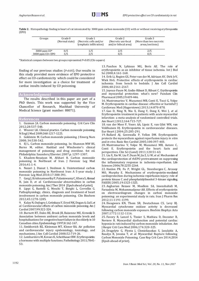
Table 2. Histopathology finding in heart of rat intoxicated by 3000 ppm carbon monoxide (CO) with or without receiving erythropoietin (EPO)
Groups Grade 0 (Normal state)
Grade 1 (Necrotic cells and/or lymphatic infiltration)
Grade 2 (Necrotic uni-focal
and/or bifocal area)
Grade 3 (More than two necrotic
areas/sections)
3000 ppm CO* 0/5 1/5 2/5 2/5 3000 ppm CO+ EPO 3/5 2/5 0/5 0/5
*Statistical compare between two groups represented P˂0.05 (Chi-square)
finding of our previous studies (P˂0.05). Our results in this study provided more evidence of EPO protective effect on CO-cardiotoxicity which could be considered for more investigation as a choice for treatment of cardiac insults induced by CO poisoning.
Acknowledgment
The results described in this paper are part of a PhD thesis. This work was supported by the Vice Chancellor of Research, Mashhad University of Medical Science (grant number: 88071).
References 1. Guzman JA. Carbon monoxide poisoning. Crit Care Clin 2012;28:537-548. 2. Weaver LK. Clinical practice. Carbon monoxide poisoning. N Engl J Med 2009;360:1217-1225. 3. Goldstein M. Carbon monoxide poisoning. J Emerg Nurs 2008; 34:538-542. 4. EJ L. Carbon monoxide poisoning. In: Shannon MW BS, Burns M, editor. Haddad and Winchester's clinical management of poisoning and drug overdose. 4st ed. Philadelphia: Saunders/Elsevier; 2007.p. 1297-1307. 5. Khadem-Rezaiyan M, Afshari R. Carbon monoxide poisoning in Northeast of Iran. J Forensic Leg Med 2016;41:1-4. 6. Nazari J, Dianat I, Stedmon A. Unintentional carbon monoxide poisoning in Northwest Iran: A 5-year study. J Forensic Leg Med 2010;17:388-391. 7. Garg J, Krishnamoorthy P, Palaniswamy C, Khera S, Ahmad H, Jain D, et al. Cardiovascular abnormalities in carbon monoxide poisoning. Am J Ther 2014 [Epub ahead of print]. 8. Lippi G, Rastelli G, Meschi T, Borghi L, Cervellin G. Pathophysiology, clinics, diagnosis and treatment of heart involvement in carbon monoxide poisoning. Clin Biochem 2012;45:1278-1285. 9. Kalay N, Ozdogru I, Cetinkaya Y, Eryol NK, Dogan A, Gul I, et al. Cardiovascular effects of carbon monoxide poisoning. Am J Cardiol 2007;99:322-324. 10. Burnett RT, Dales RE, Brook JR, Raizenne ME, Krewski D. Association between ambient carbon monoxide levels and hospitalizations for congestive heart failure in the elderly in 10 Canadian cities. Epidemiology 1997;8:162-167. 11. Simkhovich BZ, Kleinman MT, Kloner RA. Air pollution and cardiovascular injury epidemiology, toxicology, and mechanisms. J Am Coll Cardiol 2008;52:719-26. 12. Lombardero M, Kovacs K, Scheithauer BW. Erythropoietin: a hormone with multiple functions. Pathobiology 2011;78:41-53.
13. Paschos N, Lykissas MG, Beris AE. The role of erythropoietin as an inhibitor of tissue ischemia. Int J Biol Sci 2008;4:161-168. 14. Erik Li, Regien GS, Peter van der M, Adriaan AV, Dirk JvV, Wiek HvG. Protective effects of erythropoietin in cardiac ischemia: from bench to bedside. J Am Coll Cardiol 2006;48:2161-2167. 15. Joyeux-Faure M, Godin-Ribuot D, Ribuot C. Erythropoietin and myocardial protection: what's new? Fundam Clin Pharmacol 2005;19:439-446. 16. Mastromarino V, Musumeci MB, Conti E, Tocci G, Volpe M. Erythropoietin in cardiac disease: effective or harmful? J Cardiovasc Med (Hagerstown) 2013;14:870-878. 17. Gao D, Ning N, Niu X, Dang Y, Dong X, Wei J , et al. Erythropoietin treatment in patients with acute myocardial infarction: a meta-analysis of randomized controlled trials. Am Heart J 2012;164:715-727. 18. van der Meer P, Voors AA, Lipsic E, van Gilst WH, van Veldhuisen DJ. Erythropoietin in cardiovascular diseases. Eur Heart J 2004;25:285-291. 19. Bullard AJ, Govewalla P, Yellon DM. Erythropoietin protects the myocardium against reperfusion injury in vitro and in vivo. Basic Res Cardiol 2005;100:397-403. 20. Mastromarino V, Volpe M, Musumeci MB, Autore C, Conti E. Erythropoietin and the heart: facts and perspectives. Clin Sci (Lond) 2011;120:51-63. 21. Liu X, Xie W, Liu P, Duan M, Jia Z, Li W, et al. Mechanism of the cardioprotection of rhEPO pretreatment on suppressing the inflammatory response in ischemia-reperfusion. Life Sciences 2006;78:2255-2264. 22. Hanlon PR, Fu P, Wright GL, Steenbergen C, Arcasoy MO, Murphy E. Mechanisms of erythropoietin-mediated cardioprotection during ischemia-reperfusion injury: role of protein kinase C and phosphatidylinositol 3-kinase signaling. FASEB J 2005;19:1323-1325. 23. Asgharian Rezaee M, Moallem SA, Imenshahidi M, Farzadnia M, Mohammadpour AH. Effects of erythropoietin on electrocardiogram changes in carbon monoxide poisoning: an experimental study in rats. Iran J Pharm Res 2012;11:1191-1199. 24. Iheagwara KN, Thom SR, Deutschman CS, Levy RJ. Myocardial cytochrome oxidase activity is decreased following carbon monoxide exposure. Biochim Biophys Acta 2007;1772:1112-1116. 25. Favory R, Lancel S, Tissier S, Mathieu D, Decoster B, Neviere R. Myocardial dysfunction and potential cardiac hypoxia in rats induced by carbon monoxide inhalation. Am J Respir Crit Care Med 2006;174:320-325. 26. Dragelyte G, Plenta J, Chmieliauskas S, Jasulaitis A, Raudys R, Jovaisa T, et al. Myocardial Rupture following Carbon Monoxide Poisoning. Case Rep Crit Care 2014;2014 [Epub ahead of print].

EPO protective effect on CO cardiotoxicity in rat Asgharian Rezaee et al.
Iran J Basic Med Sci, Vol. 20, No. 11, Nov 2017
1193
27. Jung YS, Lee JS, Min YG, Park JS, Jeon WC, Park EJ , et al. Carbon monoxide-induced cardiomyopathy. Circ J 2014;78:1437-1444. 28. Fineschi V, Agricola E, Baroldi G, Bruni G, Cerretani D, Mondillo S, et al. Myocardial findings in fatal carbon monoxide poisoning: a human and experimental morphometric study. Int J Legal Med 2000;113:276-282. 29. Mohamadpour AH, Moallem SA, Hashemzaei M, Abnous K, Tabatabaee Yazdi SA, Imenshahidi M. Effects of granulocyte colony-stimulating factor on electrocardiogram changes after carbon monoxide poisoning in rats. Drug Chem Toxicol. 2012; 35:353-60. Effects of granulocyte colony-stimulating factor on electrocardiogram changes after carbon monoxide poisoning in rats. Drug Chem Toxicol 2012;35:353-60.
30. Vogiatzi G, Briasoulis A, Tousoulis D, Papageorgiou N, Stefanadis C. Is there a role for erythropoietin in cardiovascular disease? Expert Opin Biol Ther 2009;10:251-64. 31. Santhanam AVR, d'Uscio LV, Katusic ZS, Paul MV. Cardiovascular Effects of Erythropoietin: An Update. Adv Pharmacol 2010; 60:257-85. 32. Ammar HI, Saba S, Ammar RI, Elsayed LA, Ghaly WB, Dhingra S. Erythropoietin protects against doxorubicin-induced heart failure. Am J Physiol Heart Circ Physiol 2011;301:H2413-21. 33. Chen X, Chen Y, Bi Y, Fu N, Shan C, Wang S , et al. Preventive cardioprotection of erythropoietin against doxorubicin-induced cardiomyopathy. Cardiovasc Drugs Ther 2007;21:367-74.


