
Ionic Currents Underlying Developmental Regulation of Repetitive ...
16
Ionic Currents Underlying Developmental Regulation of Repetitive Firing in Aplysia Bag Cell Neurons Teresa A. Nick, 1 Leonard K. Kaczmarek, 1,3 and Thomas J. Carew 1,2 1 Interdepartmental Neuroscience Program, 2 Departments of Psychology and Biology, Yale University, New Haven, Connecticut 06510, and 3 Departments of Pharmacology and Cellular and Molecular Physiology, Yale University School of Medicine, New Haven, Connecticut 06510 We have investigated the developmental regulation of the abil- ity to fire repetitively in the bag cell neurons of Aplysia califor- nica, a neuronal system in which the behavioral effects of repetitive firing are well characterized. Adult bag cell neurons exhibit an afterdischarge, consisting of prolonged depolariza- tion and repetitive firing, which causes the release of several peptides from these neurons that induce egg-laying behaviors. Afterdischarge can be triggered in vitro by a variety of stimuli, including electrical stimulation and exposure to the potassium channel blocker tetraethyl ammonium chloride (TEA). In con- trast to adults, juvenile neurons did not exhibit afterdischarge in response to pleural–abdominal connective shock or TEA. Ju- venile neurons did exhibit, however, prolonged depolarizations in the presence of TEA, perhaps reflecting the anlage of the mechanism responsible for afterdischarge in the adult. To investigate developmental mechanisms underlying the regulation of repetitive firing, we compared ionic currents in adult and juvenile bag cell neurons. We found that during the period in which these neurons acquire the capacity to fire repetitively, a number of currents are regulated: (1) three K 1 currents decrease (Ca 21 -dependent K 1 and two components of voltage-dependent delayed-rectifier K 1 current); (2) A-type K 1 current increases; and (3) two Ca 21 currents increase (basal and PKC-activated). This pattern is consistent with the increase in the ability to fire repetitively that we observe during matura- tion: our results indicate that developmental control of repeti- tive firing in this system is accompanied by selective regulation of specific ionic currents which, after maturation, play important roles in generating the afterdischarge and triggering egg-laying behaviors. Key words: ion channel; bag cell neurons; Aplysia; develop- ment; neuroendocrine; delayed rectifier; potassium current; cal- cium current; culture; bursting; voltage clamp Selective developmental expression of voltage-gated ion channels controls the maturation of electrical excitability in many types of neurons (for review, see Spitzer, 1991; Ribera and Spitzer, 1992). Although the precise sequence of expression of ionic currents has been described for several neuronal types, the ontogeny of cur- rents in neurons that are capable of prolonged trains of action potentials is not well understood. Thus, in the present paper, we examined the developmental regulation of ion currents in repet- itively firing bag cell neurons that are required for the maturation of reproduction in Aplysia californica. The bag cell neurons provide several advantages as a system in which to investigate developmental regulation of ionic currents un- derlying a form of repetitive firing that has a clear role in behavior: (1) the adult form of excitability, afterdischarge, is well characterized (for review, see Conn and Kaczmarek, 1989); (2) these neurons are easily identified, even in young animals; and (3) the functional significance of the afterdischarge is well understood. Specifically, bag cell neuron afterdischarge has an essential, well characterized role in the reproduction of Aplysia: it triggers the release of several bioactive peptides, which act on a variety of neural and non-neural targets (Branton et al., 1978a,b; Dudek and Tobe, 1978; Stuart and Strum- wasser, 1980) and stimulate egg-laying behaviors (Kupfermann, 1967; Pinsker and Dudek, 1977; Dudek et al., 1979; Mackey and Carew, 1983). Thus, the bag cell neurons are an excellent model system for investigation of the developmental acquisition of repeti- tive firing capacity in the larger context of the functional analysis of neural circuits and behavior. We have identified a period in Aplysia development when the bag cell neurons are easily recognized and accessible for cellular analysis. Moreover, at this time they are capable of secreting peptide hormone in response to a depolarizing stimulus, but are not yet electrophysi- ologically mature (Nick et al., 1996). Specifically, juvenile neurons do not show the adult form of excitability, afterdischarge, which is a period of prolonged (10 – 60 min) depolarization and repetitive firing (Kupfermann and Kandel, 1970) (for review, see Conn and Kacz- marek, 1989). Using the juvenile period of development as a starting point, we systematically explored the changes in ionic currents ex- pressed by these neurons as they acquire the capacity for afterdis- charge. We found that, during the period that bag cell neurons become capable of repetitive firing, a number of specific current densities are selectively regulated: (1) three potassium currents de- crease; (2) one potassium current increases; and (3) two calcium currents increase. The overall developmental expression pattern of ion currents is consistent with the increase in electrical excitability that we observed during development. Received May 24, 1996; revised Sept. 9, 1996; accepted Sept. 9, 1996. This work was supported by U.S. Public Health Service Grant 1 F31 MH10914-01 (T.A.N.), National Science Foundation Grant BNS 8614961 (T.J.C.), and National Institutes of Health Grant NS 18492 (L.K.K.). This work was completed as part of a doctoral thesis for Yale University (T.A.N.). We gratefully acknowledge Angeles Ribera for reviewing a preliminary draft of this manuscript and providing insightful comments and suggestions. We also greatly appreciate the advice of Lesley Blair, Haig Keshishian, and Fred Sigworth throughout the progress of this study. Correspondence should be addressed to Thomas J. Carew, Department of Psy- chology, Yale University, P.O. Box 208205, New Haven, CT 06520-8205. Teresa Nick’s current address: University of Colorado Health Sciences Center, Department of Physiology, Campus Box C240, 4200 East Ninth Avenue, Denver, CO 80262. Copyright q 1996 Society for Neuroscience 0270-6474/96/167583-16$05.00/0 The Journal of Neuroscience, December 1, 1996, 16(23):7583–7598
Transcript of Ionic Currents Underlying Developmental Regulation of Repetitive ...

Ionic Currents Underlying Developmental Regulation of RepetitiveFiring in Aplysia Bag Cell Neurons
Teresa A. Nick,1 Leonard K. Kaczmarek,1,3 and Thomas J. Carew1,2
1Interdepartmental Neuroscience Program, 2Departments of Psychology and Biology, Yale University, New Haven,Connecticut 06510, and 3Departments of Pharmacology and Cellular and Molecular Physiology, Yale University School ofMedicine, New Haven, Connecticut 06510
We have investigated the developmental regulation of the abil-ity to fire repetitively in the bag cell neurons of Aplysia califor-nica, a neuronal system in which the behavioral effects ofrepetitive firing are well characterized. Adult bag cell neuronsexhibit an afterdischarge, consisting of prolonged depolariza-tion and repetitive firing, which causes the release of severalpeptides from these neurons that induce egg-laying behaviors.Afterdischarge can be triggered in vitro by a variety of stimuli,including electrical stimulation and exposure to the potassiumchannel blocker tetraethyl ammonium chloride (TEA). In con-trast to adults, juvenile neurons did not exhibit afterdischarge inresponse to pleural–abdominal connective shock or TEA. Ju-venile neurons did exhibit, however, prolonged depolarizationsin the presence of TEA, perhaps reflecting the anlage of themechanism responsible for afterdischarge in the adult.To investigate developmental mechanisms underlying the
regulation of repetitive firing, we compared ionic currents in
adult and juvenile bag cell neurons. We found that during theperiod in which these neurons acquire the capacity to firerepetitively, a number of currents are regulated: (1) three K1
currents decrease (Ca21-dependent K1 and two componentsof voltage-dependent delayed-rectifier K1 current); (2) A-typeK1 current increases; and (3) two Ca21 currents increase (basaland PKC-activated). This pattern is consistent with the increasein the ability to fire repetitively that we observe during matura-tion: our results indicate that developmental control of repeti-tive firing in this system is accompanied by selective regulationof specific ionic currents which, after maturation, play importantroles in generating the afterdischarge and triggering egg-layingbehaviors.
Key words: ion channel; bag cell neurons; Aplysia; develop-ment; neuroendocrine; delayed rectifier; potassium current; cal-cium current; culture; bursting; voltage clamp
Selective developmental expression of voltage-gated ion channelscontrols the maturation of electrical excitability in many types ofneurons (for review, see Spitzer, 1991; Ribera and Spitzer, 1992).Although the precise sequence of expression of ionic currents hasbeen described for several neuronal types, the ontogeny of cur-rents in neurons that are capable of prolonged trains of actionpotentials is not well understood. Thus, in the present paper, weexamined the developmental regulation of ion currents in repet-itively firing bag cell neurons that are required for the maturationof reproduction in Aplysia californica.The bag cell neurons provide several advantages as a system in
which to investigate developmental regulation of ionic currents un-derlying a form of repetitive firing that has a clear role in behavior:(1) the adult form of excitability, afterdischarge, is well characterized(for review, see Conn and Kaczmarek, 1989); (2) these neurons areeasily identified, even in young animals; and (3) the functionalsignificance of the afterdischarge is well understood. Specifically, bag
cell neuron afterdischarge has an essential, well characterized role inthe reproduction of Aplysia: it triggers the release of several bioactivepeptides, which act on a variety of neural and non-neural targets(Branton et al., 1978a,b; Dudek and Tobe, 1978; Stuart and Strum-wasser, 1980) and stimulate egg-laying behaviors (Kupfermann,1967; Pinsker and Dudek, 1977; Dudek et al., 1979; Mackey andCarew, 1983). Thus, the bag cell neurons are an excellent modelsystem for investigation of the developmental acquisition of repeti-tive firing capacity in the larger context of the functional analysis ofneural circuits and behavior.We have identified a period in Aplysia development when the bag
cell neurons are easily recognized and accessible for cellular analysis.Moreover, at this time they are capable of secreting peptide hormonein response to a depolarizing stimulus, but are not yet electrophysi-ologically mature (Nick et al., 1996). Specifically, juvenile neurons donot show the adult form of excitability, afterdischarge, which is aperiod of prolonged (10–60 min) depolarization and repetitive firing(Kupfermann and Kandel, 1970) (for review, see Conn and Kacz-marek, 1989). Using the juvenile period of development as a startingpoint, we systematically explored the changes in ionic currents ex-pressed by these neurons as they acquire the capacity for afterdis-charge. We found that, during the period that bag cell neuronsbecome capable of repetitive firing, a number of specific currentdensities are selectively regulated: (1) three potassium currents de-crease; (2) one potassium current increases; and (3) two calciumcurrents increase. The overall developmental expression pattern ofion currents is consistent with the increase in electrical excitabilitythat we observed during development.
Received May 24, 1996; revised Sept. 9, 1996; accepted Sept. 9, 1996.This work was supported by U.S. Public Health Service Grant 1 F31 MH10914-01
(T.A.N.), National Science Foundation Grant BNS 8614961 (T.J.C.), and NationalInstitutes of Health Grant NS 18492 (L.K.K.). This work was completed as part of adoctoral thesis for Yale University (T.A.N.). We gratefully acknowledge AngelesRibera for reviewing a preliminary draft of this manuscript and providing insightfulcomments and suggestions. We also greatly appreciate the advice of Lesley Blair,Haig Keshishian, and Fred Sigworth throughout the progress of this study.Correspondence should be addressed to Thomas J. Carew, Department of Psy-
chology, Yale University, P.O. Box 208205, New Haven, CT 06520-8205.Teresa Nick’s current address: University of Colorado Health Sciences Center,
Department of Physiology, Campus Box C240, 4200 East Ninth Avenue, Denver, CO80262.Copyright q 1996 Society for Neuroscience 0270-6474/96/167583-16$05.00/0
The Journal of Neuroscience, December 1, 1996, 16(23):7583–7598

Aspects of this work have appeared previously in abstract form(Nick et al., 1993, 1995).
MATERIALS AND METHODSAnimalsWild-caught Aplysia californica weighing 53.8–241.2 gm were obtainedfrom Marinus Inc. (Long Beach, CA). Cultured A. californica weighing2.1–77.9 gm were obtained from the Aplysia Resource Facility (RSMAS,University of Miami, Miami, FL). The age of laboratory cultured Aplysiaweighing 2.1–50 gm was 106–155 d post-hatching. Animals were main-tained in aquaria containing continuously circulating, aerated InstantOcean (Aquarium Systems, Mentor, OH) at 158C. Aplysia weighing .50gm were fed twice weekly with dried seaweed. Animals weighing ,50 gmwere given access ad libitum to laboratory-grown macroalgae from theAplysia Resource Facility.
Intact gangliaAnimals were anesthetized by injection of isotonic MgCl2 (50% bodyweight) into the body cavity. The abdominal ganglion, together with thepleural–abdominal connectives, was dissected and placed in collagenaseType IV [250 U/ml artificial seawater (ASW); Sigma, St. Louis, MO)] for30 min at 158C. The ganglion was then pinned in a SYLGARD-coated(Dow Corning, Midland, MI) Petri dish in circulating ASW (460 mMNaCl, 55 mM MgCl2, 11 mM CaCl2, 10 mM KCl, 10 mM Trizma, pH 7.6),which was chilled with ice to ;178C. In low-calcium ASW, 2 mM CaCl2was substituted for 11 mM CaCl2. Both connectives were drawn intosuction electrodes, and action potentials were recorded extracellularly forat least 5 min to ensure preparation stability. Intracellular records wereobtained using ;15 MV glass microelectrodes filled with 3 M KCl, whichwere used to penetrate the connective tissue sheath and record trans-membrane bag cell neuron activity.After microelectrode penetration of either one or two bag cell neurons,
one or both connectives were stimulated (40 V, 2.5 msec pulses at 6 Hzfor 10 sec). Occurrence of afterdischarge (an ;20 min period of pro-
longed depolarization and repetitive firing; Kupfermann and Kandel,1970; Conn and Kaczmarek, 1989) in response to connective stimulationin the bag cell neuron(s) recorded was noted. If the neuron did notafterdischarge in response to connective stimulation, 75 mM tetraethylammonium chloride (TEA; Sigma) was washed through the bath. TEAtriggers afterdischarge in adult bag cell clusters (Kaczmarek et al., 1982).Again, one or both connectives were stimulated, and occurrence ofafterdischarge or other activity was recorded. To examine the calciumsensitivity of action potential height and of the duration of juvenileTEA-induced prolonged depolarizations (described below), extracellularcalcium concentration was lowered to 2 mM after at least 5 min ofexposure to TEA and subsequently washed out. We also investigated thecalcium dependence of duration of depolarization with TEA-containingASW that had 2 mM CaCl2 and 9 mM BaCl2 substituted for 11 mM CaCl2.The duration of depolarization increases with time in TEA (see below).In an effort to study calcium sensitivity without the complicating effects ofTEA on duration of depolarization, we averaged control duration andpeak voltage values for all depolarizations that occurred in 11 mM calciumduring the 5 min period before the solution was changed to 2 mM calciumand during the 5 min recovery period after the solution was changed backto 11 mM calcium. We compared these control values to those from the10 min, 2 mM calcium period.Action potentials were recorded extracellularly through suction elec-
trodes and amplified using a Grass P5 Series Pre-Amplifier (Quincy,MA). Action potentials and prolonged depolarizations were also re-corded intracellularly through glass microelectrodes and amplified usinga Getting 5A amplifier (Iowa City, IA). Signals were monitored with aTektronix 5111A oscilloscope (Lexington, MA) and stored on reel-to-reeltapes using a Vetter Model D1 Instrumentation Recorder (Rebersburg,PA) and on chart paper using a Gould chart recorder (Valley View, OH).
Isolated neuronsAnimals were anesthetized as described above. For the “juvenile” group,only Aplysia weighing ,11 gm were included. For the “adult” group, onlyanimals weighing .100 gm were used. The abdominal ganglion, together
Figure 1. Unlike adult cells, juvenile bag cell neurons do not afterdischarge in response to pleural–abdominal connective shock. Intracellular recordsfrom bag cell neurons in intact abdominal ganglia reveal clear differences between excitability in adult (A) and juvenile (B) bag cell neurons. Each smallupward voltage deflection in B is a stimulus artifact from a shock to the pleural–abdominal connective. Juvenile bag cell neurons show small (;2 mV)depolarizations in response to connective shock that occasionally summate to trigger single action potentials.
7584 J. Neurosci., December 1, 1996, 16(23):7583–7598 Nick et al. • Developmental Regulation of Repetitive Firing

with the pleural–abdominal connectives, was dissected and placed inDispase protease (40 mg/3 ml; Boehringer Mannheim, Mannheim, Ger-many) for ;18 hr. Bag cell neurons were dissociated and plated in ASWfor neuron culture (cASW; 460 mM NaCl, 10.4 mM KCl, 55 mM MgCl2, 11mM CaCl2, 15 mM HEPES, 1 gm/l glucose, 100,000 U/l penicillin, 100 mg/lstreptomycin, pH 7.8) on Corning 35 3 10 mm polystyrene tissue culturedishes. After 18–26 hr at 158C, current- and voltage-clamp recordings ofthe neurons were made using the whole-cell patch-clamp technique(Hamill et al., 1981) at room temperature (;228C). To ensure viability,neurons selected for recording had neurites that were at least one-half thesoma diameter. Neurons with very long neurites (.2 soma diameters)were excluded to allow sufficient voltage clamp of the entire surfacemembrane. Resistance of electrodes was #1.5 MV. The current signalwas balanced to zero with the pipette immersed in the bathing solution.A 486DX computer (Zenith Data Systems) with a Digidata 1200 A/Dconverter (Axon Instruments, Foster City, CA) was used to delivercurrent pulses in current-clamp and voltage steps in voltage clamp usingpClamp 6 software (Axon Instruments).
Cells were visualized with a Zeiss IM-35 inverted microscope (Thorn-wood, NY). Whole-cell electrical signals were amplified using an EPC-7bpatch/whole-cell clamp (List-Electronic), displayed on a Tektronix T511Aoscilloscope, and stored on VCR tapes using an A/D VCR adaptor(Medical Systems Corp., Greenvale, NY) and on computer disk usingpClamp 6 software on a Zenith Data Systems 486 computer. The EPC-7b’s 0.5 GV feedback resistor was used for measurement of all currentsexcept juvenile calcium currents, for which a 50 GV resistor was used.Calcium currents were filtered at 3 kHz with an 8-pole Bessel filter builtinto the EPC-7b amplifier and sampled at 600 Hz. Potassium currentswere filtered at 5 kHz and sampled at 1 kHz. Durations of voltage stepsfor measuring currents were 300 msec, except for measuring the transientinactivating potassium current (IKA; 500 msec) and for measuring capac-itance (120 msec). Series resistance and capacitance transients werecompensated before ionic current measurement. Currents were leak-subtracted using a P/8 protocol.Current-clamp recordings were made using Standard pipette solution
(see below). While holding the neuron at 240 mV with injected current,
Figure 2. Capacity for afterdischarge increases during juvenile development. Only 1 of 39 preparations from Aplysia weighing ,11 gm afterdischarged,whereas all preparations from animals weighing.50 gm afterdischarged. There is a clear transition period between 11 and 50 gm during which the percentof preparations that afterdischarge progressively increases.
Figure 3. Juvenile bag cell neurons that do not afterdischarge nonetheless exhibit prolonged depolarizations in the presence of the potassium channelblocker TEA. TEA (75 mM) was washed into the bath;5 min before the beginning of this record. The last two spikes are followed by a prolonged plateauat 25 to 215 mV. For quantitative group results, see text.
Nick et al. • Developmental Regulation of Repetitive Firing J. Neurosci., December 1, 1996, 16(23):7583–7598 7585

a 500 msec current pulse that could trigger at least one spike (0.1 nA forjuvenile; 0.5 nA for adult) was given. Larger current injections did notyield more spikes in juvenile neurons. The number of spikes in responseto the current pulse and the most negative voltage achieved duringafterhyperpolarization (AHP) were analyzed. To be counted, minimumspike amplitude needed to be 30 mV.All currents were normalized for cell size by dividing by the membrane
capacitance, which gives the current density (in nA/nF). Membranecapacitance was measured as follows. (1) Neurons were held at 240 mVand stepped to 290 mV in five 10 mV, 120 msec steps. (2) To removecontamination of the measurement caused by leak current, the currentbaseline was adjusted post hoc such that the average of the last 50 msecof the step was set to zero. (3) The area of each capacitative transient(charge; nA z msec) was measured from 0 to 50 msec after the initiationof the voltage step. (4) These areas were plotted relative to voltage. (5)These charge–voltage curves were then fitted using linear regression. (6)The slope of the linear regression gave the membrane capacitance.Measurement of outward currents. The voltage-gated outward current of
bag cell neurons is made up of at least four potassium currents: A-typepotassium current (IKA); noninactivating delayed-rectifier (IKV1); inacti-vating delayed-rectifier (IKV2); and calcium-dependent potassium current(IKCa). Non-calcium-sensitive potassium currents were observed using apipette solution that contained the calcium chelator BAPTA: 570 mMpotassium aspartate, 10 mM HEPES, 10 mM reduced glutathione, 5 mMMgCl2, 1 mg/ml glucose, 10 mM BAPTA, 5 mM ATP, 0.1 mM GTP, pH7.3. BAPTA was used to remove IKCa. To ensure that differences seen
between adult and juvenile neurons were not caused by incompletedialysis of neurons with BAPTA, in 8 of 14 juvenile bag cell neurons andin 5 of 9 adult neurons, membrane-permeable 25 mM BAPTA-AM wasalso added to the static bath 30–60 min before recording. Because theseionic current values were not different from those from cells that had onlybeen exposed to BAPTA from the pipette, all values were combined.External solution was cASW.IKA was analyzed by holding at 280 mV and stepping to 220 mV in
three 20 mV steps with a 10 sec interpulse interval. IKV1 was revealedusing a TRAIN protocol to remove inactivating components of delayed-rectifier (see Fig. 7B): 10 prepulses (100 msec; 170 mV; 1 Hz whileholding at240 mV) preceded each of 9 test pulses from240 to1120 mVin 20 mV steps. IKCa was inferred by subtracting values obtained using theTRAIN protocol with 10 mM BAPTA pipette solution from those ob-tained from cells in the same Petri dish with Standard pipette solution,which contained 0.2 mM EGTA substituted for 10 mM BAPTA. Juvenileneurons died when the pipette was withdrawn. Thus, different popula-tions of both adult and juvenile cells were used for measurements inStandard and BAPTA solution. Thus, IKCa could only be estimated. Totaldelayed-rectifier current was revealed with the 30SEC protocol, whichwas the same as the TRAIN protocol, without prepulses and with a 30 secrecovery period between each test pulse (see Fig. 7A). Subtraction of thetraces acquired with the TRAIN protocol (IKV1) from those acquired withthe 30 SEC protocol (total delayed-rectifier) revealed IKV2 (see Fig. 7).Measurement of inward currents. Calcium currents were studied using a
pipette solution that blocked outward currents: 10 mM HEPES, 10 mM
Figure 4. Extracellular calcium modu-lates the initial spike amplitude and theduration of TEA-induced prolonged de-polarization. Artificial seawater nor-mally contains 11 mM calcium. Whenthe calcium concentration is lowered to2 mM, the prolonged depolarizations in-duced by 75 mM TEA exhibit two chang-es: (1) decreased amplitude of the initialspike, and (2) increased duration of de-polarization. Addition of barium to pre-serve the concentration of divalent cat-ions does not affect the duration ofdepolarization. For quantitative groupresults, see text.
Figure 5. TEA-triggered prolonged depolarizations in juvenile bag cell neurons occur synchronously in both bag cell clusters. Simultaneous microelec-trode recording of bag cell neurons in opposite clusters reveals that the prolonged depolarizations induced by 75 mM TEA occur coincidentally in bothclusters.
7586 J. Neurosci., December 1, 1996, 16(23):7583–7598 Nick et al. • Developmental Regulation of Repetitive Firing

reduced glutathione, 5 mM MgCl2, 1 mg/ml glucose, 20 mM EGTA, 4.14CaCl2, 470 mM CsOH, 100 mM TEA-OH, 570 mM aspartate, 5 mM ATP,0.1 mM GTP, pH 7.3. External solution was ASW in which cholinechloride had been substituted for NaCl to remove sodium current. Pi-pettes for measuring juvenile calcium currents were SYLGARD-coatedto reduce capacitative noise. Basal ICa was revealed by holding at240 mVand stepping to 190 mV in thirteen 10 mV steps with a 5 sec interpulseinterval. Protein kinase C (PKC)-sensitive calcium current in combina-tion with the basal calcium current was elicited using the same protocolafter addition of 20 mM 12-O-tetradecanoyl-phorbol-13-acetate (TPA;Sigma). Acute effects of PKC activators on bag cell neurons are preventedduring whole-cell recording (Strong et al., 1987) (T. Nick, unpublishedobservations). Thus, different sets of neurons from the same culture dishwere recorded before and after the addition of TPA. Thus, the magnitudeof PKC-activated calcium current could only be estimated. Inactive4-a-phorbol (20 mM) was used as a control phorbol ester.As described previously (Kaczmarek and Strumwasser, 1984), the
amount of sodium current expressed in bag cell neurons in culture wasquite variable. Thus, the amplitude of this current was not compared inadult and juvenile neurons. TTX-sensitive inward current was noted in asubset of both adult and juvenile neurons (T. Nick, unpublishedobservations).Values for raw currents were obtained using the ClampFit subprogram
of pClamp 6 (Axon Instruments). Subtraction of currents to observe IKV2was also achieved using ClampFit. Peak values were taken for IKV2.Because of the low signal-to-noise ratio in juvenile IKA traces, both adultand juvenile traces were smoothed with a post hoc Gaussian filter (Fre-quency 10 kHz; pClamp) before peak detection. Also because of noiseconsiderations, average current values were obtained at the end of thepulse for IKV1 and IKCa (280–300 msec). Average current values near thepeak likewise were obtained for ICa (20–40 msec). Raw current valueswere converted to current density values by dividing by the cell capaci-tance, which is a measure of membrane surface area. Current densitieswere analyzed further using Origin technical graphics and data analysisprogram (Microcal, Northampton, MA). A one-way ANOVA was usedfor analysis of calcium dependence of prolonged depolarizations and ofspike number and AHP size in response to a 500 msec current pulse incells in culture. The StatView statistics program (Abacus Concepts,Berkeley, CA) was used for calculation of two-way ANOVAs. Signifi-
cance was determined with a post hoc Bonferroni/Dunn test. Data arepresented as mean 6 SEM.
RESULTSIntact gangliaIn response to pleural–abdominal connective stimulation, all adultbag cell neurons exhibited an afterdischarge, characterized by aperiod of prolonged depolarization and repetitive firing (Fig. 1A)(Kupfermann and Kandel, 1970). Intracellular microelectroderecordings from bag cell neurons from immature Aplysia revealeda developmental period during which neurons were incapable ofafterdischarge. Unlike adult bag cell neurons, these cells did notexhibit afterdischarge in response to pleural–abdominal connec-tive stimulation. Instead, they responded with small depolariza-tions which, with repeated connective stimulation, occasionallysummated and produced single action potentials (Fig. 1B).To maximize the possibility of afterdischarge, ganglia isolated
from mature and immature Aplysia were exposed to the potassiumchannel blocker TEA (75 mM). In adult neurons, this agentpromotes the activation of afterdischarge (Kaczmarek et al.,1982). In juvenile bag cell neurons that failed to discharge in theabsence of TEA, the presence of TEA also did not promoteafterdischarge. That is, TEA did not enable afterdischarge injuvenile neurons that failed to discharge in response to connectivestimulation alone. Moreover, there was a systematic and progres-sive trend for older animals to exhibit an afterdischarge. In ver-tebrates, weight is a critical determinant of sexual development(Frisch, 1974). Using weight as a determinant of the developmen-tal status of the animals, we found that body weight accuratelypredicted the fraction of animals capable of afterdischarge in therange 2.1–241.2 gm (Fig. 2).In the presence of 75 mM TEA, juvenile bag cell neurons that
Figure 6. As in intact preparations, re-petitive firing occurs in adult, but notjuvenile bag cell neurons in dissociatedcell culture. Current pulses are shownbelow voltage traces. Because of differ-ences in input resistance, a smalleramount of current was required to trig-ger an action potential in juvenile neu-rons (B) compared to adult neurons(A). The response of a juvenile neuronthat was injected with current equal tothat used in adult neurons (0.5 nA) isshown in C.
Nick et al. • Developmental Regulation of Repetitive Firing J. Neurosci., December 1, 1996, 16(23):7583–7598 7587

did not afterdischarge did exhibit prolonged depolarizations, pre-ceded by single spikes, in the absence of repetitive firing (Fig. 3).These depolarizations occurred spontaneously in TEA, but couldalso be triggered by connective shock (in TEA). The duration ofthese depolarizations significantly increased with time in TEA(10–15 min in TEA: 2.5 6 0.2 sec; 20–25 min in TEA: 8.2 6 1.7sec, n 5 6 ganglia; p , 0.001), perhaps because of slow diffusionof TEA into the ganglion. Because voltage plateaus are calcium-dependent in many systems (see, for example, Kiehn, 1991;Barnes and Deschenes, 1992), we hypothesized that lowering theextracellular calcium concentration would decrease the voltageamplitude of the initial spike that precedes all TEA-inducedprolonged depolarizations. Indeed, lowering the concentration ofextracellular calcium significantly decreased the amplitude of theinitial action potential (Fig. 4; 11 mM Ca21: 52.3 6 3.2 mV; 2 mMCa21: 30 6 5.8 mV; n 5 3 ganglia; p , 0.005). During theseexperiments, we also noted that the duration of the depolariza-tions increased after lowering the extracellular calcium concen-tration (Fig. 4; 11 mM Ca21: 2.8 6 0.8 sec; 2 mM Ca21: 47.3 6 7.3mV; n 5 3 ganglia; p , 0.002) To investigate further the calciumdependence of depolarization duration, we repeated the aboveexperiments with 9 mM BaCl2 added to the 2 mM Ca
21 solution.Barium was used so that we could observe effects specific to thecalcium ion (although barium can interact with some potassiumchannels; Meech, 1974; Ribera and Spitzer, 1987) without adecrease in current flow carried by divalent cations. We foundthat, even with barium substitution, lowering the extracellularcalcium concentration resulted in a significant increase in dura-tion of depolarization (11 mM CaCl2: 10.0 6 1.1 sec; 2 mM CaCl2,9 mM BaCl2: 49.6 6 5.2 sec; n 5 6 ganglia; p , 0.0001). Thefinding that the duration of depolarization increased after lower-ing the extracellular calcium concentration suggests the presenceof a calcium-activated potassium current (IKCa) or an inwardcurrent that is inactivated by calcium, because lowering calciumhas the effect of prolonging depolarization in the cell. Below wereport that, as with adult bag cell neurons (Fink et al., 1988),
juvenile neurons express IKCa. Moreover, the current density ofIKCa decreases with development, such that juveniles have moreof this current than adults.Adult bag cell neurons are extensively electrically coupled
(Haskins and Blankenship, 1979; Kaczmarek et al., 1979). Lack ofcoupling between bag cell clusters might explain the lack of afterdis-charge in juvenile neurons (Fig. 1). To test this hypothesis, bag cellneurons in opposite clusters from the same juvenile ganglion wererecorded simultaneously (n 5 6 ganglia). As shown in Figure 5,depolarizations that occurred spontaneously in the presence of TEAoccurred simultaneously in two cells from opposite clusters; indicat-ing that electrical coupling between bag cell clusters is already estab-lished at this developmental stage. These data suggest that lack ofconnectivity among juvenile bag cell neurons is not sufficient toexplain the lack of afterdischarge in these cells.
Isolated neuronsTo determine whether the lack of afterdischarge capacity in juvenilebag cell neurons might be explained by regulation of intrinsic elec-trophysiological properties, we obtained whole-cell recordings fromneurons isolated in culture (18–26 hr). We found that electrophysi-ological properties of the bag cell neurons systematically changeduring the developmental onset of afterdischarge. The input resis-tance of juvenile bag cell neurons is larger than that of adults(juvenile: 26206 529 MV, n5 10; adult: 8936 347 MV, n5 8; p,0.03). Therefore, more current was required to depolarize adultneurons compared with juvenile neurons. The amount of current weinjected was determined in pilot experiments, using the criterion ofidentifying a minimum amount of current required to trigger at leastone spike. Consistent with results obtained in the intact ganglion, amajority of juvenile bag cell neurons in culture were incapable ofrepetitive firing in response to a sustained current pulse. As shown inFigure 6, in response to a 500 msec current injection (0.1 nA injuvenile neurons; 0.5 nA in adult neurons), juvenile bag cell neurons,in contrast to adult cells, rarely exhibited more than one actionpotential ( juvenile: 1.26 0.12 spikes, n5 16; adult: 3.16 0.90 spikes,
Figure 7. Stimulus protocols for revealing total and nonin-activating delayed-rectifier potassium currents. Inactivatingdelayed-rectifier was observed by subtracting the noninacti-vating (B) from the total (A) delayed-rectifier current.BAPTA (10 mM) was used in the pipette to remove calcium-dependent potassium current.
7588 J. Neurosci., December 1, 1996, 16(23):7583–7598 Nick et al. • Developmental Regulation of Repetitive Firing

n 5 9; p , 0.01). To extend the comparison, we also increased thecurrent in juvenile neurons to be equal to that used in adult cells.Even under these conditions, juvenile neurons showed reduced ex-citability compared to adults (Fig. 6C). Further, the AHP in juvenilebag cell neurons achieved significantly more hyperpolarized mem-brane potentials than in adult neurons after a 500 msec currentinjection (Fig. 6; maximum negative voltage: adult (0.5 nA),245.860.73 mV, n 5 9; juvenile (0.1 nA), 251.5 6 0.80 mV, n 5 13; p ,0.001). This may reflect the higher IKCa current density of juveniles(see below), because IKCa is thought to underlie the slow AHP insome systems (Barrett and Barrett, 1976).
The finding that, in contrast to adult neurons, juvenile bag cellneurons lack the capacity for repetitive firing in culture indicatesthat intrinsic biophysical properties of the bag cell neurons them-selves are regulated very late in development. We next sought toexamine these electrophysiological changes in detail.
Three potassium currents decrease with development ofthe afterdischargeDelayed-rectifier K1 currents were examined with 10 mM BAPTAin the pipette solution to remove IKCa. As shown in Figure 7B,
Figure 8. Noninactivating delayed-rectifier (IKV1) current density de-creases with maturation in the bag cellneurons. A and B show representativeraw current data from single cells withvoltage steps above from juvenile andadult neurons, respectively. Note thechange in current scale. C shows grouppeak current density data (mean 6SEM) versus voltage. Stimulus proto-col is depicted in Figure 7B.
Nick et al. • Developmental Regulation of Repetitive Firing J. Neurosci., December 1, 1996, 16(23):7583–7598 7589

IKV1 was studied in the absence of the inactivating IKV2 throughstimulation with a train of inactivating prepulses immediatelybefore each test pulse. The total delayed-rectifier K1 current wasobtained by waiting 30 sec between each test pulse to allow forrecovery from inactivation (Fig. 7A). Subtraction of IKV1 from thetotal delayed-rectifier K1 current revealed IKV2 (Fig. 7). Althoughthe absolute values for all currents in adult bag cell neurons weremuch larger than in juvenile neurons, when cell size was takeninto account (by dividing by the membrane capacitance), juvenileneurons were found to have greater current density values forthree potassium currents:(1) A noninactivating delayed rectifier (IKV1; juvenile peak cur-rent density at 1120 mV: 117.7 6 18.3 nA/nF, n 5 14; adult:
39.6 6 5.8 nA/nF, n 5 9; p , 0.0001 from 140 to 1120 mV) (seeFig. 8).(2) An inactivating delayed-rectifier (IKV2; Fig. 9; juvenile at1120 mV: 45.4 6 8.3 nA/nF, n 5 16; adult: 25.3 6 7.7 nA/nF,n 5 9; p , 0.001 from 160 to 1120 mV). In addition to currentdensity, the voltage dependence of IKV2 changed with develop-ment. Specifically, Boltzmann fits of the peak conductance–voltage relationship revealed that the voltage at which half-activation occurs shifts to more hyperpolarized potentials withdevelopment ( juvenile: 36.0 6 1.5 mV, n 5 12; adult: 25.4 63.2 mV, n 5 7; p , 0.005), whereas the slope factor for thesefits does not change ( juvenile: 6.3 6 0.3 mV, n 5 12; adult:7.4 6 0.9 mV, n 5 7; not significant). The rate of IKV2
Figure 9. Inactivating delayed-rectifier (IKV2) current density de-creases with development in the bagcell neurons. A and B are representa-tive raw currents. Note the change incurrent scale. C is the group peak cur-rent density–voltage relationship. Stim-ulus protocol is described in Figure 7.
7590 J. Neurosci., December 1, 1996, 16(23):7583–7598 Nick et al. • Developmental Regulation of Repetitive Firing

inactivation also appeared to change with development. How-ever, the decay phase of this current could not be fitted with asingle exponential. Therefore, we measured the percent ofcurrent left after 120–140 msec into the pulse (data pointsduring 120–140 msec were averaged and subtracted from thepeak current value). We found that this measure of inactivationis similar in adult and juvenile bag cell neurons across a rangeof voltages (juvenile at 140 mV: 59.3 6 8.5%; adult: 75.6 618.5%).(3) A calcium-dependent potassium current (IKCa; Fig. 10). IKCawas studied by subtracting noninactivating potassium currentsrecorded with pipette solution that contained 10 mM BAPTAfrom those recorded with Standard pipette solution. IKCa was seenin both juvenile (Fig. 10A; Standard at 1120 mV: 152.9 6 13.6
nA/nF, n 5 16; BAPTA: 117.7 6 18.3 nA/nF, n 5 14; p , 0.001from 140 to 1120 mV) and adult neurons (Fig. 10B; Standard at1120 mV: 53.6 6 7.9 nA/nF, n 5 9; BAPTA: 39.6 6 5.8 nA/nF,n 5 9; p , 0.004 from 140 to 1120 mV). The magnitude of IKCawas inferred by subtracting the peak current density recorded withBAPTA in the pipette from that with Standard solution (Fig.10C). The estimated IKCa with no error bars is shown becausedifferent sets of neurons were used for the BAPTA and Standardmeasurements. For both juveniles and adults, IKCa was ;35% oftotal noninactivating potassium current ( juvenile: 35.31 6 3.6%;adult: 33.7 6 2.6%; not significant). Thus, the ratio of thesecurrents appears to remain rather constant with development.The membrane capacitance of juvenile bag cell neurons was
significantly smaller than that of adult neurons ( juvenile: 35.1 6
Figure 10. Intracellular BAPTA(10 mM) decreases potassium cur-rent density in both juvenile andadult bag cell neurons. This observa-tion allows estimation and compari-son of calcium-dependent potassiumcurrent (IKCa) magnitudes. Esti-mated IKCa is shown in C. A trainprotocol (described in Fig. 7B) wasused to remove inactivating potas-sium current.
Nick et al. • Developmental Regulation of Repetitive Firing J. Neurosci., December 1, 1996, 16(23):7583–7598 7591

5.3 pF, n 5 16; adult: 271.3 6 63.9 pF, n 5 9; p , 0.001). Somatadiameters of juvenile bag cell neurons were also significantlysmaller than those of adult neurons ( juvenile: 12.5 6 0.4 mm, n 516; adult: 30.4 6 1.6 mm, n 5 9; p , 0.001), which likelycontributed to the higher input resistance of the juvenile neurons.
A-type potassium current increases with the developmentof afterdischargeIn addition to the three potassium currents described above, whichdecrease with development, we also found that a fourth potassiumcurrent, the A-type transient inactivating current (IKA; Fig. 11),
significantly increased during the developmental onset of afterdis-charge ( juvenile: 1.0 6 0.3 nA/nF, n 5 16; adult: 2.6 6 0.8 nA/nF,n5 9; p, 0.05 at240 mV), in contrast to the downregulation of theother outward currents. We found that the time constant for inacti-vation of IKA did not change during the same developmental period( juvenile: 108.9 6 24.7, n 5 8; adult: 143.4 6 13.9, n 5 9; notsignificant). Because some outward currents are downregulated dur-ing development, whereas others are upregulated, these data collec-tively show that the developmental modification of these currentsdoes not result from a general, nonspecific change.
Figure 11. A-type transient inactivatingpotassium (IKA) current density increaseswith development in the bag cell neurons.A and B are representative raw currents.Note the change in current scale. Com-parison of current densities in C revealsthat adult neurons have a larger peak Acurrent density at240 mV than juveniles.
7592 J. Neurosci., December 1, 1996, 16(23):7583–7598 Nick et al. • Developmental Regulation of Repetitive Firing

Two calcium currents increase with development ofthe afterdischargeWe also examined the developmental patterns of expression oftwo calcium currents: (1) a basal calcium current, which is ex-pressed without experimental manipulation, and (2) a PKC-sensitive calcium current, which is expressed after the activation ofPKC in bag cell neurons. As shown in Figure 12, the basalvoltage-sensitive calcium current in bag cells neurons significantlyincreases during the developmental onset of afterdischarge ( ju-
venile:21.9 nA /nF6 0.3, n5 9; adult:25.0 nA/nF6 0.7, n518; p , 0.03 from 0 to 130 mV). We also found that the timeconstant for ICa inactivation does not change with development( juvenile: 140.4 6 9.1, n 5 14; adult: 126.1 6 5.2, n 5 18; notsignificant). Because an increased calcium current would be ex-pected to increase neuronal excitability, the finding that calciumcurrent density increases with development is consistent with thefinding that adult bag cell neurons are more excitable than theirjuvenile counterparts.
Figure 12. Bag cell neuron basalcalcium (Ica) current density in-creases with development. A and Bare representative raw currents ofjuvenile and adult neurons, respec-tively. Note the change in currentscale. The current density–voltagecurves in C show that adult neuronshave larger peak calcium currentdensity than juveniles.
Nick et al. • Developmental Regulation of Repetitive Firing J. Neurosci., December 1, 1996, 16(23):7583–7598 7593

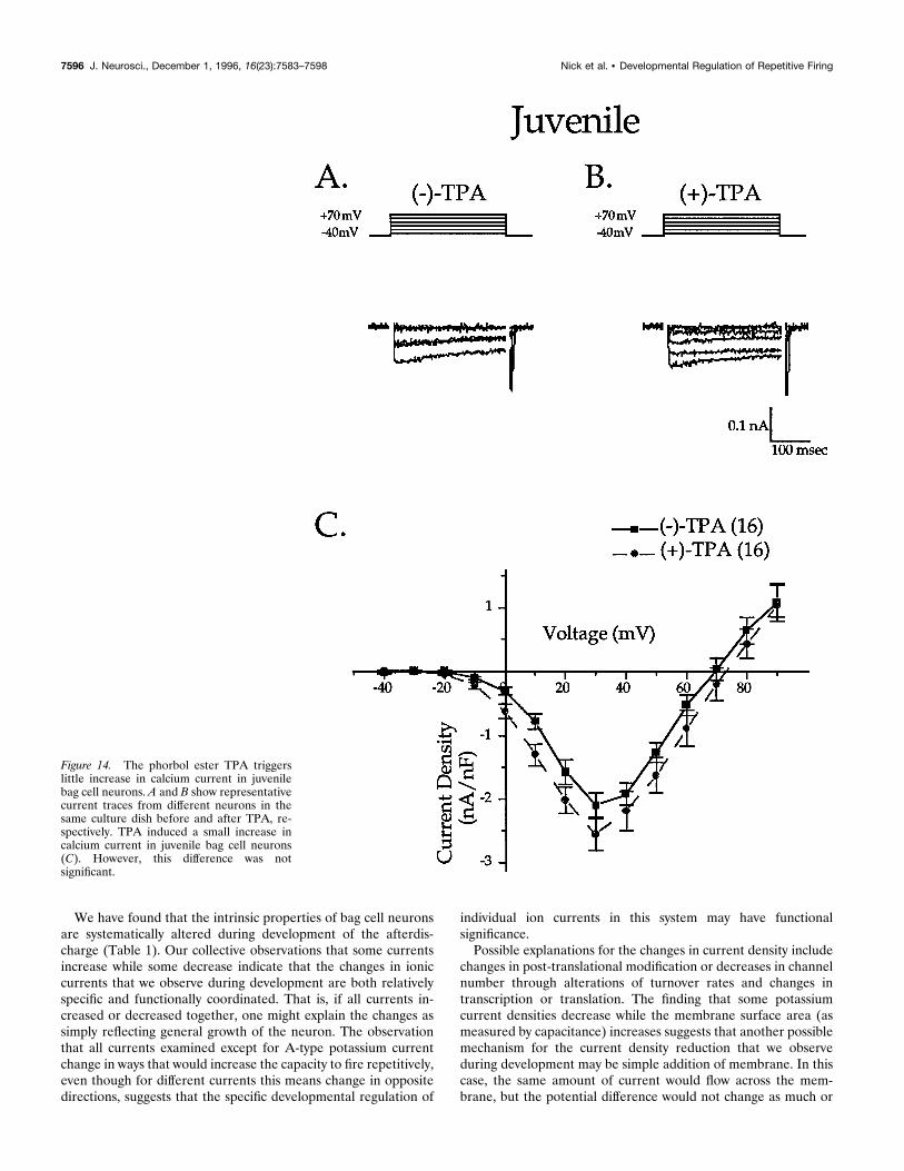
The other calcium current examined is expressed only afteractivation of PKC (DeRiemer et al., 1985). The phorbol esterTPA was used to activate PKC and induce expression of thiscalcium current [Fig. 13; (2)-TPA peak current density at 130mV: 25.0 nA/nF 6 0.7, n 5 18; (1)-TPA: 210.8 nA/nF 6 1.0,n 5 13; p , 0.0001 from 0 to 130 mV]. Juvenile bag cell neuronsdo not show a significant increase in calcium current after theaddition of TPA [Fig. 14; (2)-TPA: 21.9 nA/nF 6 0.3, n 5 9;(1)-TPA: 22.6 nA/nF 6 0.4, n 5 9; not significant]. BecausePKC-sensitive calcium current is thought to underlie the increasein action potential height observed during afterdischarge (DeRei-mer et al., 1985), lack of this current or elements in its activationpathway is consistent with a lack of afterdischarge in juvenileneurons. As reported previously (DeRiemer et al., 1985), treat-ment with an inactive phorbol ester, 4-a-phorbol, did not increasecalcium current in adults [(2)-4-a-phorbol: 25.0 nA/nF 6 0.69,n 5 18; (1)-4-a-phorbol: 26.3 nA/nF 6 1.65, n 5 4; not signifi-cant). These data thus show that both the basal and the PKC-sensitive calcium currents are upregulated during development.
DISCUSSIONWe have shown that the ability to fire repetitively is systematicallyregulated during development in the bag cell neurons of Aplysia.To investigate intrinsic mechanisms underlying the regulation ofelectrical excitability, we have compared major ionic currents inadult and juvenile neurons. Indeed, the general developmentalpattern of ionic current expression that we observe is consistentwith the increase in the ability to fire repetitively seen during thematuration of afterdischarge: three potassium currents decreaseand two calcium currents increase. In addition, a fourth potassiumcurrent (IKA) increases with development. IKA, an outward cur-rent, might be expected to decrease excitability. However, IKA hasan important role in repetitive firing in other neurons (Connorand Stevens, 1971a,b); this current dominates the interspike in-terval, whereas other currents determine the characteristics of theaction potential. Thus, increase in IKA may contribute to theregulation of repetitive firing characteristics of mature bag cellneurons. Taken collectively, the selective developmental regula-tion of the six currents that we have examined may promote theprogressive acquisition of the ability of bag cell neurons to exhibitthe afterdischarge required for egg laying in the adult.
At developmental stages when juvenile bag cell neurons areincapable of repetitive firing, they nonetheless exhibitprolonged depolarizationsAlthough juvenile bag cell neurons did not fire repetitively in re-sponse to any stimulus (e.g., nerve shock, TEA), they did exhibitprolonged depolarizations in the presence of the potassium channelblocker TEA. Thus, lack of afterdischarge in these neurons appearsto be attributable to a lack of capacity for repetitive firing, but not tothe inability to express another feature of the mature afterdischarge,prolonged depolarization. Nonspecific cation currents are thought tounderlie the depolarization that occurs during afterdischarge (Wil-son and Kaczmarek, 1993; Loechner and Kaczmarek, 1994). Becausejuvenile neurons are capable of prolonged depolarization (in thepresence of a potassium channel blocker), this suggests that theyalready express these nonspecific cation currents. This feature ofimmature neurons was advantageous, because it allowed us to de-scribe precisely a limited number of currents that would most likelybe involved in repetitive firing activity (see below). After furthercharacterization of depolarizations observed in juvenile neurons, wefound that the duration of depolarization increased with decreasedextracellular calcium concentration. Assuming that calcium activatescalcium-dependent potassium channels, leading to subsequent repo-larization of the membrane, lowering extracellular calcium shouldresult in longer depolarizations. Consistent with this idea, we findthat juvenile neurons indeed have high calcium-dependent potas-sium current density.As adult neurons are electrically coupled (Haskins and Blan-
kenship, 1979; Kaczmarek et al., 1979), regulation of connectivityamong juvenile neurons might be a potential mechanism fordevelopmental control of afterdischarge. However, our findingthat juvenile neurons that cannot afterdischarge are already elec-trically coupled does not support this hypothesis. Instead, we havefound that the acquisition of the mature phenotype reflects pro-gressive changes in the intrinsic ion current properties of theneurons. Some bag cell peptides (BCPs), which are released bythe bag cell neurons, are autoexcitatory and may have roles in thegeneration of the afterdischarge (Rothman et al., 1983; Brownand Mayeri, 1989; Loechner and Kaczmarek, 1990, 1994). Devel-opmental regulation of the expression of the BCPs or their trans-duction pathway may also contribute to intrinsic control of repet-itive firing in the bag cell neurons.
Table 1.
Juvenile(,11 gm)
Adult(.50 gm) p ,
Afterdischarge NO YESResting potential (mV) 246.9 6 4.6 254.1 6 4.6 N.S.Spike number in response to 500 msec current pulse 1.26 0.1 3.1 6 0.9 0.01Input resistance (MV) 2620 6 529 893 6 347 0.03Membrane capacitance (pF) 35.16 5.3 271.3 6 63.9 0.001Soma diameter (mm) 12.5 6 5.3 30.4 6 1.6 0.001Peak current density (nA/nF)Noninactivating delayed-rectifier 117.76 18.3 39.6 6 5.8 0.0001Inactivating delayed-rectifier 45.46 8.3 25.3 6 7.7 0.001Calcium-dependent potassium (est.) 37.6 14.0A-type potassium 1.06 0.3 2.6 6 0.8 0.05Calcium (basal) 21.9 6 0.3 25.0 6 0.7 0.03PKC-activated calcium (est.) 20.6 27.2
7594 J. Neurosci., December 1, 1996, 16(23):7583–7598 Nick et al. • Developmental Regulation of Repetitive Firing

Intrinsic properties of bag cell neurons changeduring developmentPrevious developmental studies that have examined the activity ofneurons that fire repetitively in adulthood have begun the analysisonly after the developmental onset of repetitive firing activity. Forexample, Prince and colleagues (McCormick and Prince, 1987;Hamill et al., 1991) found that neonatal neocortical pyramidalcells, like adult neurons, are capable of repetitive firing, but allmajor current densities examined were smaller than those ofadults. As the authors point out, these studies were limited by theability to recognize specific cell types and, therefore, could notexamine currents before the onset of repetitive firing capacity.One of the major advantages of the bag cell neuron system in
addressing these kinds of developmental questions is that the bagcell neurons are easily identified and can be studied before theyexpress any repetitive firing activity.In some adult systems, repetitively firing neurons that have been
examined in vitro show decreases in potassium current concomitantwith reacquisition of tonic firing and bursting capacity (Turrigiano etal., 1994, 1995; Mills and Pitman, 1995). These data are consistentwith our finding that bag cell neurons show decreases in three of fourpotassium current densities examined during the developmental ac-quisition of repetitive firing capacity. Thus, as adult neurons in vitrobegin to express the tonic firing pattern observed in vivo, they may berecapitulating a normal developmental process that underlies theemergence of repetitive firing.
Figure 13. The phorbol ester TPA triggers asignificant increase in calcium current in adultbag cell neurons. A and B show representativecurrent traces from different neurons in thesame culture dish before and after TPA, re-spectively. The effect of TPA can readily bedetected in the current density–voltage rela-tionship from adult neurons (C).
Nick et al. • Developmental Regulation of Repetitive Firing J. Neurosci., December 1, 1996, 16(23):7583–7598 7595
We have found that the intrinsic properties of bag cell neuronsare systematically altered during development of the afterdis-charge (Table 1). Our collective observations that some currentsincrease while some decrease indicate that the changes in ioniccurrents that we observe during development are both relativelyspecific and functionally coordinated. That is, if all currents in-creased or decreased together, one might explain the changes assimply reflecting general growth of the neuron. The observationthat all currents examined except for A-type potassium currentchange in ways that would increase the capacity to fire repetitively,even though for different currents this means change in oppositedirections, suggests that the specific developmental regulation of
individual ion currents in this system may have functionalsignificance.Possible explanations for the changes in current density include
changes in post-translational modification or decreases in channelnumber through alterations of turnover rates and changes intranscription or translation. The finding that some potassiumcurrent densities decrease while the membrane surface area (asmeasured by capacitance) increases suggests that another possiblemechanism for the current density reduction that we observeduring development may be simple addition of membrane. In thiscase, the same amount of current would flow across the mem-brane, but the potential difference would not change as much or
Figure 14. The phorbol ester TPA triggerslittle increase in calcium current in juvenilebag cell neurons. A and B show representativecurrent traces from different neurons in thesame culture dish before and after TPA, re-spectively. TPA induced a small increase incalcium current in juvenile bag cell neurons(C). However, this difference was notsignificant.
7596 J. Neurosci., December 1, 1996, 16(23):7583–7598 Nick et al. • Developmental Regulation of Repetitive Firing

as rapidly because of the requirement of charging and dischargingthe larger membrane capacitance in cells with a greater surfacearea. This could be a general regulatory mechanism used duringneuronal development, because many neurons show increases inmembrane surface area during maturation through the elabora-tion of dendritic trees, axonal arborizations, and synaptic contacts.This rather straightforward mechanism for modulation of currentdensity during development may not have been fully appreciatedpreviously because of the study of cultured neurons that do notpossess the extensive processes seen in vivo. Examination ofcultured bag cell neurons partially alleviates this problem becausetheir soma surface area increases with development, along withprocess elaboration (McAllister et al., 1983) (present study).However, this potential mechanism of current density reductioncould not explain the changes seen in the voltage dependence ofthe inactivating delayed rectifier. Changes in this current propertymay result from expression of different channel subunits and/orthe recombination of existing subunits. On the other hand, pos-sible mechanisms for increasing current density, which may ex-plain the changes we observe in IKA and ICa, might includeincreases in channel density and changes in post-translationalmodification.A common observation that has emerged in the analysis of
development of ionic currents is that a majority of neuronalpotassium currents that have been examined increase with devel-opment in vivo (for review, see Ribera and Spitzer, 1992). There-fore, our finding that three potassium currents decrease withdevelopment in the bag cell neurons may appear surprising. How-ever, most other neuronal cell types examined were neither re-petitively firing nor neuroendocrine. Moreover, an instructiveexception to this general observation (of increased potassiumcurrent with development) is rat pineal neurons (Aguayo, 1989)which, like the bag cell neurons, show a decrease in potassiumcurrent with development. These neurons are also neuroendo-crine in function. Premature activity of neuroendocrine cells andsubsequent expression of reproductive behaviors before sexualmaturation may cause increased mortality and decreased growthrate, without adding the benefits of reproduction (Stearns, 1976;Lima and Dill, 1990). Also, inappropriate hormone secretion mayhave teratogenic effects. Thus, neuroendocrine systems might notconform, in general, to the developmental programs of otherneurons. Our previous findings that juvenile bag cell neuronscontain and can secrete the bioactive peptide Egg Laying Hor-mone (Nick et al., 1996) support a potential role for regulation ofhormone secretion at the level of neuronal electrical excitability.Our results, in combination with data from other cell types (for
review, see Ribera and Spitzer, 1992), support the hypothesis thatneurons selectively regulate the expression of their ionic currentsduring development. Our data also suggest that the way differentneuronal types regulate their ionic currents may vary significantlyduring each specific window of development. This variation likelyreflects differences in the final neuronal electrophysiological phe-notype (e.g., repetitive firing, bursting, quiescent) and the manyaspects of development in which ion channels play a role, includ-ing cell proliferation (Gargus et al., 1993), construction of neuralcircuits (LeVay et al., 1981), regulation of neuronal differentiation(Jones and Ribera, 1994) (for review, see Spitzer, 1991), andregulation of hormone secretion (present study). Thus, examina-tion of the development of different neuronal phenotypes and thespecific changes that underlie their expression may reveal generalrules to which all neurons conform. Moreover, investigation ofexceptions to these rules may provide a better understanding of
neuronal differentiation in the context of the overall functionaldevelopment of the nervous system.
REFERENCESAguayo LG (1989) Post-natal development of K1 currents studied inisolated rat pineal cells. J Physiol (Lond) 414:283–300.
Barnes S, Deschenes MC (1992) Contribution of Ca and Ca-activated Clchannels to regenerative depolarization and membrane bistability ofcone photoreceptors. J Neurophysiol 68:745–755.
Barrett EF, Barrett JN (1976) Separation of two voltage-sensitive potas-sium currents, and demonstration of a tetrodotoxin-resistant calciumcurrent in frog motoneurons. J Physiol (Lond) 255:737–774.
Branton WD, Arch S, Smock T, Mayeri E (1978a) Evidence for media-tion of a neuronal interaction by a behaviorally active peptide. Proc NatlAcad Sci USA 75:5732–5736.
Branton WD, Mayeri E, Brownell P, Simon SB (1978b) Evidence forlocal hormonal communication between neurones in Aplysia. Nature274:70–72.
Brown RO, Mayeri E (1989) Positive feedback by autoexcitatory neu-ropeptides in neuroendocrine bag cells of Aplysia. J Neurosci9:1443–1451.
Conn PJ, Kaczmarek LK (1989) The bag cell neurons of Aplysia: a modelfor the study of the molecular mechanisms involved in the control ofprolonged animal behaviors. Mol Neurobiol 3:237–273.
Connor JA, Stevens CF (1971a) Voltage clamp studies of a transientoutward membrane current in gastropod neural somata. J Physiol(Lond) 213:21–30.
Connor JA, Stevens CF (1971b) Prediction of repetitive firing behaviorfrom voltage clamp data on an isolated neurone soma. J Physiol (Lond)213:31–53.
DeRiemer SA, Strong JA, Albert KA, Greengard P, Kaczmarek LK(1985) Enhancement of calcium current in Aplysia neurones by phorbolester and protein kinase C. Nature 313:313–316.
Dudek FE, Tobe SS (1978) Bag cell peptide acts directly on ovotestis ofAplysia californica: basis for an in vitro bioassay. Gen Comp Endocrinol36:618–627.
Dudek FE, Cobbs JS, Pinsker HM (1979) Bag cell electrical activityunderlying spontaneous egg laying in freely behaving Aplysia brasiliana.J Neurophysiol 13:319–326.
Fink LA, Connor JA, Kaczmarek LK (1988) Inositol trisphosphate re-leases intracellularly stored calcium and modulates ion channels inmolluscan neurons. J Neurosci 8:2544–2555.
Frisch RE (1974) Critical weight at menarche, initiation of the adoles-cent growth spurt, and control of puberty. In: Control of the onset ofpuberty (Grumbach MM, Grave GD, Mayer F, eds), pp 403–423.Berlin: Springer.
Gargus JJ, Frace AM, Jung F (1993) The role of a PDGF-activatednonselective cation channel in the proliferative response. EXS66:289–295.
Hamill OP, Marty A, Neher E, Sakmann B, Sigworth FJ (1981) Im-proved patch clamp techniques for high resolution current recordingfrom cells and cell-free membrane patches. Pflugers Arch 391:85–100.
Hamill OP, Huguenard JR, Prince DA (1991) Patch-clamp studies ofvoltage-gated currents in identified neurons of the rat cerebral cortex.Cereb Cortex 1:48–61.
Haskins JT, Blankenship JE (1979) Interactions between bilateral clus-ters of neuroendocrine cells in Aplysia. J Neurophysiol 42:356–367.
Jones SM, Ribera AB (1994) Overexpression of a potassium channelgene perturbs neural differentiation. J Neurosci 14:2789–2799.
Kaczmarek LK, Strumwasser F (1984) A voltage-clamp analysis of cur-rents underlying cyclic AMP-induced membrane modulation in isolatedpeptidergic neurons of Aplysia. J Neurophysiol 52:340–349.
Kaczmarek LK, Finbow M, Revel JP, Strumwasser F (1979) The mor-phology and coupling of Aplysia bag cells within the abdominal ganglionand in cell culture. J Neurobiol 10:535–550.
Kaczmarek LK, Jennings KR, Strumwasser F (1982) An early sodiumand a late calcium phase in the afterdischarge of peptide-secretingneurons of Aplysia. Brain Res 238:105–115.
Kiehn O (1991) Plateau potentials and active integration in the “finalcommon pathway” for motor behavior. Trends Neurosci 14:68–73.
Kupfermann I (1967) Stimulation of egg laying: possible neuroendocrinefunction of bag cells of abdominal ganglion of Aplysia californica.Nature 216:814–815.
Nick et al. • Developmental Regulation of Repetitive Firing J. Neurosci., December 1, 1996, 16(23):7583–7598 7597

Kupfermann I, Kandel ER (1970) Electrophysiological properties and func-tional interconnections of two symmetrical neurosecretory clusters (bagcells) in abdominal ganglion of Aplysia. J Neurophysiol 33:865–876.
LeVay S, Wiesel TN, Hubel DH (1981) The postnatal development andplasticity of ocular-dominance columns in the monkey. In: The organi-zation of the cerebral cortex (Schmitt FO, Worden FG, Adelman G,Dennis SG, eds), pp 29–45. Cambridge, MA: MIT.
Lima SL, Dill LM (1990) Behavioral decisions made under the risk ofpredation: a review and prospectus. Can J Zool 68:619–640.
Loechner KJ, Kaczmarek LK (1990) Control of potassium currents andcyclic AMP levels by autoactive neuropeptides in Aplysia neurons. BrainRes 532:1–6.
Loechner KJ, Kaczmarek LK (1994) Autoactive peptides act at threedistinct receptors to depolarize the bag cell neurons of Aplysia. J Neu-rophysiol 71:195–203.
Mackey S, Carew TJ (1983) Locomotion in Aplysia: triggering by seroto-nin and modulation by bag cell extract. J Neurosci 3:1469–1477.
McAllister LB, Scheller RH, Brownell PH, Branton WD, Padgett L(1983) In situ hybridization to study the origin and fate of identifiedneurons. Science 222:800–808.
McCormick DA, Prince DA (1987) Post-natal development of electro-physiological properties of rat cerebral cortical pyramidal neurones.J Physiol (Lond) 393:743–762.
Meech RW (1974) The sensitivity of Helix aspersa neurones to injectedcalcium ions. J Physiol (Lond) 237:259–277.
Mills JD, Pitman RM (1995) A calcium-dependent potassium conduc-tance determines electrical activity in an insect motoneurone. SocNeurosci Abstr 33:11.
Nick TA, Kaczmarek LK, Carew TJ (1993) Mechanisms underlying thedevelopment of afterdischarge in the bag cell neurons of Aplysia. SocNeurosci Abstr 708:7.
Nick TA, McKay SE, Kaczmarek LK, Carew TJ (1995) Outward currentsof Aplysia bag cell neurons decrease with development of afterdischargeand with BDNF treatment. Soc Neurosci Abstr 223:1.
Nick TA, Moreira JE, Kaczmarek LK, Carew TJ, Wayne NL (1996)Developmental dissociation of excitability and secretory ability in Aply-sia bag cell neurons. J Neurophysiol 76:5.
Pinsker HM, Dudek FE (1977) Bag cell control of egg-laying in freelybehaving Aplysia. Science 197:490–493.
Ribera AB, Spitzer NC (1987) Both barium and calcium activate neuro-nal potassium currents. Proc Natl Acad Sci USA 84:6577–6581.
Ribera AB, Spitzer NC (1992) Developmental regulation of potassiumchannels and the impact on neuronal differentiation. In: Ion channels,Vol 3 (Narahashi T, ed), pp 1–38. New York: Plenum.
Rothman BS, Mayeri E, Brown RO, Yuan P-M, Shively JE (1983) Pri-mary structure and neuronal effects of a-bag cell peptide, a secondcandidate neurotransmitter encoded by a single gene in bag cell neuronsof Aplysia. Proc Natl Acad Sci USA 80:5753–5757.
Spitzer NC (1991) A developmental handshake: neuronal control ofionic currents and their control of neuronal differentiation. J Neurobiol22:659–673.
Stearns SC (1976) Life-history tactics: a review of the ideas. Q Rev Biol51:3–47.
Strong JA, Fox AP, Tsien RW, Kaczmarek LK (1987) Stimulation ofprotein kinase C recruits covert calcium channels in Aplysia bag cellneurons. Nature 325:714–717.
Stuart DK, Strumwasser F (1980) Neuronal sites of action of a neurose-cretory peptide, egg-laying hormone in Aplysia californica. J Neuro-physiol 43:499–519.
Turrigiano GG, Abbott LF, Marder E (1994) Activity changes in theintrinsic properties of cultured neurons. Science 264:974–976.
Turrigiano GG, LeMasson G, Marder E (1995) Selective regulation ofcurrent densities underlies spontaneous changes in the activity of cul-tured neurons. J Neurosci 15:3640–3562.
Wilson GF, Kaczmarek LK (1993) Mode-switching of a voltage-gatedcation channel is mediated by a protein kinase A-regulated tyrosinephosphatase. Nature 366:433–438.
7598 J. Neurosci., December 1, 1996, 16(23):7583–7598 Nick et al. • Developmental Regulation of Repetitive Firing