Ion-Crosslinked Nanocellulose Hydrogels for Advanced Wound...
82
ACTA UNIVERSITATIS UPSALIENSIS UPPSALA 2018 Digital Comprehensive Summaries of Uppsala Dissertations from the Faculty of Science and Technology 1731 Ion-Crosslinked Nanocellulose Hydrogels for Advanced Wound Care Applications ALEX BASU ISSN 1651-6214 ISBN 978-91-513-0474-8 urn:nbn:se:uu:diva-363087
Transcript of Ion-Crosslinked Nanocellulose Hydrogels for Advanced Wound...
ACTAUNIVERSITATIS
UPSALIENSISUPPSALA
2018
Digital Comprehensive Summaries of Uppsala Dissertationsfrom the Faculty of Science and Technology 1731
Ion-Crosslinked NanocelluloseHydrogels for Advanced WoundCare Applications
ALEX BASU
ISSN 1651-6214ISBN 978-91-513-0474-8urn:nbn:se:uu:diva-363087

Dissertation presented at Uppsala University to be publicly examined in Häggsalen,Ångströmlaboratoriet, Lägerhyddsvägen 1, Uppsala, Friday, 30 November 2018 at 09:30 forthe degree of Doctor of Philosophy. The examination will be conducted in English. Facultyexaminer: Professor Pentti Tengvall (University of Gothenburg).
AbstractBasu, A. 2018. Ion-Crosslinked Nanocellulose Hydrogels for Advanced Wound CareApplications. Digital Comprehensive Summaries of Uppsala Dissertations from theFaculty of Science and Technology 1731. 81 pp. Uppsala: Acta Universitatis Upsaliensis.ISBN 978-91-513-0474-8.
A current trend in the field of wound care is the development of wound healing materials thatare designed to address specific types of wounds or underlying pathologies to achieve improvedhealing. At the same time, there is a societal drive to replace synthetic materials with renewablealternatives. The work presented in this thesis was therefore carried out to investigate the useof wood nanocellulose, produced from the world’s most abundant biopolymer, cellulose, inadvanced wound care applications.
Wood-based nanofibrillated cellulose (NFC) was chemically functionalized and crosslinkedusing calcium to obtain a self-standing hydrogel. The NFC hydrogel was evaluated in termsof its physicochemical properties, biocompatibility, blood interactions, bacterial interactions, invivo wound healing ability and, finally, as a protein carrier. Parallel with the assessment of theNFC hydrogel, modified versions of the material were tested to investigate the tunability of theabove-mentioned characteristics.
The ability of the hydrogel to maintain a moist wound bed was demonstrated. Evaluationof the biocompatibility showed that the material was cytocompatible and did not triggerinflammatory mechanisms. Furthermore, the NFC hydrogel supported cell proliferation, andwas shown to possess hemostatic properties. It was also discovered that the material had a slightbacteriostatic effect and the ability to act as a barrier against bacteria. When tested in vivo, thehydrogel was found to significantly improve wound healing.
Modifications through the incorporation of additives to the hydrogel matrix, as well asexchange of the crosslinking ion, were shown to influence the biological response to thematerial. Moreover, the results presented here demonstrate the possibility of using the NFChydrogel as a protein carrier; the easily adjustable charge property being identified as a centralparameter for manipulation to regulate the release profile.
In conclusion, this work has demonstrated the extensive wound healing ability of the calcium-crosslinked NFC hydrogel, and represents an important milestone in the research on NFCtowards advanced wound care applications. It is expected that the easily modifiable nature ofthe material can be exploited to further develop the NFC hydrogel to suit the treatment needsfor a broad range of wound types.
Keywords: nanofibrillated cellulose, wood nanocellulose, ion crosslinking, hydrogel,wound healing, biocompatibility, blood interactions, bacterial interactions, protein carrier,nanotherapeutic
Alex Basu, Department of Engineering Sciences, Nanotechnology and Functional Materials,Box 534, Uppsala University, SE-75121 Uppsala, Sweden.
© Alex Basu 2018
ISSN 1651-6214ISBN 978-91-513-0474-8urn:nbn:se:uu:diva-363087 (http://urn.kb.se/resolve?urn=urn:nbn:se:uu:diva-363087)

To my loving family


List of Papers
This thesis is based on the following papers, which are referred to in the text by their Roman numerals.
I Basu, A., Lindh, J., Ålander, E., Strømme, M., Ferraz, N.
(2017) On the use of ion-crosslinked nanocellulose hydrogels for wound healing solutions: Physicochemical properties and application-oriented biocompatibility studies. Carbohydrate Polymers, 174:299-308.
II Basu, A., Hong, J., Ferraz, N. (2017) Hemocompatibility of
Ca2+-crosslinked nanocellulose hydrogels: Toward efficient management of hemostasis. Macromolecular Bioscience, 17:1700236.
III Basu, A., Heitz, K., Strømme, M., Welch, K., Ferraz, N. (2018)
Ion-crosslinked wood-derived nanocellulose hydrogels with tunable antibacterial properties: Candidate materials for ad-vanced wound care applications. Carbohydrate Polymers, 181:345-350.
IV Basu, A., Celma, G., Strømme, M., Ferraz, N. In vitro and in
vivo evaluation of the wound healing properties of nanofibrillat-ed cellulose hydrogels. Submitted.
V Basu, A., Strømme, M., Ferraz, N. (2018) Towards tunable pro-
tein-carrier wound dressings based on nanocellulose hydrogels crosslinked with calcium ions. Nanomaterials, 8:550-563.
Reprints were made with permission from the respective publishers.

The author’s contributions to the papers included in this thesis
I I participated in the planning of the study, performed all the ex-
perimental work, analyzed the data, wrote the initial manu-script, and took part in writing the final manuscript.
II I played a major role in the planning of the study, performed all the experimental work, analyzed the data, wrote the initial man-uscript, and took part in writing the final manuscript.
III I played a major role in the planning of the study, assisted in the
bacterial studies and performed the material characterization. I analyzed the data, wrote the initial manuscript, and took part in writing the final manuscript.
IV I played a major role in the planning of the study, performed
parts of the in vitro experiments, analyzed the data, wrote the initial manuscript, and took part in writing the final manuscript.
V I had a leading role in the planning of the study, and performed
all the experimental work and the data analysis. I wrote the ini-tial manuscript, and took part in writing the final manuscript.

Conference contributions
Basu, A., Ferraz, N. (2015) Nanofibrillated cellulose hydrogels for wound healing solutions: the status quo and future prospects. Poster presentation at the Uppsala Biomaterials Conference, Uppsala, Sweden.
Basu, A., Ferraz, N. (2015) Ion-crosslinked nanocellulose hydrogels for biomedical applications. Poster presentation at the 4th EPNOE International Polysaccharide Conference, Warsaw, Poland.
Basu, A., Ferraz, N. (2016) Nanocellulose hydrogels: preparation, character-ization and cytotoxicity studies toward biomedical applications. Poster presentation at the 10th World Biomaterials Congress, Montreal, Canada. Basu, A., Ferraz, N. (2016) Nanocellulose hydrogels for wound-healing applications. Oral presentation at the 2nd International EPNOE Junior Sci-entists Meeting, Sophia Antipolis, France. Basu, A., Ferraz, N. (2016) Nanocellulose hydrogels for topical wound care applications. Oral presentation at the Uppsala Biomaterials & Bioengineer-ing Day 2016, Uppsala, Sweden. Basu, A., Tummela, G., Heitz, K., Gustafsson, S., Strietzel, C., Yang, J., Notfors, C., Huan, W., Strømme, M. (2017) Miljövänliga nanomaterial från växtriket tillåter gröna alternativ för framtiden. Poster presentation at SciFest 2017, Uppsala, Sweden. (In Swedish) Basu, A., Ferraz, N. (2018) Nanocellulose hydrogels as candidates for ad-vanced wound healing solutions. Poster presentation at the Scandinavian Society of Biomaterials 2018, Fiskebäckskil, Sweden.


Contents
1 Introduction ......................................................................................... 15
2 Aims of this work ................................................................................ 16
3 Background .......................................................................................... 173.1 Wound healing ........................................................................... 17
3.1.1 Hemostasis ............................................................................. 183.1.2 Inflammation .......................................................................... 193.1.3 Proliferation ........................................................................... 193.1.4 Remodeling ............................................................................ 203.1.5 Chronic wounds and underlying factors ................................ 20
3.2 Wound dressings ........................................................................ 213.2.1 Desirable properties ............................................................... 22
3.3 Hydrogels ................................................................................... 243.4 Nanotechnology and functional materials in wound healing ..... 25
3.4.1 Nanotechnological approaches to wound healing ................. 253.4.2 Biocompatibility aspects of nano- and biomaterials .............. 26
3.5 Cellulose and nanocellulose ....................................................... 273.5.1 Nanocellulose in biomedical applications ............................. 293.5.2 Wood NFC in wound healing ................................................ 29
4 Materials and methods ......................................................................... 314.1 Material preparation ................................................................... 31
4.1.1 Functionalization of NFC ...................................................... 314.1.2 Preparation of ion-crosslinked NFC hydrogels ..................... 32
4.2 Material characterization ............................................................ 324.2.1 Determination of functional group content and charge ......... 324.2.2 Rheology ................................................................................ 334.2.3 Water retention test ................................................................ 334.2.4 Scanning electron microscopy ............................................... 334.2.5 Fourier transform infrared spectroscopy ............................... 34
4.3 In vitro biocompatibility ............................................................. 344.3.1 In vitro models ....................................................................... 344.3.2 Evaluation of cell viability and proliferation ......................... 354.3.3 Evaluation of inflammatory response .................................... 354.3.4 Evaluation of cell morphology and migration ....................... 35
4.4 In vitro hemocompatibility ......................................................... 36
4.4.1 Complement system and coagulation activation ................... 364.4.2 Blood sampling and plasma pooling ...................................... 37
4.5 In vitro bacteria interactions ....................................................... 374.5.1 Bacterial growth models ........................................................ 374.5.2 Sessile growth pattern and bacterial impermeability ............. 38
4.6 In vivo wound healing ................................................................ 384.6.1 Study design ........................................................................... 384.6.2 Macroscopic and morphometric analysis .............................. 384.6.3 Histopathological evaluation ................................................. 39
4.7 In vitro protein interactions ........................................................ 394.7.1 Protein loading and release methods ..................................... 394.7.2 Mathematical analysis of release ........................................... 404.7.3 Protein stability investigation ................................................ 40
5 Results and discussion ......................................................................... 415.1 Overview .................................................................................... 415.2 Physicochemical properties ........................................................ 425.3 Biocompatibility ......................................................................... 43
5.3.1 Initial biocompatibility assessment ........................................ 435.3.2 Further application-oriented biocompatibility testing ........... 46
5.4 Hemocompatibility ..................................................................... 505.5 Interactions with common wound pathogens ............................. 535.6 Acute wound healing efficacy in vivo ........................................ 575.7 A way forward in addressing chronic wounds ........................... 60
6 Conclusions and future perspectives ................................................... 67
Sammanfattning på svenska .......................................................................... 69
Acknowledgements ....................................................................................... 71
References ..................................................................................................... 72

Abbreviations
a-NFC AB c-NFC BSA DMSO ECM EDTA ELISA EPTMAC FTIR-ATR FXII hDF HPTMA IL KERT LPS
Anionic nanofibrillated cellulose Alamar Blue
Cationic nanofibrillated cellulose Bovine serum albumin Dimethyl sulfoxide Extracellular matrix Ethylenediaminetetraacetic acid Enzyme-linked immunosorbent assay 2,3-epoxypropyltrimethylammonium chloride Fourier transform infrared-attenuated total reflectance Factor XII Human dermal fibroblast Hydroxypropyltrimethylammonium Interleukin Keratinocyte cell line Lipopolysaccharide
MT NFC PBS PDGF PFP PMMA PVC ROS SDS-PAGE SEM SHE SWF TAT TCC TCP TEMPO TGA TGF-β TMX TNF-α
Masson’s trichrome Nanofibrillated cellulose Phosphate buffered saline Platelet-derived growth factor Platelet-free plasma Polymethyl methacrylate Polyvinyl chloride Reactive oxygen species Sodium dodecyl sulfate-polyacrylamide gel electrophoresisScanning electron microscopy Safranin-hematoxylin-eosin Simulated wound fluid Thrombin-antithrombin Terminal complement complex Tissue culture plate 2,2,6,6-tetramethylpiperidine-1-oxyl Thrombin generation assay Transforming growth factor beta Thermanox® Tumor necrosis factor alpha

15
1 Introduction
The skin forms our largest organ, and its primary function is to protect us from pathogens, mechanical damage, hazardous substances and radiation [1, 2]. Due to its large and exposed area, it is at constant risk of injury. A break in the skin directly affects its protective properties, and the ability of our bodies to quickly repair wounded skin and fight off intruding pathogens, is thus necessary for our continued health. Acute wounds tend to heal in an orderly fashion, following the four distinct phases of hemostasis, inflamma-tion, proliferation and remodeling [3]. However, this process can be dis-turbed, for example, if the wounded area is large, by infection, or by patho-logical conditions such as diabetes, which may lead to the development of chronic wounds.
Wound dressing materials designed to aid wound healing date as far back as 2000 BC, when they were made of clay, plants and herbs. Since then, the development of wound dressings has continuously evolved with the advanc-es made by humankind, and today more than 5,000 wound care products exist on the market [4]. However, many present-day wound dressings have no better scientific rationale than the early alternatives. Our increased under-standing of many of the processes mediating wound healing, and achieve-ments in the fields of materials science and nanotechnology, now provide the opportunity to develop truly wound healing dressing materials.
The possibility of using nanoscale cellulose fibers, i.e. nanocellulose, to create a tunable platform for advanced wound healing dressings was investi-gated in the work presented in this thesis. Calcium-crosslinked nanocellulose hydrogels of different final compositions were produced and characterized in terms of physicochemical properties relevant for wound dressing materials. The candidate dressing materials were furthermore investigated using in vitro models to study the interactions with central components of the wound healing process, including human skin cells and immune cells, blood com-ponents and common wound pathogens. The ability of the nanocellulose hydrogel to heal acute wounds was assessed in an in vivo wound healing rat model, and finally, the potential of applying this hydrogel as a drug carrier in a chronic wound management context was explored.
Overall, this thesis illustrates a multidisciplinary approach to understand-ing how the world’s most abundant biopolymer, namely cellulose, can be utilized to create a new generation of renewable advanced wound healing materials.

16
2 Aims of this work
The underlying aim of the research presented in this thesis was to develop a wood-based nanocellulose hydrogel that can act as a platform in advanced wound care applications. Application-oriented characterization techniques and biocompatibility tests were employed to expand our current knowledge concerning the use of nanocellulose in biomedical applications. In addition to a calcium-crosslinked hydrogel consisting of anionic nanocellulose, re-ferred to as a nanofibrillated cellulose (NFC) hydrogel below, various modi-fied versions of the NFC hydrogel were investigated in order to explore the possibility of tuning the original composition to improve the healing of spe-cific types of wounds. The specific aims of the appended studies were as follows:
I To synthesize the NFC hydrogel and a composite hydrogel contain-ing anionic and cationic nanocellulose, and to characterize their physicochemical properties and biocompatibility.
II To investigate the hemocompatibility, with focus on the hemostatic
ability, of the NFC hydrogel and two composite hydrogels including type I collagen or kaolin.
III To investigate the interactions between the NFC hydrogel and com-
mon wound pathogens, and the possibility of tuning the bacteriostat-ic properties of the hydrogel by exchanging the crosslinking ion.
IV To study the in vitro skin cell response to the NFC hydrogel and a composite hydrogel including type I collagen, and to investigate the in vivo wound healing capability of the NFC hydrogel.
V To study the interactions between the NFC hydrogel and proteins with different physicochemical properties in order to understand how these interactions can be used in therapeutic solutions for chronic wounds.

17
3 Background
3.1 Wound healing The human skin consists of two tissue layers: an upper and a lower layer, called the epidermis and dermis, respectively. The epidermis consists mainly of keratinocyte cells and affords the skin its barrier properties. The core components of the dermis include cells such as fibroblasts, macrophages and adipocytes, together with matrix components such as collagen and elastin, which give the skin its strength and elasticity. Together, these skin layers form a vital protective barrier against the environment [5].
In the moments after a cutaneous injury, a systematic process consisting of the four overlapping phases of hemostasis, inflammation, proliferation and remodeling, is activated in order to restore tissue integrity (Figure 1) [3, 5]. As each phase is important for healing to progress successfully, a good strategy in the development of wound healing dressings is to design materi-als that target these phases. In this chapter, the wound healing process is explained to provide the reader with insight into some biological processes that can be targeted to aid or mediate wound healing.
Figure 1. Overview of the wound healing process following cutaneous injury.

18
3.1.1 Hemostasis Immediately after injury, vasocontraction (i.e. the narrowing of blood ves-sels) occurs to prevent uncontrolled bleeding [1]. As platelets escape com-promised blood vessels and come into contact with collagen in the extracel-lular matrix (ECM), they begin to aggregate and release clotting factors (e.g. polyphosphates) and growth factors (e.g. platelet-derived growth factor (PDGF) and transforming growth factor beta (TGF-β)) [1, 3].
Parallel with platelet aggregation, the blood coagulation cascade is acti-vated through two pathways (Figure 2): the intrinsic pathway (or contact activation pathway) and the extrinsic pathway (or tissue factor pathway). The intrinsic pathway is triggered by surface activation of factor XII (FXII) through blood contact with negatively charged surfaces [6, 7]. Polyphos-phates released by platelets also activate the intrinsic pathway, although this is believed to accelerate coagulation rather than to trigger it [8]. The extrin-sic pathway is activated by tissue factor, a cellular lipoprotein released by damaged cells and expressed on the surface of extravascular tissue, and is the main contributor to coagulation in normal wound healing [7, 9]. Ulti-mately, both pathways converge, where thrombin is formed from prothrom-bin. Thrombin in turn promotes the formation of fibrin and further activation of platelets [6, 7, 9].
As a result of hemostasis, a clot is created consisting largely of activated platelets and crosslinked fibrin, which acts as a temporary protective seal over the wound bed. The clot furthermore forms a stimulatory environment that activates the complement system, the recruitment of inflammatory cells and the initiation of re-epithelialization, thereby setting the stage for subse-quent phases of wound healing [5, 10, 11].
Figure 2. The coagulation and complement system cascades.
19
3.1.2 Inflammation Within a day or two, the inflammatory phase begins as neutrophils and acti-vated monocytes are recruited to the wound site. These immune cells are not only recruited in response to the degranulation of platelets, but also through activation of the complement system [11, 12].
The complement system is a central part of the innate immune system with the primary function of eliminating microorganisms and other foreign particles. As shown in Figure 2 above, three routes exist (the classical path-way, the alternative pathway and the lectin pathway) for the activation of the central complement protein C3, causing the release of the anaphylatoxin C3a and initiation of the terminal pathway. The terminal pathway ends with the formation of the terminal complement complex (TCC), also called the mem-brane attack complex, a protein complex that is able to disrupt the membrane integrity of pathogens, thereby inducing cell death. Alternatively, in the ab-sence of a biological membrane, the soluble form of TCC, sC5b-9, is formed [12-14]. Products of the complement system are central to the stimulation of inflammation and phagocytosis of foreign particles.
When neutrophils and activated monocytes are present in the wound bed, the process of “cleaning up” the area starts. Monocytes differentiate into phagocytic macrophages, perhaps the most important inflammatory cell in-volved in normal wound healing. Neutrophils and macrophages together eliminate pathogens and foreign particles, whereas macrophages have the more dominant role and are responsible for the coordination of inflammation and angiogenetic processes [1]. Macrophages are also responsible for the removal of bacteria-filled neutrophils toward the end of the inflammatory phase of wound healing [3]. At a later stage of the inflammatory phase, mac-rophages are crucial for the initiation of the proliferative phase by releasing, e.g., PDGF and TGF-β, which further stimulate fibroblasts and angiogenesis [3, 15, 16]. The transition of type M1 (pro-inflammatory) to type M2 (anti-inflammatory) macrophages is also believed to play a pivotal role in the transformation from the inflammatory phase to the proliferative phase of wound healing [16, 17].
3.1.3 Proliferation The proliferative phase, starting typically three to four days after injury, is characterized by new tissue formation, angiogenesis, wound contraction and re-epithelialization.
Granulation tissue, containing macrophages, fibroblasts, ECM compo-nents and new blood vessels, fills the wound bed, while keratinocytes mi-grate across the granulation tissue to establish a new epithelial barrier [1, 11]. Macrophages provide the area with a continuous source of growth fac-tors necessary for the stimulation of fibroblast activation and angiogenesis

20
[10]; fibroblasts and their differentiated form, myofibroblasts, synthesize new ECM with contractile properties [18]; while new blood vessels are formed through angiogenic sprouting, necessary for the provision of oxygen and nutrients to the new tissue [19]. Re-epithelialization is mediated by a process called contact guidance, in which keratinocytes change morphology to a flatter shape allowing keratinocytes from the opposing sides of the wound to move across the granulation tissue to re-establish contact [20]. This process is aided by myofibroblasts, which contract the wound, thus making the wound gap smaller [18]. Once the basal layer of keratinocytes has been formed over the wound bed, these cells start building up the height, forming the spinous, granular and cornified layer of the epidermis [21]. This process is driven by Ca2+-dependent differentiation of the keratinocytes, from the basal phenotype to the terminally differentiated and flattened corni-fied type [22].
After some days, or even weeks, when the wound bed is filled with granu-lation tissue and covered by a new epidermis, the remodeling phase starts.
3.1.4 Remodeling This last phase of wound healing, in which the provisional granulation tissue is replaced by stronger, permanent tissue, can continue for weeks or several months. Proliferation of immune and skin cells slows down and returns to normal. As the demand for nutrition decreases, the capillary density decreas-es from almost three times the normal level during the earlier phases of wound healing, to near-normal levels. With this the redness of the scar dis-appears, marking the end of the visible healing process. However, ECM breakdown and remodeling continue under the scar, with rearrangement and crosslinking of the initially deposited collagen fibers, to strengthen the new tissue. During this matrix-metalloproteinase-mediated process, type III col-lagen is replaced by the stronger type I collagen [1, 3, 10, 11]. However, remodeled skin never regains its original strength [23].
3.1.5 Chronic wounds and underlying factors Chronic wounds are defined as wounds that have failed to progress accord-ing to the normal stages of wound healing, and therefore require treatment to heal. In the US alone, health care expenditure related to chronic wound care is estimated to be USD 25 billion annually, and in developing countries about 1-2% of the population experiences a chronic wound during their life-time [24]. This type of wound thus causes tremendous patient suffering and poses a major challenge to healthcare systems worldwide.
Chronic wounds arise due to local or systemic disturbances that delay healing. Local factors directly affect the wound area (e.g. infections and

21
foreign bodies), while systemic factors are disturbances resulting from the general state of health (e.g. diseases, age or malnutrition) [25, 26].
Our skin and surrounding environment is rich in bacteria [27], thus all wounds contain some bacterial contamination, leading to the risk of infec-tion, sometimes resulting in a chronic wound. Common wound pathogens include Staphylococcus epidermidis and Pseudomonas aeruginosa. S. epi-dermidis was traditionally regarded as a non-pathogenic skin inhabitant, although it is today considered to be an opportunistic pathogen and one of the most common causes of healthcare-related infections. P. aeruginosa is commonly found in chronic wounds and poses special challenges in healthcare due to its extensive biofilm production resulting in strong antibi-otic resistance [28-30]. In the case of a microbial infection or contamination by a foreign body, the increase in reactive oxygen species (ROS) and pro-inflammatory cytokine production (e.g. interleukin (IL)-1 and tumor necrosis factor alpha (TNF-α)) which stimulates inflammation, and is meant to clear the wound area, instead causes the breakdown of surrounding tissue, thus arresting the healing process. Furthermore, an excessively inflammatory environment causes a decrease in the levels of growth factors that are essen-tial for successful wound healing [26, 31].
The reduced ability of the body to control the inflammatory phase, for ex-ample, in the elderly and individuals suffering from stress or diseases such as diabetes, may cause delay and the development of chronic wounds. Delayed wound healing in the elderly has been associated with a late inflammatory response and reduced macrophage phagocytic capacity [32]. Similarly, stress has been shown to lower the inflammatory response [33]. When the defense system against pathogens is impaired, the risk of wound infection increases, which elevates the risk of chronic wound formation. The impaired healing ability among diabetics is the result of prolonged hypoxia in the wound area due to insufficient angiogenesis, which amplifies the early inflammatory response and oxidative stress levels. Hyperglycemia also adds to increased oxidative stress levels, the combination leading to tissue destruction and inhibition of the normal wound healing process [26, 34].
Hence, both excessive and impaired inflammation can be detrimental to wound healing. While other phases of wound healing can also be disturbed by various pathological conditions, the development of chronic wounds is, in one way or another, mainly connected to disruption of the inflammatory phase.
3.2 Wound dressings Wound dressings are medical devices that aid the healing of wounds by pro-tecting the wound and providing an environment for improved healing.

22
Apart from these basic properties, modern wound dressings can have a num-ber of healing-enhancing properties.
Historically, wound dressings were made of leaves and herbal plants. It was later found that many of these herbs have antibacterial properties, af-fording these early dressings some of the desirable properties of a wound dressing (see Section 3.2.1, Table 1). However, due to a lack of knowledge concerning wound care and aseptic practice, many early remedies had no, or even deleterious, effects on the healing process [35]. Most of these treat-ments are no longer in use, although old habits prevail in some areas due to a lack of knowledge. For instance, despite strong evidence of the importance of a moist wound bed for improved wound healing, wound-drying dressings are still common [36].
It is becoming generally accepted in the scientific community that there is no generic solution for all types of wounds, i.e., the choice of dressing de-pends on the type of wound (e.g. dry, wet, acute and chronic) to be treated. As a result of increased knowledge in the fields of wound healing and mate-rials science, the number of types of wound dressings has increased steadily, and today there are over 5,000 alternatives available on the global market [4]. It is common to divide wound healing dressings into two categories: traditional and modern. Traditional dressings include cotton wool, bandages and gauzes. These simple dressings are more commonly used as secondary dressings, i.e., as the layer providing outer protection for a topical pharma-ceutical formulation or a bioactive primary dressing. Modern dressings, on the other hand, consist of more recently developed alternatives that have been designed to maintain a moist wound bed, and incorporate many of the desirable properties of a wound healing dressing. Such dressings are usually in the form of gels, thin films or foam sheets; examples being hydrocolloid dressings, alginate dressings and hydrogels [35, 37].
3.2.1 Desirable properties With advancing knowledge concerning the wound healing process, research-ers and practitioners have identified some desirable properties of a wound healing dressing. Table 1 presents a list of the generally desirable properties of such dressings. However, to provide optimal healing conditions for the specific type of wound being treated, the characteristics of that wound must be considered. For example, a highly exuding chronic wound may require a dressing with efficient liquid absorption properties to maintain a normal moisture level, rather than one that provides moisture, while the opposite is required for dry wounds.

23
Table 1. Desirable properties of a wound healing dressing (after Boateng et al. [35])
Desirable property Clinical significance
Wound cleansing Supports the accumulation of enzymes important for the auto-lytic debridement of necrotic tissue and foreign bodies
Provision and control of moisture level
Enhances epidermal migration, angiogenesis, connective tis-sue synthesis and debridement
Liquid absorption Aids hemostasis and removal of excess exudate containing tissue-degrading enzymes
Gaseous exchange Permeability to water vapor is important for exudate man-agement, and increased tissue oxygen levels stimulate epithe-lialization and fibroblast activation
Bacteriostatic and bacterial barrier
Prevents infections and protects the wound from bacterial invasion
Thermal insulation Normal tissue temperature improves blood flow and enhances epidermal migration
Low adhesion Minimizes renewed trauma upon removal of dressing material
Cost-effective Motivates users to choose the correct dressing for a particular wound type, rather than the cheapest dressing
Apart from these general properties, other, more specific properties may be vital for the treatment of, e.g., infected, inflamed or heavily bleeding wounds. For example, instead of administering antibiotics systemically to resolve a local wound infection, the use of wound dressings with antibacteri-al properties to treat infected wounds has many advantages. While the treat-ment itself may reduce the risk of chronic wound development and is less toxic to the patient, the use of antibacterial dressings, rather than systemic administration, can also contribute to the fight against antibiotic-resistant strains [38, 39]. Examples of such dressings are materials that have been chemically functionalized to make them antibacterial, or materials that are loaded with antibacterial agents (e.g. silver), which are released locally upon application to the wound [40, 41].
In addition to the delivery of antibacterial agents, the use of wound dress-ings for drug delivery can also be useful for the treatment of inflammatory wounds or for the release of therapeutic agents (e.g. growth factors) that accelerate wound healing. Desirable properties of drug delivery dressings include a uniform and sustained release, which can be obtained by tuning the swelling and diffusion parameters of the dressing material [35].
Today, up to 40% of deaths after traumatic injury are due to excessive hemorrhage [42]. While most bleeding can be stopped by manual pressure to

24
the wound, or by applying pro-coagulant minerals such as kaolin or zeolite, these methods are often unsuitable due to wound location, and can be insuf-ficient in the case of severe bleeding [42, 43]. In such cases, wound dress-ings with hemostatic properties may be useful.
3.3 Hydrogels Hydrogels are water-swollen materials made of synthetic or natural polymer-ic networks, as illustrated in Figure 3. Natural polysaccharides such as cellu-lose, chitosan, hyaluronic acid and alginate have been widely used to prepare hydrogels. The gels are held together by chemical bonding, molecular entan-glement, and/or secondary forces including ionic, hydrogen-bonding or hy-drophobic forces. Their ability to absorb large amounts of water (many times their dry weight) is due to the hydrophilic functional groups attached to the polymer backbone. Hydrogels can be designed with molecular-scale preci-sion, thus allowing control over properties such as mechanical strength, swelling, biodegradability, and chemical and biological response to stimuli [35, 44-46].
Figure 3. Illustration of hydrogel structure.
Due to their versatility, hydrogels are used in many fields and applications, including ophthalmology [47, 48], hygiene products [48], agriculture [49], biosensors [50], food additives [51] and biomedical applications [44, 48, 52]. Within the biomedical field, hydrogels are of special interest in tissue engi-neering and regenerative medicine [53, 54], drug delivery [55, 56], as well as wound healing [35, 48]. The structural similarities between hydrogels and the ECM, and their supportive matrix allowing cellular proliferation and survival, afford hydrogels advantageous properties for tissue engineering and regenerative medicine. The aqueous environment of hydrogels gives them the ability to protect and release fragile drugs (such as growth factors), which makes them interesting for drug delivery applications [57], while their high water content affords them many of the desirable properties of a wound healing dressing (e.g. promoting a moist environment and debridement, good gaseous exchange, low tissue adhesion and providing patient comfort through cooling and soft texture). Commercial hydrogels used for wound

25
healing applications are made of a variety of polymers, such as cellulose derivatives, alginate and hyaluronic acid. Examples of hydrogel dressings include Aquacel, XCell® and Silvercel™ [52, 57].
3.4 Nanotechnology and functional materials in wound healing
The main principle of nanotechnology is the engineering of materials on the nanoscale, furnishing them with characteristics such as high surface area, functional surfaces and novel structural designs. In this way, materials can be designed with unique properties not seen in their bulk counterparts, such as higher reactivity to the environment, interesting mechanical, electronic, photonic or optical properties, and bioactivity. The European Commission defines ‘nanomaterial’ as a material with at least one dimension on the 1-100 nm scale [58].
Nanotechnology offers excellent opportunities to address the problems of non- or slow-healing wounds, as wound healing solutions can be designed to be multifactorial and cell-type specific. Topical and stimuli-responsive de-livery devices are also possible, and this degree of specificity provides the unique opportunity to target specific dysfunctional processes in wound heal-ing without systemically affecting the host. The benefits of this include more efficient treatment, lower risk of systemic side-effects and reduced toxicity due to efficacy at a lower dosage. For these reasons, extensive efforts are today being devoted to research on nanotechnological wound healing solu-tions.
3.4.1 Nanotechnological approaches to wound healing Nanotechnological wound healing solutions can be roughly divided into two categories: intrinsic nanodevices and nanocarriers. Intrinsic nanodevices comprise material structures with built-in wound healing properties, while nanocarriers are devices that are employed as delivery vehicles for therapeu-tic agents [59].
Typical examples of intrinsic nanodevices include materials that can be described as porous solids, nanoparticles and nanotopological materials [59, 60]. Porous solids mimic tissue, and are often ECM-like, thus creating a native environment for skin cells, which can promote skin cell proliferation and migration. Hydrogel dressings usually belong to this category, and a common strategy for their functionalization is to modify the physicochemi-cal properties of the material [52]. Nanoparticles are endowed with proper-ties such as high surface area, which translates into higher reactivity with biological systems. In the case of metallic compounds such as silver, used in antibacterial applications, this is beneficial as the higher reactivity reduces
26
the dose required, which in turn reduces the toxicity [61]. For nanotopologi-cal materials, the aim is usually to induce cellular events by cell–material interactions upon contact. The outcomes may be altered or triggered cell adhesion, proliferation, migration or activation on the material surface [62].
An example of a nanocarrier is a stimuli-sensitive nanomaterial which, upon a biochemical trigger, undergoes structural changes leading to the re-lease of a payload. Sensoric hydrogels, for example, with crosslinkers that respond to the wound environment, leading to the release of therapeutic agents in a feedback-controlled fashion, could be envisioned [59]. Other examples of nanocarriers include nanoparticle-based and DNA-based (often referred to as nanorobots) delivery systems. The particles are small enough to penetrate wound beds and cells for targeted delivery, thus offering an interesting solution for biofilm-contaminated wounds [63]. DNA-based sys-tems can be designed to protect a therapeutic agent until it senses the cell receptors of the target site, affording this type of carrier superior specificity for targeted drug delivery [64].
3.4.2 Biocompatibility aspects of nano- and biomaterials Any biomaterial intended for use in biomedical applications must be sub-jected to biocompatibility testing before clinical testing or use. The biocom-patibility of a biomaterial is defined as the ability of the material to perform its desired function without causing any undesirable local or systemic effects [65]. Thus, the intended use of the material must be considered in the bio-compatibility study so that knowledge regarding any biological effects (in-tentional or unintentional) in the target environment can be assessed. Fur-thermore, the definition implies that the biocompatibility of a material de-pends on the application. For example, a biomaterial designed for hemostatic application and another designed for hemodialysis, in terms of activation of blood coagulation, should behave in opposite ways [66].
The first step in biocompatibility testing usually entails in vitro cytocom-patibility assessments, mainly due to ethical reasons concerning the testing of biomaterials in humans. The response of cell cultures in the presence of the biomaterial or biomaterial extract is investigated. Additional in vitro tests may be required to investigate the effects on specific biological processes and components closely related to the application. When a biomaterial has proven to be biocompatible in vitro, the same aspects are tested in more complex systems, e.g. ex vivo or in vivo, where the true effects of the bio-material are revealed.
Relevant cell models in the context of wound healing applications are human skin cells such as fibroblasts and keratinocytes, and immune cells such as monocytes and macrophages, as these constitute the main compo-nents of the skin and play a central role in the wound healing process. Thus, the response of these cells, in terms of viability, adhesion, proliferation, mi-

27
gration and cytokine release, should be tested early on in vitro. Furthermore, as blood will always come into contact with wound healing materials, the hemocompatibility of such materials should be assessed before in vivo test-ing of the material. Hemocompatibility testing comprises the assessment of coagulation and complement system activation, and in the case of wound healing dressings the lack of an effect on the complement system is desira-ble, while activation of the coagulation cascade can be regarded as benefi-cial. At the end, in vivo wound healing models are the ultimate laboratory test of the safety, tissue effect and efficacy of a wound healing dressing, and clinical assessments should only begin after in vitro and in vivo investiga-tions show that the wound healing dressing is biocompatible.
Apart from the issue of the biocompatibility of a material, the safety of nanomaterials is often the subject of debate, in terms of toxicity to the hu-man body during use, and the life cycle assessment of the material regarding its safety and environmental effects, from production to waste management [67, 68]. The reason for this debate is often the fact that nanoscale materials are small enough to penetrate cell walls and other physical barriers that sepa-rate the human host from its environment, and there is a lack of knowledge concerning the long-term effects of such exposure. Thus, it is of the utmost importance to evaluate the health effects of nanomaterials. However, it is important to distinguish between materials that comprise particles on the nanoscale or that may release nanoscale fragments, and materials on a larger scale that possess nanocharacteristics such as nanopores or a nanotopological surface. While the former type of nanomaterials should rightfully be consid-ered potentially harmful to the human body, the latter should be categorized together with more conventional types of materials.
As with biocompatibility testing, the questions concerning safety high-light the importance of case-by-case evaluation, so that materials are proper-ly tested, and their use is not prevented without scientific grounds.
3.5 Cellulose and nanocellulose Cellulose is a linear polysaccharide consisting of repeating β(1-4)-bound D-glucopyranose rings. It is biosynthesized in plants, algae, bacteria, fungi and tunicates, and is regarded as the most abundant polymer on Earth [69]. Cel-lulose has a degree of polymerization of around 10,000-15,000, and each anhydroglucose unit contains a primary alcohol at the C6 position and two secondary alcohols at the C2 and C3 positions. These hydroxyl moieties form strong intra- and intermolecular hydrogen bonds together with neigh-boring oxygen atoms, which provides stabilization to the cellulose chain, rendering it insoluble in most solvents [69, 70]. As depicted in Figure 4, the same intermolecular force gives cellulose a very hierarchical structure, in which amorphous and crystalline regions are assembled into elementary

28
fibrils that in turn form cellulose microfibrils. In the case of wood cellulose, which is of focus in this thesis, the microfibrils combined with hemicellulose and lignin form cellulosic fibers [71-73].
Figure 4. The hierarchical structure of wood cellulose.
Since cellulose was first chemically isolated in 1838 it has been the subject of extensive research to find new uses apart from traditional applications ranging from clothing to paper [69, 74]. In the wake of this, the nanoscale form of cellulose, nanocellulose, has attracted increased attention in a broad range of fields including food packaging [75-77], mechanical reinforcement [78-80], electronic devices [81, 82] and biomedical applications [47, 83-86].
In addition to the interesting properties of cellulose (e.g. renewable, good mechanical strength, biodegradable in nature and easily modifiable), nano-cellulose displays nanoscale characteristics such as a high specific surface area and good mechanical and viscoelastic properties [87-89]. Biocompati-bility is generally also included in the list of advantageous properties of cel-lulose and nanocellulose. However, despite the fact that a biomaterial is bio-compatible in one application, it is still important to test the material for other specific applications.
While nanocellulose originating from different sources (e.g. wood, bacte-ria and algae) has different supramolecular structures and different produc-tion processes are used, nanocellulose is categorized into two main types: cellulose nanocrystals and nanofibrillated cellulose (NFC) [90]. Cellulose nanocrystals are obtained by subjecting cellulose to rigorous acid hydrolysis and mechanical treatment to break down the amorphous regions of the ele-mentary fibrils, leaving the highly crystalline regions, with a rod-like struc-ture 3-30 nm wide and 100-1500 nm long. This structural type of nanocellu-lose is especially interesting as a reinforcing agent in composite materials due to its good mechanical properties [91]. NFC is generally obtained from wood pulp by homogenization in which the cellulose fibers are forced through a controlled physical geometry under high pressure, causing separa-tion of the microfibrils. The resulting NFC consists of elementary fibrils or aggregates thereof with diameters of 3-50 nm and lengths of up to several micrometers. Since it was first introduced, the production process of NFC has been modified to include a pretreatment step consisting of enzymatic
29
hydrolysis or functionalization of the hydroxyl groups to cause instabilities in the intramolecular forces between the microfibrils, to reduce the high amount of energy originally required to mechanically separate the microfi-brils by up to 98% [72, 92].
Due to the exposed C6 primary alcohols of the cellulose backbone, nano-cellulose is easily functionalized with charged chemical groups. Charged NFC can be obtained, for example, by carboxymethylation and 2,2,6,6-tetramethylpiperidine-1-oxyl (TEMPO)-mediated oxidation [72]. This offers the potential to control the bioactivity [62], to produce gels of varying vis-cosity and transparency [93, 94], and chemical sites that can be targeted to create further forms of the material, e.g., self-standing hydrogels with tuna-ble mechanical properties [95].
3.5.1 Nanocellulose in biomedical applications Among the various types of nanocellulose from different sources, bacterial cellulose has been most exploited in the biomedical field. It has been suc-cessfully used in wound healing and tissue engineering applications, where dressing materials such as XCell®, Bioprocess® and Biofilm® are examples of commercial products [96]. Algae-based nanocellulose, in contrast, has been less explored for biomedical applications. However, recent advances indicate that this type of nanocellulose could be appropriate as an immuno-sorbent due to convenient bead formation in a one-pot reaction [83, 97].
Wood-based nanocellulose for high-value biomedical applications has the advantage of being available from wood pulp, which positively affect avail-ability, price and scalability factors. In an age of digitalization, where the demand for paper is decreasing, new applications of wood pulp are becom-ing more interesting. The potential of wood-based nanocellulose for biomed-ical applications is reflected in the recent increase in research on this topic. New areas of application include wound healing [98-100], therapeutic con-tact lenses [47, 101], drug delivery systems [86, 102, 103], tissue engineer-ing and regenerative medicine [104-106].
3.5.2 Wood NFC in wound healing Little research has been carried out on the use of wood NFC in wound heal-ing. However, the use of wood NFC in the form of films/sheets [85, 107, 108], hydrogels [98-100] and aerogels [84, 109-111], as wound healing dressings has been proposed. As films and aerogels are in the dry form, they generally exhibit good moisture absorption capacity, which renders them especially suitable for highly exuding wounds (e.g. burn wounds) [35]. The dry nature of films and aerogels also affords them strong mechanical proper-ties, which are advantageous. The hydrogel form of NFC, on the other hand, have moisture donating properties beneficial for re-epithelialization, and
30
many other of the desirable properties of a wound healing dressing. Hydro-gels are thus a form of NFC with great potential in advanced wound healing applications.
In the work described in this thesis, wood-based NFC hydrogel dressings produced through the simple and reagent-free method of ion-crosslinking were studied. The method of obtaining self-standing NFC hydrogels was first demonstrated by Dong et al. in 2013 [95]. Since then, numerous studies have been performed to characterize this type of material, including tests to determine whether it is useful as a scaffold for supporting cell growth [112-114]. Liu et al. recently investigated the use of ion-crosslinked polydopa-mine/NFC composite hydrogels for drug delivery and wound healing appli-cations [102]. However, no systematic or extensive investigations of ion-crosslinked wood-based NFC hydrogels for applications in wound healing could be found in the literature. Given the renewable and easily modifiable nature of wood NFC, the simple and tunable hydrogel production through ion-crosslinking, and the beneficial effects of hydrogels on wound healing in general, a solution combining these aspects is expected to be a good candi-date for the development of a platform material for advanced wound care applications.
31
4 Materials and methods
This chapter provides brief descriptions of the methods used for material preparation and characterization, as well as the experimental models. (The reader is referred to the appended papers for more detailed information.)
4.1 Material preparation
4.1.1 Functionalization of NFC Wood-derived NFC prepared through enzymatic pretreatment of bleached softwood pulp was used in all the studies described in this thesis. In the work presented in Paper I, NFC was subjected to cationization and anionization to obtain charged materials for the production of ion-crosslinked hydrogels. In later studies, anionic NFC was the basis of the ion-crosslinked hydrogels (Papers II-V).
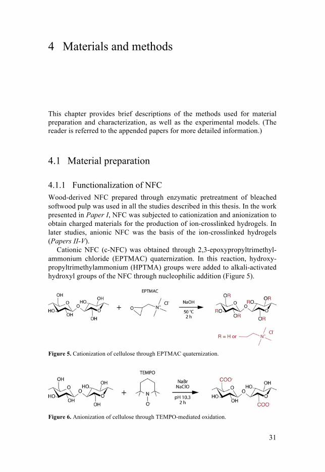
Cationic NFC (c-NFC) was obtained through 2,3-epoxypropyltrimethyl-ammonium chloride (EPTMAC) quaternization. In this reaction, hydroxy-propyltrimethylammonium (HPTMA) groups were added to alkali-activated hydroxyl groups of the NFC through nucleophilic addition (Figure 5).
Figure 5. Cationization of cellulose through EPTMAC quaternization.
Figure 6. Anionization of cellulose through TEMPO-mediated oxidation.

32
Anionic NFC (a-NFC) was prepared by selectively oxidizing the C6 hydrox-yl groups of the NFC by TEMPO-mediated oxidation (see Figure 6 above). NaBrO, generated in situ via NaClO oxidation of NaBr, was used as co-oxidant to oxidize TEMPO to an oxoammonium cation, which in turn oxi-dized the NFC.
Functionalized NFC was purified by dialysis against deionized or ul-trapure water, and the desired NFC concentration was obtained by evapora-tion of excess water.
4.1.2 Preparation of ion-crosslinked NFC hydrogels In total, five different ion-crosslinked NFC-based hydrogels were investigat-ed. Table 2 gives the composition and the names of each hydrogel used in this thesis. (Detailed information on the preparation method and composition of each hydrogel can be found in the appended papers.) Table 2. The ion-crosslinked NFC-based hydrogels studied in this work
Component 1 Component 2 Crosslinking ion
Name in thesis Paper
a-NFC Ca2+ NFC hydrogel I - V a-NFC c-NFC Ca2+ ac-NFC hydrogel I a-NFC Type I collagen Ca2+ NFC-collagen hydrogel II, IV a-NFC Kaolin Ca2+ NFC-kaolin hydrogel II a-NFC Cu2+ Cu-NFC hydrogel III Briefly, the ion-crosslinked NFC hydrogels were produced by first mixing components 1 and 2 to form a homogeneous suspension. The suspension was then poured into a mold of the desired size, and crosslinking was achieved by the drop-wise addition of an aqueous solution of the crosslinking ion. Unbound crosslinking ions were removed by washing the hydrogels with deionized or ultrapure water. Before use in biological experiments, the hy-drogels were sterilized with UV irradiation.
4.2 Material characterization
4.2.1 Determination of functional group content and charge The amino group content of c-NFC was determined by elemental analysis of the total nitrogen content (Paper I). Since unmodified NFC does not contain nitrogen, the number of HPTMA groups could be determined from the per-centage by weight of nitrogen in the studied samples.
33
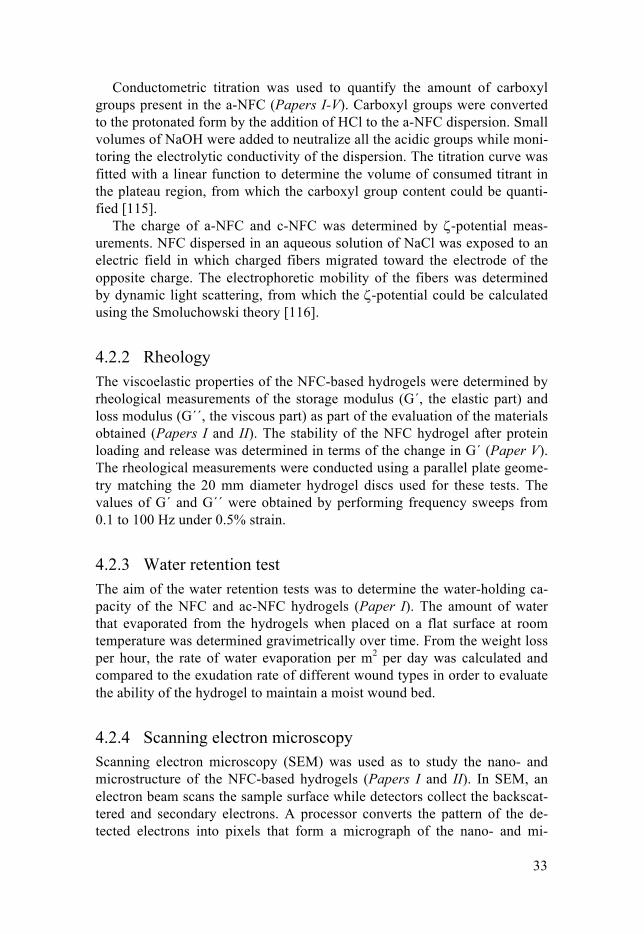
Conductometric titration was used to quantify the amount of carboxyl groups present in the a-NFC (Papers I-V). Carboxyl groups were converted to the protonated form by the addition of HCl to the a-NFC dispersion. Small volumes of NaOH were added to neutralize all the acidic groups while moni-toring the electrolytic conductivity of the dispersion. The titration curve was fitted with a linear function to determine the volume of consumed titrant in the plateau region, from which the carboxyl group content could be quanti-fied [115].
The charge of a-NFC and c-NFC was determined by ζ-potential meas-urements. NFC dispersed in an aqueous solution of NaCl was exposed to an electric field in which charged fibers migrated toward the electrode of the opposite charge. The electrophoretic mobility of the fibers was determined by dynamic light scattering, from which the ζ-potential could be calculated using the Smoluchowski theory [116].
4.2.2 Rheology The viscoelastic properties of the NFC-based hydrogels were determined by rheological measurements of the storage modulus (G´, the elastic part) and loss modulus (G´´, the viscous part) as part of the evaluation of the materials obtained (Papers I and II). The stability of the NFC hydrogel after protein loading and release was determined in terms of the change in G´ (Paper V). The rheological measurements were conducted using a parallel plate geome-try matching the 20 mm diameter hydrogel discs used for these tests. The values of G´ and G´´ were obtained by performing frequency sweeps from 0.1 to 100 Hz under 0.5% strain.
4.2.3 Water retention test The aim of the water retention tests was to determine the water-holding ca-pacity of the NFC and ac-NFC hydrogels (Paper I). The amount of water that evaporated from the hydrogels when placed on a flat surface at room temperature was determined gravimetrically over time. From the weight loss per hour, the rate of water evaporation per m2 per day was calculated and compared to the exudation rate of different wound types in order to evaluate the ability of the hydrogel to maintain a moist wound bed.
4.2.4 Scanning electron microscopy Scanning electron microscopy (SEM) was used as to study the nano- and microstructure of the NFC-based hydrogels (Papers I and II). In SEM, an electron beam scans the sample surface while detectors collect the backscat-tered and secondary electrons. A processor converts the pattern of the de-tected electrons into pixels that form a micrograph of the nano- and mi-
34
croscale structure. Hydrogel samples were lyophilized prior to imaging with SEM. In order to avoid charge build-up on the non-conducting NFC surface during imaging, samples were sputter coated with a conductive layer of gold before analysis.
4.2.5 Fourier transform infrared spectroscopy Fourier transform infrared-attenuated total reflectance (FTIR-ATR) spec-troscopy was used to evaluate the chemical interactions between the NFC hydrogel and proteins, as well as to determine the structural integrity of pro-teins (Paper V). Infrared spectroscopy exploits the fact that molecules absorb electromagnetic energy in the infrared region based on the specific excitation of vibrational and rotational modes in a fashion that is characteristic of the chemical structure. Thus, specific information regarding the chemical struc-ture is obtainable from the infrared absorption spectrum of a sample.
4.3 In vitro biocompatibility
4.3.1 In vitro models Preliminary biocompatibility screening was conducted, firstly with indirect cytotoxicity testing, in order to evaluate the potential leakage of toxic con-taminants from the NFC and ac-NFC hydrogels (Paper I). Human dermal fibroblast (hDF) and monocyte-like THP-1 cells were cultivated in extract medium for 24 h, after which cell viability was measured. This test was per-formed according to the ISO-10993-5 standard [117]. The second part of the preliminary biocompatibility screening included direct cytocompatibility tests where the response of cells cultured in direct contact with the material was evaluated (Paper I). hDF monolayers were cultured in contact with the hydrogels and cell viability and monolayer integrity were evaluated after 24-h culture, and after removal of the materials. Additionally, primary human mononuclear cells isolated from buffy coats (the cellular fraction of blood) using the Ficoll-Paque PLUS density gradient centrifugation technique, were used to evaluate immune cell response to the tested hydrogels in terms of adhesion and subsequent inflammatory response, under lipopolysaccharide (LPS)-stimulated and non-stimulated conditions.
Further application-oriented biocompatibility testing of the NFC and NFC-collagen hydrogels was performed in the work presented in Paper IV. Fibroblasts (hDF cells) and keratinocytes (an immortalized keratinocyte (KERT) cell line), which are the main cellular components of the dermal and epidermal skin layers, were cultured on the hydrogel materials and cell adhe-sion and subsequent proliferation and morphological evolution were evaluat-
35
ed for up to 7 days. The effect of the NFC and NFC-collagen hydrogels on skin cell migration was studied using a scratch assay that simulates the con-tact-guided migration of cells [118]. After scratching hDF and KERT mono-layers at the bottom of tissue culture plate (TCP) wells, hydrogels were add-ed on top of the monolayers. Scratch closure was monitored for 24 h using light and fluorescence microscopy. Aquacel® Extra, a cellulose-based hydro-fiber dressing, served as the reference material.
4.3.2 Evaluation of cell viability and proliferation The Alamar Blue (AB) assay was used to assess cell viability and prolifera-tion during indirect and direct biocompatibility studies (Papers I and IV). The AB assay detects the metabolic activity of cells via reduction of the AB reagent by the mitochondrial enzyme activity of viable cells. The redox reac-tion results in a color change of the dye solution from indigo blue to fluores-cent pink, which can be measured by coulometric or fluorometric means.
4.3.3 Evaluation of inflammatory response The inflammatory response of primary human mononuclear cells in contact with the studied hydrogels was assessed using biochemical assays (Paper I). The luminol-amplified chemiluminescence assay was used to quantify the release of ROS from activated immune cells in contact with the hydrogels. The assay measures chemically generated light emission from a reaction between intracellularly or extracellularly produced ROS and the luminol reagent. The presence of the pro-inflammatory marker TNF-α and the anti-inflammatory marker IL-10 in the culture medium after cell experiments was quantified using commercially available enzyme-linked immunosorbent assay (ELISA) kits. The ELISA technique relies on the capture of a target compound by a primary antibody that is immobilized on a solid phase. The capture of the target compound is then detected by a coulometric reaction involving, e.g. enzyme-conjugated secondary antibodies and substrate, or biotinylated antibodies with the corresponding enzyme-conjugated streptavi-din and substrate.
4.3.4 Evaluation of cell morphology and migration The morphological analysis and image acquisition of cells and cell monolay-ers were conducted using a light and fluorescence microscope equipped with a camera sensor (Papers I and IV). To obtain fluorescence images of cells, the cells were stained with calcein-AM and propidium iodine to visualize viable cells and cells with compromised membrane integrity, respectively. Quantitative migration data were obtained from the scratch assay by image analysis of micrographs obtained during the experiments (Paper IV).
36
4.4 In vitro hemocompatibility
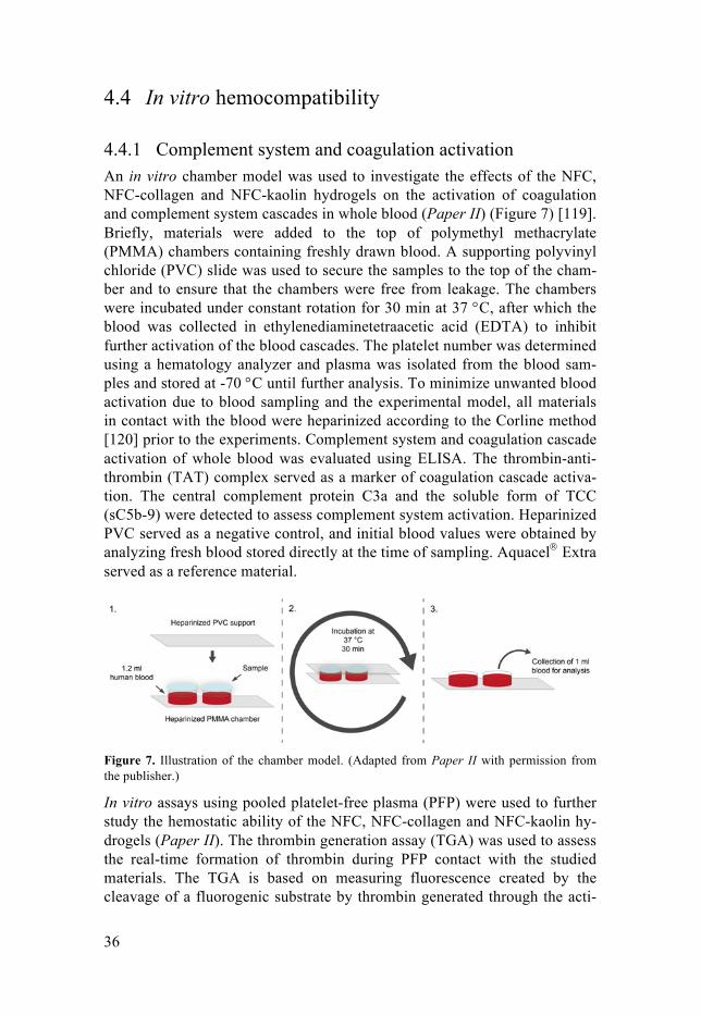
4.4.1 Complement system and coagulation activation An in vitro chamber model was used to investigate the effects of the NFC, NFC-collagen and NFC-kaolin hydrogels on the activation of coagulation and complement system cascades in whole blood (Paper II) (Figure 7) [119]. Briefly, materials were added to the top of polymethyl methacrylate (PMMA) chambers containing freshly drawn blood. A supporting polyvinyl chloride (PVC) slide was used to secure the samples to the top of the cham-ber and to ensure that the chambers were free from leakage. The chambers were incubated under constant rotation for 30 min at 37 °C, after which the blood was collected in ethylenediaminetetraacetic acid (EDTA) to inhibit further activation of the blood cascades. The platelet number was determined using a hematology analyzer and plasma was isolated from the blood sam-ples and stored at -70 °C until further analysis. To minimize unwanted blood activation due to blood sampling and the experimental model, all materials in contact with the blood were heparinized according to the Corline method [120] prior to the experiments. Complement system and coagulation cascade activation of whole blood was evaluated using ELISA. The thrombin-anti-thrombin (TAT) complex served as a marker of coagulation cascade activa-tion. The central complement protein C3a and the soluble form of TCC (sC5b-9) were detected to assess complement system activation. Heparinized PVC served as a negative control, and initial blood values were obtained by analyzing fresh blood stored directly at the time of sampling. Aquacel® Extra served as a reference material.
Figure 7. Illustration of the chamber model. (Adapted from Paper II with permission from the publisher.)
In vitro assays using pooled platelet-free plasma (PFP) were used to further study the hemostatic ability of the NFC, NFC-collagen and NFC-kaolin hy-drogels (Paper II). The thrombin generation assay (TGA) was used to assess the real-time formation of thrombin during PFP contact with the studied materials. The TGA is based on measuring fluorescence created by the cleavage of a fluorogenic substrate by thrombin generated through the acti-
37
vation of the coagulation cascade. This assay provides the time lag before thrombin formation, the thrombin peak concentration, and the time to peak concentration. In another PFP assay, the activation of the intrinsic pathway of the coagulation cascade was measured by detecting the formation of fac-tor XIIa (FXIIa) as a product of FXII activation upon PFP contact with the materials. TCP was used as the negative control and Aquacel® Extra was used as reference material for the PFP models.
4.4.2 Blood sampling and plasma pooling Blood was collected from healthy volunteers in heparinized tubes containing soluble heparin at a low concentration using an open system directly before use in the chamber model. Separately, blood was drawn from seven healthy volunteers using citrate tubes to create a plasma pool. PFP was obtained by two-step centrifugation of the blood before pooling, aliquoting and storing at -70 °C until use. The PFP was recalcified before use in the assays.
Ethical approval for blood sampling was obtained from the regional ethics committee and informed consent was obtained from the donors before the experiments.
4.5 In vitro bacteria interactions
4.5.1 Bacterial growth models S. epidermidis and P. aeruginosa were used as model bacteria to investigate the effect of the NFC and Cu-NFC hydrogels on bacterial growth (Paper III). An agitating large-volume system was used to quantify the growth of planktonic growing bacteria in the presence of the studied materials by measuring the optical density. Sessile growth was promoted in a small-volume system by seeding bacteria on top of the sample surfaces, and bacte-rial growth was quantified using measurement techniques utilizing fluores-cence (for P. aeruginosa through the use of resazurin) and luminescence (for the plasmid-modified S. epidermidis containing a luminescent system). Measurements of bacterial growth were performed continuously for up to 16 h to obtain growth curves. From these, the lag phase (i.e. the time after inoc-ulation during which the bacteria adapted to the new environment) and dou-bling time of the bacteria were determined to evaluate the effects of the ma-terials. Bacterial growth in the absence of any material served as the negative control, and the commercial antibacterial dressing Aquacel® Ag+ Extra was used as the positive control.

38
4.5.2 Sessile growth pattern and bacterial impermeability To further investigate the sessile growth of bacteria on hydrogel surfaces, samples from the small-volume system were fixated with glutaraldehyde and subsequently lyophilized to allow imaging of the growth pattern and biofilm formation using SEM. To assess bacterial impermeability, cross-sections of lyophilized hydrogels were prepared to expose possible bacterial growth in-side the materials. Hydrogels were considered impermeable to bacteria if no bacteria were observed inside the material.
4.6 In vivo wound healing
4.6.1 Study design The in vivo wound healing study presented in Paper IV was performed by NAMSA contract laboratory (France) in compliance with the ISO-10993-6 standard [121]. A total of 6 Sprague Dawley rats were used. Two paraverte-bral dermo-epidermic wounds, preserving the panniculus carnosus, were created on each side of the back of each rat on day 0. NFC hydrogel and control (saline-soaked sterile non-woven cotton gauze) primary dressings were applied to one wound each on each rat. The primary dressings were covered by a secondary dressing consisting of a sterile semi-permeable pol-yurethane adhesive dressing, sterile woven gauze and an elastic adhesive bandage. The dressings were carefully changed three times a week to allow macroscopic evaluation of the wounds and dressings. The rats were eu-thanized on day 25 and the wounds were sampled for histopathological eval-uation.
4.6.2 Macroscopic and morphometric analysis The macroscopic evaluation of dressings included examination of adhesion to the wounds and the structural stability of the dressings. Evaluation of the wounds included observation and scoring of peri-wound skin status, the presence of exudate, blood, fibrin and granulation tissue, and signs of mac-eration and/or infection and re-epithelialization. Prior to re-application of the dressings, the rats were weighed, the wounds were washed with lukewarm saline solution, and macroscopic pictures of the wounds were taken. Mor-phometric analysis of macrophotographs was conducted using morphometry software to obtain quantitative wound closure data.

39
4.6.3 Histopathological evaluation The wound samples were cut into two central sections with a microtome. One section was stained with Masson’s trichrome (MT) and the other with safranin-hematoxylin-eosin (SHE). Qualitative and semi-quantitative histo-pathological evaluation of the local tissue effects at the wound location was conducted according to the ISO-10993-6 standard [121].
4.7 In vitro protein interactions
4.7.1 Protein loading and release methods The NFC hydrogel was investigated to determine how it interacted with pro-teins, and if it had potential for use as a delivery system for therapeutic pro-teins (Paper V). Bovine serum albumin (BSA), lysozyme and fibrinogen were selected as model proteins based on their difference in size and charge at physiological pH. See Table 3 for an overview of the physicochemical properties of the proteins.
Table 3. Physicochemical properties of model proteins
MW (kDa)
Hydrodynamic radius at pH 7.4 (nm)
Isoelectric point Charge at pH 7.4
BSA 66.5 3.5 a 4.7 -
Fibrinogen 340 12.7 b 5.1 - 6.3 -
Lysozyme 14.7 1.9 c 11.1 +
MW: molecular weight a González et al. [122] b Wasilewska et al. [123] c Parmar et al. [124]
To load the NFC hydrogel with proteins, hydrogel discs were placed in phosphate buffered saline (PBS) solutions containing the respective protein and left to incubate under slight agitation for 24 h. The amount of protein loaded was determined by measuring the decrease in free protein concentra-tion in the PBS solution, by measuring the absorbance at 280 nm (A280) us-ing a spectrophotometer. To release proteins from the loaded NFC hydro-gels, the hydrogels were placed in vessels containing PBS. The release kinet-ics was determined by measuring the free protein concentration as above. Release experiments for each protein were conducted for 7 days.

40
4.7.2 Mathematical analysis of release The semi-empirical Ritger–Peppas equation was used to analyze the mecha-nism of release from the NFC hydrogel [125, 126]:
(1)
where Mt and M∞ denote the cumulative amount of protein released at time t and infinite time, respectively; k denotes a constant incorporating the struc-tural and geometrical characteristics of the device; and n is the release expo-nent, which indicates the mechanism of release. Values of n were obtained by fitting experimental release data in the region 0 < Mt/M∞ < 0.6 with Eq. 1.
The diffusion coefficients of the proteins within the NFC hydrogel were determined using the unsteady-state form of Fick’s second law of diffusion under the assumption that these systems could be described as monolithic solutions (initial drug concentration < drug solubility) with slab geometry [126, 127]:
(2)
where D is the diffusion coefficient of the protein, and L denotes the thick-ness of the hydrogel. Values of D were obtained by fitting experimental re-lease data in the region 0 < Mt/M∞ < 0.6 with Eq. 2.
4.7.3 Protein stability investigation The degradation of the released proteins was investigated using sodium do-decyl sulfate-polyacrylamide gel electrophoresis (SDS-PAGE), a method of protein separation in an electric field, which provides information regarding size of proteins, and thus reveals any degradation of proteins into smaller molecular-weight fragments.
Protein stability in terms of maintained activity was assessed using a method that quantifies the activity of a lysozyme sample by its ability to lyse the bacterium Micrococcus lysodeikticus (measured as the decrease in ab-sorption (A450) as the bacteria are lysed). To obtain information on protein activity after release, the activity of lysozyme released by the NFC hydrogel into PBS and simulated wound fluid (SWF, consisting of equal parts peptone water and fetal bovine serum) was compared to that of control solutions con-sisting of native lysozyme.
41
5 Results and discussion
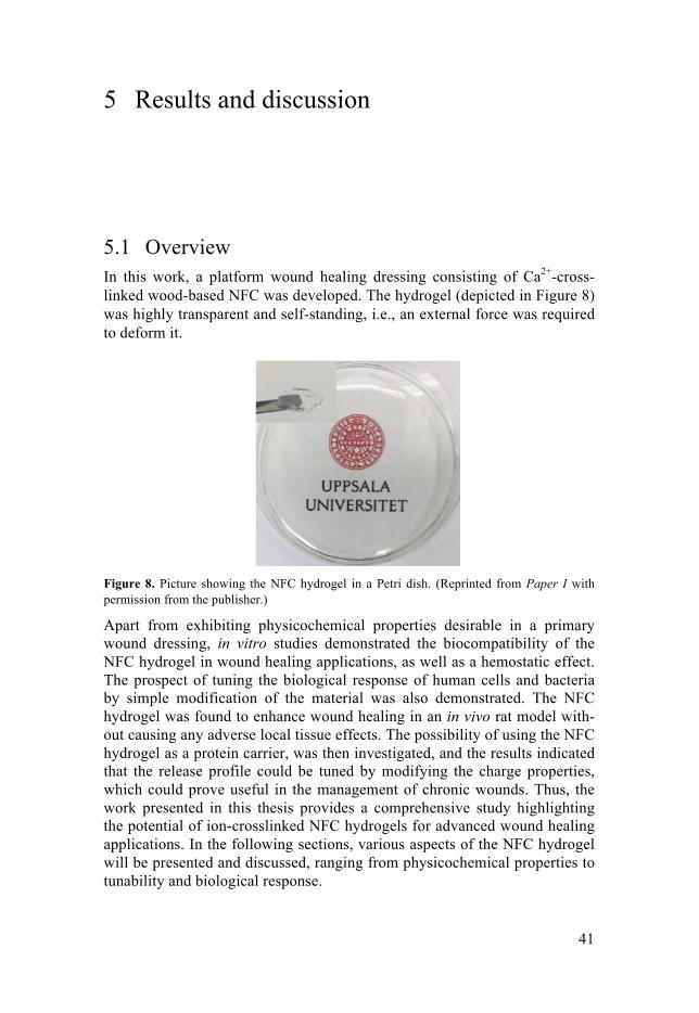
5.1 Overview In this work, a platform wound healing dressing consisting of Ca2+-cross-linked wood-based NFC was developed. The hydrogel (depicted in Figure 8) was highly transparent and self-standing, i.e., an external force was required to deform it.
Figure 8. Picture showing the NFC hydrogel in a Petri dish. (Reprinted from Paper I with permission from the publisher.)
Apart from exhibiting physicochemical properties desirable in a primary wound dressing, in vitro studies demonstrated the biocompatibility of the NFC hydrogel in wound healing applications, as well as a hemostatic effect. The prospect of tuning the biological response of human cells and bacteria by simple modification of the material was also demonstrated. The NFC hydrogel was found to enhance wound healing in an in vivo rat model with-out causing any adverse local tissue effects. The possibility of using the NFC hydrogel as a protein carrier, was then investigated, and the results indicated that the release profile could be tuned by modifying the charge properties, which could prove useful in the management of chronic wounds. Thus, the work presented in this thesis provides a comprehensive study highlighting the potential of ion-crosslinked NFC hydrogels for advanced wound healing applications. In the following sections, various aspects of the NFC hydrogel will be presented and discussed, ranging from physicochemical properties to tunability and biological response.
42
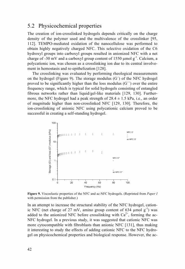
5.2 Physicochemical properties The creation of ion-crosslinked hydrogels depends critically on the charge density of the polymer used and the multivalence of the crosslinker [95, 112]. TEMPO-mediated oxidation of the nanocellulose was performed to obtain highly negatively charged NFC. This selective oxidation of the C6 hydroxyl groups into carboxyl groups resulted in anionized NFC with a net charge of -30 mV and a carboxyl group content of 1550 µmol g-1. Calcium, a polycationic ion, was chosen as a crosslinking ion due to its central involve-ment in hemostasis and re-epithelization [128].
The crosslinking was evaluated by performing rheological measurements on the hydrogel (Figure 9). The storage modulus (G´) of the NFC hydrogel proved to be significantly higher than the loss modulus (G´´) over the entire frequency range, which is typical for solid hydrogels consisting of entangled fibrous networks rather than liquid/gel-like materials [129, 130]. Further-more, the NFC hydrogel had a peak strength of 28.4 ± 1.5 kPa, i.e., an order of magnitude higher than non-crosslinked NFC [129, 130]. Therefore, the ion-crosslinking of anionic NFC using polycationic calcium proved to be successful in creating a self-standing hydrogel.
Figure 9. Viscoelastic properties of the NFC and ac-NFC hydrogels. (Reprinted from Paper I with permission from the publisher.)
In an attempt to increase the structural stability of the NFC hydrogel, cation-ic NFC (net charge of 27 mV, amino group content of 634 µmol g-1) was added to the anionized NFC before crosslinking with Ca2+, forming the ac-NFC hydrogel. In a previous study, it was suggested that cationic NFC was more cytocompatible with fibroblasts than anionic NFC [131], thus making it interesting to study the effects of adding cationic NFC to the NFC hydro-gel on physicochemical properties and biological response. However, the ac-
43
NFC hydrogel exhibited a non-significant difference in peak strength (24.7 ± 1.8 kPa) compared to the value obtained for the NFC hydrogel, indicating that it had no stabilizing effect on the anionic hydrogel.
As mentioned in Chapter 3, a key role of a modern wound dressing is to control fluid loss and maintain a moist wound bed in order to promote deb-ridement of dead tissue and epithelial regeneration. The water-holding ca-pacity of the NFC and ac-NFC hydrogels was therefore investigated. The rate of water loss from the NFC hydrogel was 362 ± 25 g m-2 per day, while that from the ac-NFC hydrogel was 301 ± 12 g m-2 per day. The ac-NFC hydrogel thus had a better water-holding capacity than the NFC hydrogel, which could stem from its more aggregated structure (as illustrated by the SEM micrograph in Figure 10), and therefore possibly smaller surface area and fibril exposure to the surrounding environment.
Figure 10. SEM micrographs of the NFC and ac-NFC hydrogels. Note the nanoscale structure of the NFC hydrogel (left) and the nano- and microscale structure of the ac-NFC hydrogel (right). (Reprinted from Paper I with permission from the publisher.)
The water vapor loss from healthy skin is about 204 g m-2 per day, whereas wounds of varying degrees of severity have water vapor transmission rates of 279 to 5138 g m-2 per day, depending on the wound type and stage [132]. The results of this work thus indicated that both the NFC hydrogel and the ac-NFC hydrogel are capable of maintaining a moist wound bed for most types of wounds. Following the demonstration of this central property of a modern wound dressing by the self-standing hydrogels produced in this work, characterization of the materials was continued by evaluating their biocompatibility with key cells in the wound healing process.
5.3 Biocompatibility
5.3.1 Initial biocompatibility assessment Initial indirect cytotoxicity screening of the NFC and ac-NFC hydrogels was performed to ensure that no substances remained in the materials after the

44
anionization and cationization processes that would affect the previously demonstrated cytocompatibility of NFC with human skin and immune cells [131, 133]. According to the ISO-10993-5 standard [117], a material can be regarded as cytocompatible if it causes a maximum of 30% decrease in cell viability compared to a negative control. The viability of hDF cells and THP-1 cells cultivated in NFC and ac-NFC hydrogel extract was above 70% (Figure 11). Hence, neither hydrogel induced cytotoxic effects through leachable compounds.
Figure 11. Indirect cytotoxicity of the NFC and ac-NFC hydrogels using a) hDF and b) THP-1 cells. The box plots represent the interquartile range of the data, where the mean is marked by a square and the median by a line. (Reprinted from Paper I with permission from the pub-lisher.)
The inflammatory response of the NFC and ac-NFC hydrogels was evaluat-ed in terms of cytokine release and ROS generation by exposing blood-isolated human mononuclear cells to the materials. Using these cells as a model system to assess the inflammatory response in vitro provides an espe-cially robust evaluation as it includes the immunological responses of indi-vidual donors. It was found that neither material promoted significant secre-tion of the pro-inflammatory cytokine TNF-α under non-stimulated condi-tions, compared to the control (the tissue culture material Thermanox®
(TMX)) (Figure 12a). Neither were the TNF-α levels significantly reduced by the hydrogels under LPS-stimulated (inflammatory) conditions. IL-10 secretion by mononuclear cells was also unaffected by the hydrogels (Figure 12b).
During wound healing, monocytes and macrophages in the wound bed re-lease the pro-inflammatory cytokine TNF-α which is crucial for the modula-tion of the early phases of healing [134]. The same cells also release the anti-inflammatory cytokine IL-10, which downregulates the production of TNF-α to limit the extent of the inflammatory response [135]. Together, these cytokines play a key role in determining the duration of the inflammatory phase of wound healing. Disrupting the balance between TNF-α and IL-10 may lead to adverse effects such as insufficient inflammation to aid healing, or the development of chronic wounds. Thus, the lack of effect of the NFC

45
and ac-NFC hydrogels on TNF-α and IL-10 can be regarded as a desirable quality, allowing these materials to function as a base from which specific inflammatory responses could be modulated, depending on the type of wound to be healed.
Figure 12. Inflammatory response of non-stimulated (-LPS) and stimulated (+LPS) mononu-clear cells upon contact with NFC and ac-NFC hydrogels as measured by the release of a) pro-inflammatory cytokine TNF-α and b) anti-inflammatory cytokine IL-10. The box plots represent the interquartile range of the data, where the mean is marked by a square and the median by a line. The asterisk indicates a statistically significant difference (p < 0.05). (Re-printed from Paper I with permission from the publisher.)
ROS also play a pivotal role in the management of the inflammatory process during normal wound healing [136]. Too low levels of ROS can trigger cell cycle arrest and insufficient assembly of the inflammatory phase of healing. In contrast, abnormally high levels of ROS increase the risk of pro-apoptotic processes and reductions in DNA, proteins and lipids, resulting in cell death and tissue damage [137]. The risk of chronic wound development also in-creases with elevated levels of ROS [138]. Figure 13 shows the results of the study on the production of ROS by mononuclear cells in contact with the NFC and ac-NFC hydrogels. Only an insignificant elevation of ROS levels, compared with the control, was observed for the NFC and ac-NFC hydrogels thus further indicating the inert nature of the materials in terms of inflamma-tory response.

46
Figure 13. Inflammatory response of mononuclear cells upon contact with the NFC and ac-NFC hydrogels expressed as the release of ROS. The box plots represent the interquartile range of the data, where the mean is marked by a square and the median by a line. (Reprinted from Paper I with permission from the publisher.)
This initial material characterization showed that the inclusion of cationic NFC in the NFC hydrogel did not improve dressing material properties to any significant degree. The ac-NFC hydrogel was therefore not included in further application-oriented biocompatibility testing. Instead, the NFC hy-drogel was supplemented with type I collagen, which is known for its bene-ficial effects on wound healing by aiding hemostasis, promoting re-epithelization and reducing scar formation [139, 140].
5.3.2 Further application-oriented biocompatibility testing The proliferation and migration of fibroblasts and keratinocytes are central to the granulation and re-epithelialization of wounds [141], thus any interac-tions between dressing materials and such processes are an important aspect that should be considered in the evaluation of materials for wound healing applications. The cell lines hDF and KERT were used as model cells for human fibroblasts and keratinocytes, respectively, in in vitro biocompatibil-ity tests. Cell adhesion, morphology and proliferation were evaluated after seeding these skin cells onto the material surfaces, and culturing for up to 7 days.
Figure 14a shows the metabolic activities of hDF cells at 1, 3 and 7 days after seeding. Measurements on day 1 showed that hDF cells seeded on the NFC hydrogel, the NFC-collagen hydrogel and Aquacel® Extra exhibited significantly lower initial adhesion than the cells seeded on TCP. However, on day 7 the metabolic activity, expressed as the percentage of that on day 1, was 380 ± 32, 426 ± 25, 394 ± 43 and 248 ± 48% for NFC, NFC-collagen, Aquacel® Extra and TCP, respectively, thus indicating that hDF cells prolif-erated on the materials.

47
Figure 14. Adhesion and proliferation of a) hDF cells and b) KERT cells after seeding onto the NFC hydrogel, the NFC-collagen hydrogel, Aquacel® Extra and TCP. Data represents mean ± standard error of the mean for n = 5. Asterisks indicate a statistically significant dif-ference (p < 0.05) compared to the previous measurement.
The fluorescence microscopy images of proliferating hDF cells in Figure 15a reflect the measured change in metabolic activity over time. The hDF cells cultured on the NFC-based hydrogels showed a spheroid morphology on day 1, which indicates that the cells had difficulty in adapting to the new envi-ronment. However, the morphology became more elongated, and compara-ble to that of hDF cells cultured on TCP from day 3 and onwards, indicating that the cells seemed to respond well to the hydrogel surface after an initial adaptation period. Note that no fluorescence microscopy images could be obtained of Aquacel® Extra due to its opaque nature.
Figure 15. Fluorescence microscopy images of a) hDF and b) KERT cells after proliferation on the NFC hydrogel, NFC-collagen hydrogel and TCP for 1-7 days. Scale bars represent 200 µm.
The results of the metabolic activity measurements on KERT cells indicated that they adhered to the NFC-based hydrogels and TCP to a similar degree (Figure 14b). The number of cells on day 3, expressed as a percentage of
48
those on day 1, was 339 ± 21, 291 ± 22 and 375 ± 16% for NFC, NFC-collagen and TCP, respectively, indicating that these cells proliferated on the various materials. The same trend was observed using fluorescence micro-copy (Figure 15b). However, KERT cells exhibited poor adhesion and pro-liferation on Aquacel® Extra. This could be due to the very soft and uneven structure of this material compared to the NFC-based hydrogels [142, 143]. Overall, the KERT cells showed faster adaptation and adhesion on the NFC and NFC-collagen hydrogels than did the hDF cells.
The physical adsorption of proteins onto a hydrogel surface is generally expected to be impaired by the low interfacial energy between the highly hydrated hydrogel phase and the aqueous biological medium [144]. As cell–surface interactions are ultimately determined by the interactions between the cell and surface-bound proteins [145], low cell adhesion to hydrogels can also be expected. Nevertheless, a number of the properties of hydrogels can influence cell adhesion, including surface wettability, the presence of func-tional groups, surface density, stiffness and roughness [142, 143, 145, 146]. For example, it has been shown that the introduction of carboxyl groups into a material improves cell adhesion [147]. In accordance with this, Rashad et al. showed that fibroblast adhesion and spreading were better on carboxylat-ed NFC than carboxymethylated NFC [113]. Based on these results, the car-boxylated (TEMPO-oxidized) NFC-based hydrogels studied in this work could be expected to allow good fibroblast adhesion. However, as variations were seen in the initial fibroblast adhesion between the studies, this shows that an individual hydrogel property, such as surface chemistry, is not the only property affecting its cell adhesion properties, and that physicochemical differences (e.g. hydrogel stiffness) resulting from changes in composition are likely to contribute to the response of the cell.
Studying the morphology of KERT cells cultured on the hydrogels and TCP revealed a marked morphological change in the cells on the NFC and NFC-collagen hydrogels compared to the morphology of the cells on TCP (Figure 15b). Islands of fused cells were observed already on day 1, and these had increased significantly in size on days 2 and 3. The fusion of keratinocytes has been described in the literature as a sign of a differentia-tion process that is tightly regulated by, amongst other mechanisms, extra-cellular calcium concentration [148, 149]. In a control experiment, KERT cells were therefore cultivated in the presence of Ca2+ (crosslinking ion), a non-crosslinked NFC suspension, and in pure culture medium as a control. Cells cultured in the presence of NFC suspension and in pure culture medi-um exhibited typical individualized and polygonal morphology, while KERT cells cultivated with the crosslinking ion underwent cell fusion (Figure 16). Despite attempts to confirm the differentiation of KERT cells by the investi-gation of cell markers associated with this process (e.g. involucrin and cy-tokeratin-10 [150]), no upregulation of such markers could be detected in either the samples or controls. This could be due to changes in the cellular
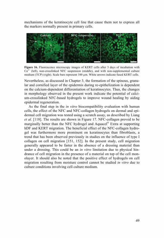
49
mechanisms of the keratinocyte cell line that cause them not to express all the markers normally present in primary cells.
Figure 16. Fluorescence microscopy images of KERT cells after 3 days of incubation with Ca2+ (left), non-crosslinked NFC suspension (middle), and with non-supplemented culture medium (TCP) (right). Scale bars represent 100 µm. White arrows indicate fused KERT cells.
Nevertheless, as discussed in Chapter 3, the formation of the spinous, granu-lar and cornified layer of the epidermis during re-epithelization is dependent on the calcium-dependent differentiation of keratinocytes. Thus, the changes in morphology observed in the present work indicate the potential of calci-um-crosslinked NFC-based hydrogels to improve wound healing by aiding epidermal regeneration.
As the final step in the in vitro biocompatibility evaluation with human cells, the effect of the NFC and NFC-collagen hydrogels on dermal and epi-dermal cell migration was tested using a scratch assay, as described by Liang et al. [118]. The results are shown in Figure 17. NFC-collagen proved to be marginally better than the NFC hydrogel and Aquacel® Extra at supporting hDF and KERT migration. The beneficial effect of the NFC-collagen hydro-gel was furthermore more prominent on keratinocytes than fibroblasts, a trend that has been observed previously in studies on the influence of type I collagen on cell migration [151, 152]. In the present study, cell migration generally appeared to be faster in the absence of a dressing material than under a dressing. This could be an in vitro limitation due to physical hin-drance of cell migration in the presence of a material on top of the cell mon-olayer. It should also be noted that the positive effect of hydrogels on cell migration resulting from moisture control cannot be studied in vitro due to culture conditions involving cell culture medium.

50
Figure 17. Migration of a) hDF and b) KERT cells after scratching cell monolayers and ap-plication of the NFC hydrogel, the NFC-collagen hydrogel and Aquacel® Extra to cover the scratch. In the TCP, the cells migrated in the absence of any dressing material. Data repre-sents mean ± standard error of the mean for n = 3. Asterisks indicate statistically significant differences (p < 0.05) between data collected at 24-h intervals.
Hydrogel adhesion to the cell monolayers was also evaluated in the scratch assay, showing good outcome. Neither of the NFC-based hydrogels adhered to the cell monolayers, and no damage was seen to the cells upon removal of the materials. Fluorescence microscopy images showing these results can be found in Figure 2 in Paper IV.
Overall, the biocompatibility of the NFC hydrogel in wound healing ap-plications was judged to be excellent as the material supported dermal and epidermal proliferation without damaging cell monolayers upon removal or inducing any adverse response of immune cells. Supplementing the NFC hydrogel with c-NFC provided no significant added value, while the addition of type I collagen proved to only marginally improve skin cell migration.
5.4 Hemocompatibility It is essential to evaluate the hemocompatibility of biomaterials that are ex-pected to come into contact with blood during use. Studies on this aspect of the NFC-based hydrogels are presented in Paper II. An additional objective of this study was to modify the NFC hydrogel for potential use as a hemo-static dressing. Therefore, the basic hydrogel was supplemented with type I collagen and kaolin, forming the NFC-collagen and the NFC-kaolin hydro-gels, respectively. Collagen was added as it is known to contribute to the primary activation of platelets, and kaolin was chosen for its strong hemo-static effect and is often used in hemostatic dressings [42, 153]. However, as negative charges (which are present in the NFC hydrogel) trigger the intrin-sic pathway through FXII activation, the NFC hydrogel itself was also ex-pected to possess hemostatic properties.
Figure 18 shows complement system activation of blood as a result of blood–material contact. The NFC, NFC-collagen and NFC-kaolin hydrogels

51
all showed C3a formation comparable to that of the negative control (hepa-rinized PVC) (Figure 18a). A similar trend was observed in sC5b-9 produc-tion (Figure 18b). However, the NFC-collagen hydrogel showed an increase in sC5b-9 production compared to the control. In contrast to the NFC-based hydrogels, Aquacel® Extra caused a significant increase in both complement system markers. Hydrogels often exhibit increased hemocompatibility due to the generally low adhesion of blood components on smooth hydrogel surfac-es [154]. The higher activation of the complement system by Aquacel® Extra could be explained by its non-crosslinked nature, which diminishes the typi-cally firm, smooth surface of hydrogels.
Figure 18. Complement system activation measured by the formation of a) C3a and b) sC5b-9 after blood contact with the studied materials. Data represents mean ± standard error of the mean for n = 5. Statistically significant differences between sample groups are indicated by asterisks (*p < 0.05, **p < 0.01 and ***p < 0.001). (Reprinted from Paper II with permission from the publisher.)
The importance of the complement system in wound healing is still the sub-ject of debate. Some authors have suggested that complement system activa-tion aids wound healing through promotion of the migration and activation of immune cells and fibroblasts and the enhancement of angiogenesis [155-157]. However, others have reported damaging effects of the complement system during the early phases of wound healing, and have suggested that inhibition of the complement system would increase the rate and efficacy of healing [158]. Regardless of whether the complement system aids or impairs wound healing, the inert nature of the NFC hydrogel is beneficial as it re-duces the risk of potential adverse effects.
The impact of the NFC, NFC-collagen and NFC-kaolin hydrogels on co-agulation was assessed using whole blood and PFP models. The NFC-based hydrogels had a clear hemostatic effect in whole blood, compared to the negative control and Aquacel® Extra. This was demonstrated by the decrease in platelet count as an effect of clotting (Figure 19a), as well as the increased concentration of TAT (Figure 19b). The NFC-kaolin hydrogel had the great-
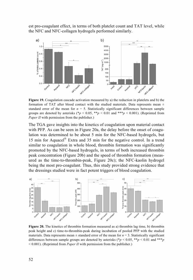
52
est pro-coagulant effect, in terms of both platelet count and TAT level, while the NFC and NFC-collagen hydrogels performed similarly.
Figure 19. Coagulation cascade activation measured by a) the reduction in platelets and b) the formation of TAT after blood contact with the studied materials. Data represents mean ± standard error of the mean for n = 5. Statistically significant differences between sample groups are denoted by asterisks (*p < 0.05, **p < 0.01 and ***p < 0.001). (Reprinted from Paper II with permission from the publisher.)
The TGA gave insights into the kinetics of coagulation upon material contact with PFP. As can be seen in Figure 20a, the delay before the onset of coagu-lation was determined to be about 5 min for the NFC-based hydrogels, but 15 min for Aquacel® Extra and 35 min for the negative control. In a trend similar to coagulation in whole blood, thrombin formation was significantly promoted by the NFC-based hydrogels, in terms of both increased thrombin peak concentration (Figure 20b) and the speed of thrombin formation (meas-ured as the time-to-thrombin-peak, Figure 20c); the NFC-kaolin hydrogel being the most pro-coagulant. Thus, this study provided strong evidence that the dressings studied were in fact potent triggers of blood coagulation.
Figure 20. The kinetics of thrombin formation measured as a) thrombin lag time, b) thrombin peak height and c) time-to-thrombin-peak during incubation of pooled PFP with the studied materials. Data represents mean ± standard error of the mean for n = 3. Statistically significant differences between sample groups are denoted by asterisks (*p < 0.05, **p < 0.01 and ***p < 0.001). (Reprinted from Paper II with permission from the publisher.)

53
To investigate whether the NFC-based hydrogels triggered coagulation through the intrinsic pathway due to the negative charges on the functional-ized nanocellulose fibrils, the activation of FXII was measured by determin-ing the concentration of FXIIa after contact between the material and PFP. The results indicated that the negatively charged NFC indeed contributed to significant FXII activation (Figure 21). Again, the NFC-kaolin hydrogel exhibited the strongest effect, as expected, and reflects the previously known potency of kaolin as an FXII activator [159].
Figure 21. FXIIa formation measured after incubation of pooled PFP with the studied materi-als. Data represents mean ± standard error of the mean for n = 5. Statistically significant differences between sample groups are denoted by asterisks (*p < 0.05, **p < 0.01 and ***p < 0.001). (Reprinted from Paper II with permission from the publisher.)
Taken together, the results presented above show that the NFC hydrogel exhibits hemostatic properties, most probably due to its negative charges, without activating the complement system. Modification with kaolin was shown to increase the hemostatic effect of the hydrogel, while the addition of collagen had no effect on the hemostatic efficiency, but marginally increased complement system activation.
5.5 Interactions with common wound pathogens In addition to studying the physicochemical characteristics, biocompatibility and hemostatic effect of the NFC hydrogel, it was deemed interesting to investigate how common wound pathogens responded to the proposed dress-ing materials. The ability to tune the antibacterial properties of the NFC hy-drogel was also studied by the parallel evaluation of a Cu2+-crosslinked NFC hydrogel. Copper was chosen as an alternative polycationic crosslinker to calcium due to its previously reported antibacterial activity [160]. The Gram-positive opportunistic pathogen S. epidermidis and the Gram-negative
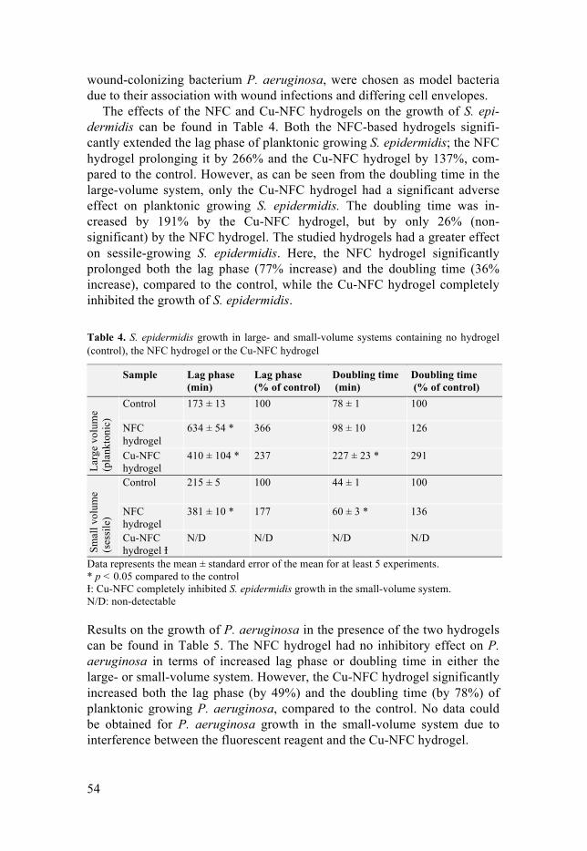
54
wound-colonizing bacterium P. aeruginosa, were chosen as model bacteria due to their association with wound infections and differing cell envelopes.
The effects of the NFC and Cu-NFC hydrogels on the growth of S. epi-dermidis can be found in Table 4. Both the NFC-based hydrogels signifi-cantly extended the lag phase of planktonic growing S. epidermidis; the NFC hydrogel prolonging it by 266% and the Cu-NFC hydrogel by 137%, com-pared to the control. However, as can be seen from the doubling time in the large-volume system, only the Cu-NFC hydrogel had a significant adverse effect on planktonic growing S. epidermidis. The doubling time was in-creased by 191% by the Cu-NFC hydrogel, but by only 26% (non-significant) by the NFC hydrogel. The studied hydrogels had a greater effect on sessile-growing S. epidermidis. Here, the NFC hydrogel significantly prolonged both the lag phase (77% increase) and the doubling time (36% increase), compared to the control, while the Cu-NFC hydrogel completely inhibited the growth of S. epidermidis.
Table 4. S. epidermidis growth in large- and small-volume systems containing no hydrogel (control), the NFC hydrogel or the Cu-NFC hydrogel
Sample Lag phase (min)
Lag phase (% of control)
Doubling time (min)
Doubling time (% of control)
Larg
e vo
lum
e (p
lank
toni
c)
Control 173 ± 13 100 78 ± 1 100
NFC hydrogel
634 ± 54 * 366 98 ± 10 126
Cu-NFC hydrogel
410 ± 104 * 237 227 ± 23 * 291
Smal
l vol
ume
(ses
sile
)
Control 215 ± 5 100 44 ± 1 100
NFC hydrogel
381 ± 10 * 177 60 ± 3 * 136
Cu-NFC hydrogel Ɨ
N/D N/D N/D N/D
Data represents the mean ± standard error of the mean for at least 5 experiments. * p < 0.05 compared to the control Ɨ: Cu-NFC completely inhibited S. epidermidis growth in the small-volume system. N/D: non-detectable Results on the growth of P. aeruginosa in the presence of the two hydrogels can be found in Table 5. The NFC hydrogel had no inhibitory effect on P. aeruginosa in terms of increased lag phase or doubling time in either the large- or small-volume system. However, the Cu-NFC hydrogel significantly increased both the lag phase (by 49%) and the doubling time (by 78%) of planktonic growing P. aeruginosa, compared to the control. No data could be obtained for P. aeruginosa growth in the small-volume system due to interference between the fluorescent reagent and the Cu-NFC hydrogel.

55
The positive control (Aquacel® Ag+ Extra) effectively inhibited the growth of both S. epidermidis and P. aeruginosa in all experiments.
Table 5. P. aeruginosa growth in large- and small-volume systems containing no hydrogel (control), the NFC hydrogel or the Cu-NFC hydrogel
Sample Lag phase (min)
Lag phase (% of control)
Doubling time (min)
Doubling time (% of control)
Larg
e vo
lum
e (p
lank
toni
c)
Control 166 ± 7 100 253 ± 52 100
NFC hydrogel
158 ± 2 95 193 ± 10 76
Cu-NFC hydrogel
248 ± 25 * 149 451 ± 19 * 178
Smal
l vol
ume
(ses
sile
)
Control 171 ± 12 100 57 ± 2 100
NFC hydrogel
195 ± 8 114 56 ± 3 98
Cu-NFC hydrogel Ɨ
- - - -
Data represents the mean ± standard error of the mean for at least 5 experiments. * p < 0.05 compared to the control Ɨ: Cu-NFC data not available for small-volume system due to interference between the mate-rial and the fluorescent reagent. The Ca-NFC hydrogel proved to have a bacteriostatic effect on S. epidermid-is, as shown by the prolonged lag phase and increased doubling time. Such an effect on this opportunistic pathogen may significantly reduce the risk of chronic wound development as a critical bacterial load and biofilm for-mation have previously been suggested to be determining factors in delayed healing due to infection [31, 161]. The absence of an inhibitory effect of the Ca-NFC hydrogel on P. aeruginosa is in line with previous reports on the increased resistance of Gram-negative bacteria to antibacterial agents, com-pared to Gram-positive bacteria [39]. Using Cu2+ as the crosslinker of the nanofibrils resulted in a clearly antibacterial nanocellulose hydrogel in terms of growth inhibition, probably resulting from the ability of copper ions to rupture bacterial cell walls and plasma membranes [162].
The sessile growth pattern of S. epidermidis and P. aeruginosa on the hy-drogels was also investigated using SEM. Typical grape-like clusters of S. epidermidis were observed on the NFC hydrogel (Figure 22a). With this additional information, the slower growth of S. epidermidis observed in the small-volume system (cf. Table 4) may be explained by the colonization of the material surface by S. epidermidis, which is expected to reduce the growth rate [163]. No S. epidermidis colonization was detected on the Cu-NFC surface, in agreement with the growth data (Figure 22b).
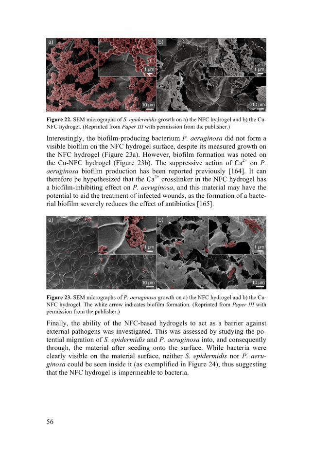
56
Figure 22. SEM micrographs of S. epidermidis growth on a) the NFC hydrogel and b) the Cu-NFC hydrogel. (Reprinted from Paper III with permission from the publisher.)
Interestingly, the biofilm-producing bacterium P. aeruginosa did not form a visible biofilm on the NFC hydrogel surface, despite its measured growth on the NFC hydrogel (Figure 23a). However, biofilm formation was noted on the Cu-NFC hydrogel (Figure 23b). The suppressive action of Ca2+ on P. aeruginosa biofilm production has been reported previously [164]. It can therefore be hypothesized that the Ca2+ crosslinker in the NFC hydrogel has a biofilm-inhibiting effect on P. aeruginosa, and this material may have the potential to aid the treatment of infected wounds, as the formation of a bacte-rial biofilm severely reduces the effect of antibiotics [165].
Figure 23. SEM micrographs of P. aeruginosa growth on a) the NFC hydrogel and b) the Cu-NFC hydrogel. The white arrow indicates biofilm formation. (Reprinted from Paper III with permission from the publisher.)
Finally, the ability of the NFC-based hydrogels to act as a barrier against external pathogens was investigated. This was assessed by studying the po-tential migration of S. epidermidis and P. aeruginosa into, and consequently through, the material after seeding onto the surface. While bacteria were clearly visible on the material surface, neither S. epidermidis nor P. aeru-ginosa could be seen inside it (as exemplified in Figure 24), thus suggesting that the NFC hydrogel is impermeable to bacteria.

57
Figure 24. SEM micrographs of the cross-section of an NFC hydrogel after incubation with S. epidermidis. The white arrow indicates (colored) surface-growing bacteria. (Reprinted from Paper III with permission from the publisher.)
In conclusion, in addition to the demonstrated bacterial impermeability of the NFC hydrogel, offering protection of a wound against infection, the re-sponse of bacteria to the NFC hydrogel was shown to be tunable by the sim-ple procedure of changing the crosslinking ion. In an extension, such tailora-ble characteristics could be exploited to meet the specific treatment needs of infected wounds.
5.6 Acute wound healing efficacy in vivo The results presented above have shown that the NFC hydrogel has physico-chemical properties, biocompatibility, a beneficial hemostatic effect and interesting bacteria–material interactions appropriate for a wound dressing. The wound-healing-enhancing properties of the Ca2+-crosslinked NFC hy-drogel, including the maintenance of a moist environment, the potential abil-ity to trigger epidermal differentiation and hemostatic behavior, were demonstrated in in vitro studies. In order to examine the acute wound heal-ing ability of the NFC hydrogel, a comprehensive study was conducted using an in vivo dermo-epidermic full-thickness wound model in rat, where the performance of the NFC hydrogel was compared to a control treatment with saline-soaked gauze. (For further details, the reader is referred to Paper IV and its supporting information.)
The results of the in vivo study showed that the NFC hydrogel conformed well to the wound bed, and that the adhesion to newly formed tissue was low, in line with the in vitro observations. This is an essential property of a good dressing material, as discussed in Chapter 3.
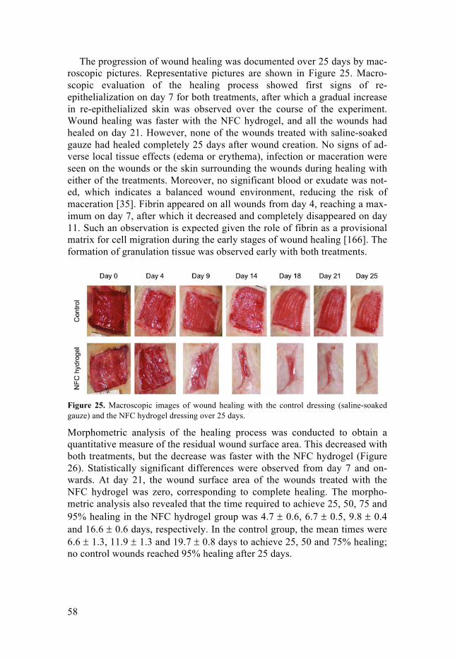
58
The progression of wound healing was documented over 25 days by mac-roscopic pictures. Representative pictures are shown in Figure 25. Macro-scopic evaluation of the healing process showed first signs of re-epithelialization on day 7 for both treatments, after which a gradual increase in re-epithelialized skin was observed over the course of the experiment. Wound healing was faster with the NFC hydrogel, and all the wounds had healed on day 21. However, none of the wounds treated with saline-soaked gauze had healed completely 25 days after wound creation. No signs of ad-verse local tissue effects (edema or erythema), infection or maceration were seen on the wounds or the skin surrounding the wounds during healing with either of the treatments. Moreover, no significant blood or exudate was not-ed, which indicates a balanced wound environment, reducing the risk of maceration [35]. Fibrin appeared on all wounds from day 4, reaching a max-imum on day 7, after which it decreased and completely disappeared on day 11. Such an observation is expected given the role of fibrin as a provisional matrix for cell migration during the early stages of wound healing [166]. The formation of granulation tissue was observed early with both treatments.
Figure 25. Macroscopic images of wound healing with the control dressing (saline-soaked gauze) and the NFC hydrogel dressing over 25 days.
Morphometric analysis of the healing process was conducted to obtain a quantitative measure of the residual wound surface area. This decreased with both treatments, but the decrease was faster with the NFC hydrogel (Figure 26). Statistically significant differences were observed from day 7 and on-wards. At day 21, the wound surface area of the wounds treated with the NFC hydrogel was zero, corresponding to complete healing. The morpho-metric analysis also revealed that the time required to achieve 25, 50, 75 and 95% healing in the NFC hydrogel group was 4.7 ± 0.6, 6.7 ± 0.5, 9.8 ± 0.4 and 16.6 ± 0.6 days, respectively. In the control group, the mean times were 6.6 ± 1.3, 11.9 ± 1.3 and 19.7 ± 0.8 days to achieve 25, 50 and 75% healing; no control wounds reached 95% healing after 25 days.

59
Figure 26. Wound closure determined by macroscopic evaluation of the residual wound surface area. Data represents mean ± standard error of the mean for n = 6. Asterisks indicate statistically significant differences (p < 0.05) between the residual wound surface area of wounds treated with the control dressing and the NFC hydrogel dressing.
The quality of healing was determined by histopathological analysis of skin samples collected 25 days after wound creation. The wounds treated with NFC hydrogel were completely healed, with newly formed epithelium com-pletely covering the granulation tissue of the wound (Figure 27a and c). The neo-epithelium was similar to the native epithelium in the adjacent intact skin, and showed advanced signs of differentiation, indentation and dermal adherence. The granulation tissue filling the wound showed signs of maturity in terms of the absence of hemorrhage and fibrin, together with low numbers of neo-vessels and acute inflammatory cells [1, 11]. It should be mentioned that macrophages were still present and appeared to be filled with residual material of either the primary or secondary dressing (Figure 27c, indicated by blue arrows).
Histopathological analysis of samples from the control group showed that all wounds were incompletely healed after 25 days, with the newly formed epithelium only partially covering the granulation tissue (Figure 27b and d). The neo-epithelium at the wound margins showed only modest signs of dif-ferentiation, indentation and adherence to the dermis. The granulation tissue was immature, as indicated by the presence of hemorrhage and fibrin, to-gether with abundant neo-vessels and acute inflammatory cells [1, 11].
Overall, the histopathological findings showed that neither treatment was associated with any adverse local tissue effects. Furthermore, the NFC hy-drogel led to better healing than the control dressing, as evidenced by the completely healed epithelium and mature granulation tissue.
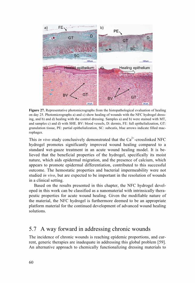
60
Figure 27. Representative photomicrographs from the histopathological evaluation of healing on day 25. Photomicrographs a) and c) show healing of wounds with the NFC hydrogel dress-ing, and b) and d) healing with the control dressing. Samples a) and b) were stained with MT, and samples c) and d) with SHE. BV: blood vessels, D: dermis, FE: full epithelialization, GT: granulation tissue, PE: partial epithelialization, SC: subcutis, blue arrows indicate filled mac-rophages.
This in vivo study conclusively demonstrated that the Ca2+-crosslinked NFC hydrogel promotes significantly improved wound healing compared to a standard wet-gauze treatment in an acute wound healing model. It is be-lieved that the beneficial properties of the hydrogel, specifically its moist nature, which aids epidermal migration, and the presence of calcium, which appears to promote epidermal differentiation, contributed to this successful outcome. The hemostatic properties and bacterial impermeability were not studied in vivo, but are expected to be important in the resolution of wounds in a clinical setting.
Based on the results presented in this chapter, the NFC hydrogel devel-oped in this work can be classified as a nanomaterial with intrinsically thera-peutic properties for acute wound healing. Given the modifiable nature of the material, the NFC hydrogel is furthermore deemed to be an appropriate platform material for the continued development of advanced wound healing solutions.
5.7 A way forward in addressing chronic wounds The incidence of chronic wounds is reaching epidemic proportions, and cur-rent, generic therapies are inadequate in addressing this global problem [59]. An alternative approach to chemically functionalizing dressing materials to
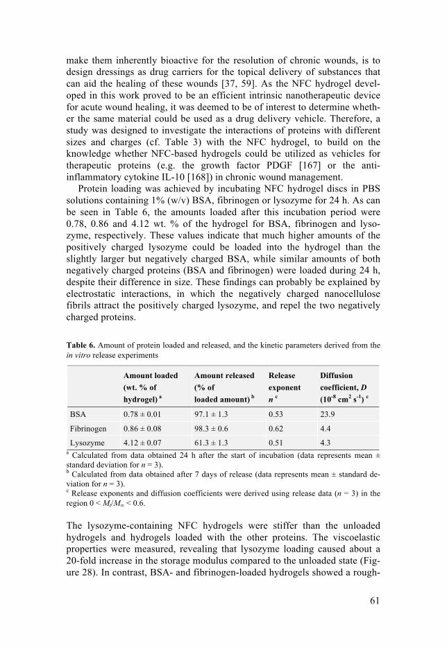
61
make them inherently bioactive for the resolution of chronic wounds, is to design dressings as drug carriers for the topical delivery of substances that can aid the healing of these wounds [37, 59]. As the NFC hydrogel devel-oped in this work proved to be an efficient intrinsic nanotherapeutic device for acute wound healing, it was deemed to be of interest to determine wheth-er the same material could be used as a drug delivery vehicle. Therefore, a study was designed to investigate the interactions of proteins with different sizes and charges (cf. Table 3) with the NFC hydrogel, to build on the knowledge whether NFC-based hydrogels could be utilized as vehicles for therapeutic proteins (e.g. the growth factor PDGF [167] or the anti-inflammatory cytokine IL-10 [168]) in chronic wound management.
Protein loading was achieved by incubating NFC hydrogel discs in PBS solutions containing 1% (w/v) BSA, fibrinogen or lysozyme for 24 h. As can be seen in Table 6, the amounts loaded after this incubation period were 0.78, 0.86 and 4.12 wt. % of the hydrogel for BSA, fibrinogen and lyso-zyme, respectively. These values indicate that much higher amounts of the positively charged lysozyme could be loaded into the hydrogel than the slightly larger but negatively charged BSA, while similar amounts of both negatively charged proteins (BSA and fibrinogen) were loaded during 24 h, despite their difference in size. These findings can probably be explained by electrostatic interactions, in which the negatively charged nanocellulose fibrils attract the positively charged lysozyme, and repel the two negatively charged proteins.
Table 6. Amount of protein loaded and released, and the kinetic parameters derived from the in vitro release experiments
Amount loaded (wt. % of hydrogel) a
Amount released
(% of loaded amount) b
Release exponent n c
Diffusion coefficient, D (10-8 cm2 s-1) c
BSA 0.78 ± 0.01 97.1 ± 1.3 0.53 23.9
Fibrinogen 0.86 ± 0.08 98.3 ± 0.6 0.62 4.4
Lysozyme 4.12 ± 0.07 61.3 ± 1.3 0.51 4.3 a Calculated from data obtained 24 h after the start of incubation (data represents mean ± standard deviation for n = 3). b Calculated from data obtained after 7 days of release (data represents mean ± standard de-viation for n = 3). c Release exponents and diffusion coefficients were derived using release data (n = 3) in the region 0 < Mt/M∞ < 0.6.
The lysozyme-containing NFC hydrogels were stiffer than the unloaded hydrogels and hydrogels loaded with the other proteins. The viscoelastic properties were measured, revealing that lysozyme loading caused about a 20-fold increase in the storage modulus compared to the unloaded state (Fig-ure 28). In contrast, BSA- and fibrinogen-loaded hydrogels showed a rough-

62
ly 10-fold decrease in strength, but were still self-standing. During loading of the NFC hydrogels with proteins, competitive displacement of the Ca2+-crosslinker could take place due to electrostatic interactions. Thus, while the negatively charged proteins would attract the Ca2+ ions and weaken the crosslinking of the nanocellulose, the positively charged protein could in-stead act as an alternative crosslinker, increasing the strength of the NFC hydrogel, as suggested by the observed change in viscoelastic properties.
Figure 28. Storage modulus (G´) of NFC hydrogel before loading (control), after loading and after 24 h release of model proteins. Data represents mean ± standard deviation for n = 3. (Reprinted from Paper V with permission from the publisher.)
To reveal any possible changes in the chemical structure of the proteins or NFC hydrogel as a result of protein loading, the unloaded and protein-loaded hydrogels were analyzed using FTIR-ATR spectroscopy (Figure 29). Spectra of unloaded TEMPO-oxidized NFC hydrogels showed the expected peaks at about 1740 cm-1 and 3300 cm-1, corresponding to free carboxyl groups and broad OH-stretch vibrations [169, 170], respectively. The same peaks asso-ciated with TEMPO-NFC were present in the protein-loaded NFC hydrogels. Additionally, distinctive bands indicating amide I and amide II vibrations of proteins were found at 1650 and 1540 cm-1, respectively, where the amide A stretching in the 3300 cm-1 region was overlapped by the OH-stretching of the cellulose [171, 172]. Together, these spectra indicate that no chemical bonding occurred between the NFC and model proteins, and that the second-ary structure of the proteins was probably maintained when the proteins were entrapped in the NFC hydrogels.
1
10
100
1000
10,000
100,000
1000,000
0 10 20 30 40 50 60 70 80 90 100
Stor
age
mod
ulus
G' (
Pa)
Frequency (Hz)
Lysozyme loaded Lysozyme released NFC control BSA loadedBSA released Fibrinogen loaded Fibrinogen released
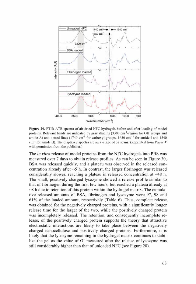
63
Figure 29. FTIR-ATR spectra of air-dried NFC hydrogels before and after loading of model proteins. Relevant bands are indicated by gray shading (3300 cm-1-region for OH groups and amide A) and dotted lines (1740 cm-1 for carboxyl groups, 1650 cm- 1 for amide I and 1540 cm-1 for amide II). The displayed spectra are an average of 32 scans. (Reprinted from Paper V with permission from the publisher.)
The in vitro release of model proteins from the NFC hydrogels into PBS was measured over 7 days to obtain release profiles. As can be seen in Figure 30, BSA was released quickly, and a plateau was observed in the released con-centration already after ~5 h. In contrast, the larger fibrinogen was released considerably slower, reaching a plateau in released concentration at ~48 h. The small, positively charged lysozyme showed a release profile similar to that of fibrinogen during the first few hours, but reached a plateau already at ~8 h due to retention of this protein within the hydrogel matrix. The cumula-tive released amounts of BSA, fibrinogen and lysozyme were 97, 98 and 61% of the loaded amount, respectively (Table 6). Thus, complete release was obtained for the negatively charged proteins, with a significantly longer release time for the larger of the two, while the positively charged protein was incompletely released. The retention, and consequently incomplete re-lease, of the positively charged protein supports the theory that attractive electrostatic interactions are likely to take place between the negatively charged nanocellulose and positively charged proteins. Furthermore, it is likely that the lysozyme remaining in the hydrogel matrix continues to stabi-lize the gel as the value of G´ measured after the release of lysozyme was still considerably higher than that of unloaded NFC (see Figure 28).

64
Figure 30. In vitro release profiles of proteins from NFC hydrogels during the first 48 h. Data represents mean ± standard deviation for n = 3. (Reprinted from Paper V with permission from the publisher.)
The protein release profiles were fitted with the semi-empirical Ritger–Peppas equation (Eq. 1) in order to extract the release exponent, which can have values between 0.5 and 1, where 0.5 is indicative of pure Fickian diffu-sion-controlled release, and 1 of swelling- or erosion-controlled release [125-127]. Values between these indicate anomalous release, where both diffusion and swelling/erosion influence release [173, 174]; or the presence of heterogeneities in the distribution of molecules within the delivery vehicle [175]. The release exponents derived for BSA and lysozyme were 0.53 and 0.51 (Table 6), respectively, thus suggesting almost pure Fickian diffusion-driven release, as was expected due to the non-swelling nature of the NFC hydrogel. For fibrinogen, however, the release exponent was 0.62 (Table 6), which suggests slightly anomalous release. This is most likely due to the larger size of fibrinogen compared to BSA and lysozyme, causing heteroge-neities in the protein distribution throughout the hydrogel, with “point sources” of proteins being released rather than a continuum of sources, as would be the case for homogeneously distributed releasing entities.
Diffusion coefficients were obtained for the model proteins by fitting ex-perimental release data in the region 0 < Mt/M∞ < 0.6 with Eq. 2. As ex-pected from the obtained release exponents, the fits were excellent for BSA and lysozyme (R2 values of 0.999 and 0.995, respectively), and acceptable for fibrinogen (R2 = 0.989). The extracted diffusion coefficients were 23.9, 4.4 and 4.3 × 10 cm² s-1 for BSA, fibrinogen and lysozyme, respectively (Table 6). Hence, as observed in the release curves, the size of the molecule had a clear effect on the diffusion coefficients in the case of the anionic pro-teins; the smaller BSA (hydrodynamic radius 3.5 nm, Table 3) showing a more than 5 times higher diffusion coefficient than the larger fibrinogen
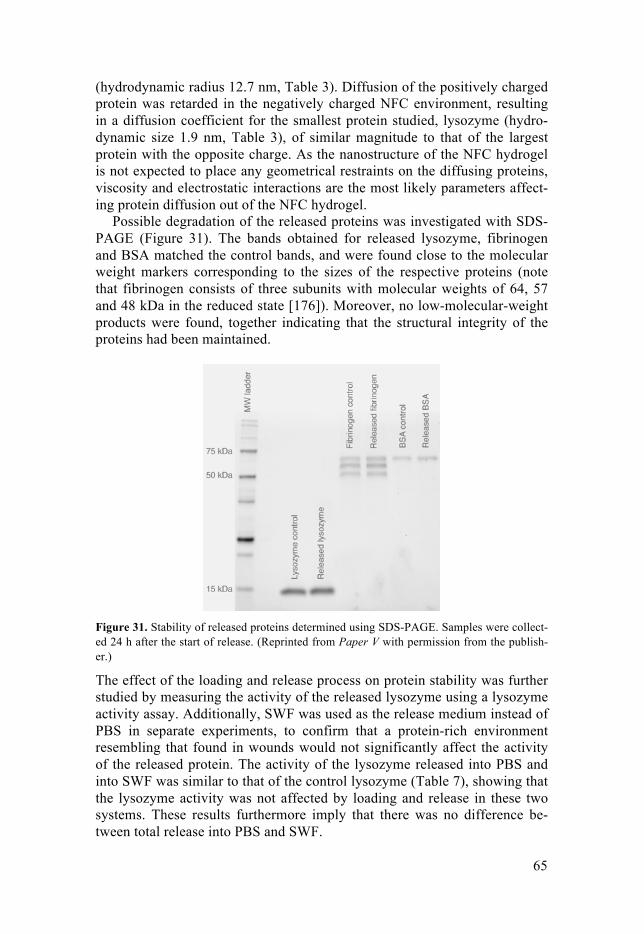
65
(hydrodynamic radius 12.7 nm, Table 3). Diffusion of the positively charged protein was retarded in the negatively charged NFC environment, resulting in a diffusion coefficient for the smallest protein studied, lysozyme (hydro-dynamic size 1.9 nm, Table 3), of similar magnitude to that of the largest protein with the opposite charge. As the nanostructure of the NFC hydrogel is not expected to place any geometrical restraints on the diffusing proteins, viscosity and electrostatic interactions are the most likely parameters affect-ing protein diffusion out of the NFC hydrogel.
Possible degradation of the released proteins was investigated with SDS-PAGE (Figure 31). The bands obtained for released lysozyme, fibrinogen and BSA matched the control bands, and were found close to the molecular weight markers corresponding to the sizes of the respective proteins (note that fibrinogen consists of three subunits with molecular weights of 64, 57 and 48 kDa in the reduced state [176]). Moreover, no low-molecular-weight products were found, together indicating that the structural integrity of the proteins had been maintained.
Figure 31. Stability of released proteins determined using SDS-PAGE. Samples were collect-ed 24 h after the start of release. (Reprinted from Paper V with permission from the publish-er.)
The effect of the loading and release process on protein stability was further studied by measuring the activity of the released lysozyme using a lysozyme activity assay. Additionally, SWF was used as the release medium instead of PBS in separate experiments, to confirm that a protein-rich environment resembling that found in wounds would not significantly affect the activity of the released protein. The activity of the lysozyme released into PBS and into SWF was similar to that of the control lysozyme (Table 7), showing that the lysozyme activity was not affected by loading and release in these two systems. These results furthermore imply that there was no difference be-tween total release into PBS and SWF.

66
Table 7. Lysozyme activity in samples collected after 24 h release of lysozyme into PBS and SWF
Lysozyme activity (A450 decrease min-1)
% of control
Lysozyme, control 0.020 ± 0.004 -
Released into PBS 0.019 ± 0.001 95 ± 5
Released into SWF 0.019 ± 0.001 95 ± 5
Data represents mean ± standard deviation for n = 3.
Together, the results of these in vitro experiments show that proteins can be loaded into the anionic NFC hydrogel by a simple soaking procedure, and that diffusion is the main mechanism of release. The viscosity of the gel environment and electrostatic interactions affected the release of the pro-teins; the larger anionic protein showing lower diffusivity than the smaller protein of same charge, and the diffusion of the small cationic protein being similar to that of the largest protein of the opposite charge. While the viscoe-lastic properties of the hydrogel were affected by proteins in the hydrogel matrix, the self-standing characteristics of the hydrogel were maintained. However, the process of loading and release from the NFC hydrogel did not affect the structure or activity of the proteins.
The results of this study indicate that the challenge of chronic wound healing could be addressed using NFC hydrogels, and that their charge prop-erties could be easily adjusted to design a vehicle with release profiles suita-ble for the delivery of therapeutic proteins. Combined with the intrinsic wound healing properties of the NFC hydrogel, such a solution could prove a potent tool in treatment of chronic wounds by 1) resolving the chronic state of the wound, and 2) protecting the wound and promoting healing to achieve complete skin tissue regeneration.

67
6 Conclusions and future perspectives
In this work, a platform wound healing dressing comprised of a Ca2+-cross-linked NFC hydrogel was studied. Key findings at the physicochemical level illustrating the suitability of this NFC hydrogel for wound healing applica-tions were its self-standing nature, created by the Ca2+-crosslinking, and its moisture donating property, which stems from the highly hydrated fibril structure of the hydrogel.
In the evaluation of the biocompatibility with key cells in the wound heal-ing process, it was found that the NFC hydrogel induced no cytotoxic effects on human skin- or immune cells, nor did it affect the inflammatory response of the immune cells. Furthermore, biocompatibility studies showed that the hydrogel supports dermal and epidermal proliferation, which are key pro-cesses in skin regeneration.
In studies with human blood and plasma, the NFC hydrogel was discov-ered to have a hemostatic effect, due mainly to the negatively charged sur-face of NFC. Such a hemostatic property of the NFC hydrogel is foreseen to be of significant importance in the treatment of heavily bleeding wounds. Additionally, as the material neither suppressed nor stimulated complement activation, it was deemed to be hemocompatible in wound healing applica-tions. A balance in inflammatory processes is necessary during wound heal-ing, and a lack of effect on the inflammatory mechanisms studied is thus highly desirable.
Investigations of the interaction of the NFC hydrogel with bacteria showed that it is impermeable to bacteria, and can thus act as a protective barrier to external pathogens. Furthermore, an inhibitory effect of the Ca2+ crosslinker on the formation of a biofilm by the wound pathogen P. aeru-ginosa was observed, which could aid chronic wound treatment as biofilms protect bacteria against antibiotics.
In vivo, the NFC hydrogel exhibited considerable beneficial effects on acute wound healing without causing any adverse local tissue effects, com-pared to a standard wet-gauze treatment. The results indicated that the Ca2+ crosslinker and the moist nature of the hydrogel probably contributed to the advanced regeneration of the epidermis. Thus, the NFC hydrogel material itself proved to be a potent intrinsic nanodevice for wound care applications.
Simple modifications of the composition of the NFC hydrogel were shown to offer tunability of the material. For example, bioactive components could be mixed into the nanocellulose matrix prior to crosslinking to elicit

68
specific cellular responses. This was demonstrated by the inclusion of type I collagen, which improved in vitro cell migration, and by the incorporation of kaolin, which significantly improved the hemostatic effect of the material. An additional simple modification investigated was the substitution of the crosslinker. Using Cu2+ instead of Ca2+ endowed the hydrogel with antibac-terial properties against common wound pathogens.
This tunability will be of significant importance considering the highly individual treatment needs that arise from the different pathologies that may be the underlying cause of slow- or non-healing wounds. Thus, the NFC hydrogel described here provides a promising platform for the further devel-opment of materials suited to various wound types.
A way forward for chronic wound treatment was suggested, employing this intrinsically wound healing NFC hydrogel for the delivery of therapeutic proteins. Such a solution would have considerable potential for the resolu-tion of chronic wounds, apart from protecting the wound and supporting subsequent wound healing. Fundamental interactions between proteins and the NFC hydrogel were investigated providing important knowledge for further developments in this direction. It was found that viscosity and elec-trostatic interactions are key parameters affecting protein loading and deliv-ery. Based on these findings, it was suggested that the easily tunable charge properties of the NFC hydrogel could provide an intriguing means of opti-mizing protein release profiles for chronic wound management.
In conclusion, the work presented in this thesis provides valuable insight into the ways in which NFC-based hydrogels can be used in advanced wound care applications. The results presented here are expected to contrib-ute significantly to further research in this field, and it is suggested that fu-ture research on NFC-based hydrogels be focused on developing next-generation treatments for the societally problematic chronic wounds through the modification and optimization of the NFC hydrogel platform described here.
69
Sammanfattning på svenska
Huden är människans största organ och dess huvudsaklig funktion är att skydda oss från bland annat skadlig mekanisk påverkan, bakterier, giftiga substanser och strålning. Givet dess stora exponering mot kringliggande miljö är huden i konstant risk för skada som medför att hudens skyddande egenskap försvagas. Kroppens förmåga att snabbt och effektivt reparera ska-dad hud är därför av oerhörd vikt. I normala fall läker sår enlig en sekventi-ellt noggrant orkestrerad process bestående av fyra huvudsakliga faser: koagulation, inflammation, nybildning och ombyggnad. I vissa fall är denna naturliga läkningsprocess dock inte tillräcklig. Till exempel komplicerade sår med stor öppen yta, infekterade sår eller sår hos patienter med immunför-svagande sjukdomar riskerar att inte läkas och istället bli kroniska. För dessa sår är behandling nödvändig.
Människan har sedan lång tid tillbaka använt förband för att behandla sår. Tidiga versioner var förband skapade av bland annat lera och växter. Senare har förbanden utvecklats och idag finns över 5000 alternativ på marknaden. Tyvärr visar forskning att många av dagens alternativ är ineffektiva och en-bart används på grund av vana. Dessutom har man etablerat att ett optimalt sårläkningsförband som är lämplig för alla typer av sår inte existerar och att moderna förband snarare bör designas för att vara bäst på en kategori av sår.
Den mest uppenbara vägen framåt för utvecklingen av verkligt sårläkande förband ligger i att kombinera förståelsen av biokemiska och cellulära pro-cesser kopplade till problematisk sårläkning med avancerad materialforsk-ning, där material skapas för att specifikt påverka dessa processer. Tack vare nanoteknologins framfart är det idag möjligt att manipulera material på en nivå där cellinteraktioner direkt kan påverkas av materialets egenskaper. Ur detta uppstår nya möjligheter för utvecklingen av moderna sårläkningspro-dukter.
I denna avhandling undersöks möjligheten att använda träbaserad nano-cellulosa för sårläkningsapplikationer. Nanocellulosa skapas via nedbrytning av cellulosafibrer till nanofibrer som har en diameter på några tiotal nanome-ter. Med sin relativt stora och exponerade ytarea kan nanocellulosan enkelt modifieras med funktionella kemiska grupper vilket gör den väldigt formbar för framställningen av olika typer av material med unika egenskaper. Ef-tersom cellulosa är en viktig svensk råvara, och givet digitaliseringens fram-fart, är det idag viktigare än någonsin att hitta nya applikationsområden för detta förnybara material. Inom kategorin medicinteknik finns många till-

70
lämpningsområden för nanocellulosa, där material för läkemedelsleverans, blodrening, diagnostik, vävnadsrekonstruktion och sårläkning är några ex-empel.
För sårläkning har materialtypen hydrogeler särskild potential. Hydroge-ler består av ett tredimensionellt nätverk av tvärbundna polymerer som, likt namnet föreslår, binder in stora mängder vatten. Dess höga vattenbestånds-del gör att materialet kan donera fukt till ett läkande sår, en egenskap som i sig har visats förbättra sårläkningen. Nanocellulosa utgör ett utmärkt materi-al för tillverkning av hydrogeler. Därför har nanocellulosabaserade hydroge-ler för avancerade sårläkningsapplikationer varit i fokus i denna avhandling.
Tidigt i projektet påvisades möjligheten att skapa en stabil nanocellulosa-baserad hydrogel genom en enkel metod som endast använder kalciumjoner för att binda ihop nanocellulosafibrerna. Genom specifika experiment sågs även att materialet kan skapa en fuktig miljö för främjad sårläkning. Bio-kompatibilitetsstudier med humana hud- och immunceller gav goda resultat, där materialet stödde celltillväxt utan att det störde viktiga inflammatoriska processer. Vidare bidrog materialet till blodkoagulation, vilket bedöms vara främjande för behandling av starkt blödande sår. Det visades även att nano-cellulosahydrogelen kan stänga ute bakterier och därmed minska risken för sårinfektion vid behandling. Efter att materialet hade testats med positiva resultat i modellsystem undersöktes hur materialet påverkade sårläknings-processen i djurförsök. I detta försök bevisades att det utvecklade materialet starkt förbättrade sårläkningen i jämförelse med en standardbehandling. Den förbättrade sårläkningen bedömdes till stor del vara på grund av kalciumet i hydrogelen som bidrar till hudvävnadsåterskapning samt den fuktiga miljön som skapades av materialet. Utifrån detta fastslogs att projektet varit lyckat i att framställa ett avancerat sårläkningsmaterial gjort av träbaserad nanocellu-losa.
Under projektets gång undersöktes parallellt hur materialet som en platt-form för sårläkningsapplikationer kan modifieras för att förändra den biolo-giska responsen. Ur dessa undersökningar sågs att hydrogelen enkelt kan modifieras genom att blanda in aktiva komponenter eller genom att nyttja andra metalljoner för tvärbindningen, för att på så sätt förstärka materialets påverkan på specifika processer som kan skapa kroniska sår. Vidare demon-strerades möjligheten att använda denna plattform för läkemedelsleverans, där materialets egenskaper kan justeras för att påverka hur snabbt läkemedel frisätts.
Överlag bedöms den framställda nanocellulosahydrogelen vara ett enkelt modifierbart material som i grunden främjar akut sårläkning. Genom möjlig-heten att använda materialet för till exempel leverans av läkemedel som främjar upplösning av skadliga processer, för att därefter via materialets egenskaper stödja den strukturerade sårläkningsprocessen, ses en stor pot-ential för detta material att framgent vidareutvecklas för behandling av de samhällsmässigt problematiska kroniska såren.

71
Acknowledgements
Marie Curie once said, “Nothing in life is to be feared, it is only to be under-stood. Now is the time to understand more, so that we may fear less.”. If I were to find one expression that describes my greatest learning from my main supervisor, Assoc. Prof. Natalia Ferraz, the above quote would proba-bly be it. You invited me to take part in a journey of discovery, for which I will be forever grateful. You taught me to look beyond the scope of the cur-riculum to promote understanding, and from that I’ve learned a philosophy for life. Thank you for these years, and thank you for your supervision.
I would also like to thank my co-supervisors, Dr. Jonas Lindh, Prof. Ma-ria Strømme and Prof. Albert Mihranyan; your help has meant a lot to me. To know that I always had your support if I needed it was a source of great comfort. Thank you.
All my colleagues at the Nano group; it has been great working with you, and I enjoyed your company during the breaks from the lab. Specifically, I would like to thank Simon, Maria V, Christian, Mia and Lisa for our time together, and I wish you all well in completing your PhDs and in your future endeavors. Igor, my officemate and travel-mate, it’s been a pleasure, and I hope to see you again soon. Gopi, Petter, Levon, Viviana, Teresa, Kai, Hao, Changqing, Jiaojiao, Martin, Ken, Thanos, Huan, Rikard, Ocean, Rui, Chao, Lulu, Michelle, Shenyang and other past and present members of the Nano group; thank you, and I wish you the best of luck in the future.
To Xi, Shiuli, Michael and everyone at the cell lab and in the MiM group, and to Jaan at IGP; it has been a privilege working with you.
To my friends, thank you for having brought enjoyment, relaxation and much needed distraction into my life during these years. It has meant the world to me.
Finally, I would like to thank my dear family, especially you, Eve. I am forever grateful for your faithful encouragement, support and patience, which began long before I started my PhD and will continue long after I have written these words. You are truly my life and my universe. Thank you.
72
References
1. Delavary, M.B., et al., Macrophages in skin injury and repair. Immunobiology, 2011. 216(7): p. 753-762.
2. Tortora, G.J. and Derrickson, B.H., Principles of Anatomy and Physiologi. Vol. 200. 2014, New York: John Wiley & Sons Inc. 140-154.
3. Diegelmann, R.F. and Evans, M.C., Wound healing: an overview of acute, fibrotic and delayed healing. Front. Biosci., 2004. 9: p. 283-289.
4. Shah, J.B., The history of wound care. J. Am. Col. Certif. Wound Spec., 2011. 3(3): p. 65-66.
5. Martin, P., Wound healing--aiming for perfect skin regeneration. Science, 1997. 276(5309): p. 75-81.
6. Derkx, F.H.M., et al., An intrinsic factor XII–prekallikrein-dependent pathway activates the human plasma renin–angiotensin system. Nature, 1978. 280: p. 315-316.
7. Gale, A.J., Continuing education course #2: current understanding of hemostasis. Toxicol. Pathol., 2011. 39(1): p. 273-280.
8. Morrissey, J.H., Choi, S.H., and Smith, S.A., Polyphosphate: an ancient molecule that links platelets, coagulation, and inflammation. Blood, 2012. 119(25): p. 5972-5979.
9. Lane, D.A., Philippou, H., and Huntington, J.A., Directing thrombin. Blood, 2005. 106(8): p. 2605-2612.
10. Singer, A.J. and Clark, R.A., Cutaneous wound healing. New Engl. J. Med., 1999. 341(10): p. 738-746.
11. Gurtner, G.C., et al., Wound repair and regeneration. Nature, 2008. 453(7193): p. 314-321.
12. Sinno, H. and Prakash, S., Complements and the wound healing cascade: an updated review. Plast. Surg. Int., 2013. 2013.
13. Meri, S. and Jarva, H., Complement Regulation. Vox Sang, 1998. 74: p. 291-392.
14. Kinoshita, T., Biology of complement: the overture. Immunol. Today, 1991. 12: p. 291-295.
15. Leibovich, S. and Ross, R., The role of the macrophage in wound repair. A study with hydrocortisone and antimacrophage serum. Am. J. Pathol., 1975. 78(1): p. 71.
16. Lucas, T., et al., Differential roles of macrophages in diverse phases of skin repair. J. Immunol., 2010. 184(7): p. 3964-3977.
17. Porcheray, F., et al., Macrophage activation switching: an asset for the resolution of inflammation. Am. J. Clin. Exp. Immunol., 2005. 142(3): p. 481-489.
18. Werner, S., Krieg, T., and Smola, H., Keratinocyte–fibroblast interactions in wound healing. J. Investig. Dermatol., 2007. 127(5): p. 998-1008.

73
19. Tonnesen, M.G., Feng, X., and Clark, R.A., Angiogenesis in wound healing. J. Investig. Dermatol. Symp. Proc., 2000. 5(1): p. 40-46.
20. Santoro, M.M. and Gaudino, G., Cellular and molecular facets of keratinocyte reepithelization during wound healing. Exp. Cell. Res., 2005. 304(1): p. 274-286.
21. Pastar, I., et al., Epithelialization in wound healing: a comprehensive review. Adv. Wound Care, 2014. 3(7): p. 445-464.
22. Morasso, M.I. and Tomic Canic, M., Epidermal stem cells: the cradle of epidermal determination, differentiation and wound healing. Biol. Cell, 2005. 97(3): p. 173-183.
23. Levenson, S., et al., Healing of rat skin wounds. Ann. Surg., 1965. 161(2): p. 293.
24. Siddiqui, A.R. and Bernstein, J.M., Chronic wound infection: facts and controversies. Clin. Dermatol., 2010. 28(5): p. 519-526.
25. Menke, N.B., et al., Impaired wound healing. Clin. Dermatol., 2007. 25(1): p. 19-25.
26. Guo, S.A. and DiPietro, L.A., Factors affecting wound healing. J. Dent. Res., 2010. 89(3): p. 219-229.
27. Evans, C.A., et al., Bacterial flora of the normal human skin. J. Investig. Dermatol., 1950. 15(4): p. 305-324.
28. Otto, M., Staphylococcus epidermidis - the 'accidental' pathogen. Nat. Rev. Microbiol., 2009. 7(8): p. 555-567.
29. Bjarnsholt, T., et al., Why chronic wounds will not heal: a novel hypothesis. Wound Repair Regen., 2008. 16(1): p. 2-10.
30. Streeter, K. and Katouli, M., Pseudomonas aeruginosa: A review of their Pathogenesis and Prevalence in Clinical Settings and the Environment. Infect. Epidemiol. Med., 2016. 2(1): p. 25-32.
31. Edwards, R. and Harding, K.G., Bacteria and wound healing. Curr. Opin. Infect. Dis., 2004. 17(2): p. 91-96.
32. Swift, M.E., et al., Age-related alterations in the inflammatory response to dermal injury. J. Investig. Dermatol., 2001. 117(5): p. 1027-1035.
33. Godbout, J.P. and Glaser, R., Stress-induced immune dysregulation: implications for wound healing, infectious disease and cancer. J. Neuroimmune Pharmacol., 2006. 1(4): p. 421-427.
34. Sibbald, R.G. and Woo, K.Y., The biology of chronic foot ulcers in persons with diabetes. Diabetes Metab. Res. Rev., 2008. 24(S1).
35. Boateng, J.S., et al., Wound healing dressings and drug delivery systems: a review. J. Pharm. Sci., 2008. 97(8): p. 2892-2923.
36. Goldsmith, S.P., Wound Care: Combining Three Classification Systems to Select Dressings. Home Health Care Manag. Pract., 1996. 8(6): p. 17-27.
37. Boateng, J. and Catanzano, O., Advanced therapeutic dressings for effective wound healing—a review. J. Pharm. Sci., 2015. 104(11): p. 3653-3680.
38. White, R., Wound dressings and other topical treatment modalities in bioburden control. J. Wound Care, 2011. 20(9): p. 431-439.
39. Stadler, M. and Dersch, P., How to Overcome the Antibiotic Crisis: Facts, Challenges, Technologies and Future Perspectives. Vol. 398. 2016: Springer.
40. Abdelrahman, T. and Newton, H., Wound dressings: principles and practice. Surgery, 2011. 29(10): p. 491-495.
74
41. Ovington, L.G., Advances in wound dressings. Clin. Dermatol., 2007. 25(1): p. 33-38.
42. Smith, A.H., et al., Haemostatic dressings in prehospital care. Emerg. Med. J., 2013. 30(10): p. 784-789.
43. Achneck, H.E., et al., A comprehensive review of topical hemostatic agents: efficacy and recommendations for use. Ann. Surg., 2010. 251(2): p. 217-228.
44. Ahmed, E.M., Hydrogel: Preparation, characterization, and applications: A review. J. Adv. Res., 2015. 6(2): p. 105-121.
45. Hoffman, A.S., Hydrogels for biomedical applications. Adv. Drug Deliv. Rev., 2012. 64: p. 18-23.
46. Alexandre, N., et al., Biocompatibility and hemocompatibility of polyvinyl alcohol hydrogel used for vascular grafting--In vitro and in vivo studies. J. Biomed. Mater. Res. A, 2014. 102(12): p. 4262-4275.
47. Tummala, G.K., et al., Hyperelastic Nanocellulose-Reinforced Hydrogel of High Water Content for Ophthalmic Applications. ACS Biomater. Sci. Eng., 2016. 2(11): p. 2072-2079.
48. Caló, E. and Khutoryanskiy, V.V., Biomedical applications of hydrogels: A review of patents and commercial products. Eur. Polym. J., 2015. 65: p. 252-267.
49. Rudzinski, W., et al., Hydrogels as controlled release devices in agriculture. Des. Monomers Polym., 2002. 5(1): p. 39-65.
50. Andrus, L.P., et al., Characterization of lactate sensors based on lactate oxidase and palladium benzoporphyrin immobilized in hydrogels. Biosensors, 2015. 5(3): p. 398-416.
51. Chen, X., et al., Enzymatic and chemoenzymatic approaches to synthesis of sugar-based polymer and hydrogels. Carbohydr. Polym., 1995. 28(1): p. 15-21.
52. del Valle, L.J., Díaz, A., and Puiggalí, J., Hydrogels for Biomedical Applications: Cellulose, Chitosan, and Protein/Peptide Derivatives. Gels, 2017. 3(3): p. 27.
53. Lee, K.Y. and Mooney, D.J., Hydrogels for tissue engineering. Chem. Rev., 2001. 101(7): p. 1869-1880.
54. Slaughter, B.V., et al., Hydrogels in regenerative medicine. Adv. Mater., 2009. 21(32 33): p. 3307-3329.
55. Kashyap, N., Kumar, N., and Kumar, M.R., Hydrogels for pharmaceutical and biomedical applications. Crit. Rev. Ther. Drug Carrier Syst., 2005. 22(2).
56. Hamidi, M., Azadi, A., and Rafiei, P., Hydrogel nanoparticles in drug delivery. Adv. Drug Deliv. Rev., 2008. 60(15): p. 1638-1649.
57. Fernandes, E.M., et al., Bionanocomposites from lignocellulosic resources: Properties, applications and future trends for their use in the biomedical field. Progr. Polym. Sci., 2013. 38(10-11): p. 1415-1441.
58. Commission Recommendation of 18 October 2011 on the definition of nanomaterial, in Off. J. Eur. Union. 2011, European Commission. p. 38.
59. Hamdan, S., et al., Nanotechnology-Driven Therapeutic Interventions in Wound Healing: Potential Uses and Applications. ACS Cent. Sci., 2017. 3(3): p. 163-175.
60. Kalashnikova, I., Das, S., and Seal, S., Nanomaterials for wound healing: scope and advancement. Nanomedicine, 2015. 10(16): p. 2593-2612.
75
61. Adhya, A., et al., Healing of burn wounds by topical treatment: A randomized controlled comparison between silver sulfadiazine and nano-crystalline silver. J. Basic Clin. Pharm., 2014. 6(1): p. 29.
62. Hua, K., et al., Transition from bioinert to bioactive material by tailoring the biological cell response to carboxylated nanocellulose. Biomacromolecules, 2016. 17(3): p. 1224-1233.
63. Tocco, I., et al., Nanotechnology-based therapies for skin wound regeneration. J. Nanomater., 2012. 2012: p. 4.
64. Bradley, C.A., Drug delivery: DNA nanorobots—seek and destroy. Nat. Rev. Drug Discov., 2018. 17(4): p. 242.
65. Williams, D.F., On the mechanisms of biocompatibility. Biomaterials, 2008. 29(20): p. 2941-2953.
66. Ratner, B.D., et al., Biomaterials science: an introduction to materials in medicine. 2004: Elsevier.
67. Shatkin, J.A. and Kim, B., Cellulose nanomaterials: life cycle risk assessment, and environmental health and safety roadmap. Environ. Sci. Nano, 2015. 2(5): p. 477-499.
68. Krug, H.F., Nanosafety research—are we on the right track? Angew. Chem. Int. Ed., 2014. 53(46): p. 12304-12319.
69. Klemm, D., et al., Cellulose: fascinating biopolymer and sustainable raw material. Angew. Chem. Int. Ed., 2005. 44(22): p. 3358-3393.
70. Medronho, B. and Lindman, B., Brief overview on cellulose dissolution/regeneration interactions and mechanisms. Adv. Colloid Interface Sci., 2015. 222: p. 502-508.
71. Moon, R.J., et al., Cellulose nanomaterials review: structure, properties and nanocomposites. Chem. Soc. Rev., 2011. 40(7): p. 3941-2994.
72. Lavoine, N., et al., Microfibrillated cellulose - its barrier properties and applications in cellulosic materials: a review. Carbohydr. Polym., 2012. 90(2): p. 735-764.
73. Isogai, A., Saito, T., and Fukuzumi, H., TEMPO-oxidized cellulose nanofibers. Nanoscale, 2011. 3(1): p. 71-85.
74. Payen, A., Mémoire sur la composition du tissu propre des plantes et du ligneux. Comptes Rendus, 1838. 7: p. 1052-1056.
75. Ghaderi, M., et al., All-cellulose nanocomposite film made from bagasse cellulose nanofibers for food packaging application. Carbohydr. Polym., 2014. 104: p. 59-65.
76. Noorbakhsh-Soltani, S., Zerafat, M., and Sabbaghi, S., A comparative study of gelatin and starch-based nano-composite films modified by nano-cellulose and chitosan for food packaging applications. Carbohydr. Poylm., 2018. 189: p. 48-55.
77. Niu, X., et al., Rosin modified cellulose nanofiber as a reinforcing and co-antimicrobial agents in polylactic acid/chitosan composite film for food packaging. Carbohydr. Polym., 2018. 183: p. 102-109.
78. Yin, Y., et al., Modification of cellulose nanocrystal via SI-ATRP of styrene and the mechanism of its reinforcement of polymethylmethacrylate. Carbohydr. Polym., 2016. 142: p. 206-212.
79. da Costa Correia, V., et al., Nanofibrillated cellulose and cellulosic pulp for reinforcement of the extruded cement based materials. Constr. Build. Mater., 2018. 160: p. 376-384.
76
80. Alloin, F., et al., Nanocomposite polymer electrolyte based on whisker or microfibrils polyoxyethylene nanocomposites. Electrochim. Acta, 2010. 55(18): p. 5186-5194.
81. Razaq, A., et al., Paper Based Energy Storage Devices Comprising Carbon Fiber Reinforced Polypyrrole Cladophora Nanocellulose Composite Electrodes. Adv. Energy Mater., 2012. 2(4): p. 445-454.
82. Hoeng, F., Denneulin, A., and Bras, J., Use of nanocellulose in printed electronics: a review. Nanoscale, 2016. 8(27): p. 13131-13154.
83. Rocha, I., et al., Sulfonated nanocellulose beads as potential immunosorbents. Cellulose, 2018. 25(3): p. 1899-1910.
84. Rees, A., et al., 3D Bioprinting of Carboxymethylated-Periodate Oxidized Nanocellulose Constructs for Wound Dressing Applications. Biomed. Res. Int., 2014: p. 8.
85. Hakkarainen, T., et al., Nanofibrillar cellulose wound dressing in skin graft donor site treatment. J. Contr. Release, 2016. 244(Pt B): p. 292-301.
86. Paukkonen, H., et al., Nanofibrillar cellulose hydrogels and reconstructed hydrogels as matrices for controlled drug release. Int. J. Pharm., 2017. 532(1): p. 269-280.
87. Siró, I. and Plackett, D., Microfibrillated cellulose and new nanocomposite materials: a review. Cellulose, 2010. 17(3): p. 459-494.
88. Klemm, D., et al., Nanocelluloses as innovative polymers in research and application, in Polysaccharides II. 2006, Springer. p. 49-96.
89. Kangas, H., et al., Characterization of fibrillated celluloses. A short review and evaluation of characteristics with a combination of methods. Nord. Pulp Pap. Res. J., 2014. 29(1): p. 129-143.
90. Lin, N. and Dufresne, A., Nanocellulose in biomedicine: Current status and future prospect. Eur. Polym. J., 2014. 59: p. 302-325.
91. Grishkewich, N., et al., Recent advances in the application of cellulose nanocrystals. Curr. Opin. Colloid Interface Sci., 2017. 29: p. 32-45.
92. Lindström, T., et al., Microfibrillated cellulose. Encycl. Polym. Sci. Tech., 2002: p. 1-34.
93. Fukuzumi, H., et al., Transparent and high gas barrier films of cellulose nanofibers prepared by TEMPO-mediated oxidation. Biomacromolecules, 2008. 10(1): p. 162-165.
94. Saito, T., et al., Cellulose nanofibers prepared by TEMPO-mediated oxidation of native cellulose. Biomacromolecules, 2007. 8(8): p. 2485-2491.
95. Dong, H., et al., Cation-induced hydrogels of cellulose nanofibrils with tunable moduli. Biomacromolecules, 2013. 14(9): p. 3338-3345.
96. Jorfi, M. and Foster, E.J., Recent advances in nanocellulose for biomedical applications. J. Appl. Polym. Sci., 2015. 132(14): p. 1-19.
97. Lindh, J., et al., Convenient one-pot formation of 2, 3-dialdehyde cellulose beads via periodate oxidation of cellulose in water. Biomacromolecules, 2014. 15(5): p. 1928-1932.
98. Jack, A.A., et al., The interaction of wood nanocellulose dressings and the wound pathogen P. aeruginosa. Carbohydr. Polym., 2017. 157: p. 1955-1962.
99. Liu, J., et al., Hemicellulose-reinforced nanocellulose hydrogels for wound healing application. Cellulose, 2016. 23(5): p. 3129-3143.
100. Liu, R., et al., Antibacterial and Hemostatic Hydrogel via Nanocomposite from Cellulose Nanofibers. Carbohydr. Polym., 2018: p. 63-70.
77
101. Tummala, G.K., Rojas, R., and Mihranyan, A., Poly (vinyl alcohol) Hydrogels Reinforced with Nanocellulose for Ophthalmic Applications: General Characteristics and Optical Properties. J. Phys. Chem. B, 2016. 120(51): p. 13094-13101.
102. Liu, Y., et al., A physically crosslinked polydopamine/nanocellulose hydrogel as potential versatile vehicles for drug delivery and wound healing. Carbohydr. Polym., 2018. 188: p. 27-36.
103. Mertaniemi, H., et al., Human stem cell decorated nanocellulose threads for biomedical applications. Biomaterials, 2016. 82: p. 208-220.
104. Lou, Y.-R., et al., The use of nanofibrillar cellulose hydrogel as a flexible three-dimensional model to culture human pluripotent stem cells. Stem Cells Dev., 2013. 23(4): p. 380-392.
105. Malinen, M.M., et al., Differentiation of liver progenitor cell line to functional organotypic cultures in 3D nanofibrillar cellulose and hyaluronan-gelatin hydrogels. Biomaterials, 2014. 35(19): p. 5110-5121.
106. Markstedt, K., et al., 3D bioprinting human chondrocytes with nanocellulose–alginate bioink for cartilage tissue engineering applications. Biomacromolecules, 2015. 16(5): p. 1489-1496.
107. Tehrani, Z., et al., Translucent and ductile nanocellulose-PEG bionanocomposites—A novel substrate with potential to be functionalized by printing for wound dressing applications. Ind. Crop. Prod., 2016. 93: p. 193-202.
108. Maver, T., et al., Cellulose based thin films as a platform for drug release studies to mimick wound dressing materials. Cellulose, 2014. 22(1): p. 749-761.
109. Lu, T., et al., Composite aerogels based on dialdehyde nanocellulose and collagen for potential applications as wound dressing and tissue engineering scaffold. Compos. Sci. Tech., 2014. 94: p. 132-138.
110. Nordli, H.R., et al., Producing ultrapure wood cellulose nanofibrils and evaluating the cytotoxicity using human skin cells. Carbohydr. Polym., 2016. 150: p. 65-73.
111. Uddin, K.M., et al., Retention of lysozyme activity by physical immobilization in nanocellulose aerogels and antibacterial effects. Cellulose, 2017. 24(7): p. 2837-2848.
112. Zander, N.E., et al., Metal cation cross-linked nanocellulose hydrogels as tissue engineering substrates. ACS Appl. Mater. Interfaces, 2014. 6(21): p. 18502-18510.
113. Rashad, A., et al., Cytocompatibility of Wood-Derived Cellulose Nanofibril Hydrogels with Different Surface Chemistry. Biomacromolecules, 2017. 18(4): p. 1238-1248.
114. Yang, J., Xu, F., and Han, C.R., Metal Ion Mediated Cellulose Nanofibrils Transient Network in Covalently Cross-linked Hydrogels: Mechanistic Insight into Morphology and Dynamics. Biomacromolecules, 2017. 18(3): p. 1019-1028.
115. Katz, S. and Beatson, R.P., The determination of strong and weak acidic groups in sulfite pulps. Svensk Papperstid., 1984. 87(6): p. 48-53.
116. Von Smoluchowski, M., Zur kinetischen theorie der brownschen molekularbewegung und der suspensionen. Ann. Phys., 1906. 326(14): p. 756-780.

78
117. ISO, in ISO 10993-5:2009. 2009, International Standard Organisation: Geneva, Switzerland.
118. Liang, C.C., Park, A.Y., and Guan, J.L., In Vitro Scratch Assay: a Convenient and Inexpensive Method for Analysis of Cell Migration In Vitro. Nat. Protoc., 2007. 2(2): p. 329-333.
119. Hong, J., et al., A new in vitro model to study interaction between whole blood and biomaterials. Studies of platelet and coagulation activation and the effect of aspirin. Biomaterials, 1999. 20: p. 603-611.
120. Biran, R. and Pond, D., Heparin coatings for improving blood compatibility of medical devices. Adv. Drug Deliv. Rev., 2017. 112: p. 12-23.
121. ISO, in ISO 10993-6: Biological Evaluation of Medical Devices. Tests for Local Effects after Implantation; Part-6. 2007: Geneva, Switzerland.
122. González Flecha, F.L. and Levi, V., Determination of the molecular size of BSA by fluorescence anisotropy. Biochem. Mol. Biol. Educ., 2003. 31(5): p. 319-322.
123. Wasilewska, M., Adamczyk, Z., and Jachimska, B., Structure of fibrinogen in electrolyte solutions derived from dynamic light scattering (DLS) and viscosity measurements. Langmuir, 2009. 25(6): p. 3698-3704.
124. Parmar, A.S. and Muschol, M., Hydration and hydrodynamic interactions of lysozyme: effects of chaotropic versus kosmotropic ions. Biophys. J., 2009. 97(2): p. 590-598.
125. Ritger, P.L. and Peppas, N.A., A simple equation for description of solute release I. Fickian and non-fickian release from non-swellable devices in the form of slabs, spheres, cylinders or discs. J. Control. Release, 1987. 5(1): p. 23-36.
126. Siepmann, J. and Peppas, N., Modeling of drug release from delivery systems based on hydroxypropyl methylcellulose (HPMC). Adv. Drug Deliv. Rev., 2001. 48(2-3): p. 139-157.
127. Siepmann, J. and Siepmann, F., Modeling of diffusion controlled drug delivery. J. Control. Release, 2012. 161(2): p. 351-362.
128. Lansdown, A., Calcium: a potential central regulator in wound healing in the skin. Wound Repair Regen., 2002. 10: p. 271-285.
129. Nechyporchuk, O., Belgacem, M.N., and Pignon, F., Rheological properties of micro-/nanofibrillated cellulose suspensions: wall-slip and shear banding phenomena. Carbohydr. Polym., 2014. 112: p. 432-439.
130. Benchabane, A. and Bekkour, K., Rheological properties of carboxymethyl cellulose (CMC) solutions. Colloid Polym. Sci., 2008. 286(10): p. 1173-1180.
131. Hua, K., et al., Translational study between structure and biological response of nanocellulose from wood and green algae. RSC Adv., 2014. 4(6): p. 2892-2903.
132. Wu, Y.-B., et al., Preparation and characterization on mechanical and antibacterial properties of chitsoan/cellulose blends. Carbohydr. Polym., 2004. 57(4): p. 435-440.
133. Hua, K., et al., Surface Chemistry of Nanocellulose Fibers Directs Monocyte/Macrophage Response. Biomacromolecules, 2015. 16(9): p. 2787-2795.
134. Barrientos, S., et al., Growth factors and cytokines in wound healing. Wound Repair Regen., 2008. 16(5): p. 585-601.

79
135. Wanidworanun, C. and Strober, W., Predominant role of tumor necrosis factor-alpha in human monocyte IL-10 synthesis. J. Immunol., 1993. 151(2): p. 6853-6861.
136. Dunnill, C., et al., Reactive oxygen species (ROS) and wound healing: the functional role of ROS and emerging ROS-modulating technologies for augmentation of the healing process. Int. Wound J., 2015: p. 1-8.
137. Trachootham, D., et al., Redox regulation of cell survival. Antioxid. Redox Signal., 2008. 10(8): p. 1343-1374.
138. Schafer, M. and Werner, S., Oxidative stress in normal and impaired wound repair. J. Pharmacol. Res., 2008. 58(2): p. 165-171.
139. Chattopadhyay, S. and Raines, R.T., Review collagen-based biomaterials for wound healing. Biopolymers, 2014. 101(8): p. 821-833.
140. Silverstein, M., et al., Collagen fibers as a fleece hemostatic agent. J. Trauma, 1980. 20(8): p. 688-694.
141. Strodtbeck, F., Physiology of wound healing. Newborn Infant Nurs. Rev., 2001. 1(1): p. 43-52.
142. Wang, L.-S., Chung, J.E., and Kurisawa, M., Controlling fibroblast proliferation with dimensionality-specific response by stiffness of injectable gelatin hydrogels. J. Biomater. Sci. Polym. Ed., 2012. 23(14): p. 1793-1806.
143. Guvendiren, M. and Burdick, J.A., Stiffening Hydrogels to Probe Short-and Long-Term Cellular Responses to Dynamic Mechanics. Nat. Commun., 2012. 3(792): p. 1-9.
144. Tan, J., et al., Improved cell adhesion and proliferation on synthetic phosphonic acid-containing hydrogels. Biomaterials, 2005. 26(17): p. 3663-3671.
145. Kasemo, B., Biological surface science. Surf. Sci., 2002. 500(1-3): p. 656-677.
146. Norde, W., Adsorption of proteins from solution at the solid-liquid interface. Adv. Colloid. Interface Sci., 1986. 25: p. 267-340.
147. Faucheux, N., et al., Self-assembled monolayers with different terminating groups as model substrates for cell adhesion studies. Biomaterials, 2004. 25(14): p. 2721-2730.
148. Micallef, L., et al., Effects of extracellular calcium on the growthdifferentiation switch in immortalized keratinocyte HaCaT cells compared with normal human keratinocytes. Exp. Dermatol., 2009. 18(2): p. 143-151.
149. Bikle, D.D., Xie, Z., and Tu, C.-L., Calcium regulation of keratinocyte differentiation. Expert Rev. Endocrinol. Metab., 2012. 7(4): p. 461-472.
150. Watt, F.M., Involucrin and other markers of keratinocyte terminal differentiation. J. Investig. Dermatol., 1983. 81(1): p. S100-S103.
151. Woodley, D.T., Bachmann, P.M., and O'Keefe, E.J., Laminin inhibits human keratinocyte migration. J. Cell Physiol., 1988. 136(1): p. 140-146.
152. O’Toole, E., Extracellular matrix and keratinocyte migration. Clin. Exp. Dermatol., 2001. 26(6): p. 525-530.
153. Farndale, R., et al., The role of collagen in thrombosis and hemostasis. J. Thromb. Haemost., 2004. 2(4): p. 561-573.
154. Shtilman, M., Polymeric Biomaterials. Part 1. Polymer Implants. 1 ed. 2003, Boston: CRC Press.
80
155. Sinno, H., et al., Topical application of complement C3 in collagen formulation increases early wound healing. J. Dermatolog. Treat., 2013. 24(2): p. 141-147.
156. Bossi, F., et al., C1q as a unique player in angiogenesis with therapeutic implication in wound healing. Proc. Natl. Acad. Sci. U. S. A., 2014. 111(11): p. 4209-4214.
157. Cazander, G., Jukema, G.N., and Nibbering, P.H., Complement activation and inhibition in wound healing. Clin. Dev. Immunol., 2012. 2012.
158. Rafail, S., et al., Complement deficiency promotes cutaneous wound healing in mice. J. Immunol., 2015. 194(3): p. 1285-1291.
159. Granville-Chapman, J., Jacobs, N., and Midwinter, M.J., Pre-hospital haemostatic dressings: a systematic review. Injury, 2011. 42(5): p. 447-459.
160. Hajipour, M.J., et al., Antibacterial properties of nanoparticles. Trends Biotechnol., 2012. 30(10): p. 499-511.
161. Rahim, K., et al., Bacterial Contribution in Chronicity of Wounds. Microb. Ecol., 2016: p. 1-12.
162. Ruparelia, J.P., et al., Strain specificity in antimicrobial activity of silver and copper nanoparticles. Acta Biomater., 2008. 4(3): p. 707-716.
163. Marić, S. and Vraneš, J., Characteristics and significance of microbial biofilm formation. Period. Biol., 2007. 109: p. 115-121.
164. Pier, G.B., Matthews Jr, W.J., and Eardley, D.D., Immunochemical characterization of the mucoid exopolysaccharide of Pseudomonas aeruginosa. J. Infect. Dis., 1983. 147(3): p. 494-503.
165. Landini, P., et al., Molecular mechanisms of compounds affecting bacterial biofilm formation and dispersal. Appl. Microbiol. Biotechnol., 2010. 86(3): p. 813-823.
166. Clark, R.A., Fibrin and wound healing. Ann. N. Y. Acad. Sci., 2001. 936(1): p. 355-367.
167. Smiell, J.M., et al., Efficacy and safety of becaplermin (recombinant human platelet derived growth factor BB) in patients with nonhealing, lower extremity diabetic ulcers: a combined analysis of four randomized studies. Wound Repair Regen., 1999. 7(5): p. 335-346.
168. Eming, S.A., Krieg, T., and Davidson, J.M., Inflammation in wound repair: molecular and cellular mechanisms. J. Investig. Dermatol., 2007. 127(3): p. 514-525.
169. Fujisawa, S., et al., Preparation and characterization of TEMPO-oxidized cellulose nanofibril films with free carboxyl groups. Carbohyr. Polym., 2011. 84(1): p. 579-583.
170. Lee, C.M., et al., Hydrogen-bonding network and OH stretch vibration of cellulose: comparison of computational modeling with polarized IR and SFG spectra. J. Phys. Chem. B, 2015. 119(49): p. 15138-15149.
171. Miyazawa, T. and Blout, E., The infrared spectra of polypeptides in various conformations: amide I and II bands1. J. Am. Chem. Soc., 1961. 83(3): p. 712-719.
172. Jackson, M. and Mantsch, H.H., The use and misuse of FTIR spectroscopy in the determination of protein structure. Crit. Rev. Biochem. Mol. Biol., 1995. 30(2): p. 95-120.
173. Vermonden, T., Censi, R., and Hennink, W.E., Hydrogels for protein delivery. Chem. Rev., 2012. 112(5): p. 2853-2888.
81
174. Censi, R., et al., Hydrogels for protein delivery in tissue engineering. J. Control. Release, 2012. 161(2): p. 680-692.
175. Müller, A., et al., The biopolymer bacterial nanocellulose as drug delivery system: investigation of drug loading and release using the model protein albumin. J. Pharm. Sci., 2013. 102(2): p. 579-592.
176. McDonagh, J., et al., Molecular weight analysis of fibrinogen and fibrin chains by an improved sodium dodecyl sulfate gel electrophoresis method. Biochim. Biophys. Acta, 1972. 257(1): p. 135-142.
Acta Universitatis UpsaliensisDigital Comprehensive Summaries of Uppsala Dissertationsfrom the Faculty of Science and Technology 1731
Editor: The Dean of the Faculty of Science and Technology
A doctoral dissertation from the Faculty of Science andTechnology, Uppsala University, is usually a summary of anumber of papers. A few copies of the complete dissertationare kept at major Swedish research libraries, while thesummary alone is distributed internationally throughthe series Digital Comprehensive Summaries of UppsalaDissertations from the Faculty of Science and Technology.(Prior to January, 2005, the series was published under thetitle “Comprehensive Summaries of Uppsala Dissertationsfrom the Faculty of Science and Technology”.)
Distribution: publications.uu.seurn:nbn:se:uu:diva-363087
ACTAUNIVERSITATIS
UPSALIENSISUPPSALA
2018