Investigation of the Antibacterial Effect of Venom of the ...
8
Iranian Journal of Veterinary Science and Technology Vol. 2, No. 2, 2010, 93-100 Iranian Journal of Veterinary Science and Technology, Vol. 2, No. 2 IJVST Investigation of the Antibacterial Effect of Venom of the Iranian Snake Echis carinatus Atena Jami al ahmadi 1 , Behrooz Fathi 1 *, Abdoula Jamshidi 2 , Hosein Zolfagharian 3 and Abbase Zare Mirakabbadi 3 1 Department of Basic Sciences and 2 Department of Food Hygiene and Aquaculture, School of Veterinary Medicine, Ferdowsi University of Mashhad, Mashhad, Iran 3 Razi Vaccine and Serum Research Institute, Tehran, Iran Received: August 28, 2010 Accepted: November 8, 2010 Abstract Although some venoms and their isolated compounds have been shown to have antibacterial properties, most have not been investigated for such activity. Echis carinatus is one of the most venomous snakes in the world and has an effective haematotoxic venom that destroys endothelial cells and causes haemorrhagia. In this study, the antibacterial activity of Iranian snake Echis carinatus venom against six different bacteria (Staphylococcus aureus, Methicillin Resistant Staphylococcus aureus (MRSA), Listeria monocytogenes, Bacillus subtilis, Salmonella typhimurium and Escherichia coli O157:H7), were investigated. Crude venom (100μg/ml) and different standard antibiotic disks as positive controls were tested by the gel diffusion method. Since the results showed that Echis carinatus venom has a significant antibacterial effect against S. aureus and MRSA, the minimum inhibitory concentrations (MIC) were also determined for these two susceptible bacteria: this was 80μg ml -1 against both strains. Also, the results determined that Echis carinatus venom dose not have a noticeable effect on other tested bacteria. Keywords: antibacterial, venom, Echis carinatus. *Corresponding author: Behrooz Fathi Email: [email protected] Telfax: +98 511 876 3655 Mobile: +98 915 976 5651
Transcript of Investigation of the Antibacterial Effect of Venom of the ...

Iranian Journal of Veterinary Science and TechnologyVol. 2, No. 2, 2010, 93-100
Iranian Journal of Veterinary Science and Technology, Vol. 2, No. 2
IJVST
Investigation of the Antibacterial Effect of Venom
of the Iranian Snake Echis carinatus
Atena Jami al ahmadi 1, Behrooz Fathi 1*, Abdoula Jamshidi 2, Hosein Zolfagharian 3 andAbbase Zare Mirakabbadi 3
1Department of Basic Sciences and 2Department of Food Hygiene and Aquaculture,School of Veterinary Medicine, Ferdowsi University of Mashhad, Mashhad, Iran
3 Razi Vaccine and Serum Research Institute, Tehran, Iran
Received: August 28, 2010 Accepted: November 8, 2010
Abstract
Although some venoms and their isolated compounds have been shown to have antibacterial properties, most have not been investigated for such activity. Echis carinatus is one of the most venomous snakes in the world and has an effective haematotoxic venom that destroys endothelial cells and causes haemorrhagia.
In this study, the antibacterial activity of Iranian snake Echis carinatus venom against sixdifferent bacteria (Staphylococcus aureus, Methicillin Resistant Staphylococcus aureus (MRSA), Listeria monocytogenes, Bacillus subtilis, Salmonella typhimurium and Escherichia coli O157:H7), were investigated. Crude venom (100µg/ml) and different standard antibiotic disks as positive controls were tested by the gel diffusion method. Since the results showed that Echis carinatus venom has a significant antibacterial effect against S. aureus and MRSA, the minimum inhibitory concentrations (MIC) were also determined for these two susceptible bacteria: this was 80µg ml-1 against both strains. Also, the results determined that Echis carinatus venom dose not have a noticeable effect on other tested bacteria.
Keywords: antibacterial, venom, Echis carinatus.
*Corresponding author: Behrooz FathiEmail: [email protected]: +98 511 876 3655Mobile: +98 915 976 5651

94 Fathi B., et al.
Iranian Journal of Veterinary Science and Technology, Vol. 2, No. 2
IntroductionThe discovery of penicillin by Alexander
Fleming in 1928 changed the world of medicine. From that time, the challenge between bacteria and antibiotics started and still continues (Raghunath, 2008). The majority of bacteria such as Pseudomonas, Klebsiella, Entrobacter, Acinobacter, Salmonella, Staphylococcus, MethicillinResistant Staphylococcus aureus (MRSA), Enterococcus and penicillin-resistantStreptococcus pneumoniae (PRSP) vancomycin-resistant enterococci have developed several ways to resist antibiotics. Such bacteria are becoming a serious clinical problem throughout the world (e.g., Ang et al., 2004). Therefore, the discovery of new effective antibacterial agents or developing antibacterials with a new mechanism is continuously necessary.
Natural products are an important source of medicinal compounds. A wide variety of organisms produce such bioactive compounds and some of these natural substances have been shown to be able to kill bacteria (Wenhua et al., 2006; Jensen et al., 2006; Perumal Samy et al., 2006 and Shittu et al., 2007). Snake venoms contain a great variety of biologically active proteins responsible for various pathological effects. Venoms include toxins which are high potency compounds with selective and specific activities. They can be useful and valuable as pharmacological tools in drug research, as potential drug design templates and as therapeutic agents (Harvey et al., 2004 and Koh et al., 2006). In recent years, venoms and venom components from different venomous animals have shown potential antibacterial activity. This includes snake (Stiles et al., 1991 and Perumal Samy et al., 2007) and scorpion venoms (Haeberli et al., 2000; Conde et al., 2000 and Tores-Larios et al., 2002). In addition, venom of the common honey bee (Apis mellifera) displays antimicrobial properties (Fennell et al., 1967, 1968). Its major component, mellitin, is more active against gram-positive than gram-negative bacteria. Also, venom of wasps has
been reported to have antibacterial properties (Dani et al., 2003 and Perumal Samy et al., 2007), and spider peptide toxins have also been recognized for their antimicrobial properties (Yan and Adams, 1998; Corzo et al., 2002; Budnik et al., 2004; Benli and Yigit, 2008). Recently described potent antimicrobial peptide Latarcin 2a (Ltc2a) from Lachesana tarabaevi spider venom showed a broad-spectrum antibacterial activity (Kozlov et al., 2006).
To date, only a few studies have been made on the antimicrobial activities of snake venoms (Perumal Samy et al., 2006). In 1948, Glaser investigated antibacterial activity of Crotalusvenom (Glaser, 1948) and then in 1968 Aloof Hirsch and his colleagues reported an antibacterial lytic factor from the venom of the cobra Hemachatus haemachatus (Aloof-Hirsch, 1968).
Echis carinatus is one of the most venomous viper snakes in the world, found specifically in India, Pakistan, Afghanistan and Iran (Backshall, 2007). It has also shown a potential therapeutic ability. Echistatinas and Ecarin are two medicinal drugs, isolated from Echis carinatus venom. Echistatin is one of the most potent disintegrin polypeptide which has platelet aggregation inhibitor activity and used as an anticoagulant while Ecarin is an enzyme used in the ecarin clotting time (ECT) test to monitor anticoagulation during treatment with hirudin. (Garsky et al., 1989; Tans and Rosing, 2001; Guerranti et al., 2002; Nowak G., 2004 and Gawade, 2007).
The aim of the present study was to investigate the antibacterial activity of Echis carinatus crude venom against Gram-negative bacteria (E.coli O157:H7, Salmonella typhimurium), as well as Gram-positive (Staphylococcus aureus, Methicillin-Resistant Staphylococcus aureus (MRSA), Bacillus subtillis, Listeria monocytogenes) bacteria.
Materials and Methods
Venom:Lyophilized crude venom of Echis carinatus

Antibacterial Effect of Venom 95
Iranian Journal of Veterinary Science and Technology, Vol. 2, No. 2
was a kind gift of the Razi Vaccine and Serum research Institute, Tehran-Iran.
Micro-organismsThe Gram-positive bacteria includingStaphylococcus aurues (ATCC: 25923), Listeria monocytogenes (ATCC: 7644),Bacillus subtilis (ATCC: 6633) and Gram-negative bacteria including Salmonella typhimurium (ATCC: 14028) and E.coli O157.H7 (ATCC: 35150) were purchased from Mast Laboratories LTD-UK andMethicillin-Resistant Staphylococcus aureus (MRSA) bacteria obtained from RCC (Razi Culture Collection).
Standard antibiotics The standard antibiotics including tetracycline, neomycin and streptomycin were purchased from Liofilchem S.r.l Diagnostic Company (Italy) and used for comparison with the venom.
Methods
Disc-diffusionLyophilized crude venom (100 µg) was dissolved in 1ml of 50mM Tris-HCl buffer (pH 7.4) and stored at 4°C for the assay. Antibacterial susceptibility tests were performed by the disc-diffusion method (Bauer et al., 1966). Pure cultures were prepared by subculturing the test strain into 10 ml of Brain Heart Infusion broth (BHI broth) (Merck), following incubation at 37°C for 24 h. The concentration of the resulting culture was determined by preparing serial dilutions and surface plating on SPC agar (Merck). The culture media was diluted and adjusted to 0.5 McFarland standards, containing 1.5x10⁸ cfu ml -1, in order to inoculate the same dose of bacteria in repeating the experiment. The absorbance of the cultured media was also determined at 600 nm, using a spectrophotometer apparatus (Jenway 6105, Essex, England).
A sterile cotton swab was used for spreading diluted cultures on Mueller-Hinton (MH) agar plates. Sterile blank paper discs
(7mm diameter) were then placed on MH agar surface and 20µl of venom sample were added per disc in five replicates. Antibiogram disks including, Streptomycin (10 µg/disk),tetracycline (30 µg/disk) and neomycin (30 µg/disk) were used as positive controls. The plates were incubated at 37°C for 24h and the zones of inhibition were measured. The experiments were performed at least in seven replicates.
MICMinimum inhibitory concentration (MIC) of Echis carinatus venom was determined by broth tube dilution technique (Wu and Hancock, 1999). Cultures of bacteria were prepared by inoculating of stock bacteria on MH agar at 37C° for 24h. In the Muller Hinton broth (MHB) 3-4 colonies were grown to a mid logarithmic phase with absorbance of 0.1 at 600 nm (equal to 1.5×10⁶ CFU ml-1).
The venom was prepared in the range of 1.25-160 µg ml-1 concentrations using Tris-HCl buffer (pH 7.4) (1mol l-1). Twenty micro liter of reconstituted venom was added to 200µl of mid-logarithmic phase of bacterial culture in a 96 well plate.
The bacterial control was made by adding 200 µl inoculated bacterial culture in one well, while negative control was prepared by adding 20 µl of 1mol l-1 of Tris-HCl buffer (pH 7.4) to 200 µl uninoculated MH broth.
In order to compare the antibacterial effect of the venom with a standard antibiotic, 20µl of tetracycline solution in Tris-HCl buffer with the same concentrations of the venom (from 160 to1.25 µg ml-1), were added to 200µl of cultured media. The microplates were incubated at 37°C for 24h. The inhibition of bacterial growth was determined by ELISA reader apparatus (ELx 800-Bio-TEK Instrument, INC) measuring the absorbance at 560 nm.
Results
In order to evaluate the antibacterial effects of Echis carinatus venom, we used the disc-diffusion method with 20 l of venom (100
96 Fathi B., et al.
Iranian Journal of Veterinary Science and Technology, Vol. 2, No. 2
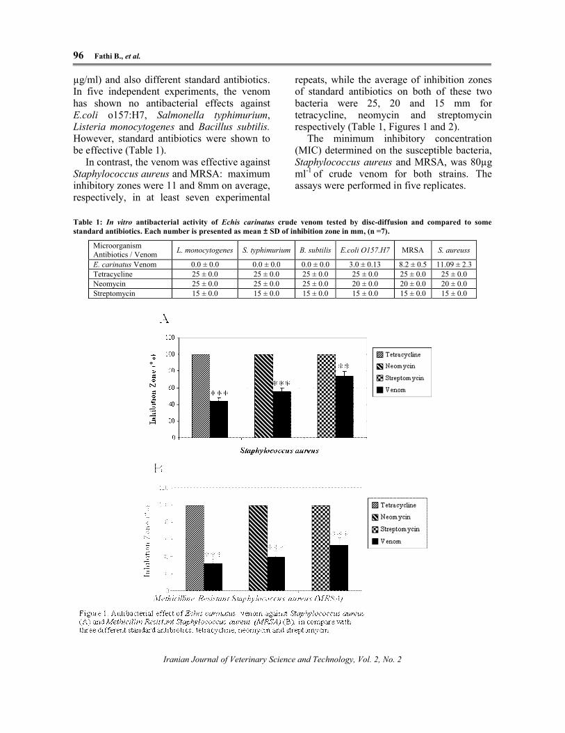
µg/ml) and also different standard antibiotics. In five independent experiments, the venom has shown no antibacterial effects against E.coli o157:H7, Salmonella typhimurium, Listeria monocytogenes and Bacillus subtilis.However, standard antibiotics were shown to be effective (Table 1).
In contrast, the venom was effective against Staphylococcus aureus and MRSA: maximum inhibitory zones were 11 and 8mm on average, respectively, in at least seven experimental
repeats, while the average of inhibition zones of standard antibiotics on both of these two bacteria were 25, 20 and 15 mm for tetracycline, neomycin and streptomycin respectively (Table 1, Figures 1 and 2).
The minimum inhibitory concentration (MIC) determined on the susceptible bacteria, Staphylococcus aureus and MRSA, was 80µg ml-1 of crude venom for both strains. The assays were performed in five replicates.
Table 1: In vitro antibacterial activity of Echis carinatus crude venom tested by disc-diffusion and compared to some standard antibiotics. Each number is presented as mean ± SD of inhibition zone in mm, (n =7).
S. aureussMRSAE.coli O157.H7B. subtilisS. typhimuriumL. monocytogenesMicroorganismAntibiotics / Venom
11.09 ± 2.38.2 ± 0.53.0 ± 0.130.0 ± 0.00.0 ± 0.00.0 ± 0.0E. carinatus Venom25 ± 0.025 ± 0.025 ± 0.025 ± 0.025 ± 0.025 ± 0.0Tetracycline20 ± 0.020 ± 0.020 ± 0.025 ± 0.025 ± 0.025 ± 0.0Neomycin15 ± 0.015 ± 0.015 ± 0.015 ± 0.015 ± 0.015 ± 0.0Streptomycin

Antibacterial Effect of Venom 97
Iranian Journal of Veterinary Science and Technology, Vol. 2, No. 2
Figure 2: Antibacterial effect of Echis carinatus venom (V) against Staphylococcus aureus (Left) and Methicillin ResistantStaphylococcus aureus (MRSA) (Right), in compare with two and three different standard antibiotics; neomycin (A),tetracycline (B), streptomycin (C).
Discussion
Researchers have reported antibacterial effects of some venoms including Echis carinatus venom against some bacteria (Perumal Samy et al., 2006; 2007). Herein it was clear that Echis carinatus venom has not a wide spectrum antibacterial effect against the mentioned bacteria, although a significant activity against S. aureus and MRSA in comparison with the standard antibiotics, tetracycline, streptomycin and neomycin has been observed. This is in agreement with the results of Gopalakrishnakone (Perumal Samy et al., 2006).
As described in the literature, venom consists of many different substances like proteins and enzymes. Which responsible for its biological activities. Therefore, these compounds may interact with specific molecules of some bacteria while not affecting other strains. Herein we conclude that the venom of Echis carinatus lacks effective proteins responsible for its antibacterial activity for some specific strains while it waseffective against S. aureus and MRSA.
It has been reported that phospholipase A2
(PLA2) can have antibacterial effects (Nunez et al., 2004; Barbosa et al., 2005; Perumal Samy et al., 2006 and Xu et al., 2007), whileEchis carinatus venom contains PLA2 activity (Kemparaju et al., 1994), and this may be responsible for its antibacterial properties.
However, Ferreira and his colleagues reported that, Lachesis muta venom had PLA2 activity, with no antibacterial effect. They concluded that presence of PLA2 in the venom does not guarantee the antibacterial activity. Therefore, it is possible that Echis carinatus venom may have a specific mechanism or unknown molecule that exhibit antibacterial effect on the susceptible bacteria.
To determine whether Echis carinatusvenom can influence other pathogens, further studies are needed using a wider spectrum of Gram-positive and Gram-negative bacteria and also other concentrations of this venom. Fractionated venom for detection of active components can improve investigation of its antibacterial activity.
References
Aloof-Hirsch, S., Devries, A. and Berger, A. (1968) The direct lytic factor of cobra venom: Purification and chemical characterization. Biochimistry and Biophysics Acta. 154, 53-60.
Ang, J.Y., Ezike, E. and Asmar, B.I. (2004) Antibacterial resistance. Symposium series Society for Applied Microbiology Ser Soc Appl Microbiol. 3, 229-239.
Backshall, S. (2007) Steve Backshall's venom: Poisonous creatures in the natural world. New Holland publishers (UK) Ltd, London.

98 Fathi B., et al.
Iranian Journal of Veterinary Science and Technology, Vol. 2, No. 2
Barbosa, P.S. Martins, A.M. Havt, A. Toyama, D.O. Evangelhista, J.S. Ferreira, D.P. Joazeiro, P.P. Beriam, I.O. Toyama, M.H. Fonteles, M.C. and Monterio, H.S. (2005) A renal and antibacterial effects induced by myotoxin I and II isolated from Bothrps jararacussu venom. Toxicon 46, 376-386.
Bauer, A.W. Kirby, W.M. Sherris, J.C. and Turck, M. (1966) Antibiotic susceptibility testing by a standardized single disk method. American Journal of Clinical Pathology 45(4), 493-496.
Benli, M. and Yigit, N. (2008) Antibacterial activity of venom from funnel web spider Agelena labyrinthica (Araneae: Agelenidae).Journal of Venomous Animals and Toxins including Tropical Diseases 17 (4), 641-650.
Budnik, B.A. Olsen, J.V. Egorov, T.V. Anisimova, V.E. Galkina, TG. Musolyamov, A.K. Grishin E.V. and Zubarev, R.A. (2004) De novo sequencing of antimicrobialpeptides isolated from the venom glands of the wolfspider Lycosa singoriensis. Journal of Mass Spectrometry 39, 193-201.
Conde, R. Zamudio, F.Z. Rodtiguez, M.H. and Possani, L.D. (2000) Scorpine, an anti-malaria and anti-bacterial agent purified from scorpion venom. Federation of European Biochemical Societies 471, 165-168.
Corzo, G. Villegas, E. Gó mez-Lagunas, F. Possani, L.D. Belokoneva, O.S. and Nakajima, T. (2002) Oxyopinins, large amphipathic peptides isolated from the venom of the wolf spider Oxyopes kitabensiswith cytolytic properties and positive insecticidal cooperativity with spider neurotoxins. Journal of biological chemistry277, 23627-23637.
Dani, M.P. Richards, E.H. Isaac, R.E. and Edwards, J.P. (2003) Antibacterial proteolytic activity in venom from the endoparasitic wasp Pimpla hypochondriaca(Hymenoptera: Ichneumonidae). Journal of Insect Physiology 49, 945-954.
Fennell, J.F. Shipman, W.H. and Cole, L.J. (1967) Antibacterial action of a bee venom fraction (melittin) against a penicillin-resistant Staphylococcus and other microorganisms. Research and Development Technical Report 5, 1-13.
Fennell, J.F., Shipman, W.H. and Cole, L.J. (1968) Antibacterial action of melittin, polypeptide from bee venom (32779). The actions of melittin on membranes. Proceedings of the Society for Experimental Biology and Medicine 127 (3), 707-710.
Garesky, V. M., Lumma, P. K., Freidinger, R. M., Pitzenberger, S. M., Randall, W. C., Veber, D. F., Gould, R. J., and Friedman, P. A., 1989. Chemical synthesis of echistatin, a potent inhibitor of platelet aggregation from Echis carinatus: synthesis and biological activity of selected analogs. Proceeding of the National Academy Sciences of the United States of America 86, 4022-4026.
Gawade, P.S., 2007. Therapeutic alternatives from venoms and toxins. Indian Journal of Pharmacology 39 (6), 260-264.
Glasser, H.R.S., 1948. Bactericidal activity of Crotalus venom in vitro. Copeia. 4, 245-247.
Guerranti, R., Aguiyi, J.C., Neri, S., Leoncini, R., Pagani, R., Marinello, E., 2002. Proteins from Mucuna pruriens and enzymes from Echis carinatus venom: characterization and cross-reactions. The Journal of Biological Chemistry 277, 17072-17078.
Haeberli, S., Kuhn-Nentwing, L., Schaller, J., Nentwig, W., 2000. Characterisation of antibacterial activity of peptides isolated from the venom of the spider Cupiennius Salei. Toxicon 38, 373-380.
Harvey,A.L.,Robertson,B.,2004.Dendrotoxins: structure-activity relationships and effects on potassium ion channels. Current Medicinal Chemistry 11, 3065-3072.
Jenssen, H., Hamill, P., Hancock, R.E.W., 2006. Peptide antimicrobial agents. Clinical Microbiology Review 19 (3), 491-511.
Kemparaju, K., Prasad, N.B., Gowda, V.T.,

Antibacterial Effect of Venom 99
Iranian Journal of Veterinary Science and Technology, Vol. 2, No. 2
1994. Purification of a basic phospholipase A2 from Indian saw-scaled viper (Echis carinatus) venom:
characterization of antigenic, catalytic and pharmacological properties. Toxicon 32 (10), 1187-1196.
Koh, D.C., Armugam, A., Jeyaseelan, K., 2006. Snake venom components and their applications in biomedicine. Cellular and Molecular Life Sciences 63, 3030-3041.
Kozlov, S.A., Vassilevski, A.V., Feofanov, A.Y., Surovoy, D.V., Karpunin, E.V., Grishin, E., 2006. Latarcins antimicrobial and cytolytic peptides from venom of the spider Lachesana tarabaevi (Zodariidae) that exemplify biomolecular diversity. Journal of Biological Chemistry 281 (30), 20983-20992.
Nowak, G., 2004. The Ecarin clotting time, a universal methos to quantify direct thrombin inhibitors. Pathophysiology of Haemostasis and Thrombosis 4 (33), 173-183.
Nunez, V.,Arce, V., Gutierrez, J.M., Lomonte, B., 2004. Structural and functional
characterization of myotoxin I, a Lys49 phospholipase A2 homologue from the of the snake Bothrops atrox venom. Toxicon 44 (1), 91-101.
Permual Samy, R., Gopalakrishnakone, P., Thwin, M.M., Chow, T.K.V., Bow, H., Hain, Y.E., Thong, T.W.J., 2007. Antibacterial activity of snake, scorpion and bee venoms: a comparison with purified venom phospholipase A2 enzymes. Journal of Applied Microbiology 102, 650-659.
Permual Samy, R., Pachiappan, A., Gopalakrishnakone, P., Thwin, M.M., Hian, Y.E., Chow, T.K.V., Bow, H., Weng, J.T., 2006. In vitro antibacterial activity of natural toxins and animal venoms tested against Burkholderia pseudomallei. BMC Infectious Diseases 6 (100), 1-16.
Raghunath, D., 2008. Emerging antibiotic resistance in bacteria with special reference to Indian. Journal of Biosciences. 33 (4),
593-603.
Shittu, L.A.J., Bankole, M.A., Ahmed, T., Bankole, M.N., Shittu, R.K., Saalu, C.L., Ashiru, O.A., 2007. Antibacterial and antifungal activities of essential oils of crude extracts of Sesame Radiatum against some common pathogenic micro-organism. Iranian Journal of pharmacology and Therapeutics (IJPT) 6, 165-170.
Stiles, B.G., Sexton, F.W., Weinstein, S.A., 1991. Antibacterial effects of different snake venoms: purification and characterization of antibacterial proteins from Pseudechis australis (Australian king brown or Mulga snake) venom. Toxicon 29, 1129-1141.
Tans, G., Rosing, J., 2001. Snake venom activators of factor X: an overview. Haemostasis 31, 225-233.
Torres-Larios, A., Gurrola, G.B., Zamudio, F.Z., Possani, L.D., 2002. Hadrurin, a new antimicrobial peptide from the venom of the scorpion Hadrurus aztecus. European Journal of Biochemistry 267, 5023-5031.
Wenhua, R., Shuangquan, Z., Daxiang, S., Kaiya, Z., Guang, Y., 2006. Induction, purification and characterization of an antibacterial peptide scolopendrin I from the venom of centipede Scolopendra subspinipes multilans. Indian Journal of Biochemistry & Biophysics 43, 88-93.
Wu, M., Hancock, R.E.W., 1999. Improved Derivatives of Bactenecin, a Cyclic Dodecameric Antimicrobial Cationic Peptide. American Society for Microbiology43 (5), 1274-1276.
Xu, C., Ma, D., Yu, H., Li, Z., Liang, J., Lin, G., Zhang, Y., Lai, R., 2007. A bactericidal homodimeric phospholipases A2 from Bungarus Fasciatus venom. Peptides 28 (5), 969-973.
Yan, L., Adams, M.E., 1998. Lycotoxins, antimicrobial peptides from venom of the wolf spider Lycosa carolinensis. Journal of Biological Chemistry 273, 2059-2066.
Iranian Journal of Veterinary Science and TechnologyVol. 2, No. 2, 2010, 93-100
Iranian Journal of Veterinary Science and Technology, Vol. 2, No. 2
IJVST
بررسی اثر ضد باکتریایی زهر مار جعفري ایران
3و عباس زارع 3ذوالفقاریان ن، حسی2جمشیدي.، عبدا1بهروز فتحی ،1آتنا جامی الاحمدي
ایران، مشهد، دانشکده دامپزشکی، دانشگاه فردوسی مشهد، بهداشت مواد غذایی و آبزیان 2و علوم پایه 1 گروه هاي
کرج، ایران سرم سازي رازي،واکسن و تحقیقات موسسه 3
17/8/89: نهایی پذیرش 6/6/89: مقاله دریافت
چکیده
اگر چه برخی از زهرها و ترکیبات مشتق شده از آنها، نشان داده اند که داراي خواص ضد باکتریایی هستند اما بیشـتر آنهـا بـراي یـافتن
ها نیز واجـد هاي جدید، باکتريبیوتیکآنتی رماکولوژي، همزمان با کشف و مصرفدر دنیاي فا. رسی قرار نگرفته اندچنین فعالیتهایی مورد بر
این امر باعث تحقیق و بررسی بیشـتر . گردداثر بوده و مسئله مقاومت باکتریایی مطرح میها بیها بر آنبیوتیکشوند که آنتیهایی میویژگی
زهرهاي جانوران به دلیل اثرات مختلـف مشـاهده . شودهاي موثرتر و جدیدتر میبیوتیکیعی مختلف براي کشف آنتیدانشمندان در منابع طب
اي از انواع پپتیـدها و مـواد غیـر پپتیـدي بـا زهر ترکیب بسیار پیچیده. اندباکتریال بسیار مورد توجه محققین قرار گرفتهشده از جمله اثر آنتی
در ایـن . سازي زهـر مارهـا صـورت گرفتـه اسـت باکتریال و خالصمطالعات بسیار کمی براي بررسی اثر آنتی. باشدهاي گوناگون میفعالیت
استافیلوکوکوس اورئوس، استافیلوکوکوس اورئوس مقاوم : باکتریال زهر مارجعفري ایران برعلیه شش گونه باکتري از جملهمطالعه اثرات آنتی
ونلا تایفی موریوم، باسیلوس سوبتیلیس و لیستریا مونوسایتوژنز با اسـتفاده از تسـت انتشـار در دیسـک و به متیسیلین، اشریشیا کولاي، سالم
بیوتیک استرپتومایسـین، نئومایسـین در این آزمایشات از سه آنتی. تست بررسی حداقل غلظت ممانعت کننده از رشد، مورد بررسی قرار گرفت
-باکتریال بوده و بـر بـاکتري نتایج نشان داد که زهر مار جعفري ایران داراي اثر آنتی. ستفاده گردیدبه عنوان کنترل مثبت انیز و تتراسایکلین
. باشدهاي استافیلوکوك اورئوس واستافیلوکوك اورئوس مقاوم به متیسیلین بطور معنی داري موثر می
ي، زهر، مار جعفريضد باکتر :واژگان کلیدي