
INVESTIGATING THE ROLE OF ZOLEDRONIC ACID ON...
74
INVESTIGATING THE ROLE OF ZOLEDRONIC ACID ON INTERLEUKIN-6 CYTOKINE EXPRESSION IN PROSTATE CANCER CELL LINES A Thesis Submitted to the Graduate School of Engineering and Science of zmir Institute of Technology in Partial Fulfillment of the Requirements for the Degree of MASTER OF SCIENCE in Biotechnology by Layka ABBAS ASBAGH June 2006 ZMR
Transcript of INVESTIGATING THE ROLE OF ZOLEDRONIC ACID ON...

INVESTIGATING THE ROLE OF ZOLEDRONIC ACID ON
INTERLEUKIN-6 CYTOKINE EXPRESSION IN PROSTATE CANCER CELL LINES
A Thesis Submitted to the Graduate School of Engineering and Science of
�zmir Institute of Technology in Partial Fulfillment of the Requirements for the Degree of
MASTER OF SCIENCE
in Biotechnology
by Layka ABBAS� ASBAGH
June 2006 �ZM�R
We approve the thesis of Layka ABBAS� ASBAGH Date of Signature ………………………………………… 15 June 2006 Asst. Prof. Dr. Fuat DOYMAZ Supervisor Department of Chemical Engineering �zmir Institute of Technology ………………………………………… 15 June 2006 Assoc. Prof. Dr. Ça� ÇAL Co-Supervisor Department of Urology Ege University ………………………………………… 15 June 2006 Asst. Prof. Dr. Ça�lar KARAKAYA Department of Biology �zmir Institute of Technology …………………………………………. 15 June 2006 Prof. Dr. Erdem GÖKER Department of Medical Oncology Ege University ………………………………………… 15 June 2006 Prof. Dr. Semra ÜLKÜ Head of Department �zmir Institute of Technology
................................................... Assoc. Prof. Dr. Semahat ÖZDEM�R
Head of the Graduate School

iii
ACKNOWLEDGEMENTS
First I would like to thank to everyone who had contributed to my education
troughout my life.
Many thanks go to my advisor, Assist. Prof. Dr. Fuat DOYMAZ. Thank you
for your knowledge, concern, humor, friendship and patience during my studies in
�YTE.
I would like to thank Assoc. Prof. Dr. Ça� ÇAL for his inavulable ideas,
supervision, throughout this study.I am grateful to Assist.Prof.Dr.Selim UZUNO�LU
for his valuble suggestions and supports in my thesis.
I would also like to thank Assist. Prof. Dr. O�uz BAYRAKTAR for his ever-
lasting encouragement and support in my life. I am particularly grateful to my friends
Yonca BA�SOY and Çı�ır B�RAY for their understanding, and endless patience.
Finally, I want to express my gratitude to my dear family for their excellent
support, understanding and encouragement in all my life.
�
�
�

iv
ABSTRACT
Investigating The Role Of Zoledronic Acid On Interleukin-6 Cytokine
Expression In Prostate Cancer Cell Lines Prostate cancer (CaP) is a common disease in aged men. Androgen deprivation
therapy is the first line treatment for advanced disease. Following this therapy, within
few months CaP cells become androgen independent in a few mounths. The mechanism
responsible for development of androgen-independent cancer is not known. Evidences
suggest that growth factors and cytokines including Interleukin–6 (IL-6) cast a main
role in this mechanism. IL–6 is an important cytokine on CaP progression.
Bisphosphonates inhibit the proliferation and induce the apoptosis of certain prostate
cancer cell lines, in vitro. However, their mechanism of action is not clearly
understood. One possible mechanism might be the IL-6 mediated pathway. This
relation between zoledronic acid (ZA) and IL-6 hasn’t been studied.
The objective of this study is to evaluate the cytotoxic and apoptotic effect of
ZA on hormone independent CaP cell line (PC-3) and to understand the relation of
underlying mechanism of ZA-mediated cytotoxicity and IL-6 expression.
PC-3 cells were treated with different concentrations of ZA. Drug-mediated
cytotoxicity and apoptosis were determined. IL-6 levels were measured from the
supernatants of PC-3 cells at 24, 48, 72 hrs.
Maximum cytotoxicity was achieved at 90 and 100 µM of ZA. ZA produced
the classical DNA ladders on gel synonymous of apoptosis. The decrease in IL-6
secretion was found to be statistically significant compared to controls. The decreasing
effect of ZA on IL-6 expression might suggest a new mechanism of action of ZA’s
antitumor activity. This outcome may offer a novel therapy that utilizes IL-6 antagonists
along with ZA in CaP patients.

v
ÖZET
PROSTAT KANSER� HÜCRE HATLARINDA ZOLEDRON�K AS�D�N �NTERLÖK�N-6 S�TOK�N�N�N EKSPRESYONU ÜZER�NDEK�
ROLÜNÜN ARA�TIRILMASI
Prostat kanseri günümüzde ileri ya� erkekler arasında en sık görülen kanser
tipidir. Hastalı�ın ileri evresinde kullanılan tedavi androjen baskılanmasıdır. Androjen
baskılanmasından kısa bir süre sonra prostat kanseri hücrelerinin birço�u hormondan
ba�ımsız ço�almaya devam ederler. Bu ço�almanın mekanizması henüz bilinmemekle
birlikte çe�itli büyüme faktörleri ve sitokinlerin bu mekanizmada önemli rol aldı�ı
dü�ünülmektedir. �nterlökin–6 (IL–6) prostat kanserinin ilerlemesinde önemli bir
sitokindir. Bifosfonatlar, çe�itli prostat kanser hücre hatlarında ço�almayı önleyici ve
apoptozisi tetikleyici bir etki göstermektedir, fakat etki mekanizmaları henüz
bilinmemektedir. Olası mekanizmalardan biri IL–6 yola�ı üzerinden olabilir. �u ana
kadar Zoledronik asit (ZA) ile IL–6 ili�kisini ortaya koyan bir çalı�ma yapılmamı�tır.
Bu çalı�manın amacı, hormon-dirençli prostat kanseri hücre hattı (PC–3)
üzerinde ZA’nın sitotoksik ve apoptotik etkisini göstererek ilaca ba�lı meydana gelen
bu sitotoksisitenin mekanizmasının IL–6 ile ili�kisini saptamaktır.
PC–3 hücreleri farklı konsantrasyonlarda ZA ile inkübe ediltikten sonra
ZA’nın PC–3 hücreleri üzerindeki sitotoksisitesi ve apoptotik etkisi ölçüldü. Hücrelerin
süpernatantlarındaki IL–6 düzeyleri 24, 48 ve 72 saatlerde ölçüldü.
Maksimum sitotoksisiteye ZA’nın 90 ve 100 µM konsantrasyonunda ula�ıldı.
ZA ile inkübasyon sonrasında apoptozisin göstergesi olan DNA merdiven olu�umu jel
üzerinde gözlendi. Kontrol grubuna oranla IL–6 salınımındaki azalmalar istatistiksel
açıdan anlamlı bulundu. ZA’nın IL–6 ekspresyonu üzerindeki azaltıcı etkisi, ZA’nın
antitümör aktivitesinde yeni bir mekanizmanın varlı�ına i�aret etmektedir. Bu sonuç,
prostat kanserli hastalarda zoledronik asitle birlikte IL–6 antagonistlerinin kullanılması
ile yeni bir tedavi seçene�ine kapı açmaktadır.

vi
To Selim Uzuno�lu, with all my respect and special thanks.

vii
TABLE OF CONTENTS
LIST OF FIGURES ........................................................................................................ vii
LIST OF TABLES......................................................................................................... viii
CHAPTER 1. INTRODUCTION .................................................................................. 1
1.1. Prostate Cancer and Treatment ............................................................ 3
1.2. Zoledronic Acid ................................................................................... 9
1.3. Interleukin- 6 in Prostate Cancer ....................................................... 14
1.4. Characterization of PC-3 Prostate Cancer Cell Line ......................... 19
1.5. Methods for studying cytotoxicity..................................................... 20
1.6. Methods for studying apoptosis in cell populations .......................... 22
1.7. Methods for measuring Interleukin-6 levels ...................................... 26
CHAPTER 2. MATERIALS AND METHODS.......................................................... 28
2.1. Materials ............................................................................................ 28
2.2. Methods ............................................................................................. 30
2.2.1. Cell Culture of PC-3 Cell Line .................................................... 30
2.2.2. Assessment of Cytotoxicity Produced by Zoledronic Acid ....... 30
2.2.2.1. Trypan –Blue Dye Exclusion Assay................................... 30
2.2.2.2. XTT Proliferation Assay..................................................... 31
2.2.3. Evaluation of Apoptosis Induced by Zoledronic Acid ................ 33
2.2.3.1. Mono-Oligo Nucleosome Detection-based ELISA ............ 33
2.2.3.2. DNA Fragmentation on Simple Agarose Gel ..................... 34
2.2.4. Detection of IL-6 Levels by ELISA ........................................... 35
2.3. Statistical Analysis............................................................................. 38
CHAPTER 3. RESULTS AND DISCUSSIONS......................................................... 39
3.1. Results................................................................................................ 39
3.2. Discussions ........................................................................................ 45

viii
CHAPTER 4. CONCLUSIONS ..................................................................................... 50
REFERENCES .............................................................................................................. 51

ix
LIST OF FIGURES
Figure Page
Figure 1.1. Chemical structure of zoledronic acid ....................................................... 10
Figure 1.2. IL–6 and signaling sathways...................................................................... 17
Figure 1.3. Morphology of PC-3 cells under a microscope ......................................... 20
Figure 1.4. Metabolization of XTT to a water soluble formazan salt by
viable cells ................................................................................................. 22
Figure 1.5. The biochemistry of DNA fragmentation and the appearance of
the “DNA ladder” ...................................................................................... 25
Figure 1.6. ELISA microplate reader ........................................................................... 27
Figure 2.1. Measurement of cytotoxicity by counting the cells with a
hemocytometer........................................................................................... 31
Figure 2.2. Measurement of metabolic activity using the tetrazolium salt
XTT............................................................................................................ 32
Figure 2.3. How the Cell Death Detection ELISAPLUS works. .................................... 34
Figure 2.4. Human IL-6 assay summary ...................................................................... 37
Figure 3.1. Trypan blue dye exclusion assay ............................................................... 39
Figure 3.2. Cell count of viable PC-3 cells as measured by trypan blue
exclusion assay with zoledronic acid (24, 48 and 72 hr) ........................... 40
Figure 3.3. Dose –dependent cytotoxic activities of zoledronic acid against
PC-3 cells ................................................................................................... 41
Figure 3.4. Evidence of apoptotic effects of zoledronic acid....................................... 42
Figure 3.5. DNA Fragmentation analysis of zoledronic acid treated PC-3
cells using simple agarose gel electrophoresis .......................................... 42
Figure 3.6. Time and dose dependent decrease of IL-6 levels in the
supernatants of PC-3 cells.......................................................................... 44

x
LIST OF TABLES
Table Page
Table 1.1. Differential features and significance of necrosis and apoptosis................... 23
Table 2.1. List of chemicals used in experiments........................................................... 29
Table 2.2. List of equipments used in experiments ........................................................ 29
Table 3.1. IL-6 Levels in supernatants of PC-3 cells...................................................... 43

1
CHAPTER 1
INTRODUCTION
Our growing understanding of cancer cell biology should lead to better ways of
diagnosis and treating this disease. Anticancer therapies can be designed to destroy
cancer cells preferentially by exploiting the properties that distinguish them from
normal cells, including the defects they harbor in their DNA repair mechanisms, cell
cycle check points, and apoptosis pathways. Tumors can also be attacked through their
dependence on their blood supply. By understanding the normal control mechanisms
and how they are subverted in specific cancers, it becomes possible to devise drugs to
target cancers more precisely. Cancer cells are defined by two heritable properties: they
and their progeny reproduce in defiance of the normal restraints on cell division and
invade and colonize territories normally reserved for other cells. It is the combination of
these actions that makes cancer dangerous. An isolated abnormal cell that does not
proliferate more than its neighbors does no significant damage, no matter what other
disagreeable properties it may have; but if its proliferation is out of control, it will give
rise to a tumor, or neoplasm- a growing mass of abnormal cells. As long as the
neoplastic cells remain clustered together in a single mass, the tumor is said to be
benign. At this stage, a complete cure can usually be achieved by removing the mass
surgically. A tumor is considered a cancer only if it is malignant, that is, only if its cells
have acquired the ability to invade surrounding tissue. Invasiveness usually implies an
ability to break loose, enter the bloodstream or lymphatic vessels, and form secondary
tumors, called metastases, at other sites in the body. The more widely a cancer spreads,
the harder it becomes to eradicate.
Prostate cancer is a common disease in aged men. Normal and early stage
prostate cancer cells require androgen for growth and survival, which led to the
formulation and successful application of androgen ablation and anti-androgen therapies
as principal treatment modalities for the disease. However, beneficial effects of the
hormonal therapies are often temporary, and the cancer regularly progresses to the
androgen-refractory stage characterized by recurrent growth and metastasis,
predominantly to bone (Feldman et al, 2001, Nelson et al, 2003). Despite decades of

2
intense laboratory and clinical research, to date there is no cure for androgen-refractory
prostate cancer. The existence of androgen-independent prostate tumors suggests that
release of locally produced and/or circulating growth factors such as Interleukin-6,
which work through cellular receptors, can switch the prostate cells from an inactive to
an activated phenotype leading to cellular proliferation. The majority of prostate cancers
arise from epithelial cells, and in tissue cultures, epithelial prostate cancer cells produce
factors that act in an autocrine fashion to regulate their growth and survival (Xie et al,
2002, Berger et al, 2003).
Interleukin-6 (IL-6) is a multifunctional cytokine which is involved in regulation
of growth of various malignant tumors. IL-6 also regulates gene expression in a number
of different organs, modulates immune function, stimulates the hypothalamic-pituitary
axis, promotes osteoclasting resorption in bone, and stimulates bone marrow (Ershler et
al, 2000). The biological activities of IL-6 are mediated by the IL-6 receptor, which
binds IL-6 specifically and with low affinity, and gp130, which associates with the IL-
6-IL-6 receptor complex, resulting in high-affinity binding and activation of
intracellular signaling. The evidences were shown that IL-6 may be an important
autocrine and/or paracrine growth factor for prostate cancer and may contribute to
cancer progression (Siegall et al, 1990, Okamoto et al, 1997, Siegsmund MJ et al, 1994,
Hobisch A et al, 2000). For example, inhibition of IL-6, with anti-IL-6 antibody,
sensitizes androgen-independent prostate cancer cells to chemotherapeutic agents in
vitro. Additionally, IL-6 induces androgen receptor (AR) activity through both
increasing AR gene expression and activating the AR in the absence of androgen in
prostate cancer cells (Lin et al, 2001). These data suggest that targeting IL-6 may have
multiple benefits in prostate cancer patients.
Bisphosphonates inhibits the proliferation and induces the apoptosis of certain
prostate cancer cell lines, in vitro (Lee et al, 2001). Zoledronic acid, nitrogen-containing
bisphosphonate, is used in prostate cancer patients according to laboratory evidence
documenting that prostate cancer bone metastases are associated with an increased
osteoclast-mediated bone resorption (Ikeda et al, 1996, Maeda et al, 1997). Furthermore,
preclinical studies suggest that bisphosphonates possess antitumor activity and can
inhibit proliferation and induce apoptosis of tumor cell lines (Lipton et al, 2004).
In this study, we discuss prostate cancer as a model and try to find out drug-
mediated cytotoxicity of zoledronic acid on hormone independent prostate cancer cell
line and its mechanism of action. Thus, we first considered more closely what prostate

3
cancer is and its current treatments. Then we reviewed zoledronic acid and its role in
prostate cancer treatment. Finally, we examined the function of interleukin-6 in prostate
cancer cells. The objective of the study is to confirm the known apoptotic and cytotoxic
effect of zoledronic acid in PC–3 prostate cell line and investigate the role of zoledronic
acid on the expression of IL–6 in this cell line.
1.1 Prostate Cancer and Treatment
Prostate cancer is the fourth most common cancer in men, worldwide, with
incidence and mortality rates that vary markedly among and within different countries.
Since the early 1990s, new screening tests and improved treatments have been
associated with dramatic shifts in the incidence, stage at diagnosis, and mortality of this
disease. Major advances in molecular biology and epidemiology have provided new
insights into the etiology and biology of prostate cancer. These developments promise
to transform our understanding of this disease and will likely lead to new and better
ways to prevent and treat prostate cancer in the future.Incidence and mortality rates vary
tremendously among countries. Incidence and mortality rates are generally higher in
Western countries than in developing countries. There are multiple complex causes for
the worldwide and ethnic variations in prostate cancer incidence. The two major factors
are genetics and environment. Prostate cancer is predominantly a disease of the elderly
man, with more than 75% of new prostate cancers being diagnosed in men older than 65
years. The incidence of prostate cancer in men age 50 to 59 years, however, has
increased substantially since the 1970s.
Although the specific causes of prostate cancer initiation and progression are not
yet known, considerable evidence suggests that both genetics and environment play a
role in the evolution of this disease. Classic and molecular epidemiology studies have
identified a number of potential risk factors associated with the development of prostate
cancer. A number of case-control studies have demonstrated familial clustering of
prostate cancer, suggesting that some prostate cancers may be inherited. The clinical
characteristics of familial prostate cancers have also been characterized to determine if
hereditary prostate cancer may be more aggressive.
Prostate cancer initiation and progression are influenced by androgens. Prostate
tumors are exquisitely sensitive to androgens and regress after medical or surgical

4
castration. Ross and colleagues hypothesized that the higher incidence of prostate
cancer observed in African Americans may be related to elevated levels of circulating
androgen (Ross et al, 1998).
Although the exact etiology of prostate cancer remains unknown, there has been
tremendous progress in defining the molecular events responsible for the initiation and
progression of this disease. This progress has resulted from a number of factors.
Increased funding for prostate cancer research has focused and accelerated efforts in the
field. Prostate cancers develop from the accumulation of genetic alterations that result in
an increase in cell proliferation relative to cell death, arrest differentiation, and confer
the ability to invade, metastasize, and proliferate in a distant site. Histologic changes are
present in the prostates of men in their 20s, yet the diagnosis is typically made 3 to 4
decades later, which suggests that the development of the disease is a multistep process
(Sakr et al, 1993). The alterations include somatic point mutations, gene deletions,
amplifications, chromosomal rearrangements, and changes in DNA methylation (Nelson
et al, 2003). It is believed that the accumulation of changes acting synergistically is
more critical than the order in which the alterations occur. Identifying and
understanding the events has implications for control of the disease at the earliest stages
of transformation, for progression as an invasive tumor, for prognostication, and for
points of therapeutic attack. Men who are castrated before the age of 40 years rarely
develop prostate cancer (Montie et al, 1994). The evolution of the tumor is influenced
by hormonal factors; it is also influenced by environmental, infectious/inflammatory
factors, and, given the long history once the diagnosis is established, the specific
therapy (ies) used to treat the disease. The alterations include a reduction in defense
against carcinogen-induced damage, inflammation, and changes in androgen signaling
and changes in growth-regulatory genes that contribute to cell proliferation, survival,
and spread.
Neoplastic growth is the result of genetic, hormonal, environmental, and
possibly infectious factors that modulate the expression of specific genes. The normal
mutation frequency of DNA (1 x 10⁻¹⁰) is too low to produce significant changes in
overall gene expression. It is now believed that tumor cells themselves have an inherent
genetic instability that results in the coexistence of multiple genetically related, yet
distinct, clones within a tumor mass (Sarasin et al, 2003). Clones with a survival
advantage continue to proliferate, whereas those that acquire changes that reduce
viability undergo cell death.

5
Overexpression of certain growth factors, such as epidermal growth factor
(EGF), basic fibroblast growth factor, and platelet-derived growth factor (PDGF), has
been reported to be involved in prostate cancer as autocrine and paracrine signaling
loops together with their corresponding receptors (EGFR, fibroblast growth factor
receptor, and IL–6) (Scher et al, 1995, Uehara et al, 2003, Culig et al, 1996). The
insulin-like growth factor–1 and -2 (IGF–1, IGF–2), and transforming growth factor-
and - (TGF- , TGF- ) and their receptors (IGFR and TGFR, respectively) have also
been implicated (Cardillo et al, 2003, Tu et al, 2003). IGFs are potent mitogens for
human prostate cancer cells and osteoblasts via interaction with IGF receptors. Elevated
levels of serum IGF-binding proteins have been reported in metastatic human prostate
cancer. HER–2/neu protein is a transmembrane tyrosine kinase receptor with strong
homology to EGFR. Amplification of the HER–2/neu gene and overexpression of the
protein have prognostic significance in breast cancer and are used to select therapy.
HER–2/neu amplification in prostate cancer is uncommon. Nevertheless, increased
HER2/neu protein was found in 20% of untreated hormone-naive primary tumors,
whereas overexpression was observed in 80% of metastatic cases and more than 60% of
primary tumors surviving after androgen ablation. Investigators have also reported
HER2 can activate AR independent of ligand (Craft et al, 1999).
The major cause of death from prostate cancer is progressive castration-resistant
disease, a tumor that continues to grow despite castrate levels of testosterone. As
prostate cancers evolve to castration resistance, PSA synthesis resumes. The current
view is that prostatic cancers at the time of diagnosis are composed of cells with three
distinct cellular phenotypes: androgen-dependent, androgen-sensitive, and androgen-
independent cells. Androgen-dependent cancer cells continuously require a critical level
of androgenic stimulation for maintenance and growth (i.e. without adequate androgenic
stimulation, these cells die) and, in this regard, are very similar to the androgen-
dependent non-neoplastic cells of the normal prostate. The growth of androgen-sensitive
cancer cells slows when androgens are withdrawn and they become dormant. They do
not die. In contrast, the growth of androgen-independent cells does not change after
androgen deprivation, no matter how complete; these cells are completely free of
androgenic effects on growth (Arnold et al, 2002, Isaacs et al, 1992). In contrast to what
can be accomplished in vitro using charcoal-stripped serum to eliminate androgens
completely, in vivo it is virtually impossible to eliminate all androgens completely.

6
How do resistant tumor cells emerge during androgen ablation therapy? Some
researchers theorize that selection of resistant cells occurs, whereas others suggest that,
under the pressure of androgen-ablative therapies, cells that were sensitive only to the
point at which growth was slowed without cell death adapt to the low androgen
environment and, over time, acquire additional somatic changes that result in tumor
regrowth (Isaacs et al, 1981, Craft et al, 1999, Bruchovsky et al, 1990). Clinically,
selection of resistant or insensitive cells may be more relevant immediately after
androgen withdrawal and adaptation more important later. The observation that basal
epithelial cells preferentially survive androgen ablation (in contrast to secretory
epithelial cells) demonstrates that intrinsically resistant cells do exist even in the normal
prostate gland, in line with the selection hypothesis. This is consistent with the theorized
role of basal cells as the stem cells for the prostatic epithelium (De Marzo et al, 1998,
Bui et al, 1998, Isaacs et al, 1999). Unknown is whether the first transformed cell that
ultimately develops into the self-renewing stem cell of a prostate cancer is dependent,
sensitive, or insensitive to androgens. It is also difficult to determine whether the
resistant/surviving cell population has a more basal or stem cell genotype, or a basal or
a more differentiated cell that has been transformed. That hormonal ablation alone
cannot eradicate the disease completely in either the primary site or in a metastatic focus
suggests an intrinsic resistance and, at best, partial androgen sensitivity. Many pathways
associated with resistance involve inhibition of proapoptotic molecules or the up-
regulation of cell-survival molecules.
The androgen receptor (AR) is a member of a super-family of ligand-dependent
transcription factors. Alterations in AR signaling that have been identified in human
prostate cancer include alterations in steroid metabolism, an increase in the level of the
protein, changes in co-regulator profiles, and ligand-independent activation. Changes in
AR occur as the disease progresses from a clinically localized lesion in a non-castrate
environment to a castrate metastatic lesion (Buchanan et al, 2001, Grossmann et al,
2001, Scher et al, 2001). All of these mechanisms are consistent with continued
signaling through the receptor in castration-resistant lesions. The AR protein is
expressed in prostate cancers of all clinical states. In upwards of 30% of cases, levels
are higher in castration-resistant as opposed to non-castrate tumors (Culig et al, 1998,
Tilley et al, 1994). Amplification of the AR gene itself has also been reported in
approximately 22% of castration-resistant metastases (Bubendorf et al, 1999) and in
7
23% to 28% of recurrent primary tumors and is associated with increased levels of the
AR and the proteins it regulates (Koivisto et al, 1996, Linja et al, 2001).
The most common mutations in the AR that have been identified in clinical
specimens are in the ligand-binding domain. Virtually all are associated with a gain of
function, as opposed to the loss of function mutations, which are most common among
patients with androgen-insensitivity syndromes. Most disrupt a protein–protein
interaction surface and result in an increase in the transactivation activity of the receptor
in response to a range of classical and nonclassical ligands (Buchanan et al, 2000)
In addition to steroid hormones, growth factors, such as keratinocyte growth
factor, IGF–1, and EGF; HER2; and cytokines, such as interleukin–6 (IL–6), can
activate the AR independent of ligand (Culig et al, 1994, Ueda et al, 2002). Exactly how
these factors activate signaling is an area of active study. This can also contribute to
progression in castration-resistant disease.
Inhibitors of apoptosis are also implicated in the acquisition of the castration-
resistant phenotype. Blocking cell death pathways that are normally induced by
androgen ablation allows cells to survive. bcl–2, which inhibits the death of cancer cells
without affecting their rate of proliferation, is essentially undetectable in most non-
castrate lesions but is highly expressed in castration-resistant disease (McDonnell et al,
1992). Similarly, survivin, a member of the class of proteins called inhibitors of
apoptosis, is highly expressed in benign and malignant prostate neuroendocrine cells
(Xing et al, 2001).
Bone Metastasis
Bone metastases represent a major health and financial burden because of their
frequency and the considerable morbidity they generate in cancer patients. They occur
commonly in patients with advanced prostate carcinoma, affecting at least 70% of
patients with metastatic disease (Adami et al, 1997).
Prostate cancer cells that have gained access to the circulation have a unique
predilection for bone. The establishment of a metastatic focus in bone involves multiple
steps, including adhesion of the tumor cells to endothelial cells in the marrow and
migration through fenestrations in the endothelial cell layer. This migration is driven, in
part, by a chemo attractant gradient of marrow- and stromal-derived growth factors.
Once established, tumor cells and marrow-derived cells develop a bidirectional
interaction that protects the epithelial cells and promotes tumor cell survival and
proliferation (Cher et al, 2001).

8
Factors that contribute to cancer growth in bone are broadly divided into
osteoblastic and osteolytic factors. Sources include the tumor cells themselves, normal
bone cells, and reserves in the bone matrix that are released as part of the remodeling
process. Many of the factors contributing to the osteoblastic phenotype, such as
endothelin–1 (ET–1) (Nelson et al, 2003) and IL–6 can be targeted directly. ET–1,
produced by prostate cells, stimulates the differentiation of osteoblast precursors,
decreases osteoclastic bone resorption and motility, and augments the mitogenic effects
of IGF–1, IGF–2. In normal bone, osteoblasts regulate osteoclastogenesis by interacting
with mononuclear hematopoietic precursors. Osteoblasts express receptor activator of
nuclear factor B ligand (RANKL) and osteoprotegerin. Osteoprotegerin level increases
in patients with bone metastases (Jung et al, 2003). IL–6 is another cytokine released by
prostate cancer cells that contributes to increased bone resorption (Keller et al, 2004).
Binding of RANKL to RANK on osteoclastic precursors initiates intracellular signals
that activate an osteoclastic phenotype.
Management of Prostate Cancer
The management of all stages of prostate cancer is sometimes highly
contraversial. This disease often has a long natural history; therefore, substantial
numbers of patients survive 15 years or longer after the diagnosis (even without
treatment). Furthermore, because the disease occurs in older men, a large number of
patients die from these conditions before they suffer symptoms or die from prostate
cancer. Investigators and clinicians vary widely in their use of surgery, radiotherapy,
hormonal manipulation, and other measures for treating each stage of disease. Most
clinicians agree, however, that treatment of early stage disease with either surgery or
radiotherapy results in comparable survival. It is unclear at time whether similar
survival rates could be achieved with systemic therapies.
Endocrine therapy is mainstay of treatment for symptomatic advanced prostate
cancer. Patients with asymptomatic advanced disease do not appear to have improved
survival with treatment when compared with untreated cells. Thus, treatment of patients
with asymptomatic, advanced disease is not essential. Orchiectomy (removal of testicles
by surgery), luteinizing hormone-releasing hormone (LHRH) agonists, and
antiandrogens are the available treatments. After androgen ablation treatment, usually
prostate cancer cells undergo an active process of programmed cell death (apoptosis).
Unfortunately, within 18–24 months after starting this treatment %80 relapse occurs and
cells will be adept to survive without androgenic stimulation and become unresponsive

9
to androgen withdrawal.. The mechanism responsible for development of androgen-
independent cancer is not clear yet. Accumulating evidence suggests that growth factors
and cytokines play an important role in this mechanism.�
Other agents that may be helpful in management of prostate cancer include the
following; progestins, corticosteroids, zoledronic acid for reduction in bone pain, in
time to first skeletal related events and other drugs that inhibit androgen synthesis such
as aminoglutethimide or ketoconazole. Chemotherapy also provides relief in 20 % to
30% of symptomatic patients with prostate cancer. Estramustine, cisplatine, 5-
fluorouracil, vinorelbine, gemcitabine, paclitaxel are the most popular agents.
1.2 Zoledronic Acid
Bisphosphonates are pyrophosphate analogues in which a carbon atom has
replaced the oxygen bridge. They bind strongly to hydroxyapatite in bone. The addition
of different carbon side chains has generated a diverse group of compounds that can
inhibit bone resorption via actions on the osteoclast. The more recently developed
nitrogen containing bisphosphonates; such as pamidronate and zoledronic acid, have
greatly enhanced potency. Zoledronic acid is a nitrogen-containing bisphosphonate that
inhibits osteoclast activity and bone resorption. It is indicated for the treatment of
hypercalcemia of malignancy and for the treatment of patients with multiple myeloma
and patients with documented metastasis from solid tumors, in conjunction with
standard antineoplastic therapy. Zoledronic acid is structurally similar to other
bisphosphonates, having the required phosphorus-carbon-phosphorus core and a
hydroxyl group at the R1 position (Green et al, 2001). However, it is the heterocyclic
imidazole group attached to the R2 position that distinguishes zoledronic acid from
other bisphosphonates (Widler et al, 2002). The chemical structure of zoledronic acid is
shown in the Figure 1.1.

10
Figure 1.1. Chemical structure of zoledronic acid
(Source: Zometa®, insert package)
Bone metastases represent a major health and financial burden because of their
frequency and the considerable morbidity they generate in cancer patients. They occur
commonly in patients with advanced prostate carcinoma, affecting at least 70% of
patients with metastatic disease (Adami et al, 1997). After development of hormone-
refractory metastatic disease, prostate cancer is incurable. The metastasis of prostate
cancer cells to the bone marrow constitutes the major source of morbidity and mortality
in prostate cancer (Singh et al, 2006). Excessive osteoclast activity plays a central role
in the pathophysiology of bone disease at each stage of prostate cancer disease
progression (Saad et al, 2006). The bone marrow microenvironment is strictly involved
in the evolution of the prostate disease progression by supporting cell growth and
survival (Corso et al, 2005). Tumor cells then release a variety of growth factors that
promote bone resorption and increase the risk of skeletal complications (Lipton, 2004).
The activation of some signaling pathways within the bone stromal cells increases the
productions of several cytokines which in turn favors the cell proliferation and survival
and enhance the drug resistance by anti-apoptotic mechanisms (Corso et al, 2005).
Bisphosphonates have become a new form of medical therapy for tumor bone
disease (Body et al, 1998, Powles et al, 2002). They are successfully used to treat the
symptoms of metastatic bone disease, especially bone pain, and they reduce by up to
40% the long-term skeletal morbidity rate. Bisphosphonates interfere as well with bone
microenvironment inhibiting the survival of stromal cells and hampering the contact
between plasma and stromal cells (Corso et al, 2005). Zoledronic acid, a new-generation
bisphosphonate is mainly osteolysis-inhibitory agent (Brubaker et al, 2006). These
agents act primarily by initiating biochemical processes that ultimately result in
apoptosis of osteoclasts, but they also have a number of other antitumor functions (eg,
inhibition of angiogenesis) (Conte et al, 2004). Osteoclast inhibition was obtained with

11
bone-targeted zoledronic acid (Gao et al, 2005). Bisphosphonates are potent inhibitors
of osteoclast activity that have demonstrated efficacy in the treatment of bone
metastases (Lipton, 2004). Bisphosphonates bind eagerly to the bone matrix, are
released during bone resorption, and are subsequently internalized by osteoclasts, where
they interfere with biochemical pathways and induce osteoclast apoptosis (Lipton,
2004). Bisphosphonates also antagonize osteoclastogenesis and promote the
differentiation of osteoblasts. As a result, bisphosphonates inhibit tumor-induced
osteolysis and reduce skeletal morbidity (Lipton, 2004).
Zoledronic acid, a highly potent inhibitor of osteoclast-mediated bone
resorption, has increased bone mineral density in men receiving androgen deprivation
therapy and is the only bisphosphonate that has shown statistically significant
reductions in skeletal morbidity in patients with bone metastases from prostate cancer
(Saad et al, 2006). Due to its unique properties, zoledronic acid is a breakthrough in the
management of metastatic bone disease in patients with advanced prostate cancer
(Hoesl et al, 2006). Zoledronic acid should be considered for the prevention of skeletal
morbidity in patients with prostate cancer throughout their treatment continuum (Saad et
al, 2006).
Nitrogen containing bisphosphonates (e.g., pamidronate, ibandronate, zoledronic
acid) inhibit protein prenylation, thus affecting cell function and survival (Green, 2003).
In vitro, submicromolar concentrations of bisphosphonates inhibited tumor cell
adhesion and reduced invasion through extracellular matrix. At higher concentrations,
antiproliferative and proapoptotic effects have been reported (Green, 2003).
Bisphosphonates exert their effects on osteoclasts and tumor cells by inhibiting a key
enzyme in the mevalonate pathway, farnesyl diphosphate synthase, thus preventing
protein prenylation and activation of intracellular signaling proteins such as Ras (Green,
2004). Recent evidence suggests that bisphosphonates also induce production of a
unique adenosine triphosphate analogue (Apppi) that can directly induce apoptosis
(Green, 2004). Their biochemical effects on protein prenylation, they induce caspase-
dependent apoptosis, inhibit matrix metalloproteinase activity, and down regulate alpha
(v) beta (3) and alpha (v) beta (5) integrins (Green, 2004).
Zoledronic acid also inhibits tumor cell adhesion to the extracellular matrix and
has anti-angiogenic activity (Green, 2004).
Preclinical evidence suggests that zoledronic acid has antitumor activity in
prostate cancer models probably due to their ability to interfere with several intracellular

12
signaling molecules (Melis et al, 2005, Saad et al, 2006, Green, 2005). Furthermore,
preclinical studies suggest that bisphosphonates possess antitumor activity and can
inhibit proliferation and induce apoptosis of tumor cell lines (Lipton, 2004). In addition,
zoledronic acid, a new-generation bisphosphonate, appears to inhibit tumor cell
invasion of the extracellular matrix (Lipton, 2004).
Bisphosphonates work by inhibiting osteoclast-mediated bone resorption and
have also demonstrated antitumor activity in preclinical models (Saad, 2005).
Specifically, zoledronic acid inhibits proliferation and induces apoptosis of human
prostate cancer cell lines in vitro (Green, 2005). In a model of prostate cancer,
zoledronic acid significantly inhibited growth of both osteolytic and osteoblastic tumors
and reduced circulating levels of prostate-specific antigen. Bisphosphonates block the
development of monocytes into osteoclasts and are thought to promote apoptosis of
osteoclasts (Berenson, 2001). These agents prevent osteoclasts from moving to the bone
surface and seem to inhibit the production of bone-resorbing cytokines such as
interleukin-6 (IL-6) by bone marrow stromal cells (Berenson, 2001).
The therapeutic benefits of biphosphonates in patients with advanced prostate
were proved in several studies. Zoledronic acid (Zometa; Novartis Pharmaceuticals
Corp.; East Hanover, NJ and Basel, Switzerland) exerts synergistic antitumor activity
when combined with other anticancer agents (Green, 2004). It was shown that the
combination of zoledronic acid with selective EGFR-tyrosine kinase inhibitor (gefitinib)
causes a cooperative antitumor effect accompanied by induction of apoptosis and
regulation of the expression of mitogenic factors, proangiogenic factors and cell cycle
controllers both in vitro and in xenografted nude mice (Melisi et al, 2005). These results
provide new insights into the mechanism of action of zoledronic acid and a novel
rationale to translate this feasible combination treatment strategy into a clinical setting
(Melisi et al, 2005). The experimental data based on combinations of zoledronic acid
with either gemcitabine or fluvastatin indicates its therapeutic role in treatment of bone
metastasis of selected malignancies (Budman et al, 2006). The recent history of
chemotherapy has shown that combinatorial therapy by using specific inhibitors of
dominantly active signaling pathways opens new treatment perspectives in metastastic
tumors. These studies suggest that zoledronic acid has the potential to inhibit bone
metastasis and bone lesion progression in patients with prostate cancer (Green, 2005).
Recent studies have suggested that bisphosphonates may also exert direct
antitumor effects on myeloma cells (Avcu et al, 2005). In vitro studies have

13
demonstrated the antitumor potential of zoledronic acid on myeloma cell lines (Corso et
al, 2005). Zoledronic acid is able to affect the isoprenylation of intracellular small G
proteins (Caraglia et al, 2004). The antitumor activity of Zoledronic acid combined with
R115777 farnesyl transferase inhibitor (FTI) against epidermoid cancer cells was
increased (Caraglia et al, 2004). In human epidermoid head and neck KB and lung
H1355 cancer cells, 48 h exposure to pamidronate and zoledronic acid induced growth
inhibition (25 and 10 µM, respectively) and apoptosis and abolished the proliferative
and antiapoptotic stimuli induced by epidermal growth factor (EGF) (Caraglia M et al,
2004).
Zoledronic acid induced apoptosis through the activation of caspase 3. A strong
decrease of basal ras activity was observed. These effects were paralleled by impaired
activation of the survival enzymes extracellular signal regulated kinase 1 and 2 (Erk-
1/2) and Akt that were not restored by EGF (Caraglia et al, 2004).
The combined treatment with Zoledronic acid and R115777 resulted in a strong
synergism both in growth inhibition and apoptosis in KB and H1355 cells. The
combination was highly effective in the inhibition of ras, Erk and Akt activity, while
farnesol again antagonized these effects (Caraglia et al, 2004).
Zoledronic acid has been shown to inhibit the oncogenicity of Ras through the
inhibition of prenylation of Ras and Ras-related proteins by the induction of S-phase
cell cycle arrest and apoptosis (Chuah et al, 2005). Nitrogen containing-
bisphosphonates have direct antitumor effects via the inactivation of Ras proteins (Sato
et al, 2005). Zoledronic acid also can exert an antiangiogenic activity and inhibition of
tumor cell bone invasiveness by a transient reduction of VEGF, bFGF circulating levels
after infusion (Ferretti et al, 2005).
Although zoledronic acid is widely used in metastatic prostate cancer
management, few data are available about its molecular effects in prostatic cells. The
antitumor activity of biphosphonates that found in vitro and in vivo is interesting and
has to be further assessed in laboratory and clinical studies. Further studies are needed
to fully elucidate these biochemical mechanisms and to determine if the antitumor
potential of bisphosphonates translates to the clinical setting.
Bone metastasis microenvironment-related growth factors such as insulin-like
growth factor 1 (IGF-1), transforming growth factor beta 1 (TGF-beta1), basic
fibroblast growth factor (bFGF) and interleukin 6 (IL-6) show survival factor activity by
inhibiting chemotherapy-induced apoptosis of PC-3 prostate cancer cells in vitro (Tenta

14
et al, 2005). Recently, zoledronic acid has been shown to induce apoptosis in PC-3
prostate cancer cells (Tenta et al, 2005). These bone microenvironment-related growth
factors, also can modify the actions of various pharmaceutical agents, including
cytotoxic drugs in malignant cell lines (Tenta et al, 2006). Since bisphosphonates inhibit
the proliferation and induce the apoptosis of certain prostate cancer cell lines, its
mechanism of action is not clearly understood. IL-6 is an important cytokine on prostate
cancer progression. One possible mechanism might be the IL-6 mediated pathway. The
relation between zoledronic acid and IL-6 has not been known since there is no
experimental data.
1.3. Interleukin-6 in Prostate Cancer
IL-6 is a 21–28 Kd cytokine containing 184 amino acids following cleavage of a
28 aa signal peptide (May et al, 1988). IL-6 belongs to the ‘‘IL-6 type cytokine’’ family
that also includes leukemia inhibitory factor, IL-11, ciliary neurotrophic factor,
cardiotrophin-1 and oncostatin M (Sehgal et al, 1995). In the normal homeostatic state,
IL-6 levels are typically very low. However, in response to the appropriate stimulus
(e.g. inflammation); a wide variety of cells produce IL-6. Many physiologic functions
are attributed to IL-6 including promotion of antibody production from B lymphocytes,
modulation of hepatic acute phase reactant synthesis, promotion of osteoclastic-
mediated bone resorption, and induction of thrombopoiesis (Hirano et al, 1992). In
addition, it is implicated in development and progression of tumors of various organs, in
particular myeloma, renal and prostate cancer, and melanoma.
Multiple studies have demonstrated that IL-6 is elevated in the sera of patients
with metastatic prostate cancer (Adler et al, 1999; Hoosein N et al, 1995; Twillie et al,
1995). Adler et al demonstrated that serum levels of IL-6 and transforming growth
factor-b1 are elevated in patients with metastatic prostate cancer, and that these levels
correlate with tumor burden as assessed by serum PSA or clinically evident metastases.
In a similar fashion, Drachenberg et al reported elevated serum IL-6 levels in men with
hormone-refractory prostate cancer compared to normal controls, benign prostatic
hyperplasia, prostatitis, and localized or recurrent disease (Drachenberg et al, 1999).
These observations suggest that IL-6 may be a surrogate marker of the androgen
independent phenotype. IL-6 has been shown to be a candidate mediator of prostate

15
cancer morbidity and a candidate marker of disease activity for prospective clinical
testing (Twillie et al, 1995). In addition to its role as a mediator of morbidity, IL-6 may
also act as a growth factor, and protect prostate cancer cells from cell death induced by
certain chemotherapeutic agents. IL-6 has been implicated in the modulation of growth
and differentiation in many malignant tumors and is associated with poor prognosis in
several solid and hematopoietic neoplasms such as renal cell carcinoma, ovarian cancer,
lymphoma, and melanoma (Siegall et al, 1990). Thus, taken together, these data provide
a large body of evidence that IL-6 is associated with prostate cancer in the clinical
arena.
The androgen receptor (AR), which is expressed in normal prostate tissue and
heterogeneously in prostate cancers, is a key transcription factor in the prostate
(Hobisch et al, 1996; Hobisch et al, 1991; Van der Kwast et al, 1991). Activation of the
AR in prostate cancer is being intensively investigated, and there is evidence that the
AR could be stimulated by a number of nonsteroidal compounds, such as polypeptide
growth factors, protein kinase A activators, vitamin D and neuropeptides (Culig et al,
1994, 1997; Nakhla et al, 1997; Nazareth et al, 1996; Zhao et al, 1997). IL-6 activates
the AR in a ligand-independent manner and induces a synergistic AR response with
very low concentrations of androgen (Chen et al, 2000; Hobisch et al, 1998). The
interaction between IL-6 and AR might be particularly important in patients with
advanced prostate cancer who have elevated serum levels of IL-6 (Adler et al, 1999;
Hoosein et al, 1995; Twillie et al, 1995).
IL-6 and Signaling Pathways
IL-6 signals through the membrane receptor that is composed of the ligand-
binding subunit gp80 which forms a low affinity complex with the cytokine and the
signal transduction subunit gp130. The gp130 subunit’s action is redundant since it is
activated in response to IL-6-related cytokines, such as leukemia inhibitory factor (LIF)
and IL-11. IL-6 receptors are expressed in a variety of benign and malignant cells.
Following homodimerization of gp130, there is a formation of a hexameric complex
consisting of two molecules each of IL-6, gp80, and gp130. This complex forms a high-
affinity binding site for IL-6. IL-6 signaling is enhanced by the soluble IL-6 receptor,
which can form a fully hexameric complex. One reason for induction of different
responses is the ability of IL-6 to activate signaling through distinct pathways.
IL–6 is capable of activating three major proliferative pathways as shown in the
Figure 1.2. The MAPK pathways and STAT transcription factor activation serves to

16
drive proliferation, as does the PI3K pathways. Activation of AKT inactivates many
pro-apoptotic mediators. While this leads to cell survival in an immune related crisis, it
can also lead to survival of DNA damaged cells, and potentially to neoplastic growth.
In various cell types, IL-6 binding to its receptor leads to phosphorylation of
Janus kinases (JAK) that in turn phosphorylate the gp130 receptor subunits. The next
step in IL-6 signaling is translocation and phosphorylation of signal transducers and
transcription (STAT) factors by JAK. Among these transcription factors, STAT3 has a
predominant role in IL-6 signal transduction. The role of STAT3 in malignant
transformation of several cell lines has been well documented (Horiguchi et al, 2002).
Upon tyrosine phosphorylation, STATs (including STAT1, STAT3, and STAT5)
translocate to the nucleolus as homodimers or heterodimers and bind to specific
consensus DNA sequences of target-gene promoters and activate transcription (Fukada
et al, 1996; Fourcin et al, 1996).
Another signaling pathway of IL-6 involves the GTP binding protein Ras, which
may also be involved in other cytokine systems. GTP-binding motifs are present in the
gp130 intracytoplasmic region; however, their precise role is unclear (Hibi et al, 1990).
This Ras-dependent pathway includes intermediate steps involving Raf, MEK and
MAPK (Chen-Kiang et al, 1995, Ernst et al, 1996, Kishimoto et al, 1994, Kishimoto et
al, 1995, Akira et al, 1990). Following translocation into the nucleus, it is believed that
MAP kinase activates the nuclear factor for IL-6 (NF-IL6) transcription factor to act on
its target genes (Akira et al, 1990). The binding activity of NF-IL6 is most likely
induced by IL-6 through the increased expression of the NF-IL6 gene, rather than
through post-translational modification (Matsumoto et al, 1998). Other serine: threonine
protein kinases can also be activated by IL-6 (Yin et al, 1994).
Moreover, IL-6 also activates phosphatidylinositol (PI3)-kinase through the
activation of the p-85 subunit of PI3-kinase and contributes to the complexity of the
cellular response to this cytokine (Boulton et al, 1994, Chen et al, 1999, Takahashi-
Tezuka et al, 1997). Regulation of cell death by IL-6 is a subject of great importance. A
typical feature of prostate cancer is a decreased rate of cell death. The PI3-K pathway is
activated in response to IL-6 in PC-3 cells. This was demonstrated in experiments in
which the tyrosine phosphorylation of p85, the regulatory subunit of the PI3-K was
studied (Chung et al, 2000).
P85 coprecipitated with the signal-transducing subunit of the IL-6 receptor. IL-6
thus contributes to an increase in phosphorylation of Akt, which is a typical feature of

17
aggressive prostate cancer. Akt phosphorylation in prostate cancer Gleason scores 6 or 7
have a prognostic significance (Ayala et al, 2004).
Figure 1.2. IL–6 and signaling pathways
(Source: David et al, 2005.)
Effects of IL- 6 on human prostate cancer cell lines
Defining mechanisms to control IL-6 or IL-6R expression may prove useful for
therapy of the many clinical disorders in which IL-6 plays a role (Keller et al, 1996).
Addition of anti-IL-6 antibody to the growth medium of the hormone independent cell
lines DU145, PC-3 (Chung et al, 1999, Borsellino et al, 1995) and TSU (Chung et al,
1999) inhibits cell growth. In vitro studies have shown that the addition of anti-IL-6
enhances the cytotoxicity of certain chemotherapeutic agents in PC-3 cells, which are
resistant to the drugs alone (Borsellino et al, 1995). Borsellino et al. later showed that
the activity of IL-6 was more efficiently blocked with an IL-6R antagonist, Sant7, and
that this too potentiated the sensitivity of PC-3 cells to etoposide-mediated cytotoxicity
(Borsellino et al, 1999). In this study, the investigators also blocked signaling through
gp130 in PC-3 using a gp130 antisense oligodeoxynucleotide. This inhibited cell growth
and viability by about 20% and increased sensitivity to etoposide, confirming the

18
positive role of endogenous IL-6 in cell survival (Borsellino et al, 1999). These data
suggest that endogenous IL-6 acts to protect tumor cells from drug induced cell death,
and its neutralization may be a useful adjuvant to chemotherapy.
Studies of IL-6 expression in prostate cancer were initially carried out in a
variety of prostate cancer cell lines. The androgen-refractory cell lines PC-3, DU145,
and TSU secrete a number of cytokines including high levels of IL-6 (Chung et al,
1999). IL-6 was also detected in the supernatants of cultured prostatic stromal and
epithelial cells (Hobisch et al, 2000, Chung et al, 1999, Degeorges et al, 1996). There
have been conflicting reports as to whether or not the androgen-responsive cell line
LNCaP secretes IL-6. Some investigators have observed IL-6 production in this cell line
(Keller et al, 1996, Siegall et al, 1990) while others have reported minimal or no IL-6
production by LNCaP cells (Chung et al, 1999, Okamoto et al, 1997). In contrast to the
conflicting data regarding IL-6, the presence of IL-6 receptor is more consistent.
Specifically, the hormone refractory cell lines DU-145, PC-3 and TSU, and the
hormone-dependent cell lines LNCaP, LNCaP-ATCC, and LNCaP-GW have been
shown to express both components of the IL-6 receptor complex.
Proliferation studies carried out with prostate cancer cell lines revealed different
effects of IL-6 on androgen-sensitive and insensitive cells (Chung et al, 1999). Just as
there have been contrasting results regarding the production of IL-6 by LNCaP cells, a
number of studies from various laboratories have yielded contrasting results regarding
the effects of IL-6 on the growth of these cells. Chung et al showed that inhibition of
IL-6 resulted in decreased cell growth of hormone-refractory cells, but had no effect on
the growth of hormone-dependent cell lines (Chung et al, 1999). Addition of exogenous
IL-6 to the culture media of LNCaP cells by several groups has resulted in a dose-
dependent inhibition of cell growth (Chung et al, 1999, Degeorges et al, 1996, Ritchie et
al, 1997, Hobisch et al, 2000). On the other hand, some researchers observed a
stimulatory response after treatment with IL-6 (Qiu et al, 1998, Okamoto et al, 1997).
The reasons for these differences have not been clarified to date but it seems that IL-6 in
human prostate cancers exerts divergent effects and therefore it will be interesting to
learn more about its co-localization with molecules that regulate cellular proliferation. It
thus appears that IL-6 acts as an autocrine and paracrine growth factor in PC-3, TSU,
and DU145 cells (Chung et al, 1999) and as a paracrine growth inhibitor in LNCaP cells
(Chung et al, 1999, Degeorges et al, 1996). However, in the presence of androgen, IL-6
acts as an autocrine growth factor in LNCaP cells (Okamoto et al, 1997). Applying

19
these findings to behavior of prostate cancer in vivo, Chung et al. have suggested that
IL-6 may undergo a functional transition from paracrine growth inhibitor to autocrine
growth stimulator during the progression of CaP to the hormone refractory phenotype
(Chung et al, 1999).
1.4. Characterization of PC-3 Prostate Cancer Cell Line
The human hormone-independent prostate cancer cell line PC-3 was used
(Figure 1.3.) which is derived from a bone metastasis of a grade IV prostatic
adenocarcinoma from a 62-year-old male Caucasian (Kaighn et al, 1979). The cultured
cells show anchorage-independent growth in both monolayers and in soft agar
suspension and produce subcutaneous tumors in nude mice. Culture of the transplanted
tumor yielded a human cell line with characteristics identical to those used initially to
produce the tumor. Drug-resistant prostatic carcinoma cell-line PC-3 has a greatly
reduced dependence upon serum for growth when compared to normal prostatic
epithelial cells and does not respond to androgens, glucocorticoids, or epidermal or
fibroblast growth factors. Karyotypic analysis by quinacrine banding revealed the cells
to be completely aneuploid with a modal chromosome number in the hypotriploid
range. At least 10 distinctive marker chromosomes were identified (Chen et al, 1993).
The overall karyotypes as well as the marker chromosomes are distinct from those of
the HeLa cell. Electron microscopic studies revealed many features common to
neoplastic cells of epithelial origin including numerous microvilli, junctional
complexes, abnormal nuclei and nucleoli, abnormal mitochondria, annulate lamellae,
and lipoidal bodies. They have non-functional androgen receptor (AR), express the IL–6
receptor (Tilley et al, 1990) also secrete IL–6 (Chung et al, 1999).
Overall, the functional and morphologic characteristics of PC-3 are those of a
poorly-differentiated adenocarcinoma. These cells should be useful in investigating the
biochemical changes in advanced prostatic cancer cells and in assessing their response
to chemotherapeutic agents.

20
Figure 1.3. Morphology of PC-3 cells under a microscope (10x)
1.5. Methods for Studying Cytotoxicity
Cytotoxicity is the cell-killing property of a chemical compound (such as a food,
cosmetic, or pharmaceutical) or a mediator cell (cytotoxic T cell). In contrast to necrosis
and apoptosis, the term cytotoxicity does not indicate a specific cellular death
mechanism. For example, cell-mediated cytotoxicity (that is, cell death mediated by
either cytotoxic T lymphocytes [CTL] or natural killer [NK] cells) combines some
aspects of both necrosis and apoptosis (Berke et al, 1991; Krahenbuhl et al, 1991).
Most current assays for measuring cytotoxicity are based on alterations of
plasma membrane permeability and the consequent release (leakage) of components
into the supernatant or the uptake of dyes, normally excluded by viable cells (“Dye
exclusion method”). This permeability assays involve staining damaged cells with a dye
and counting viable cells that exclude the dye. Counts can be performed manually using
a hemocytometer. Counting cells by the use of a hemocytometer is a convenient and
practical method of determining cell numbers and it gives us the opportunity to see what
we are counting. If the cells were previously mixed with an equal volume of viability
stain (e.g trypan blue) a viability determination may be performed at the same time.
Most of the errors in this procedure occur by incorrect sampling and transfer of cells to

21
the chamber. Although the trypan stain distinction has been questioned, it is simple and
gives a good approximation.
This method is also inexpensive, and requires only a small fraction of total cells
from a cell population. Therefore, it is generally used to determine the cell
concentration (cell number/ml) in batch cell cultures. This is helpful in ensuring that
cell cultures have reached the optimal level of growth and cell density before routine
sub-culture, freezing, or any experiment (Berridge et al, 1993).
A serious disadvantage of permeability assays is that the initial sites of damage
of many, if not most cytotoxic agents are intracellular. Therefore, cells may be
irreversibly damaged and committed to die and the plasma membrane is still intact.
Thus, these assays tend to underestimate cellular damage when compared to other
methods. Despite this fact, permeability assays have been widely accepted for the
measurement of cytotoxicity.
Alternatively, metabolic activity can be measured by adding tetrazolium salts to
cells. These salts are converted by viable cells (metabolically active cells) to colored
formazan dyes that are measured spectrophotometrically. In contrast dead cells are
unable to metabolize tetrazolium salts. Thus, tetrazolium salt-based colorimetric assays
detect viable cells exclusively. This allows the use of the colorimetric assays MTT,
XTT, or WST-1 to measure cell survival. Because they are sensitive, these assays can
readily be performed in a microplate with relatively few cells. Since a cytotoxic factor
will reduce the rate of tetrazolium salt cleavage by a population of cells, these metabolic
activity assays are frequently used to measure factor-induced cytotoxicity or cell
necrosis (Mosmann et al, 1983, Mosmann et al 1989). Applications include: Assessment
of growth-inhibitory or cytotoxic effects of physiological mediators, analysis of the
cytotoxic and cytostatic effects of potential anti-cancer and other drugs, analysis of
cytopathic effects of viruses and screening of compounds with potential anti-viral
activity and screening of antibodies for growth-inhibiting potential.
The most common used microplate-based metabolic activity assays are;
• MTT Cell Proliferation Kit, in which metabolically active cells cleave the
tetrazolium salt MTT to a water-insoluble formazan that can be solubilized and
quantitated with an ELISA plate reader
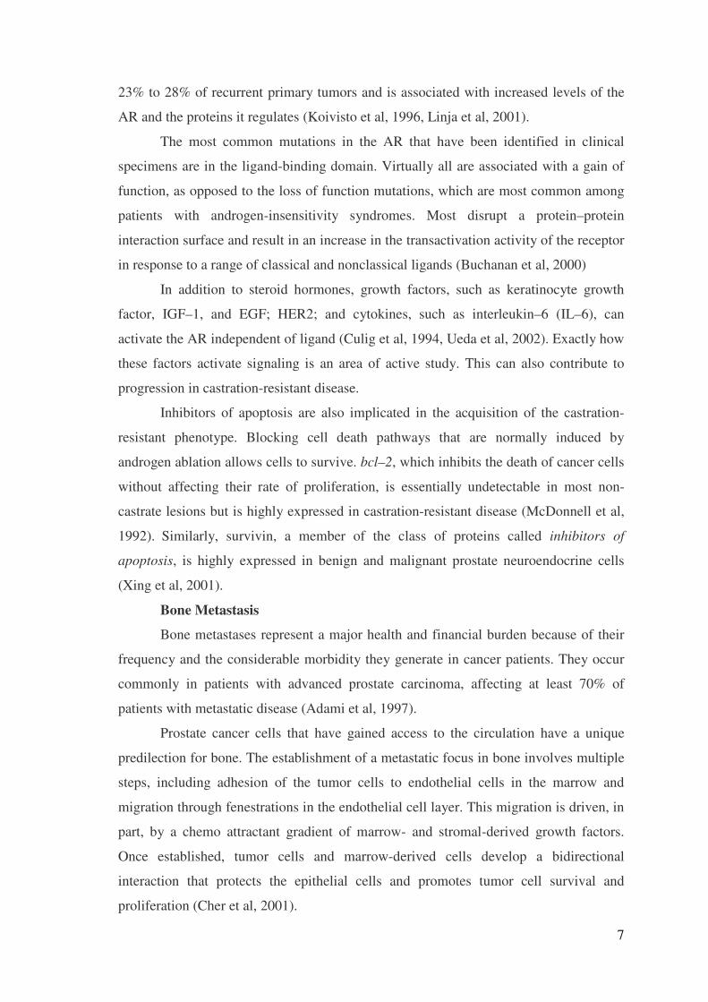
• XTT Cell Proliferation Kit, in which metabolically active cells cleave the
modified tetrazolium salt XTT to a water-soluble formazan, which may be directly
quantitated with an ELISA plate reader (Figure 1.4.)
22
• Cell Proliferation Reagent WST-1, a modified tetrazolium salt that can be
cleaved by metabolically active cells to a water-soluble formazan, which may be
directly quantitated with an ELISA plate reader
Figure 1.4. Metabolization of XTT to a water soluble formazan salt by viable cells.
(Source: Roche Applied Science®)
1.6. Methods for Studying Apoptosis in Cell Populations
Cell death can occur by either of two distinct mechanisms (Schwartzman et al,
1993, Vermes et al, 1994) necrosis or apoptosis. The two mechanisms of cell death may
briefly be defined:
Necrosis (“accidental” cell death) is the pathological process which occurs when
cells are exposed to a serious physical or chemical insult. Apoptosis (“normal” or
“programmed” cell death) is the physiological process by which unwanted or useless
cells are eliminated during development and other normal biological processes.
There are many observable morphological and biochemical differences (Table
1.1.) between necrosis and apoptosis (Vermes et al, 1994). Necrosis occurs when cells
are exposed to extreme variance from physiological conditions (e.g., hypothermia,
hypoxia) which may result in damage to the plasma membrane. Under physiological
conditions direct damage to the plasma membrane is evoked by agents like complement
and lytic viruses.

23
Table 1.1. Differential features and significance of necrosis and apoptosis.
Necrosis Apoptosis
Loss of membrane integrity Membrane blebbing, but no loss of integrity
Begins with swelling of cytoplasm and
mitochondria
Begins with shrinking of cytoplasm and condensation of nucleus
Ends with total cell lysis Ends with fragmentation of cell into smaller bodies
No vesicle formation, complete lysis Formation of membrane bound vesicles
Random digestion of DNA (smear of DNA after
agarose gel electrophoresis)
Non-random mono- and oligonucleosomal length
fragmentation of DNA (Ladder pattern after
agarose gel electrophoresis)
Postlytic DNA fragmentation Prelytic DNA fragmentation
Affects groups of contiguous cells Affects individual cells
Phagocytosis by macrophages Phagocytosis by adjacent cells or macrophages
Significant inflammatory response No inflammatory response
Loss of regulation of ion homeostasis Activation of caspase cascade
Necrosis begins with an impairment of the cell’s ability to maintain homeostasis,
leading to an influx of water and extracellular ions. Intracellular organelles, most
notably the mitochondria, and the entire cell swell and rupture (cell lysis). Due to the
ultimate breakdown of the plasma membrane, the cytoplasmic contents including
lysosomal enzymes are released into the extracellular fluid. Therefore, in vivo, necrotic
cell death is often associated with extensive tissue damage resulting in an intense
inflammatory response (Van Furth et al, 1988).
Apoptosis, in contrast, is a mode of cell death that occurs under normal
physiological conditions and the cell is an active participant in its own demise (“cellular
suicide”). It is most often found during normal cell turnover and tissue homeostasis,
embryogenesis, induction and maintenance of immune tolerance, development of the
nervous system and endocrine-dependent tissue atrophy.
Cells undergoing apoptosis show characteristic morphological and biochemical
features (Cohen et al, 1993). These features include chromatin aggregation, nuclear and
cytoplasmic condensation, partition of cytoplasm and nucleus into membrane bound-
vesicles (apoptotic bodies) which contain ribosomes, morphologically intact
mitochondria and nuclear material. In vivo, these apoptotic bodies are rapidly
recognized and phagocytized by either macrophages or adjacent epithelial cells (Savill
et al, 1989). Due to this efficient mechanism for the removal of apoptotic cells in vivo

24
no inflammatory response is elicited. In vitro, the apoptotic bodies as well as the
remaining cell fragments ultimately swell and finally lyse.
Scientists now recognize that most, if not all, physiological cell death occurs by
apoptosis, and that alteration of apoptosis may result in a variety of malignant disorders.
Consequently, in the last few years, interest in apoptosis has increased greatly. Great
progress has been made in the understanding of the basic mechanisms of apoptosis and
the gene products involved. Key elements of the apoptotic pathway include:
Death receptors:
Apoptosis has been found to be induced via the stimulation of several different
cell surface receptors in association with caspase activation. For example, the CD95
(APO-1, Fas) receptor ligand system is a critical mediator of several physiological and
pathophysiological processes, including homeostasis of the peripheral lymphoid
compartment and CTL mediated target cell killing. Upon cross-linking by ligand or
agonist antibody, the Fas receptor initiates a signal transduction cascade which leads to
caspase-dependent programmed cell death.
Membrane alterations:
In the early stages of apoptosis, changes occur at the cell surface and plasma
membrane. One of these plasma membrane alterations is the translocation of
phosphatidylserine (PS) from the inner side of the plasma membrane to the outer layer,
by which PS becomes exposed at the external surface of the cell.
Protease cascade:
Signals leading to the activation of a family of intracellular cysteine proteases,
the caspases, (Cysteinyl-aspartate-specific proteinases) play a pivotal role in the
initiation and execution of apoptosis induced by various stimuli. Different members of
caspases in mammalian cells have been identified. Among the best-characterized
caspases is caspase-1 or ICE (Interleukin-1-Converting Enzyme), which was originally
identified as a cysteine protease responsible for the processing of interleukin-1 (IL-1).
Mitochondrial changes:
Mitochondrial physiology is disrupted in cells undergoing either apoptosis or
necrosis. During apoptosis mitochondrial permeability is altered and apoptosis specific
protease activators are released from mitochondria. Specifically, the discontinuity of the
outer mitochondrial membrane results in the redistribution of cytochrome C to the
cytosol followed by subsequent depolarization of the inner mitochondrial membrane.
Cytochrome C (Apaf-2) release further promotes caspase activation by binding to Apaf-

25
1 and therefore activating Apaf-3 (caspase 9). AIF (apoptosis inducing factor), released
in the cytoplasm, has proteolytic activity and is by itself sufficient to induce apoptosis.
DNA fragmentation:
The biochemical hallmark of apoptosis is the fragmentation of the genomic
DNA, an irreversible event that commits the cell to die and occurs before changes in
plasma membrane permeability (prelytic DNA fragmentation). In many systems, this
DNA fragmentation has been shown to result from activation of an endogenous Ca2+
and Mg2+ - dependent nuclear endonuclease. This enzyme selectively cleaves DNA at
sites located between nucleosomal units (linker DNA) generating mono- and
oligonucleosomal DNA fragments (Figure 1.4). In contrast, the DNA of the
nucleosomes is tightly complexed with the core histones H2A, H2B, H3 and H4 and
therefore is protected from cleavage by endonuclease (Burgoyne et al, 1974, Stach et al,
1979). The DNA fragments yielded are discrete multiples of a 180 bp subunit which is
detected as a “DNA-ladder” on agarose gels after extraction and separation of the
fragmented DNA. The enrichment of mono- and oligonucleosomes in the cytoplasm of
the apoptotic cells is due to the fact that DNA degradation occurs several hours before
plasma membrane breakdown (Duke et al, 1986, Bonfoco et al, 1995).
Histone octomer of nucleosome core
Endonuclease
Histone octomerof nucleosome
Linker DNA
360
180
520
700
“ Beads on string” form ofchromotin
Mono-oligonucleosomes
DNA subjected togel electrophoresis
Distance between cuts:multiple of 180 pairs
DNA Ladder aftergel electrophoresis
Figure 1.5. The biochemistry of DNA fragmentation and the appearance of the “DNA
ladder”. (Source: Roche Applied Science®)

26
A number of methods have now been developed to study apoptosis in cell
populations. Because DNA cleavage is a hallmark for apoptosis, assays which measure
prelytic DNA fragmentation are especially attractive for the determination of apoptotic
cell death. The DNA fragments may be assayed in either of two ways:
-As “ladders” (with the 180 bp multiples as “rungs” of the ladder) derived from
populations of cells. In this assay, DNA has to be isolated and analyzed by simple
agarose gel in order to detect fragmented DNA in cells.
-By quantification of histone-complexed DNA fragments (mono- and
oligonucleosomes) with an ELISA. It is an alternative method which circumvents the
isolation and electrophoretic analysis of DNA, and is the immunological detection of
DNA fragments by an immunoassay.
1.7. Methods for Measuring Interleukin-6 in the Supernatant of Cell
Culture
Enzyme Linked Immunosorbent Assay (ELISA) combine the specificity of
antibodies with the sensitivity of simple enzyme assays, by using antibodies or antigens
coupled to an easily assayed enzyme that possesses a high turnover number. ELISA can
provide a useful measurement of antigen or antibody concentration.
One of the most useful of the immunoassays is the two antibody “sandwich”
ELISA. This assay is used to determine the antigen concentration in unknown samples.
This ELISA is fast and accurate, and if a purified antigen standard is available, the assay
can determine the absolute amount of antigen in an unknown sample. The sandwich
ELISA requires two antibodies that bind to epitopes that do not overlap on the antigen.
This can be accomplished with either two monoclonal antibodies that recognize discrete
sites or one batch of affinity-purified polyclonal antibodies.
To utilize this assay, one antibody is purified and bound to a solid phase
typically attached to the bottom of a plate well. Antigen is then added and allowed to
complex with the bound antibody. Unbound products are then removed with a wash,
and a labeled second antibody is allowed to bind to the antigen, thus completing the
“sandwich”. The assay is then quantitated by measuring the amount of labeled second
antibody bound to the matrix, through the use of a colorimetric substrate by ELISA
microplate reader (Figure 1.6). Major advantages of this technique are that the antigen

27
does not need to be purified prior to use, and that these assays are very specific.
However, one disadvantage is that not all antibodies can be used. Monoclonal antibody
combinations must be qualified as “matched pairs”, meaning that they can recognize
separate epitopes on the antigen so they do not hinder each other’s binding.
Figure 1.6. ELISA microplate reader

28
CHAPTER 2
MATERIALS AND METHODS
In this study, cell culture technique was used in order to grow prostate cancer
cells, in vitro. The term cell culture refers to a culture derived from dispersed cells taken
from original tissues, from a primary culture, or from a cell line or cell strain by
enzymatic, mechanical, or chemical disaggregation. The major advantage of cell culture
is control of the physiochemical environment (pH, temperature, osmotic pressure) also
large quantities of well-defined cells can be obtained by using this technique.
2.1 Materials
The chemicals, reagents and kits used in our experiments are shown in Table
2.1. In The human hormone-independent prostate cancer cell line PC-3 was generously
supplied by Dr Levent Türkeli Marmara University, �stanbul, Turkey. Zoledronic acid
was obtained from Novartis Pharmaceuticals (Basel, Switzerland). 1mM stock solution
of zoledronic acid was prepared by dissolving 4,016 mg dry powder zoledronic acid in
10 mL distilled water and aliquots were stored in -20º C. Because zoledronic acid acts
as a chelating agent on Ca++ in cell culture media, also we assessed the effects of the
knowing chelating agent, EDTA (1-100µM) on the growth of PC-3 cells, under
identical experimental conditions. Laboratory equipments required in this study are
shown in Table 2.2.

29
Table 2.1. List of chemicals used in experiments.
Chemicals Trademark
RPMI 1640 with L-glutamine Biological Industries
DMSO Sigma Chemical Co.
Heat inactivated fetal bovine serum Biological Industries
EDTA Sigma Chemical Co.
Tyripsin-EDTA Sigma Chemical Co.
Penicillin Biological Industries
Streptomycin Biological Industries
Trypan-Blue Sigma Chemical Co.
Zoledronic acid Novartis Pharma AG
Cell Proliferation Kit II (XTT) Roche Diagnostic GmBH
Apoptosis Cell Death Detection ELISA PLUS Kit Roche Diagnostic GmBH
Apoptotic DNA Ladder Kit Roche Diagnostic GmBH
Human IL-6 ELISA Kit Biosource Inc.
Table 2.2. List of equipments used in experiments.
Equipments Trademark
Laminar-Flow Hood NuAire,USA
CO2 Incubator NuAire,USA
Inverted Microscope Olympus,Japan
Light Microscope Olympus,Japan
Hemocytometer Neubauer
Pipettors ( 10,20,100,1000µl) Eppendorf
Plate Reader Medispec ESR 200
Culture vessels Corning
Pipetting Aid Eppendorf

30
2.2 Methods
2.2.1 Cell Culture of PC-3 Cell Line
The human hormone-independent and drug-resistant prostatic carcinoma cell-
line PC-3 was maintained in culture as adherent cells and cultured in Roswell Park
Memorial Institute 1640 (RPMI 1640, Biological Industries, Israel) plus 5% heat
inactivated fetal bovine serum (Biological Industries, Israel) added to 1% L-glutamine
(Biological Industries, Israel) 1% nonessential amino acids (Biological Industries,
Israel), 10.000 U/mL penicillin (Biological Industries, Israel) and 10 mg/mL
streptomycin (Biological Industries, Israel). All cells were grown in humidified
atmosphere at 37C° in 5% CO2. When the tumor cells were used as target cells, they
were treated with trypsin–EDTA (Sigma Chemical Co.), washed, and resuspended in
complete medium.
2.2.2 Assessment of Cytotoxicity Produced by Zoledronic Acid.
Trypan blue dye exclusion assay and XTT proliferation assay was used to
confirm the drug-mediated cytotoxicity of zoledronic acid on PC-3 cells.
2.2.2.1 Trypan Blue Dye Exclusion Assay
Trypan Blue Dye Exclusion assay was used to assess the cytotoxic effect of
zoledronic acid on PC-3 cells. This assay was also used to measure the proportion of
viable cells following a potentially traumatic procedure, such as disaggregation, cell
separation, or freezing and thawing during the cell culture. Briefly, assay involves
staining damaged cells with a dye and counting viable cells that exclude the dye. Counts
were performed manually using a hemocytometer and trypan blue (Figure 2.1).
The hemocytometer consists of two chambers, each of which is divided into nine
1.0 mm squares. A cover glass is supported 0.1 mm over these squares so that the total
volume over each square is 1.0 mm x 0.1 mm or 0.1 mm3, or 10-4 cm3. Since 1 cm3 is
approximately equivalent to 1 ml, the cell concentration per ml will be the average
count per square x 104. In order to determine the ability of zoledronic acid to inhibit the

31
growth of PC-3 cells, cells were plated at a cell density of 6x 10³ cells/well in 24-well
plates and grown with RPMI containing 5% FBS. Cells then treated with zoledronic
acid in a dose- (10, 60, and100 µM) and time-dependent manner (24, 48, 72 hr). After
incubation period, 50 µL cells were taken and mixed with an equal volume of trypan
blue [0.4% (w/v) trypan blue in PBS]. Then this suspension was loaded to the counting
chamber of hemocytometer. Using the microscope with a 10X ocular, the numbers of
unstained cells (viable cells) in the 4 outer squares were counted. Then the numbers of
viable cells per mL were calculated. Counting was repeated in order to check
reproducibility. The cell concentration was calculated as follows:
Cell concentration per milliliter = average count per square x 104
Figure 2.1. Measurement of cytotoxicity by counting the cells with a hemocytometer.
(Source: Roche Applied Science®)
2.2.2.2 XTT Proliferation Assay
The effect of zoledronic acid (1-100µM) on PC-3 cell proliferation was
investigated using a commercially available XTT proliferation kit (Roche Applied
Science, Penzberg, Germany). The assay is based on the cleavage of the yellow
tetrazolium salt XTT to form an orange dye by metabolic active cells (Gerlier et al,
1986). While living (metabolically active) cells reduce tetrazolium salt to colored

32
formazan compound; dead cells do not. The formazan dye formed is soluble in aqueous
solutions and is directly quantified using an ELISA microplate reader.
Briefly, the cells were plated in 96-well culture plates (Corning Glass Works,
Corning, N.Y, USA) at a density of 1000 cells/well in 200-µl RPMI culture medium
after verifying cell viability by trypan blue (Sigma Chemical Co.) dye exclusion test.
After incubation with zoledronic acid for 48 and 72 hr, 50 µl of XTT labeling mixture
(sodium3’-[1-(phenyl-aminocarbonyl)-3,4-tetrazolium]-bis(4-methoxy-6-nitro) benzene
sulfonic acid hydrate and N-methyl dibenzopyrazine methyl sulfate; mixed in
proportion 50:1) was added to the each well. Then cells were incubated with this
mixture for 4 hr in a humidified atmosphere. The optical density was measured at 450
nm with a reference wavelength at 650 nm in a Medispec ESR 200 microplate reader.
The percentage of cytotoxicity was calculated as follows:
% Cytotoxicity = 1- (A of experimental well/ A of positive control well) x100
Figure 2.2. Measurement of metabolic activity using the tetrazolium salt XTT
(Source: Roche Applied Science®)
+ XTT
ELISA Reader

33
2.2.3 Evaluation of Apoptosis Induced by Zoledronic Acid
Mono-oligo nucleosome detection-based ELISA and DNA fragmentation on
simple agarose gel electrophoresis assays were used to evaluate the apoptosis.
2.2.3.1 Mono-Oligo Nucleosome Detection-based ELISA
The commercially available Cell Death Detection ELISA kit (Roche Applied
Science, Penzberg, Germany) was used to detect DNA fragmentation according to
manufacturer’s instructions (Aragene et al, 1998). This nonradioactive immunoassay
has been designed for relative quantification of DNA fragmentation in cells and it
measures the enrichment of histone-complexed DNA fragments (mono- and
oligonucleosomes) in the cytoplasm of apoptotic cells.
Briefly, cytoplasmic lysates from control and zoledronic acid treated cells were
transferred to a streptavidin-coated plate that was supplied with the kit. They were
incubated for 2 hr in the presence of the immune reagent containing the antibodies
against the histone proteins and DNA fragments (Anti-histone-biotin and Anti-DNA-
POD). During the incubation period, the Anti-histone antibody bound to the histone-
component of the nucleosomes and simultaneously captured the immunocomplex to the
streptavidin-coated plate via its biotinylation. Additionally, the Anti-DNA-POD
antibody reacted with the DNA component of the nucleosomes. After the removal of
unbound components by a washing step, quantitative determination of the amount of
nucleosomes was measured by the POD retained in the immunocomplex. POD is
determined photometrically with ABTS as substrate, which then was read for optical
density at 405 nm with a reference wavelength at 490 nm (Figure 2.3.). The enrichment
of histones and DNA fragments in cell lysates were evaluated as indicator of apoptosis.
The degree of apoptosis was displayed as percentage according to positive control cells.

34
Figure 2.3. How the Cell Death Detection ELISAPLUS works. (Sample preparation and
ELISA) (Source: Roche Applied Science®)
2.2.3.2 DNA Fragmentation on Simple Agarose Gel
The Apoptotic DNA Ladder Kit (Roche Diagnostic, Mannheim, Germany) was
used to detect the typical DNA ladder formation in zoledronic acid treated cells, which
is the hallmark of apoptotic cells. DNA fragments reveal, upon agarose gel
After incubating cells with an apoptosis-inducing agent; Zoledronic acid, pellet the cells by centrifugation. Discard the supernatant, which may contain necrotic DNA that leaked through the membrane during the incubation.
Incubate cells with lysis buffer.
Pellet the intact nuclei by centrifugation. Take an aliquot of the supernatant (cell lysate) and determine the amount of apoptotic nucleosomes present.
Streptavidin coated well
Anti-histone biotine
Sample containing nucleosomes
Anti- DNA Peroxidase
ABTS Substrate

35
electrophoresis, a distinctive ladder pattern consisting of multiples of an approximately
180 bp subunit.
Firstly PC-3 cells were plated at a cell density of 2x 10⁶ cells/well in 24-well
plates and grown with RPMI containing 5% FBS and then treated with different
concentration of zoledronic acid (1,10,60 and 90 µM) for 48 and 72 hr. After lysis of
cultured cells in binding buffer, the cell lysates is applied to a filter tube with glass fiber
fleece and passaged through the glass fiber by centrifugation. During this procedure
nucleic acids bind specifically to the surface of glass fibers in the presence of chaotropic
salts (Vogelstein et al, 1979). Residual impurities were removed by a wash step and
subsequently DNA was eluted in elution buffer. The DNA is stable and can be used
directly or stored at -20º C for later analysis.
For gel electrophoresis, DNA samples and positive control (apoptotic U937
cells) were run on 1% agarose gel stained with ethidium bromide. The voltage was set
to 75V for 2 hours. DNA ladders indicative of nucleosomal DNA fragmentation became
apparent after 2 hours. DNA ladders were visualized by placing gel onto an UV light
source and photographed.
2.2.4 Detection of IL–6 Levels by ELISA
The Human IL-6 ELISA Kit (Biosource Inc., California, USA) was used for the
quantititative determination of IL-6 in the supernatant from zoledronic acid treated PC-3
cells. Hu IL-6 kit is a solid phase sandwich ELISA. A monoclonal antibody specific for
Hu IL-6 has been coated onto the wells of the microtiter strips provided. Samples,
including standards of known Hu IL-6 content, control specimens, and unknowns, are
pipetted into these wells, followed by the addition of a biotinylated monoclonal second
antibody. During the first incubation, the Hu IL-6 antigen binds simultaneously to the
immobilized antibody on one site, and to the solution phase biotinylated antibody on a
second site. After removal of excess second antibody, Streptavidin- Peroxidase enzyme
is added. This binds to the biotinylated antibody to complete the four-member
sandwich. After a second incubation and washing to remove the entire unbound
enzyme, a substrate solution is added, which is acted upon by the bound enzyme to
produce color. The intensity of this colored product is directly proportional to the
concentration of Hu IL-6 present in our sample.

36
Briefly, PC-3 cells were cultured for 24,48,72 hr at a concentration of 1x10⁵
cells/ml of RPMI medium supplemented with 5% FBS. Then cells were incubated in the
same medium in the absence or presence of zoledronic acid (1, 10, 60, 90,100µM).
Supernatants were collected for all culture conditions and stored at -20°C until the day
of the assay. Before thawing the supernatants, Hu IL-6 standards was prepared by
making serial dilutions of 500, 250, 125, 62.5, 31.2, 15.6 and 7.8 pg/mL Hu IL-6. 100
µL of supernatants and standards were added to the microplate wells. Then 50 µL of
biotinylated anti-IL-6 solution was added into each well and plate was incubated for 2
hours at RT. After the washing, 100 µL Streptavidin-HRP working solution was added
to each well and plate was incubated with this solution 30 minutes at RT. After washing
thoroughly, 100 µL of Stabilized Chromogen was added to each well. The liquids in the
wells began to turn blue. Plate was incubated for 30 minutes at room temperature and in
the dark. By adding 100 µL of Stop Solution to each well process is completed and the
solutions in the wells changed from blue to yellow (Figure 2.4.). The absorbance was
measured at 450nm with ELISA microplate reader. The absorbance of the standards
against the standard concentration was plotted on a graph paper and the best smooth
curve trough these points was drawn to construct the standard curve. The Hu IL-6
concentrations for unknown samples were read from the standard curve.

37
Figure 2.4. Human IL-6 assay summary

38
2.3. Statistical Analysis
All assays were set up in triplicate and the results were expressed as the
mean+SD. Analysis of variance (one-way ANOVA) statistical methodology was
performed to elucidate the mean effect of ZA treatment on IL-6 levels.

39
CHAPTER 3
RESULTS AND DISCUSSIONS
3.1. Results
Two different methods were used to confirme the cytotoxic effect of zoledronic
acid on PC-3 cells. First, trypan blue dye exclusion assay was used to determine the
inhibition of proliferation of PC-3 cells by zoledronic acid. After treatment of various
concentration of zoledronic acid, cells were stained with trypan blue and then counted
with hemocytometer. The addition of trypan blue helps to distinguish viable, unstained
cells from non-viable, blue-stained cells (Figure 3.1.). The number of viable cells was
calculated as previously described. Our data proved that the cytotoxic effect of
zoledronic acid was dose and time dependent in PC-3 cells (Figure 3.2.).
Figure 3.1. Trypan blue dye exculision assay (VC: Viable cell, DC: Dead cell)
���
���

40
In concert with trypan blue exclusion assay, XTT proliferation assay have
confirmed the ability of zoledronic acid to inhibit the growth of PC-3 cells in a dose and
time dependent manner (48 and 72 hr). Identical experiments with increasing doses of
zoledronic acid produced an inhibition of the growth of PC-3 cells, at drug
concentrations as low as 10 µM (48 and 72 hr) (Figure 3.3.).
Figure 3.2. Cell counts of viable PC-3 cells as measured by trypan blue exclusion
assay with zoledronic acid (100 µM for 24, 48 and 72 hr)

41
Figure 3.3. Dose–dependent cytotoxic activities of zoledronic acid against PC-3 cells
(48 and 72 hrs)
We tested the apoptotic effect of zoledronic acid on PC3- cells by using two
different apoptosis measurement assay. It was shown that zoledronic acid induces
apoptosis and this effect was time and dose dependent manner as displayed in Figure
3.3. and 3.4.
First, the apoptotic effect of zoledronic acid was measured by Cell Death
Detection ELISA kit (Roche Applied Science, Penzberg, Germany), and displayed as
the percentage values based on positive control data. Apoptotic effect was dependent on
time and dose exposure (1, 10, 60, 90, 100 µM) in PC-3 cells (Figure 3.4.).

42
Figure 3.4. Evidence of the apoptotic effect of zoledronic acid (48 hrs).
Then apoptotic effect of zoledronic acid was assessed by Apoptotic DNA Ladder
Kit (Roche Applied Science, Penzberg, Germany). The cells were cultured for 48 and
72 hr with various concentrations of zoledronic acid (1, 10, 60, 90, 100 µM).
Fragmented DNA extracted from the cells was analyzed by agarose gel electrophoresis.
Zoledronic acid (90 and100 µM for 72 hr) produced the classical DNA ladders on
agarose gel characteristic of PC-3 cells undergoing apoptosis (Figure 3.4.).
Figure 3.5. DNA fragmentation analysis of zoledronic acid treated PC-3 cells using
simple agarose gel electrophoresis.

43
IL-6 levels produced in vitro by PC-3 cells were determined using The Human
IL-6 ELISA Kit (Biosource Inc., California, USA). PC-3 cells produced 24.04 ± 2.6
pg/ml IL-6 (72 hr), under normal culture conditions in the absence of zoledronic acid.
PC-3 cells were incubated with various concentrations of zoledronic acid (1, 10,30 , 60,
90, 100 µM ) for 24,48 and 72 hrs. Then IL-6 levels were measured in supernatants of
cell culture. IL-6 concentrations secreted by PC3 cells in the treatment of various
zoledronic acid concentrations were lowered from 24 ± 0.4 pg/ml to 7 ± 0.8 pg/ml
(Table 3.1.).
Table 3.1. IL-6 Levels in supernatants of PC-3 cells.
Time Point
Control (pg/ 105
viable cell)
Zoldronic Acid 1µM (pg/ 105
viable cell)
ZA 10µM
(pg/105)
ZA 60 µM (pg/105)
Z A 90µM
(pg/ 105)
ZA 100µM
(pg/ 105 )
24 hrs 10.10±0.84
3.57±1.04
2.31±0.40
2.33±1.17
1.93 ±1.08 3.45 ± 1.09
48 hrs 20.01± 0.33
11.12 ±0.31
9.16±0.41
10.44±0.61 10.72±1.53 8.9 ±0.94
72 hrs 24.04±2.60
10.99±0.62
8.36±1.10
8.16±0.80 7.76±0.41 7.24±0.85
There was significant decrease in IL-6 secretion in the 1-100 �M concentrations
in PC-3 cells (Table 3.1.). The decrease in IL-6 levels in the presence of zoledronic acid
concentrations comparing to control group was statistically significant (p< 0.05).
However there was no significant difference between 1 µM and 100 µM zoledronic acid
concentration except in 72 hours exposure. There was slide difference between 1 µM
and 100 µM in 72 hours.
IL-6 levels were decreased by various zoledronic acid concentrations (1, 10, 60,
90, 100 µM) in time and dose dependent manner in PC-3 cell lines. The displayed data
was average values from 24, 48 and 72 hr exposure time points (Figure 3.6.).

44
Figure 3.6. Time and dose dependent decrease of IL-6 levels in the supernatants of
PC-3 cells. (24, 48 and 72 hrs)

45
These data suggest that zoledronic acid inhibits the expression and secretion of
IL-6 normally produced in high levels in PC-3 cell lines. These data imply that anti
tumor effects of zoledronic acid could be also mediated by interfering IL-6 signaling
pathways in prostate cells.
3.2 Discussions
In the literature the first data on cytotoxic effect of zoledronic acid was produced
by Aparicio A et al in myeloma cell lines (Aparici et al, 1998). Then several studies
(Lee et al, 2001, Witters et al, 2002; Fromigue et al, 2000; Mundy et al, 2001, Jagdev et
al, 2000, Jagdev et al, 2001, Derenne et al, 1999, Tassone et al, 2000, Corey et al, 2003,
Lee et al, 2002) have suggested direct antiproliferative and pro-apoptotic effects of the
bisphosphonates on breast, prostate, and myeloma cancer cells.
Lee et al studied the effects of pamidronate and zoledronic acid on prostate
cancer cell growth in vitro. Three separate prostate cancer cell lines were seeded in 96-
well plates and incubated in serum-free media with or without the bisphosphonates at
various concentrations for up to 72 hours. The numbers of cells remaining were
determined indirectly by measuring the absorbance of crystal violet dye stain in viable
cells. In all cell lines, treatment with 100-�mol/L pamidronate induced significant cell
death compared with untreated cells, whereas lower concentrations (25 and 50 �mol/L)
caused cell stasis. In contrast, zoledronic acid 100 �mol/L did not cause cell death but
caused cell cytostasis, even at concentrations as low as 10 �mol/L. In the presence of
serum, both bisphosphonates inhibited prostate cancer cell growth, but only zoledronic
acid was effective at lower concentrations (25–50 �mol/L). In a separate in vitro study
of zoledronic acid with a cyclooxygenase inhibitor in the DU-145 human prostate
carcinoma cell line (Witters et al, 2002) the effects on cell growth were determined
using the 3,[4,5-dimethylthiazol-2-41]-2,5-diphenyltetrazolium bromide (MTT) dye
assay. Cell exposure to zoledronic acid (1–10 �mol/L) resulted in a dose-dependent
growth inhibition (8%–70%). The combination gave an additive inhibitory effect on cell
number compared with zoledronic acid treatment (Witters et al, 2002). Studies have also
shown that the combination of zoledronic acid plus docetaxel resulted in additive
apoptotic effects on PC–3 and DU–145 prostate cancer cells (Wei et al, 1989).
Two in vitro studies (Corey et al, 2003, Lee et al, 2002) have further examined
the exposed 2 prostate cancer cell lines—LnCaP and PC-3—to zoledronic acid to

46
determine its effects on apoptosis and cell cycle progression. Compared with unexposed
cells, the proportion of apoptotic cells among the PC-3 cells was increased after 1 day
of treatment, but there was no significant difference among the LnCaP cells.The
proliferation of PC-3 cells was inhibited by either continuous or pulsatile exposures of
zoledronic acid in a dose-dependent manner (Tenta et al, 2004). Tenta et al also showed
that zoledronic acid induces apoptosis in PC-3 prostate cancer cells (Tenta et al, 2005).
In a recent study of the effects of several bisphosphonates on PC–3 prostate
cancer cells, zoledronic acid markedly inhibited cell growth through a combination of
cytostatic and apoptotic effects (Dumon et al, 2004). Notably, zoledronic acid was the
most potent bisphosphonate, exerting profound antiproliferative effects more rapidly
than either pamidronate or ibandronate.
The known effects of zoledronic acid were confirmed in our experimental
conditions. Our data also proved that the cytotoxic effect of zoledronic acid was dose-
and time- dependent in PC-3 cell line as displayed in several studies (Figure 3.3.). We
also tested the apoptotic effect of zoledronic acid on PC-3 cell lines by using two
different apoptosis measurement assay. It was shown that zoledronic acid induces
apoptosis and this effect was also time and dose dependent manner (Figure 3.4. and
3.5.). In summary we found that zoledronic acid have two main effects on PC-3 cell
lines. The cytotoxic and apoptotic effect of zoledronic acid was dependent on time and
concentration. The second finding was that the expression of IL-6 was downregulated in
time and dose dependent manner by zoledronic acid.
IL-6 is produced by lymphoid, non-lymphoid cells, and cancer cells (Kishimoto
et al, 1989). IL-6 is a multifunctional cytokine that plays an important role in the
regulation of hematopoiesis, immune response, inflammation, bone metabolism, and
neural development (Kishimoto et al, 1992). It has been demonstrated that normal
prostate epithelial cells, cells derived from benign prostatic hyperplasia and primary
prostate tumors secrete IL-6 in cell culture (Twillie et al, 1995). IL-6 can increase the proliferation of prostate cancer cells (McCarty et al, 2004)
and treatment of cultures of androgen insensitive prostatic cancer cell lines PC-3 and
DU145 with anti-IL-6 antibodies decreases growth rates of both cell lines (Borsellino et
al, 1995). In contrast, androgen sensitive cell line LNCaP showed some divergence in
reported effects of IL-6. Both stimulation and inhibition of proliferation being reported
in this cell line (Okamoto et al, 1997; Levesque et al, 1998).

47
It was shown that bisphosphonates induce both myeloma cell and bone marrow
stromal cells (BMSCs) apoptosis. Furthermore, at lower concentrations, they induce a
significant inhibition (40% and 60%, respectively) of the constitutive production of
interleukin-6 (IL-6) by BMSCs. In conclusion, the apoptosis of myeloma cells and
BMSCs and the inhibition of IL-6 production induced by bisphosphonates, mainly
zoledronate, could have antitumoral effects in patients with MM (Derenne et al, 1999).
Interleukin-6 (IL-6) is the major growth and survival factor for multiple
myeloma (MM), and has been shown to protect MM cells from apoptosis induced by a
variety of agents (Tassone et al, 2002). IL-6 receptor antagonists, which prevent the
assembly of functional IL-6 receptor complexes, inhibit cell proliferation and induce
apoptosis in MM cells (Tassone et al, 2002).
It was shown that the IL-6 receptor super-antagonist Sant7 might enhance the
antiproliferative and apoptotic effects induced by the combination of dexamethasone
and zoledronic acid on human multiple myeloma (MM) cell lines. Sant7 significantly
enhanced growth inhibition and apoptosis induced by dexamethasone and zoledronic
acid on both MM cell lines. These results indicate that overcoming IL-6 mediated cell
resistance by Sant7 potentiates the effect of bisphosphonates on MM cell growth and
survival, providing a rationale for therapies including IL-6 antagonists in MM. (Tassone
et al, 2002). Furthermore, exogenous administration of interleukin 6 (IL-6) inhibited the
doxorubicin-induced apoptosis of PC-3 cells (Tenta et al, 2004).
In vitro studies have demonstrated the antitumor potential of zoledronic acid on
myeloma cell lines, but few data are available on its effects on bone marrow stromal
cells (BMSCs). In vitro exposure to the lowest concentrations of zoledronic acid
decreased IL-6 production by on bone marrow stromal cells (BMSCs) (Corso et al,
2005).
In our study, we confirmed that zoledronic acid also inhibited the expression of
IL-6 significantly in PC-3 prostate cell lines. There is no data on the decreasing of IL-6
levels induced by zoledronic acid in PC-3 cell lines in current literature. In addition we
also found that decreasing of IL-6 expression by various zoledronic acid concentrations
was not dose dependent. The data on the 1 µM and 100 µM zoledronic acid
concentrations caused the more or less the same amount of decrease in IL-6 level. This
was so significant that the 1 µM which is also achieved serum level in prostate patients
switch off IL-6 secretion in significant level. This might be directly inactivation of IL-6

48
nuclear transcription factor by zoledronic acid. This new data implies new hypothesis
that zoledronic acid may exert its antitumor effects via interfering IL-6 expression.
We speculate the effects of zoledronic acid on PC-3 cell lines might be mediated
by interfering IL-6 signaling pathways. IL-6 has different effects on prostate cancer cell
lines. The bases of these differences can be originated from multistep and alternative
signal flow pathways used by IL-6 in different cells. For instance, there are two forms
(membrane bound and soluble) of IL-6 receptor regulated tightly. Signal transducer gp-
130 in membrane is common substrate for several growth factors. Therefore its
activation is not only dependent on IL-6.
Moreover activated gp130 either simultaneously or preferentially triggers three
intracellular pathways by altering its intracellular domain. IL-6 signaling was mediated
by JAK-STAT, ras-raf-MAPK and PI3K-Akt signaling. It is suggested that, one or two
alternative pathways are preferentially more active in different cell lines. IL-6 is also
capable of up and down regulation by autocrine or paracrine effect and feed-back (Klein
et al, 1989, Miki et al, 1989, Yee et al, 1989, Okamoto et al, 1997, Chung et al, 1999).
The expression of IL-6 is regulated by several transcription factors such as AP-1,
NFKapaB, CREB and c/EBP. It is considered that intracellular signaling pathways of
IL-6 also regulate these transcription factors. Therefore mentioned IL-6 signaling
pathways might be affected by zoledronic acid. The possible interacting proteins with
zoledronic acid might be either the structural or regulatory component of the following
signaling pathways. These are Ras-Raf-MAPK cascade involving cell proliferation,
tumor suppressor and apoptotic pathways, cell cycle proteins (cyclins), posttranslational
mechanisms of intracellular signaling proteins. Moreover, there may be not only one
substrate for zoledronic acid; it may have multiple interacting substrates within the cell.
At present, it is not certain which pathways in the upstream or downstream signal flow
are disrupted by zoledronic acid. However, it was shown that zoledronic acid interferes
with isoprenylation of intracellular small G proteins such as ras (Caraglia et al, 2004)
by inhibiting the farnesyl pyrophosphate (FPP) synthase enzyme (Rogers et al, 2000),
which is required for producing FPP and subsequently geranylgeranyl pyrophosphate
(GGPP).
It is also shown that zoledronic acid induced apoptosis by either caspase 3 or
caspase 9 (Oades et al, 2003). Although the apoptotic effect of zoledronic acid was
diminished by co-treating with a broad spectrum caspase inhibitor, Z-VAD-FMK
(Coxon et al, 2004), it is not clear whether zoledronic acid binds to one of caspases.

49
On the other hand the apoptotic effect of zoledronic acid may not be the
premature action of zoledronic acid, since it is possible that zoledronic acid first inhibits
signal transduction in the upstream regulation of apoptotic switch mechanism. This
possibility may explain the apoptotic effect and decrease of IL-6 in PC-3 cells which
was shown in our study. The observed apoptosis in PC-3 cells and decreases in IL-6
expression seem to be paralel events. But these two effects of zoledronic acid might be
either independent or associated. However it is uncertain whether the apoptotic pathway
was triggered by zoledronic acid mediated-decrease of IL-6 level. It is also unknown
whether the decrease in IL-6 expression is caused by zoledronic acid induced apoptosis.
There is also another hypothesis that if these two events are independently controlled in
prostate cancer cells, then it might be interpreted that zoledronic acid can exert its effect
by inhibiting IL-6 autocrine effect mechanism. Therefore, there is a need to carry out
further studies in order to define the new zoledronic acid substrates mediating apoptotic
effects.
Moreover it is also not certain that apoptotic pathway in these cell lines and IL-6
down regulation pathway are independently active or related each other. As displayed in
several studies including our’s, zoledronic acid switch on apoptotic pathway, therefore
one of the outcome of apoptosis might lead to decreasing in expression of IL-6. But it
has to be investigated further.
This study indicates that the molecular target of zoledronic acid is likely to be
one of the molecules involving in IL-6 expression or signaling pathway. If it is true, it
could be possible to develop new treatment strategies for prostate cancer. It suggests
that the inhibition of IL-6 may be a useful adjunct to prostate cancer therapy regarding
to in vitro studies. It could be speculated that a therapeutic intervention that interferes
with IL-6 signaling may be justified both at early and late stages of prostate
carcinogenesis and could decrease morbidity in patients with advanced disease. On the
other hand, the importance of IL-6 for androgen independent growth of tumor cells
makes it a good candidate for targeted therapy in human carcinoma of the prostate.
50
CHAPTER 5
CONCLUSIONS
Zoledronic acid is a potent, nitrogen-containing bisphosphonate that inhibits
bone resorption by reducing osteoclast proliferation and inducing osteoclast apoptosis.
Zoledronic acid exerts direct antitumor effects by inducing apoptosis, inhibiting cell
adhesion molecules, preventing prostate and breast cancer cell adherence to bone,
reducing IL-6 secretion from myeloma stromal cells, and affecting angiogenesis.
However, zoledronic acid’s mechanism of action is not clearly understood. Also, its
molecular substrates in PC-3 cell lines are not known.
In this study we have showed that zoledronic acid reduces IL-6 expression in
significant level in prostate cancer cell line PC-3. The ability of IL-6 to activate
signaling through several distinct pathways may exert differential effects on various cell
lines. IL-6 is using three intracellular pathways to transducing IL-6 signaling by binding
gp130 then activating signal transducer and activator of transcription (STAT)-3,
mitogen activated protein kinases (MAPK), and phosphatidylinositol 3-kinase (PI3K)
signaling pathways, each depends upon different regions of gp130.
Additional research is required to elucidate further the action of IL-6 and its role
in the pathogenesis of advanced prostate cancer at the cellular and molecular levels. At
present, it is not certain which pathways in the upstream or downstream signal flow are
disrupted by zoledronic acid. New therapeutic strategies using anti-IL-6 monoclonal
antibodies seem warranted in such patients for which conventional treatment holds little
and may afford improved quality of life and survival benefit.
These original data on the decreasing of IL-6 secretion by zoledronic acid in PC-
3 cells suggest that targeting IL-6 in future may have multiple benefits in prostate
cancer patients in terms of developing alternative combined threapies in hormone
refractory prostate cancer patients.

51
REFERENCES
Adami, S., 1997."Bisphosphonates in prostate carcinoma," Cancer, Vol. 80, No. 8
Suppl, pp. 1674-1679. Adler, H. L., McCurdy, M. A., Kattan, M. W., Timme, T. L., Scardino, P. T., and
Thompson, T. C., 1999. "Elevated levels of circulating interleukin-6 and transforming growth factor-beta1 in patients with metastatic prostatic carcinoma," J.Urol., Vol. 161, No. 1, pp. 182-187.
Akira, S., Isshiki, H., Sugita, T., Tanabe, O., Kinoshita, S., Nishio, Y., Nakajima, T.,
Hirano, T., and Kishimoto, T., 1990. "A nuclear factor for IL-6 expression (NF-IL6) is a member of a C/EBP family," EMBO J., Vol. 9, No. 6, pp. 1897-1906.
Aparicio, A., Gardner, A., Tu, Y., Savage, A., Berenson, J., and Lichtenstein, A., 1998.
"In vitro cytoreductive effects on multiple myeloma cells induced by bisphosphonates," Leukemia, Vol. 12, No. 2, pp. 220-229.
Aragane, Y., Kulms, D., Metze, D., Wilkes, G., Poppelmann, B., Luger, T. A., and
Schwarz, T., 1998. "Ultraviolet light induces apoptosis via direct activation of CD95 (Fas/APO-1) independently of its ligand CD95L," J.Cell Biol., Vol. 140, No. 1, pp. 171-182.
Arnold, J. T. and Isaacs, J. T., 2002. "Mechanisms involved in the progression of
androgen-independent prostate cancers: it is not only the cancer cell's fault," Endocr.Relat Cancer, Vol. 9, No. 1, pp. 61-73.
Avcu, F., Ural, A. U., Yilmaz, M. I., Ozcan, A., Ide, T., Kurt, B., and Yalcin, A., 2005.
"The bisphosphonate zoledronic acid inhibits the development of plasmacytoma induced in BALB/c mice by intraperitoneal injection of pristane," Eur.J.Haematol., Vol. 74, No. 6, pp. 496-500.
Ayala, G., Thompson, T., Yang, G., Frolov, A., Li, R., Scardino, P., Ohori, M.,
Wheeler, T., and Harper, W., 2004. "High levels of phosphorylated form of Akt-1 in prostate cancer and non-neoplastic prostate tissues are strong predictors of biochemical recurrence," Clin.Cancer Res., Vol. 10, No. 19, pp. 6572-6578.
Berenson, J. R., 2001. "New advances in the biology and treatment of myeloma bone
disease," Semin.Hematol., Vol. 38, No. 2 Suppl 3, pp. 15-20. Berger, A. P., Kofler, K., Bektic, J., Rogatsch, H., Steiner, H., Bartsch, G., and Klocker,
H., 2003. "Increased growth factor production in a human prostatic stromal cell culture model caused by hypoxia," Prostate, Vol. 57, No. 1, pp. 57-65.
Berger, A. P., Kofler, K., Bektic, J., Rogatsch, H., Steiner, H., Bartsch, G., and Klocker,
H., 2003. "Increased growth factor production in a human prostatic stromal cell culture model caused by hypoxia," Prostate, Vol. 57, No. 1, pp. 57-65.

52
Berke, G., 1991."Debate: the mechanism of lymphocyte-mediated killing. Lymphocyte-triggered internal target disintegration," Immunol.Today, Vol. 12, No. 11, pp. 396-399.
Berridge, M. V. and Tan, A. S., 1993. "Characterization of the cellular reduction of 3-
(4,5-dimethylthiazol-2-yl)-2,5-diphenyltetrazolium bromide (MTT): subcellular localization, substrate dependence, and involvement of mitochondrial electron transport in MTT reduction," Arch.Biochem.Biophys., Vol. 303, No. 2, pp. 474-482.
Body, J. J., Bartl, R., Burckhardt, P., Delmas, P. D., Diel, I. J., Fleisch, H., Kanis, J. A.,
Kyle, R. A., Mundy, G. R., Paterson, A. H., and Rubens, R. D., 1998. "Current use of bisphosphonates in oncology. International Bone and Cancer Study Group," J.Clin.Oncol., Vol. 16, No. 12, pp. 3890-3899.
Bonfoco, E., Krainc, D., Ankarcrona, M., Nicotera, P., and Lipton, S. A., 1995.
"Apoptosis and necrosis: two distinct events induced, respectively, by mild and intense insults with N-methyl-D-aspartate or nitric oxide/superoxide in cortical cell cultures," Proc.Natl.Acad.Sci.U.S.A, Vol. 92, No. 16, pp. 7162-7166.
Borsellino, N., Belldegrun, A., and Bonavida, B., 1995. "Endogenous interleukin 6 is a
resistance factor for cis-diamminedichloroplatinum and etoposide-mediated cytotoxicity of human prostate carcinoma cell lines," Cancer Res., Vol. 55, No. 20, pp. 4633-4639.
Borsellino, N., Bonavida, B., Ciliberto, G., Toniatti, C., Travali, S., and D'Alessandro,
N., 1999. "Blocking signaling through the Gp130 receptor chain by interleukin-6 and oncostatin M inhibits PC-3 cell growth and sensitizes the tumor cells to etoposide and cisplatin-mediated cytotoxicity," Cancer, Vol. 85, No. 1, pp. 134-144.
Boulton, T. G., Stahl, N., and Yancopoulos, G. D., 1994. "Ciliary neurotrophic
factor/leukemia inhibitory factor/interleukin 6/oncostatin M family of cytokines induces tyrosine phosphorylation of a common set of proteins overlapping those induced by other cytokines and growth factors," J.Biol.Chem., Vol. 269, No. 15, pp. 11648-11655.
Brubaker, K. D., Brown, L. G., Vessella, R. L., and Corey, E., 2006. "Administration of
zoledronic acid enhances the effects of docetaxel on growth of prostate cancer in the bone environment," BMC.Cancer, Vol. 6, pp. 15.
Bruchovsky, N., Rennie, P. S., Coldman, A. J., Goldenberg, S. L., To, M., and Lawson,
D., 1990. "Effects of androgen withdrawal on the stem cell composition of the Shionogi carcinoma," Cancer Res., Vol. 50, No. 8, pp. 2275-2282.
Bubendorf, L., Kononen, J., Koivisto, P., Schraml, P., Moch, H., Gasser, T. C., Willi,
N., Mihatsch, M. J., Sauter, G., and Kallioniemi, O. P., 1999. "Survey of gene amplifications during prostate cancer progression by high-throughout fluorescence in situ hybridization on tissue microarrays," Cancer Res., Vol. 59, No. 4, pp. 803-806.

53
Buchanan, G., Irvine, R. A., Coetzee, G. A., and Tilley, W. D., 2001. "Contribution of the androgen receptor to prostate cancer predisposition and progression," Cancer Metastasis Rev., Vol. 20, No. 3-4, pp. 207-223.
Buchanan, G., Yang, M., Harris, J. M., Nahm, H. S., Han, G., Moore, N., Bentel, J. M.,
Matusik, R. J., Horsfall, D. J., Marshall, V. R., Greenberg, N. M., and Tilley, W. D., 2001. "Mutations at the boundary of the hinge and ligand binding domain of the androgen receptor confer increased transactivation function," Mol.Endocrinol., Vol. 15, No. 1, pp. 46-56.
Budman, D. R. and Calabro, A., 2006. "Zoledronic Acid (Zometa) Enhances the
Cytotoxic Effect of Gemcitabine and Fluvastatin: In vitro Isobologram Studies with Conventional and Nonconventional Cytotoxic Agents," Oncology, Vol. 70, No. 2, pp. 147-153.
Burgoyne, L. A., 1974. "Effect of cytosine arabinoside triphosphate on
deoxyribonucleic acid synthesis in permealysed cells from Ehrlich ascites tumour. Studies of phosphorylated drub metabolites on quasi-normal deoxyribonucleic acid replication," Biochem.Pharmacol., Vol. 23, No. 11, pp. 1619-1627.
Caraglia, M., D'Alessandro, A. M., Marra, M., Giuberti, G., Vitale, G., Viscomi, C.,
Colao, A., Prete, S. D., Tagliaferri, P., Tassone, P., Budillon, A., Venuta, S., and Abbruzzese, A., 2004. "The farnesyl transferase inhibitor R115777 (Zarnestra) synergistically enhances growth inhibition and apoptosis induced on epidermoid cancer cells by Zoledronic acid (Zometa) and Pamidronate," Oncogene, Vol. 23, No. 41, pp. 6900-6913.
Cardillo, M. R., Monti, S., Di, S. F., Gentile, V., Sciarra, F., and Toscano, V., 2003.
"Insulin-like growth factor (IGF)-I, IGF-II and IGF type I receptor (IGFR-I) expression in prostatic cancer," Anticancer Res., Vol. 23, No. 5A, pp. 3825-3835.
Chen-Kiang, S., 1995. "Regulation of terminal differentiation of human B-cells by IL-
6," Curr.Top.Microbiol.Immunol., Vol. 194, pp. 189-198. Chen, R. H., Chang, M. C., Su, Y. H., Tsai, Y. T., and Kuo, M. L., 1999. "Interleukin-6
inhibits transforming growth factor-beta-induced apoptosis through the phosphatidylinositol 3-kinase/Akt and signal transducers and activators of transcription 3 pathways," J.Biol.Chem., Vol. 274, No. 33, pp. 23013-23019.
Chen, T., Wang, L. H., and Farrar, W. L., 2000. "Interleukin 6 activates androgen
receptor-mediated gene expression through a signal transducer and activator of transcription 3-dependent pathway in LNCaP prostate cancer cells," Cancer Res., Vol. 60, No. 8, pp. 2132-2135.
Chen, T. R., 1993. "Chromosome identity of human prostate cancer cell lines, PC-3 and
PPC-1," Cytogenet.Cell Genet., Vol. 62, No. 2-3, pp. 183-184.

54
Cher, M. L., 2001. "Mechanisms governing bone metastasis in prostate cancer," Curr.Opin.Urol., Vol. 11, No. 5, pp. 483-488.
Chuah, C., Barnes, D. J., Kwok, M., Corbin, A., Deininger, M. W., Druker, B. J., and
Melo, J. V., 2005. "Zoledronate inhibits proliferation and induces apoptosis of imatinib-resistant chronic myeloid leukaemia cells," Leukemia, Vol. 19, No. 11, pp. 1896-1904.
Chung, T. D., Yu, J. J., Spiotto, M. T., Bartkowski, M., and Simons, J. W., 1999.
"Characterization of the role of IL-6 in the progression of prostate cancer," Prostate, Vol. 38, No. 3, pp. 199-207.
Cohen, J. J., 1993. "Apoptosis," Immunol.Today, Vol. 14, No. 3, pp. 126-130. Conte, P. and Coleman, R., 2004. "Bisphosphonates in the treatment of skeletal
metastases," Semin.Oncol., Vol. 31, No. 5 Suppl 10, pp. 59-63. Corey, E., Brown, L. G., Quinn, J. E., Poot, M., Roudier, M. P., Higano, C. S., and
Vessella, R. L., 2003. "Zoledronic acid exhibits inhibitory effects on osteoblastic and osteolytic metastases of prostate cancer," Clin.Cancer Res., Vol. 9, No. 1, pp. 295-306.
Corso, A., Ferretti, E., Lunghi, M., Zappasodi, P., Mangiacavalli, S., De, A. M.,
Rusconi, C., Varettoni, M., and Lazzarino, M., 2005. "Zoledronic acid down-regulates adhesion molecules of bone marrow stromal cells in multiple myeloma: a possible mechanism for its antitumor effect," Cancer, Vol. 104, No. 1, pp. 118-125.
Corso, A., Ferretti, E., and Lazzarino, M., 2005. "Zoledronic acid exerts its antitumor
effect in multiple myeloma interfering with the bone marrow microenvironment," Hematology., Vol. 10, No. 3, pp. 215-224.
Coxon, J. P., Oades, G. M., Kirby, R. S., and Colston, K. W., 2004. "Zoledronic acid
induces apoptosis and inhibits adhesion to mineralized matrix in prostate cancer cells via inhibition of protein prenylation," BJU.Int., Vol. 94, No. 1, pp. 164-170.
Craft, N., Chhor, C., Tran, C., Belldegrun, A., DeKernion, J., Witte, O. N., Said, J.,
Reiter, R. E., and Sawyers, C. L., 1999. "Evidence for clonal outgrowth of androgen-independent prostate cancer cells from androgen-dependent tumors through a two-step process," Cancer Res., Vol. 59, No. 19, pp. 5030-5036.
Craft, N., Shostak, Y., Carey, M., and Sawyers, C. L., 1999. "A mechanism for
hormone-independent prostate cancer through modulation of androgen receptor signaling by the HER-2/neu tyrosine kinase," Nat.Med., Vol. 5, No. 3, pp. 280-285.

55
Culig, Z., Hobisch, A., Cronauer, M. V., Radmayr, C., Trapman, J., Hittmair, A., Bartsch, G., and Klocker, H., 1994. "Androgen receptor activation in prostatic tumor cell lines by insulin-like growth factor-I, keratinocyte growth factor, and epidermal growth factor," Cancer Res., Vol. 54, No. 20, pp. 5474-5478.
Culig, Z., Hobisch, A., Cronauer, M. V., Radmayr, C., Hittmair, A., Zhang, J., Thurnher, M.,
Bartsch, G., and Klocker, H., 1996. "Regulation of prostatic growth and function by peptide growth factors," Prostate, Vol. 28, No. 6, pp. 392-405.
Culig, Z., Hobisch, A., Hittmair, A., Cronauer, M. V., Radmayr, C., Zhang, J., Bartsch, G., and Klocker, H., 1997. "Synergistic activation of androgen receptor by androgen and luteinizing hormone-releasing hormone in prostatic carcinoma cells," Prostate, Vol. 32, No. 2, pp. 106-114.
Culig, Z., Hobisch, A., Hittmair, A., Peterziel, H., Cato, A. C., Bartsch, G., and
Klocker, H., 1998. "Expression, structure, and function of androgen receptor in advanced prostatic carcinoma," Prostate, Vol. 35, No. 1, pp. 63-70.
Degeorges, A., Tatoud, R., Fauvel-Lafeve, F., Podgorniak, M. P., Millot, G., de, C. P.,
and Calvo, F., 1996. "Stromal cells from human benign prostate hyperplasia produce a growth-inhibitory factor for LNCaP prostate cancer cells, identified as interleukin-6," Int.J.Cancer, Vol. 68, No. 2, pp. 207-214.
Derene, S., Amiot, M., Barille, S., Collette, M., Robillard, N., Berthaud, P., Harousseau,
J.L., Bataille, R., 1999. “Zoledronate is a potent inhibitor of myeloma cell growth and secretion of IL-6 and MMP-1 by the tumoral environment.” J Bone Miner Res., Vol.14, pp. 2048-2056.
Drachenberg, D. E., Elgamal, A. A., Rowbotham, R., Peterson, M., and Murphy, G. P.,
1999. "Circulating levels of interleukin-6 in patients with hormone refractory prostate cancer," Prostate, Vol. 41, No. 2, pp. 127-133.
Duke, R. C. and Cohen, J. J., 1986. "IL-2 addiction: withdrawal of growth factor
activates a suicide program in dependent T cells," Lymphokine Res., Vol. 5, No. 4, pp. 289-299.
Dumon, J. C., Journe, F., Kheddoumi, N., Lagneaux, L., and Body, J. J., 2004.
"Cytostatic and apoptotic effects of bisphosphonates on prostate cancer cells," Eur.Urol., Vol. 45, No. 4, pp. 521-528.
Ernst, M., Oates, A., and Dunn, A. R., 1996. "Gp130-mediated signal transduction in
embryonic stem cells involves activation of Jak and Ras/mitogen-activated protein kinase pathways," J.Biol.Chem., Vol. 271, No. 47, pp. 30136-30143.
Ershler, W. B. and Keller, E. T., 2000. "Age-associated increased interleukin-6 gene
expression, late-life diseases, and frailty," Annu.Rev.Med., Vol. 51, pp. 245-270. Feldman, B. J., and Feldman, D., 2001. “The development of androgen-independent
prostate cancer.” Nat. Rev. Cancer., Vol. 1, pp. 34–45.

56
Ferretti, G., Fabi, A., Carlini, P., Papaldo, P., Cordiali, F. P., Di, C. S., Salesi, N., Giannarelli, D., Alimonti, A., Di, C. B., D'Agosto, G., Bordignon, V., Trento, E., and Cognetti, F., 2005. "Zoledronic-acid-induced circulating level modifications of angiogenic factors, metalloproteinases and proinflammatory cytokines in metastatic breast cancer patients," Oncology, Vol. 69, No. 1, pp. 35-43.
Fourcin, M., Chevalier, S., Guillet, C., Robledo, O., Froger, J., Pouplard-Barthelaix, A.,
and Gascan, H., 1996. "gp130 transducing receptor cross-linking is sufficient to induce interleukin-6 type responses," J.Biol.Chem., Vol. 271, No. 20, pp. 11756-11760.
Fromigue, O., Lagneaux, L., and Body, J. J., 2000. "Bisphosphonates induce breast
cancer cell death in vitro," J.Bone Miner.Res., Vol. 15, No. 11, pp. 2211-2221. Fukada, T., Hibi, M., Yamanaka, Y., Takahashi-Tezuka, M., Fujitani, Y., Yamaguchi,
T., Nakajima, K., and Hirano, T., 1996. "Two signals are necessary for cell proliferation induced by a cytokine receptor gp130: involvement of STAT3 in anti-apoptosis," Immunity., Vol. 5, No. 5, pp. 449-460.
Gao, L., Deng, H., Zhao, H., Hirbe, A., Harding, J., Ratner, L., and Weilbaecher, K.,
2005. "HTLV-1 Tax transgenic mice develop spontaneous osteolytic bone metastases prevented by osteoclast inhibition," Blood, Vol. 106, No. 13, pp. 4294-4302.
Gerlier, D. and Thomasset, N., 1986. "Use of MTT colorimetric assay to measure cell
activation," J.Immunol.Methods, Vol. 94, No. 1-2, pp. 57-63. Green, J. R., 2001. "Chemical and biological prerequisites for novel bisphosphonate
molecules: results of comparative preclinical studies," Semin.Oncol., Vol. 28, No. 2 Suppl 6, pp. 4-10.
Green, J. R., 2003. "Antitumor effects of bisphosphonates," Cancer, Vol. 97, No. 3
Suppl, pp. 840-847. Green, J. R., 2004. "Bisphosphonates: preclinical review," Oncologist., Vol. 9 Suppl 4,
pp. 3-13. Green, J. R., 2005. "Skeletal complications of prostate cancer: pathophysiology and
therapeutic potential of bisphosphonates," Acta Oncol., Vol. 44, No. 3, pp. 282-292.
Grossmann, M. E., Huang, H., and Tindall, D. J., 2001. "Androgen receptor signaling in
androgen-refractory prostate cancer," J.Natl.Cancer Inst., Vol. 93, No. 22, pp. 1687-1697.
Hibi, M., Murakami, M., Saito, M., Hirano, T., Taga, T., and Kishimoto, T., 1990.
"Molecular cloning and expression of an IL-6 signal transducer, gp130," Cell, Vol. 63, No. 6, pp. 1149-1157.

57
Hobisch, A., Culig, Z., Radmayr, C., Bartsch, G., Klocker, H., and Hittmair, A., 1996. "Androgen receptor status of lymph node metastases from prostate cancer," Prostate, Vol. 28, No. 2, pp. 129-135.
Hobisch, A., Eder, I. E., Putz, T., Horninger, W., Bartsch, G., Klocker, H., and Culig,
Z., 1998. "Interleukin-6 regulates prostate-specific protein expression in prostate carcinoma cells by activation of the androgen receptor," Cancer Res., Vol. 58, No. 20, pp. 4640-4645.
Hobisch, A., Rogatsch, H., Hittmair, A., Fuchs, D., Bartsch, G., Jr., Klocker, H.,
Bartsch, G., and Culig, Z., 2000. "Immunohistochemical localization of interleukin-6 and its receptor in benign, premalignant and malignant prostate tissue," J.Pathol., Vol. 191, No. 3, pp. 239-244.
Hodge, D. R., Hurt, E. M., and Farrar, W. L., 2005. "The role of IL-6 and STAT3 in
inflammation and cancer," Eur.J.Cancer, Vol. 41, No. 16, pp. 2502-2512. Hoesl, C. E. and Altwein, J. E., 2006. "Biphosphonates in advanced prostate and renal
cell cancer--current status and potential applications," Urol.Int., Vol. 76, No. 2, pp. 97-105.
Horiguchi, A., Oya, M., Marumo, K., and Murai, M., 2002. "STAT3, but not ERKs,
mediates the IL-6-induced proliferation of renal cancer cells, ACHN and 769P," Kidney Int., Vol. 61, No. 3, pp. 926-938.
Hoosein, N., Abdul, M., McCabe, R., Gero, E., Deftos, L., Banks, M., 1995. “Clinical
significance of elevation in neuroendocrine factors and interleukin-6 in metastatic prostatic carcinoma.” Urol Oncol., Vol. 1, pp. 246–51.
Ikeda, I., Miura, T., and Kondo, I., 1996. "Pyridinium cross-links as urinary markers of
bone metastases in patients with prostate cancer," Br.J.Urol., Vol. 77, No. 1, pp. 102-106.
Isaacs, J. T. and Coffey, D. S., 1981. "Adaptation versus selection as the mechanism
responsible for the relapse of prostatic cancer to androgen ablation therapy as studied in the Dunning R-3327-H adenocarcinoma," Cancer Res., Vol. 41, No. 12 Pt 1, pp. 5070-5075.
Isaacs, J. T., Lundmo, P. I., Berges, R., Martikainen, P., Kyprianou, N., and English, H.
F., 1992. "Androgen regulation of programmed death of normal and malignant prostatic cells," J.Androl, Vol. 13, No. 6, pp. 457-464.
Jagdev, S.P., Croucher, P.I., Coleman, R.E., 2000. “Zoledronate induces apoptosis of
breast cancer cells in vitro, evidence for additive and synergistic effects with taxol and tamoxifen.”Proc Am Soc Clin Oncol., Vol. 20, pp. 664.
Jagdev, S. P., Coleman, R. E., Shipman, C. M., Rostami, H., and Croucher, P. I., 2001.
"The bisphosphonate, zoledronic acid, induces apoptosis of breast cancer cells: evidence for synergy with paclitaxel," Br.J.Cancer, Vol. 84, No. 8, pp. 1126-1134.

58
Jung, K., Stephan, C., Semjonow, A., Lein, M., Schnorr, D., and Loening, S. A., 2003. "Serum osteoprotegerin and receptor activator of nuclear factor-kappa B ligand as indicators of disturbed osteoclastogenesis in patients with prostate cancer," J.Urol., Vol. 170, No. 6 Pt 1, pp. 2302-2305.
Keller, E. T., Wanagat, J., and Ershler, W. B., 1996. "Molecular and cellular biology of
interleukin-6 and its receptor," Front Biosci., Vol. 1, pp. d340-d357. Keller, E. T. and Brown, J., 2004. "Prostate cancer bone metastases promote both
osteolytic and osteoblastic activity," J.Cell Biochem., Vol. 91, No. 4, pp. 718-729.
Kishimoto, T., 1989. "The biology of interleukin-6," Blood, Vol. 74, No. 1, pp. 1-10. Kishimoto, T., Akira, S., and Taga, T., 1992. "Interleukin-6 and its receptor: a paradigm
for cytokines," Science, Vol. 258, No. 5082, pp. 593-597. Kishimoto, T., Taga, T., and Akira, S., 1994. "Cytokine signal transduction," Cell, Vol.
76, No. 2, pp. 253-262. Kishimoto, T., Taniguchi, T., 1995. “Cytokine signaling through nonreceptor protein
tyrosine kinases.” Science, Vol. 268, pp. 251–5. Klein, B., Zhang, X. G., Jourdan, M., Content, J., Houssiau, F., Aarden, L., Piechaczyk,
M., and Bataille, R., 1989. "Paracrine rather than autocrine regulation of myeloma-cell growth and differentiation by interleukin-6," Blood, Vol. 73, No. 2, pp. 517-526.
Koivisto, P., Visakorpi, T., and Kallioniemi, O. P., 1996. "Androgen receptor gene
amplification: a novel molecular mechanism for endocrine therapy resistance in human prostate cancer," Scand.J.Clin.Lab Invest Suppl, Vol. 226, pp. 57-63.
Krahenbuhl, O. and Tschopp, J., 1991. "Debate: the mechanism of lymphocyte-
mediated killing. Perforin-induced pore formation," Immunol.Today, Vol. 12, No. 11, pp. 399-402.
Lee, M. V., Fong, E. M., Singer, F. R., and Guenette, R. S., 2001. "Bisphosphonate
treatment inhibits the growth of prostate cancer cells," Cancer Res., Vol. 61, No. 6, pp. 2602-2608.
Lee, Y. P., Schwarz, E. M., Davies, M., Jo, M., Gates, J., Zhang, X., Wu, J., and
Lieberman, J. R., 2002. "Use of zoledronate to treat osteoblastic versus osteolytic lesions in a severe-combined-immunodeficient mouse model," Cancer Res., Vol. 62, No. 19, pp. 5564-5570.
Levesque, E., Beaulieu, M., Guillemette, C., Hum, D. W., and Belanger, A., 1998.
"Effect of interleukins on UGT2B15 and UGT2B17 steroid uridine diphosphate-glucuronosyltransferase expression and activity in the LNCaP cell line," Endocrinology, Vol. 139, No. 5, pp. 2375-2381.

59
Lin, D. L., Whitney, M. C., Yao, Z., and Keller, E. T., 2001. "Interleukin-6 induces androgen responsiveness in prostate cancer cells through up-regulation of androgen receptor expression," Clin.Cancer Res., Vol. 7, No. 6, pp. 1773-1781.
Linja, M. J., Savinainen, K. J., Saramaki, O. R., Tammela, T. L., Vessella, R. L., and
Visakorpi, T., 2001. "Amplification and overexpression of androgen receptor gene in hormone-refractory prostate cancer," Cancer Res., Vol. 61, No. 9, pp. 3550-3555.
Lipton, A., 2004. "Pathophysiology of bone metastases: how this knowledge may lead
to therapeutic intervention," J.Support.Oncol., Vol. 2, No. 3, pp. 205-213. Maeda, H., Koizumi, M., Yoshimura, K., Yamauchi, T., Kawai, T., and Ogata, E., 1997.
"Correlation between bone metabolic markers and bone scan in prostatic cancer," J.Urol., Vol. 157, No. 2, pp. 539-543.
Matsumoto, M., Sakao, Y., Akira, S., 1998, “Inducible expression of nuclear factor IL-6
increases endogenous gene expression of macrophage inflammatory protein-1 alpha, osteopontin and CD14 in a monocytic leukemia cell line.” Int Immunol., Vol.10, pp. 1825–35.
May, L. T., Ghrayeb, J., Santhanam, U., Tatter, S. B., Sthoeger, Z., Helfgott, D. C.,
Chiorazzi, N., Grieninger, G., and Sehgal, P. B., 1988. "Synthesis and secretion of multiple forms of beta 2-interferon/B-cell differentiation factor 2/hepatocyte-stimulating factor by human fibroblasts and monocytes," J.Biol.Chem., Vol. 263, No. 16, pp. 7760-7766.
McCarty, M.F., 2004, “Integrative Cancer Therapies Targeting Multiple Signaling
Pathways as a Strategy for Managing Prostate Cancer.” Multifocal Signal Modulation Therapy, Vol. 4, pp. 349-380.
McDonnell, T. J., Troncoso, P., Brisbay, S. M., Logothetis, C., Chung, L. W., Hsieh, J.
T., Tu, S. M., and Campbell, M. L., 1992. "Expression of the protooncogene bcl-2 in the prostate and its association with emergence of androgen-independent prostate cancer," Cancer Res., Vol. 52, No. 24, pp. 6940-6944.
Melisi, D., Caputo, R., Damiano, V., Bianco, R., Veneziani, B. M., Bianco, A. R., De,
P. S., Ciardiello, F., and Tortora, G., 2005. "Zoledronic acid cooperates with a cyclooxygenase-2 inhibitor and gefitinib in inhibiting breast and prostate cancer," Endocr.Relat Cancer, Vol. 12, No. 4, pp. 1051-1058.
Miki, S., Iwano, M., Miki, Y., Yamamoto, M., Tang, B., Yokokawa, K., Sonoda, T.,
Hirano, T., and Kishimoto, T., 1989. "Interleukin-6 (IL-6) functions as an in vitro autocrine growth factor in renal cell carcinomas," FEBS Lett., Vol. 250, No. 2, pp. 607-610.
Montie, J. E. and Pienta, K. J., 1994. "Review of the role of androgenic hormones in the
epidemiology of benign prostatic hyperplasia and prostate cancer," Urology, Vol. 43, No. 6, pp. 892-899.

60
Mosmann, T., 1983. "Rapid colorimetric assay for cellular growth and survival: application to proliferation and cytotoxicity assays," J.Immunol.Methods, Vol. 65, No. 1-2, pp. 55-63.
Mosmann, T. R. and Fong, T. A., 1989. "Specific assays for cytokine production by T
cells," J.Immunol.Methods, Vol. 116, No. 2, pp. 151-158. Mundy, G., 2001. "Preclinical models of bone metastases," Semin.Oncol., Vol. 28, No.
4 Suppl 11, pp. 2-8. Nakhla, A. M., Romas, N. A., and Rosner, W., 1997. "Estradiol activates the prostate
androgen receptor and prostate-specific antigen secretion through the intermediacy of sex hormone-binding globulin," J.Biol.Chem., Vol. 272, No. 11, pp. 6838-6841.
Nazareth, L. V. and Weigel, N. L., 1996. "Activation of the human androgen receptor
through a protein kinase A signaling pathway," J.Biol.Chem., Vol. 271, No. 33, pp. 19900-19907.
Nelson, J., Bagnato, A., Battistini, B., and Nisen, P., 2003. "The endothelin axis:
emerging role in cancer," Nat.Rev.Cancer, Vol. 3, No. 2, pp. 110-116. Nelson, W. G., De Marzo, A. M., and Isaacs, W. B., 2003. "Prostate cancer,"
N.Engl.J.Med., Vol. 349, No. 4, pp. 366-381. Oades, G. M., Senaratne, S. G., Clarke, I. A., Kirby, R. S., and Colston, K. W., 2003.
"Nitrogen containing bisphosphonates induce apoptosis and inhibit the mevalonate pathway, impairing Ras membrane localization in prostate cancer cells," J.Urol., Vol. 170, No. 1, pp. 246-252.
Okamoto, M., Lee, C., and Oyasu, R., 1997. "Autocrine effect of androgen on
proliferation of an androgen responsive prostatic carcinoma cell line, LNCAP: role of interleukin-6," Endocrinology, Vol. 138, No. 11, pp. 5071-5074.
Okamoto, M., Lee, C., and Oyasu, R., 1997. "Interleukin-6 as a paracrine and autocrine
growth factor in human prostatic carcinoma cells in vitro," Cancer Res., Vol. 57, No. 1, pp. 141-146.
Powles, T., Paterson, S., Kanis, J. A., McCloskey, E., Ashley, S., Tidy, A., Rosenqvist,
K., Smith, I., Ottestad, L., Legault, S., Pajunen, M., Nevantaus, A., Mannisto, E., Suovuori, A., Atula, S., Nevalainen, J., and Pylkkanen, L., 2002. "Randomized, placebo-controlled trial of clodronate in patients with primary operable breast cancer," J.Clin.Oncol., Vol. 20, No. 15, pp. 3219-3224.
Qiu, Y., Robinson, D., Pretlow, T. G., and Kung, H. J., 1998. "Etk/Bmx, a tyrosine
kinase with a pleckstrin-homology domain, is an effector of phosphatidylinositol 3'-kinase and is involved in interleukin 6-induced neuroendocrine differentiation of prostate cancer cells," Proc.Natl.Acad.Sci.U.S.A, Vol. 95, No. 7, pp. 3644-3649.

61
Rogers, M. J., Gordon, S., Benford, H. L., Coxon, F. P., Luckman, S. P., Monkkonen, J., and Frith, J. C., 2000. "Cellular and molecular mechanisms of action of bisphosphonates," Cancer, Vol. 88, No. 12 Suppl, pp. 2961-2978.
Ross, R. K., Pike, M. C., Coetzee, G. A., Reichardt, J. K., Yu, M. C., Feigelson, H.,
Stanczyk, F. Z., Kolonel, L. N., and Henderson, B. E., 1998. "Androgen metabolism and prostate cancer: establishing a model of genetic susceptibility," Cancer Res., Vol. 58, No. 20, pp. 4497-4504.
Saad, F., 2005. “Zoledronic acid: past, present and future roles in cancer treatment.”
Future Oncol., Vol. 1, pp. 149-159. Saad, F., McKiernan, J., and Eastham, J., 2006. "Rationale for zoledronic acid therapy
in men with hormone-sensitive prostate cancer with or without bone metastasis," Urol.Oncol., Vol. 24, No. 1, pp. 4-12.
Sadi, M. V., Walsh, P. C., and Barrack, E. R., 1991. "Immunohistochemical study of
androgen receptors in metastatic prostate cancer. Comparison of receptor content and response to hormonal therapy," Cancer, Vol. 67, No. 12, pp. 3057-3064.
Sakr, W. A., Haas, G. P., Cassin, B. F., Pontes, J. E., and Crissman, J. D., 1993. "The
frequency of carcinoma and intraepithelial neoplasia of the prostate in young male patients," J.Urol., Vol. 150, No. 2 Pt 1, pp. 379-385.
Sarasin, A., 2003. "An overview of the mechanisms of mutagenesis and
carcinogenesis," Mutat.Res., Vol. 544, No. 2-3, pp. 99-106. Sato, K., Kimura, S., Segawa, H., Yokota, A., Matsumoto, S., Kuroda, J., Nogawa, M.,
Yuasa, T., Kiyono, Y., Wada, H., and Maekawa, T., 2005. "Cytotoxic effects of gammadelta T cells expanded ex vivo by a third generation bisphosphonate for cancer immunotherapy," Int.J.Cancer, Vol. 116, No. 1, pp. 94-99.
Savill, J. S., Wyllie, A. H., Henson, J. E., Walport, M. J., Henson, P. M., and Haslett,
C., 1989. "Macrophage phagocytosis of aging neutrophils in inflammation. Programmed cell death in the neutrophil leads to its recognition by macrophages," J.Clin.Invest, Vol. 83, No. 3, pp. 865-875.
Scher, H. I., Sarkis, A., Reuter, V., Cohen, D., Netto, G., Petrylak, D., Lianes, P., Fuks,
Z., Mendelsohn, J., and Cordon-Cardo, C., 1995. "Changing pattern of expression of the epidermal growth factor receptor and transforming growth factor alpha in the progression of prostatic neoplasms," Clin.Cancer Res., Vol. 1, No. 5, pp. 545-550.
Scher, H. I., Buchanan, G., Gerald, W., Butler, L. M., and Tilley, W. D., 2004.
"Targeting the androgen receptor: improving outcomes for castration-resistant prostate cancer," Endocr.Relat Cancer, Vol. 11, No. 3, pp. 459-476.
Schwartzman, R. A. and Cidlowski, J. A., 1993. "Apoptosis: the biochemistry and
molecular biology of programmed cell death," Endocr.Rev., Vol. 14, No. 2, pp. 133-151.

62
Sehgal, P. B., Wang, L., Rayanade, R., Pan, H., and Margulies, L., 1995. "Interleukin-6-
type cytokines," Ann.N.Y.Acad.Sci., Vol. 762, pp. 1-14. Siegall, C. B., Schwab, G., Nordan, R. P., FitzGerald, D. J., and Pastan, I., 1990.
"Expression of the interleukin 6 receptor and interleukin 6 in prostate carcinoma cells," Cancer Res., Vol. 50, No. 24, pp. 7786-7788.
Siegall, C. B., Nordan, R. P., FitzGerald, D. J., and Pastan, I., 1990. "Cell-specific
toxicity of a chimeric protein composed of interleukin-6 and Pseudomonas exotoxin (IL6-PE40) on tumor cells," Mol.Cell Biol., Vol. 10, No. 6, pp. 2443-2447.
Siegsmund, M. J., Yamazaki, H., and Pastan, I., 1994. "Interleukin 6 receptor mRNA in
prostate carcinomas and benign prostate hyperplasia," J.Urol., Vol. 151, No. 5, pp. 1396-1399.
Singh, A. S., Macpherson, G. R., Price, D. K., Schimel, D., and Figg, W. D., 2006.
"Evaluation of human fetal bone implants in SCID mice as a model of prostate cancer bone metastasis," Oncol.Rep., Vol. 15, No. 3, pp. 519-524.
Stach, R. W., Garian, N., and Olender, E. J., 1979. "Biological activity of the beta nerve
growth factor: the effects of various added proteins," J.Neurochem., Vol. 33, No. 1, pp. 257-261.
Takahashi-Tezuka, M., Hibi, M., Fujitani, Y., Fukada, T., Yamaguchi, T., and Hirano,
T., 1997. "Tec tyrosine kinase links the cytokine receptors to PI-3 kinase probably through JAK," Oncogene, Vol. 14, No. 19, pp. 2273-2282.
Tassone, P., Forciniti, S., Galea, E., Morrone, G., Turco, M. C., Martinelli, V.,
Tagliaferri, P., and Venuta, S., 2000. "Growth inhibition and synergistic induction of apoptosis by zoledronate and dexamethasone in human myeloma cell lines," Leukemia , Vol. 14, No. 5, pp. 841-844.
Tassone, P., Galea, E., Forciniti, S., Tagliaferri, P., and Venuta, S., 2002. "The IL-6
receptor super-antagonist Sant7 enhances antiproliferative and apoptotic effects induced by dexamethasone and zoledronic acid on multiple myeloma cells," Int.J.Oncol., Vol. 21, No. 4, pp. 867-873.
Tenta, R., Tiblalexi, D., Sotiriou, E., Lembessis, P., Manoussakis, M., and Koutsilieris,
M., 2004. "Bone microenvironment-related growth factors modulate differentially the anticancer actions of zoledronic acid and doxorubicin on PC-3 prostate cancer cells," Prostate, Vol. 59, No. 2, pp. 120-131.
Tenta, R., Sourla, A., Lembessis, P., and Koutsilieris, M., 2006. "Bone-related growth
factors and zoledronic acid regulate the PTHrP/PTH.1 receptor bioregulation systems in MG-63 human osteosarcoma cells," Anticancer Res., Vol. 26, No. 1A, pp. 283-291.
63
Tenta, R., Sourla, A., Lembessis, P., Luu-The, V., and Koutsilieris, M., 2005. "Bone microenvironment-related growth factors, zoledronic acid and dexamethasone differentially modulate PTHrP expression in PC-3 prostate cancer cells," Horm.Metab Res., Vol. 37, No. 10, pp. 593-601.
Tilley, W. D., Wilson, C. M., Marcelli, M., and McPhaul, M. J., 1990. "Androgen
receptor gene expression in human prostate carcinoma cell lines," Cancer Res., Vol. 50, No. 17, pp. 5382-5386.
Tilley, W.D., Buchanan, G., Hickey, T.E., 1994. “Prostate cancer is associated with a
high frequency of mutations in the androgen receptor gene.” Proc Am Assoc Cancer Res Special Conference on Basic and Clinical Aspects of Prostate Cancer.
Tu, W. H., Thomas, T. Z., Masumori, N., Bhowmick, N. A., Gorska, A. E., Shyr, Y.,
Kasper, S., Case, T., Roberts, R. L., Shappell, S. B., Moses, H. L., and Matusik, R. J., 2003. "The loss of TGF-beta signaling promotes prostate cancer metastasis," Neoplasia., Vol. 5, No. 3, pp. 267-277.
Twillie, D. A., Eisenberger, M. A., Carducci, M. A., Hseih, W. S., Kim, W. Y., and
Simons, J. W., 1995. "Interleukin-6: a candidate mediator of human prostate cancer morbidity," Urology, Vol. 45, No. 3, pp. 542-549.
Ueda, T., Mawji, N. R., Bruchovsky, N., and Sadar, M. D., 2002. "Ligand-independent
activation of the androgen receptor by interleukin-6 and the role of steroid receptor coactivator-1 in prostate cancer cells," J.Biol.Chem., Vol. 277, No. 41, pp. 38087-38094.
Uehara, H., Kim, S. J., Karashima, T., Shepherd, D. L., Fan, D., Tsan, R., Killion, J. J.,
Logothetis, C., Mathew, P., and Fidler, I. J., 2003. "Effects of blocking platelet-derived growth factor-receptor signaling in a mouse model of experimental prostate cancer bone metastases," J.Natl.Cancer Inst., Vol. 95, No. 6, pp. 458-470.
Van der Kwast, T. H., Schalken, J., Ruizeveld de Winter, J. A., van Vroonhoven, C. C.,
Mulder, E., Boersma, W., and Trapman, J., 1991. "Androgen receptors in endocrine-therapy-resistant human prostate cancer," Int.J.Cancer, Vol. 48, No. 2, pp. 189-193.
Van Furth, R. and Van Zwet, T. L., 1988. “Immunocytochemical detection of 5-bromo-
2-deoxyuridine incorporation in individual cells.” J. Immunol. Methods, Vol. 108, pp. 45
Vermes, I. and Haanen, C., 1994. "Apoptosis and programmed cell death in health and
disease," Adv.Clin.Chem., Vol. 31, pp. 177-246. Vogelstein, B. and Gillespie, D., 1979. "Preparative and analytical purification of DNA
from agarose," Proc.Natl.Acad.Sci.U.S.A, Vol. 76, No. 2, pp. 615-619.

64
Wei, L.J., Lin, D.Y., Weissfeld, L., 1989. “Regression analysis of multivariate incomplete failure time data by modeling marginal distributions.” J Amer Stat Assoc, Vol. 84, pp. 1065–73.
Widler, L., Jaeggi, K. A., Glatt, M., Muller, K., Bachmann, R., Bisping, M., Born, A.
R., Cortesi, R., Guiglia, G., Jeker, H., Klein, R., Ramseier, U., Schmid, J., Schreiber, G., Seltenmeyer, Y., and Green, J. R., 2002. "Highly potent geminal bisphosphonates. From pamidronate disodium (Aredia) to zoledronic acid (Zometa)," J.Med.Chem., Vol. 45, No. 17, pp. 3721-3738.
Witters, L., Crispino J., Javeed M., 2002, “Inhibition of growth of a human prostate
cancer cell line with the combination of zoledronic acid and a COX-2 inhibitor.” Proc Am Soc Clin Oncol., Vol. 21, pp. 5.
Xie, Y., Gibbs, T. C., Mukhin, Y. V., and Meier, K. E., 2002. "Role for 18:1
lysophosphatidic acid as an autocrine mediator in prostate cancer cells," J.Biol.Chem., Vol. 277, No. 36, pp. 32516-32526.
Xing, N., Qian, J., Bostwick, D., Bergstralh, E., and Young, C. Y., 2001.
"Neuroendocrine cells in human prostate over-express the anti-apoptosis protein survivin," Prostate, Vol. 48, No. 1, pp. 7-15.
Yee, C., Biondi, A., Wang, X. H., Iscove, N. N., de, S. J., Aarden, L. A., Wong, G. G.,
Clark, S. C., Messner, H. A., and Minden, M. D., 1989. "A possible autocrine role for interleukin-6 in two lymphoma cell lines," Blood, Vol. 74, No. 2, pp. 798-804.
Yin, T. and Yang, Y. C., 1994. "Mitogen-activated protein kinases and ribosomal S6
protein kinases are involved in signaling pathways shared by interleukin-11, interleukin-6, leukemia inhibitory factor, and oncostatin M in mouse 3T3-L1 cells," J.Biol.Chem., Vol. 269, No. 5, pp. 3731-3738.
Zhao, X. Y., Ly, L. H., Peehl, D. M., and Feldman, D., 1997. "1alpha,25-
dihydroxyvitamin D3 actions in LNCaP human prostate cancer cells are androgen-dependent," Endocrinology, Vol. 138, No. 8, pp. 3290-3298.
Zometa® [package insert], 2003, East Hanover, NJ: Novartis Pharmaceuticals
Corporation.


