investigating the effects and mechanisms of interaction - Deep Blue
Investigating the Mechanisms of Amylolysis of Starch Granules bySolution-State NMRAndrew J. Baldwin,*,†,‡ Danielle L. Egan,§ Fredrick J. Warren,§ Paul D. Barker,† Christopher M. Dobson,†
Peter J. Butterworth,*,§ and Peter R. Ellis*,§
†Department of Chemistry, University of Cambridge, Lensfield Road, Cambridge, CB2 1EW, United Kingdom§Biopolymers Group, Diabetes and Nutritional Sciences Division, King’s College London, Franklin-Wilkins Building, 150 StamfordStreet, London, SE1 9NH, United Kingdom
*S Supporting Information
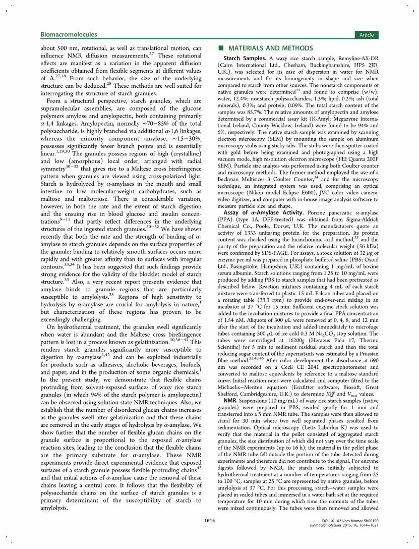
ABSTRACT: Starch is a prominent component of the humandiet and is hydrolyzed by α-amylase post-ingestion. Probingthe mechanism of this process has proven challenging, due tothe intrinsic heterogeneity of individual starch granules. Bymeans of solution-state NMR, we demonstrate that flexiblepolysaccharide chains protruding from the solvent-exposedsurfaces of waxy rice starch granules are highly mobile and thatduring hydrothermal treatment, when the granules swell, thenumber of flexible residues on the exposed surfaces increasesby a factor of 15. Moreover, we show that these flexible chainsare the primary substrates for α-amylase, being cleaved in the initial stages of hydrolysis. These findings allow us to conclude thatthe quantity of flexible α-glucan chains protruding from the granule surface will greatly influence the rate of energy acquisitionfrom digestion of starch.
■ INTRODUCTION
Starch granules are energy storage components formed withinplant cells. The granules are insoluble in water yet stillaccessible to the plant’s metabolic enzymes.1 With thedevelopment of cooking methods by hominids, the emergenceof the ability to hydrolyze most of any starch ingested fromplants with the enzyme α-amylase and therefore benefit fromthe energy source, represents a major development in thecourse of human evolution.2−5 The energetic advantages arisingfrom digestion of starch are believed to be of great importancein supporting the evolution of increased brain size.6 In contrastto animal feedstuff, nearly all ingested starch in human diets willhave been subjected to hydrothermal treatment duringdomestic and commercial food processing. Such starch isnormally a main source of exogenous glucose produced duringdigestion that subsequently appears at high concentrations inthe blood circulation.7
Cooked starch is digested rapidly in the gastrointestinal tractcausing peaks in glycemia and insulinemia within 1 h afteringestion, but the rates of digestion of different starch sourcescan differ noticeably.8 Controlling the variations in postprandialglycemia and insulinemia is of great importance in theprevention and treatment of diabetes mellitus and cardiovas-cular disease and also has implications for obesity manage-ment.9−14 Thus, it is important that the relatively early stages ofinteraction between starch and α-amylase, the enzyme thatcatalyzes the first stage in the intestinal digestion of starch, arefully understood so that better predictions can be made of
intestinal digestibility rates of particular starch sources and ofthe subsequent glycemia and insulinemia.Solution-state NMR is uniquely powerful for observing
dynamic segments of macromolecular assemblies.15−17 If asegment within a complex assembly has a high degree ofconformational flexibility, the effective local tumbling rate canbe substantially greater than that expected for a rigid system ofthe same size, a phenomenon that results in significantlynarrower NMR signals from such regions. A particularlyinteresting situation arises when flexible segments of amacromolecular assembly give rise to sharp NMR signals,whereas signals from the rigid core are too broad to bedetected. This situation has been observed in studies of largefunctional biomolecular complexes, including ribosomes andprotein aggregates, such as amyloid fibrils.18−24 NMR experi-ments that are particularly useful for characterizing theresonances from flexible regions of significantly larger structuresuse pulsed field gradients (PFGs) to measure diffusioncoefficients.25 Such experiments measure physical displacementof nuclear spins during a delay denoted by Δ. Translationaldiffusion occurring in solution leads to the displacement ofnuclear spins and so NMR diffusion experiments havetraditionally been interpreted in terms of this phenomenon.26
In recent years, however, it has been shown that forbiomolecular assemblies with at least one dimension exceeding
Received: February 9, 2015Revised: March 26, 2015Published: March 27, 2015
Article
pubs.acs.org/Biomac
© 2015 American Chemical Society 1614 DOI: 10.1021/acs.biomac.5b00190Biomacromolecules 2015, 16, 1614−1621
This is an open access article published under a Creative Commons Attribution (CC-BY)License, which permits unrestricted use, distribution and reproduction in any medium,provided the author and source are cited.
about 500 nm, rotational, as well as translational motion, caninfluence NMR diffusion measurements.27 These rotationaleffects are manifest as a variation in the apparent diffusioncoefficients obtained from flexible segments at different valuesof Δ.27,28 From such behavior, the size of the underlyingstructure can be deduced.28 These methods are well suited forinterrogating the structure of starch granules.From a structural perspective, starch granules, which are
supramolecular assemblies, are composed of the glucosepolymers amylose and amylopectin, both containing primarilyα-1,4 linkages. Amylopectin, normally ∼70−85% of the totalpolysaccharide, is highly branched via additional α-1,6 linkages,whereas the minority component amylose, ∼15−30%,possesses significantly fewer branch points and is essentiallylinear.1,29,30 The granules possess regions of high (crystalline)and low (amorphous) local order, arranged with radialsymmetry30−32 that gives rise to a Maltese cross birefringencepattern when granules are viewed using cross-polarized light.Starch is hydrolyzed by α-amylases in the mouth and smallintestine to low molecular-weight carbohydrates, such asmaltose and maltotriose. There is considerable variation,however, in both the rate and the extent of starch digestionand the ensuing rise in blood glucose and insulin concen-trations8−11 that partly reflect differences in the underlyingstructures of the ingested starch granules.30−32 We have shownrecently that both the rate and the strength of binding of α-amylase to starch granules depends on the surface properties ofthe granule; binding to relatively smooth surfaces occurs morerapidly and with greater affinity than to surfaces with irregularcontours.33,34 It has been suggested that such findings providestrong evidence for the validity of the blocklet model of starchstructure.33 Also, a very recent report presents evidence thatamylase binds to granule regions that are particularlysusceptible to amylolysis.35 Regions of high sensitivity tohydrolysis by α-amylase are crucial for amylolysis in nature,1
but characterization of these regions has proven to beexceedingly challenging.On hydrothermal treatment, the granules swell significantly
when water is abundant and the Maltese cross birefringencepattern is lost in a process known as gelatinization.30,36−41 Thisrenders starch granules significantly more susceptible todigestion by α-amylase1,42 and can be exploited industriallyfor products such as adhesives, alcoholic beverages, biofuels,and paper, and in the production of some organic chemicals.1
In the present study, we demonstrate that flexible chainsprotruding from solvent-exposed surfaces of waxy rice starchgranules (in which 94% of the starch polymer is amylopectin)can be observed using solution-state NMR techniques. Also, weestablish that the number of disordered glucan chains increasesas the granules swell after gelatinization and that these chainsare removed in the early stages of hydrolysis by α-amylase. Weshow further that the number of flexible glucan chains on thegranule surface is proportional to the exposed α-amylasereaction sites, leading to the conclusion that the flexible chainsare the primary substrate for α-amylase. These NMRexperiments provide direct experimental evidence that exposedsurfaces of a starch granule possess flexible protruding chains43
and that initial actions of α-amylase cause the removal of thesechains leaving a central core. It follows that the flexibility ofpolysaccharide chains on the surface of starch granules is aprimary determinant of the susceptibility of starch toamylolysis.
■ MATERIALS AND METHODSStarch Samples. A waxy rice starch sample, Remyline-AX-DR
(Cairn International Ltd., Chesham, Buckinghamshire, HP5 2JD,U.K.), was selected for its ease of dispersion in water for NMRmeasurements and for its homogeneity in shape and size whencompared to starch from other sources. The nonstarch components ofnative granules were determined44 and found to comprise (w/w):water, 12.4%; nonstarch polysaccharides, 1.3%; lipid, 0.2%; ash (totalminerals), 0.3%; and protein, 0.09%. The total starch content of thesamples was 85.7%. The relative amounts of amylopectin and amylosedetermined by a commercial assay kit (K-Amyl; Megazyme Interna-tional Ireland, County Wicklow, Ireland) were found to be 94% and6%, respectively. The native starch sample was examined by scanningelectron microscopy (SEM) by mounting the sample on aluminummicroscopy stubs using sticky tabs. The stubs were then sputter coatedwith gold before being examined and photographed using a highvacuum mode, high resolution electron microscope (FEI Quanta 200FSEM). Particle size analysis was performed using both Coulter counterand microscopy methods. The former method employed the use of aBeckman Multisizer 3 Coulter Counter,34 and for the microscopytechnique, an integrated system was used, comprising an opticalmicroscope (Nikon model Eclipse E600), JVC color video camera,video digitizer, and computer with in-house image analysis software tomeasure particle size and shape.
Assay of α-Amylase Activity. Porcine pancreatic α-amylase(PPA) (type 1A, DFP-treated) was obtained from Sigma-AldrichChemical Co., Poole, Dorset, U.K. The manufacturers quote anactivity of 1333 units/mg protein for the preparation. Its proteincontent was checked using the bicinchoninic acid method,33 and thepurity of the preparation and the relative molecular weight (56 kDa)were confirmed by SDS-PAGE. For assays, a stock solution of 32 μg ofenzyme per ml was prepared in phosphate buffered saline (PBS; OxoidLtd., Basingstoke, Hampshire, U.K.) containing 1 mg/mL of bovineserum albumin. Starch solutions ranging from 1.25 to 10 mg/mL wereproduced by adding PBS to starch samples that had been pretreated asdescribed below. Reaction mixtures containing 4 mL of each starchmixture were transferred to plastic 15 mL Falcon tubes and placed ona rotating table (33.3 rpm) to provide end-over-end mixing in anincubator at 37 °C for 15 min. Sufficient enzyme stock solution wasadded to the incubation mixtures to provide a final PPA concentrationof 1.54 nM. Aliquots of 300 μL were removed at 0, 4, 8, and 12 minafter the start of the incubation and added immediately to microfugetubes containing 300 μL of ice cold 0.3 M Na2CO3 stop solution. Thetubes were centrifuged at 16200g (Heraeus Pico 17, ThermoScientific) for 5 min to sediment residual starch and then the totalreducing sugar content of the supernatants was estimated by a PrussianBlue method.33,45,46 After color development the absorbance at 690nm was recorded on a Cecil CE 2041 spectrophotometer andconverted to maltose equivalents by reference to a maltose standardcurve. Initial reaction rates were calculated and computer fitted to theMichaelis−Menten equation (Enzfitter software, Biosoft, GreatShelford, Cambridgeshire, U.K.) to determine KM
exp and Vmax values.NMR. Suspensions (10 mg/mL) of waxy rice starch samples (native
granules) were prepared in PBS, swirled gently for 1 min andtransferred into a 5 mm NMR tube. The samples were then allowed tostand for 30 min where two well separated phases resulted fromsedimentation. Optical microscopy (Leitz Laborlux K) was used toverify that the material in the pellet consisted of aggregated starchgranules, the size distribution of which did not vary over the time scaleof the NMR experiments (up to 16 h); the material in the pellet phaseof the NMR tube fell outside the portion of the tube detected duringexperiments and therefore did not contribute to the signal. For enzymedigests followed by NMR, the starch was initially subjected tohydrothermal treatment at a number of temperatures ranging from 25to 100 °C; samples at 25 °C are represented by native granules, beforeamylolysis at 37 °C. For this processing, starch−water samples wereplaced in sealed tubes and immersed in a water bath set at the requiredtemperature for 10 min during which time the contents of the tubeswere mixed continuously. The tubes were then removed and allowed
Biomacromolecules Article
DOI: 10.1021/acs.biomac.5b00190Biomacromolecules 2015, 16, 1614−1621
1615
to stand for 10 min at room temperature (20 °C) before examinationby NMR. When the concentration of flexible chains was to bequantified, the heating process was conducted within the NMRspectrometer. Optical microscopy was used to determine that theabundance of starch granules in the aqueous phase was not obviouslyaffected by heating up to 80 °C.NMR data were acquired at 500 MHz on Bruker Avance
instruments with a cryogenic TCI probe and ATM-TXI probe foruse above 37 °C. A pulsed field gradient stimulated echo (PFGSE)sequence with a 3−9−19 watergate solvent suppression (Bruker pulsesequence stbpgp 1s19) was used for the diffusion experiments at 27 °Cwith δ = 5.4 ms.28 Diffusion coefficients (Deff) were obtained fordiffusion delays, Δ, between 50 ms and 1 s, as described in the Resultsand Discussion. Sample viscosity was monitored by measuring the self-diffusion of water and was found not to vary substantially between thestarch preparations. Processing and fitting of such data have beendescribed in greater detail elsewhere.28 Experimental uncertaintieswere estimated by comparing the variation of measured decayconstants between individually recorded frequencies in the region ofthe peak of interest. In a PFGSE NMR diffusion experiment, theobserved integrated signal Si is attenuated due to translational diffusionfrom a reference intensity S0 by a factor given by the Stejskal−Tannerrelation26 Si = S0 exp(−Deffα
2βG†2), where α = γδGmax, γ is thegyromagnetic ratio of the observed nucleus, δ is the duration of theapplied gradient, Gmax is the maximum strength of the appliedgradients, β = Δ − δ/3, Δ is the diffusion delay, G† = G/Gmax, whereG is the experimentally applied gradient. Thus, a plot of ln Si/S0(α2β)−1 versus G†2 has a gradient of −Deff. For solutions of smallmolecules such as maltose or even of globular proteins, onlytranslational diffusion, DT, contributions will be significant, and soDeff = DT. DT can be interpreted using hydrodynamic models such asthe Einstein−Stokes relation for a continuum solvent.47 DT = kBT/6πηRH, where kB is Boltzmann’s constant, T is the thermodynamictemperature, and η is the sample viscosity. A more general expressionfor the decay of signal, including the effects of rotational diffusion,27
shows that in the case of a sphere
β= +D D R /3eff T H2 (1)
Through studying the variation of Deff with Δ and by using a modelfor DT, the effective hydrodynamic radius of the underlying particle RHcan be determined.27,28
■ RESULTS AND DISCUSSION
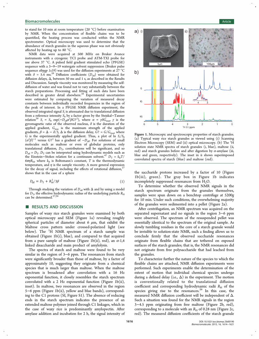
Samples of waxy rice starch granules were examined by bothoptical microscopy and SEM (Figure 1a) revealing roughlyspherical particles of diameter about 6 μm, that exhibit theMaltese cross pattern under crossed-polarized light (seebelow). The 1H NMR spectrum of a starch sample wasobtained (Figure 1b(i), blue), and compared to that acquiredfrom a pure sample of maltose (Figure 1b(ii), red), an α-1,4linked disaccharide and main product of amylolysis.The spectra of starch and maltose were found to be very
similar in the region of 3−6 ppm. The resonances from starchwere significantly broader than those of maltose, by a factor ofapproximately 10, suggesting they originate from a chemicalspecies that is much larger than maltose. When the maltosespectrum is broadened after convolution with a 16 Hzexponential function, it closely resembles the starch spectrumconvoluted with a 2 Hz exponential function (Figure 1b(ii),inset). In maltose, two resonances are observed in the region5−6 ppm (Figure 1b(ii), yellow and blue bands), correspond-ing to the C1 protons (SI, Figure S1). The absence of reducingends in the starch spectrum indicates the presence of anextended maltose polymer joined through C1 linkages, which inthe case of waxy rice is predominantly amylopectin. Afteramylase addition and incubation for 2 h, the signal intensity of
the saccharide protons increased by a factor of 10 (Figure1b(iii), green). The gray box in Figure 1b indicatesincompletely suppressed resonances from H2O.To determine whether the observed NMR signals in the
starch spectrum originate from the granules themselves,samples were spun down on a benchtop centrifuge at 5200gfor 10 min. Under such conditions, the overwhelming majorityof the granules were sedimented into a pellet (Figure 2a).After centrifugation, an NMR spectrum was acquired on the
separated supernatant and no signals in the region 3−6 ppmwere observed. The spectrum of the resuspended pellet wasessentially identical to the spectrum of the original sample. Asslowly tumbling residues in the core of a starch granule wouldbe invisible to solution-state NMR, such a finding allows us toconclude firmly that the observed saccharide resonancesoriginate from flexible chains that are tethered on exposedsurfaces of the starch granules; that is, the NMR resonances didnot originate from free polysaccharide that had leached fromthe granules.To characterize further the nature of the species to which the
flexible chains are attached, NMR diffusion experiments wereperformed. Such experiments enable the determination of theextent of motion that individual chemical species undergoduring a defined delay (i.e., Δ) in the experiment. The motionis conventionally related to the translational diffusioncoefficient and corresponding hydrodynamic radii RH of thespecies giving rise to the resonances.26 In this case, themeasured NMR diffusion coefficient will be independent of Δ.Such a situation was found for the NMR signals in the region3−4.5 ppm originating from free maltose (Figure 2b, red),corresponding to a molecule with an RH of 0.28 nm (Figure 2c,red). The measured diffusion coefficients of the starch granule
Figure 1. Microscopic and spectroscopic properties of starch granules.(a) Typical waxy rice starch granules as viewed using (i) ScanningElectron Microscopy (SEM) and (ii) optical microscopy. (b) The 1Hsolution state NMR spectra of starch granules (i, blue), maltose (ii,red) and starch granules before and after digestion by α-amylase (iii,blue and green, respectively). The inset in ii shows superimposedconvoluted spectra of starch (blue) and maltose (red).
Biomacromolecules Article
DOI: 10.1021/acs.biomac.5b00190Biomacromolecules 2015, 16, 1614−1621
1616
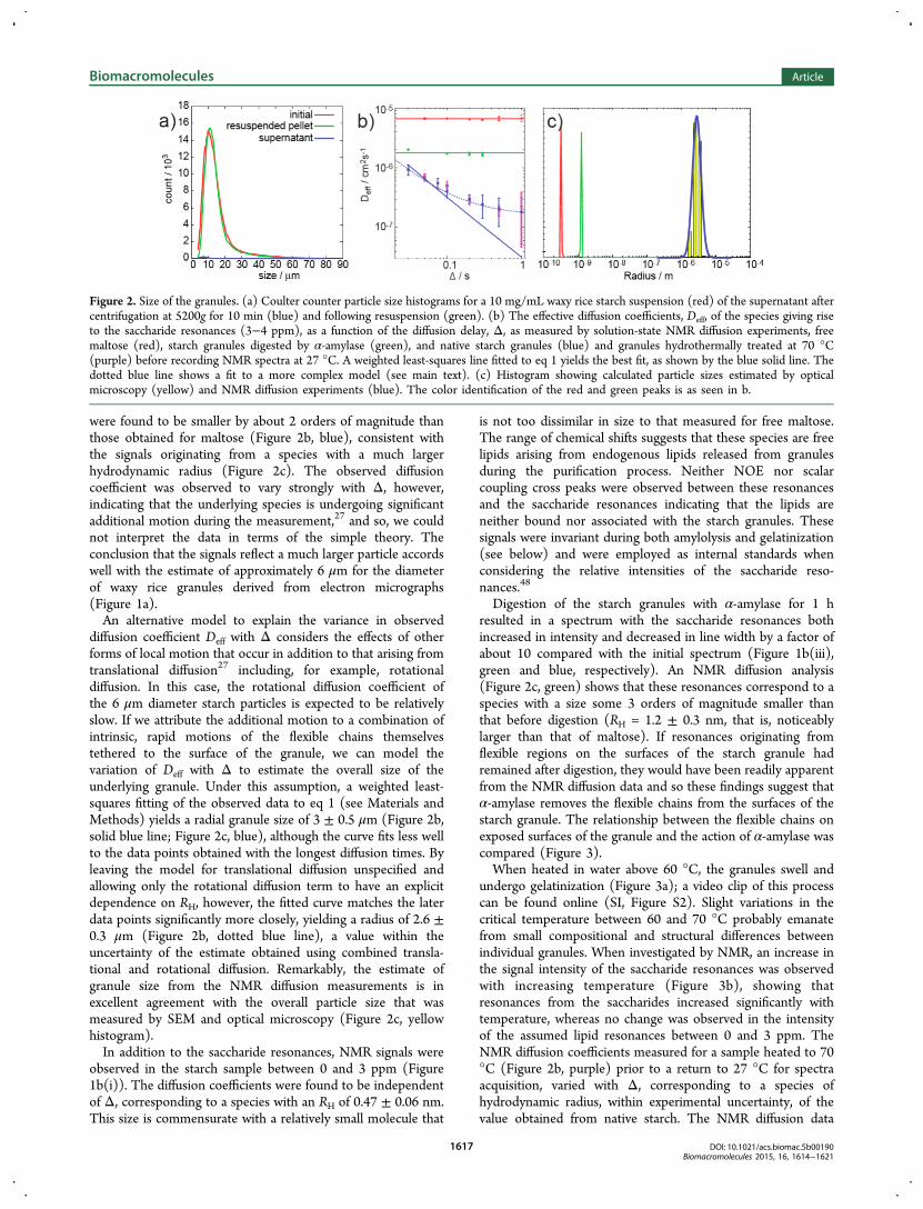
were found to be smaller by about 2 orders of magnitude thanthose obtained for maltose (Figure 2b, blue), consistent withthe signals originating from a species with a much largerhydrodynamic radius (Figure 2c). The observed diffusioncoefficient was observed to vary strongly with Δ, however,indicating that the underlying species is undergoing significantadditional motion during the measurement,27 and so, we couldnot interpret the data in terms of the simple theory. Theconclusion that the signals reflect a much larger particle accordswell with the estimate of approximately 6 μm for the diameterof waxy rice granules derived from electron micrographs(Figure 1a).An alternative model to explain the variance in observed
diffusion coefficient Deff with Δ considers the effects of otherforms of local motion that occur in addition to that arising fromtranslational diffusion27 including, for example, rotationaldiffusion. In this case, the rotational diffusion coefficient ofthe 6 μm diameter starch particles is expected to be relativelyslow. If we attribute the additional motion to a combination ofintrinsic, rapid motions of the flexible chains themselvestethered to the surface of the granule, we can model thevariation of Deff with Δ to estimate the overall size of theunderlying granule. Under this assumption, a weighted least-squares fitting of the observed data to eq 1 (see Materials andMethods) yields a radial granule size of 3 ± 0.5 μm (Figure 2b,solid blue line; Figure 2c, blue), although the curve fits less wellto the data points obtained with the longest diffusion times. Byleaving the model for translational diffusion unspecified andallowing only the rotational diffusion term to have an explicitdependence on RH, however, the fitted curve matches the laterdata points significantly more closely, yielding a radius of 2.6 ±0.3 μm (Figure 2b, dotted blue line), a value within theuncertainty of the estimate obtained using combined transla-tional and rotational diffusion. Remarkably, the estimate ofgranule size from the NMR diffusion measurements is inexcellent agreement with the overall particle size that wasmeasured by SEM and optical microscopy (Figure 2c, yellowhistogram).In addition to the saccharide resonances, NMR signals were
observed in the starch sample between 0 and 3 ppm (Figure1b(i)). The diffusion coefficients were found to be independentof Δ, corresponding to a species with an RH of 0.47 ± 0.06 nm.This size is commensurate with a relatively small molecule that
is not too dissimilar in size to that measured for free maltose.The range of chemical shifts suggests that these species are freelipids arising from endogenous lipids released from granulesduring the purification process. Neither NOE nor scalarcoupling cross peaks were observed between these resonancesand the saccharide resonances indicating that the lipids areneither bound nor associated with the starch granules. Thesesignals were invariant during both amylolysis and gelatinization(see below) and were employed as internal standards whenconsidering the relative intensities of the saccharide reso-nances.48
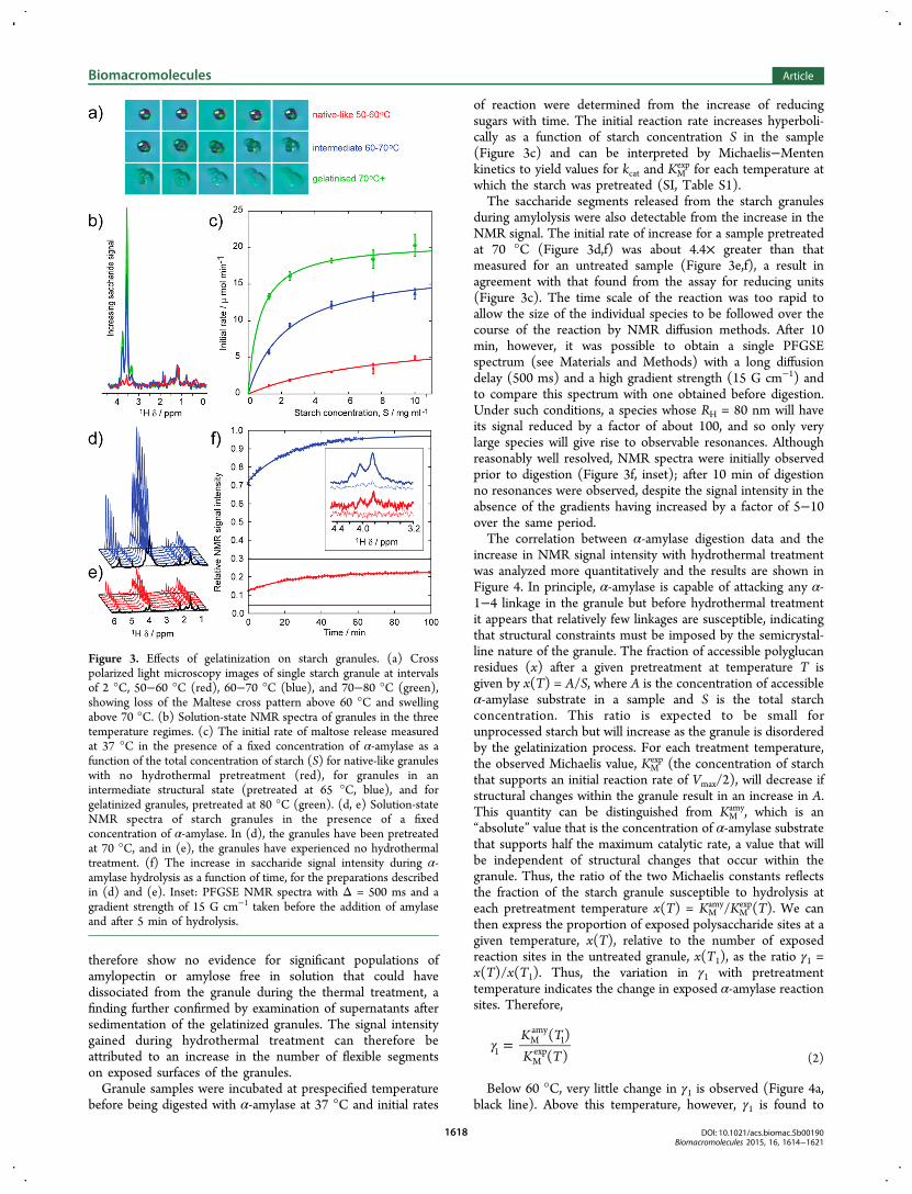
Digestion of the starch granules with α-amylase for 1 hresulted in a spectrum with the saccharide resonances bothincreased in intensity and decreased in line width by a factor ofabout 10 compared with the initial spectrum (Figure 1b(iii),green and blue, respectively). An NMR diffusion analysis(Figure 2c, green) shows that these resonances correspond to aspecies with a size some 3 orders of magnitude smaller thanthat before digestion (RH = 1.2 ± 0.3 nm, that is, noticeablylarger than that of maltose). If resonances originating fromflexible regions on the surfaces of the starch granule hadremained after digestion, they would have been readily apparentfrom the NMR diffusion data and so these findings suggest thatα-amylase removes the flexible chains from the surfaces of thestarch granule. The relationship between the flexible chains onexposed surfaces of the granule and the action of α-amylase wascompared (Figure 3).When heated in water above 60 °C, the granules swell and
undergo gelatinization (Figure 3a); a video clip of this processcan be found online (SI, Figure S2). Slight variations in thecritical temperature between 60 and 70 °C probably emanatefrom small compositional and structural differences betweenindividual granules. When investigated by NMR, an increase inthe signal intensity of the saccharide resonances was observedwith increasing temperature (Figure 3b), showing thatresonances from the saccharides increased significantly withtemperature, whereas no change was observed in the intensityof the assumed lipid resonances between 0 and 3 ppm. TheNMR diffusion coefficients measured for a sample heated to 70°C (Figure 2b, purple) prior to a return to 27 °C for spectraacquisition, varied with Δ, corresponding to a species ofhydrodynamic radius, within experimental uncertainty, of thevalue obtained from native starch. The NMR diffusion data
Figure 2. Size of the granules. (a) Coulter counter particle size histograms for a 10 mg/mL waxy rice starch suspension (red) of the supernatant aftercentrifugation at 5200g for 10 min (blue) and following resuspension (green). (b) The effective diffusion coefficients, Deff, of the species giving riseto the saccharide resonances (3−4 ppm), as a function of the diffusion delay, Δ, as measured by solution-state NMR diffusion experiments, freemaltose (red), starch granules digested by α-amylase (green), and native starch granules (blue) and granules hydrothermally treated at 70 °C(purple) before recording NMR spectra at 27 °C. A weighted least-squares line fitted to eq 1 yields the best fit, as shown by the blue solid line. Thedotted blue line shows a fit to a more complex model (see main text). (c) Histogram showing calculated particle sizes estimated by opticalmicroscopy (yellow) and NMR diffusion experiments (blue). The color identification of the red and green peaks is as seen in b.
Biomacromolecules Article
DOI: 10.1021/acs.biomac.5b00190Biomacromolecules 2015, 16, 1614−1621
1617
therefore show no evidence for significant populations ofamylopectin or amylose free in solution that could havedissociated from the granule during the thermal treatment, afinding further confirmed by examination of supernatants aftersedimentation of the gelatinized granules. The signal intensitygained during hydrothermal treatment can therefore beattributed to an increase in the number of flexible segmentson exposed surfaces of the granules.Granule samples were incubated at prespecified temperature
before being digested with α-amylase at 37 °C and initial rates
of reaction were determined from the increase of reducingsugars with time. The initial reaction rate increases hyperboli-cally as a function of starch concentration S in the sample(Figure 3c) and can be interpreted by Michaelis−Mentenkinetics to yield values for kcat and KM
exp for each temperature atwhich the starch was pretreated (SI, Table S1).The saccharide segments released from the starch granules
during amylolysis were also detectable from the increase in theNMR signal. The initial rate of increase for a sample pretreatedat 70 °C (Figure 3d,f) was about 4.4× greater than thatmeasured for an untreated sample (Figure 3e,f), a result inagreement with that found from the assay for reducing units(Figure 3c). The time scale of the reaction was too rapid toallow the size of the individual species to be followed over thecourse of the reaction by NMR diffusion methods. After 10min, however, it was possible to obtain a single PFGSEspectrum (see Materials and Methods) with a long diffusiondelay (500 ms) and a high gradient strength (15 G cm−1) andto compare this spectrum with one obtained before digestion.Under such conditions, a species whose RH = 80 nm will haveits signal reduced by a factor of about 100, and so only verylarge species will give rise to observable resonances. Althoughreasonably well resolved, NMR spectra were initially observedprior to digestion (Figure 3f, inset); after 10 min of digestionno resonances were observed, despite the signal intensity in theabsence of the gradients having increased by a factor of 5−10over the same period.The correlation between α-amylase digestion data and the
increase in NMR signal intensity with hydrothermal treatmentwas analyzed more quantitatively and the results are shown inFigure 4. In principle, α-amylase is capable of attacking any α-1−4 linkage in the granule but before hydrothermal treatmentit appears that relatively few linkages are susceptible, indicatingthat structural constraints must be imposed by the semicrystal-line nature of the granule. The fraction of accessible polyglucanresidues (x) after a given pretreatment at temperature T isgiven by x(T) = A/S, where A is the concentration of accessibleα-amylase substrate in a sample and S is the total starchconcentration. This ratio is expected to be small forunprocessed starch but will increase as the granule is disorderedby the gelatinization process. For each treatment temperature,the observed Michaelis value, KM
exp (the concentration of starchthat supports an initial reaction rate of Vmax/2), will decrease ifstructural changes within the granule result in an increase in A.This quantity can be distinguished from KM
amy, which is an“absolute” value that is the concentration of α-amylase substratethat supports half the maximum catalytic rate, a value that willbe independent of structural changes that occur within thegranule. Thus, the ratio of the two Michaelis constants reflectsthe fraction of the starch granule susceptible to hydrolysis ateach pretreatment temperature x(T) = KM
amy/KMexp(T). We can
then express the proportion of exposed polysaccharide sites at agiven temperature, x(T), relative to the number of exposedreaction sites in the untreated granule, x(T1), as the ratio γ1 =x(T)/x(T1). Thus, the variation in γ1 with pretreatmenttemperature indicates the change in exposed α-amylase reactionsites. Therefore,
γ =K TK T
( )( )1
Mamy
1
Mexp
(2)
Below 60 °C, very little change in γ1 is observed (Figure 4a,black line). Above this temperature, however, γ1 is found to
Figure 3. Effects of gelatinization on starch granules. (a) Crosspolarized light microscopy images of single starch granule at intervalsof 2 °C, 50−60 °C (red), 60−70 °C (blue), and 70−80 °C (green),showing loss of the Maltese cross pattern above 60 °C and swellingabove 70 °C. (b) Solution-state NMR spectra of granules in the threetemperature regimes. (c) The initial rate of maltose release measuredat 37 °C in the presence of a fixed concentration of α-amylase as afunction of the total concentration of starch (S) for native-like granuleswith no hydrothermal pretreatment (red), for granules in anintermediate structural state (pretreated at 65 °C, blue), and forgelatinized granules, pretreated at 80 °C (green). (d, e) Solution-stateNMR spectra of starch granules in the presence of a fixedconcentration of α-amylase. In (d), the granules have been pretreatedat 70 °C, and in (e), the granules have experienced no hydrothermaltreatment. (f) The increase in saccharide signal intensity during α-amylase hydrolysis as a function of time, for the preparations describedin (d) and (e). Inset: PFGSE NMR spectra with Δ = 500 ms and agradient strength of 15 G cm−1 taken before the addition of amylaseand after 5 min of hydrolysis.
Biomacromolecules Article
DOI: 10.1021/acs.biomac.5b00190Biomacromolecules 2015, 16, 1614−1621
1618
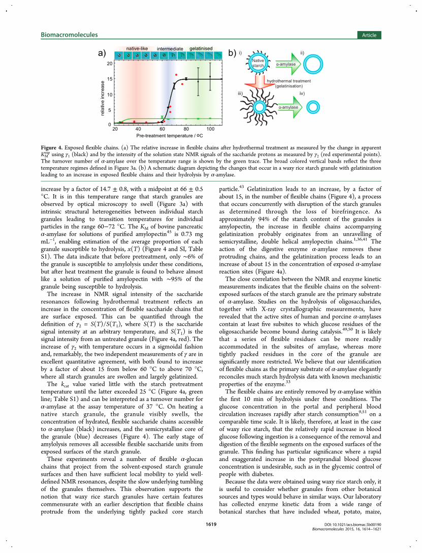
increase by a factor of 14.7 ± 0.8, with a midpoint at 66 ± 0.5°C. It is in this temperature range that starch granules areobserved by optical microscopy to swell (Figure 3a) withintrinsic structural heterogeneities between individual starchgranules leading to transition temperatures for individualparticles in the range 60−72 °C. The KM of bovine pancreaticα-amylase for solutions of purified amylopectin45 is 0.73 mgmL−1, enabling estimation of the average proportion of eachgranule susceptible to hydrolysis, x(T) (Figure 4 and SI, TableS1). The data indicate that before pretreatment, only ∼6% ofthe granule is susceptible to amylolysis under these conditions,but after heat treatment the granule is found to behave almostlike a solution of purified amylopectin with ∼95% of thegranule being susceptible to hydrolysis.The increase in NMR signal intensity of the saccharide
resonances following hydrothermal treatment reflects anincrease in the concentration of flexible saccharide chains thatare surface exposed. This can be quantified through thedefinition of γ2 = S(T)/S(T1), where S(T) is the saccharidesignal intensity at an arbitrary temperature, and S(T1) is thesignal intensity from an untreated granule (Figure 4a, red). Theincrease of γ2 with temperature occurs in a sigmoidal fashionand, remarkably, the two independent measurements of γ are inexcellent quantitative agreement, with both found to increaseby a factor of about 15 from below 60 °C to above 70 °C,where all starch granules are swollen and largely gelatinized.The kcat value varied little with the starch pretreatment
temperature until the latter exceeded 25 °C (Figure 4a, greenline; Table S1) and can be interpreted as a turnover number forα-amylase at the assay temperature of 37 °C. On heating anative starch granule, the granule visibly swells, theconcentration of hydrated, flexible saccharide chains accessibleto α-amylase (black) increases, and the semicrystalline core ofthe granule (blue) decreases (Figure 4). The early stage ofamylolysis removes all accessible flexible saccharide units fromexposed surfaces of the starch granule.These experiments reveal a number of flexible α-glucan
chains that project from the solvent-exposed starch granulesurfaces and then have sufficient local mobility to yield well-defined NMR resonances, despite the slow underlying tumblingof the granules themselves. This observation supports thenotion that waxy rice starch granules have certain featurescommensurate with an earlier description that flexible chainsprotrude from the underlying tightly packed core starch
particle.43 Gelatinization leads to an increase, by a factor ofabout 15, in the number of flexible chains (Figure 4), a processthat occurs concurrently with disruption of the starch granulesas determined through the loss of birefringence. Asapproximately 94% of the starch content of the granules isamylopectin, the increase in flexible chains accompanyinggelatinization probably originates from an unravelling ofsemicrystalline, double helical amylopectin chains.1,36,41 Theaction of the digestive enzyme α-amylase removes theseprotruding chains, and the gelatinization process leads to anincrease of about 15 in the concentration of exposed α-amylasereaction sites (Figure 4a).The close correlation between the NMR and enzyme kinetic
measurements indicates that the flexible chains on the solvent-exposed surfaces of the starch granule are the primary substrateof α-amylase. Studies on the hydrolysis of oligosaccharides,together with X-ray crystallographic measurements, haverevealed that the active sites of human and porcine α-amylasescontain at least five subsites to which glucose residues of theoligosaccharide become bound during catalysis.49,50 It is likelythat a series of flexible residues can be more readilyaccommodated in the subsites of amylase, whereas moretightly packed residues in the core of the granule aresignificantly more restricted. We believe that our identificationof flexible chains as the primary substrate of α-amylase elegantlyreconciles much starch hydrolysis data with known mechanisticproperties of the enzyme.33
The flexible chains are entirely removed by α-amylase withinthe first 10 min of hydrolysis under these conditions. Theglucose concentration in the portal and peripheral bloodcirculation increases rapidly after starch consumption9,51 on acomparable time scale. It is likely, therefore, at least in the caseof waxy rice starch, that the relatively rapid increase in bloodglucose following ingestion is a consequence of the removal anddigestion of the flexible segments on the exposed surfaces of thegranule. This finding has particular significance where a rapidand exaggerated increase in the postprandial blood glucoseconcentration is undesirable, such as in the glycemic control ofpeople with diabetes.Because the data were obtained using waxy rice starch only, it
is useful to consider whether granules from other botanicalsources and types would behave in similar ways. Our laboratoryhas collected enzyme kinetic data from a wide range ofbotanical starches that have included wheat, potato, maize,
Figure 4. Exposed flexible chains. (a) The relative increase in flexible chains after hydrothermal treatment as measured by the change in apparentKMexp using γ1 (black) and by the intensity of the solution state NMR signals of the saccharide protons as measured by γ2 (red experimental points).
The turnover number of α-amylase over the temperature range is shown by the green trace. The broad colored vertical bands reflect the threetemperature regimes defined in Figure 3a. (b) A schematic diagram depicting the changes that occur in a waxy rice starch granule with gelatinizationleading to an increase in exposed flexible chains and their hydrolysis by α-amylase.
Biomacromolecules Article
DOI: 10.1021/acs.biomac.5b00190Biomacromolecules 2015, 16, 1614−1621
1619
normal rice and wild type, lam and r pea mutants, and wherethe amylopectin content has ranged from the 98% of waxy ricedown to 28% for the pea r mutant.33,34,42,52 In all cases,measured KM values for hydrothermally treated starch are muchsmaller than the value found for native granules suggesting thatthe affinity for amylase is increased. It seems pertinent to theargument that we have shown a direct linear relationshipbetween KM values for starch hydrolysis and the dissociationconstant (Kd) for starch binding to granules of various sizes andamylopectin content.34 Kd is also related to the surfaceproperties of granules.33 It is not unreasonable therefore, tosuppose that the change in kinetic and NMR signals that weobserve for waxy rice starch are representative of a generalgranule property.Using conventional NMR diffusion methodology, it is
common to employ a single short delay, Δ, of about 50 msin experiments. Under such conditions, the apparent diffusioncoefficients for the hydrolyzed α-glucan chains in the digestionreaction and those from the starch granules are comparable(Figure 2b, green and blue, respectively). As the NMRexperiment is sensitive to physical displacements, this findingis a consequence of the similarities in the relative motions onthis time scale; the displacement of a flexible chain iscomparable to the displacement of the hydrolysis productsundergoing translational diffusion. As found with previousstudies on amyloid fibrils,28 only by considering the change indiffusion coefficient with diffusion delay can the true nature ofthe underlying dynamics be elucidated.
■ CONCLUSIONSOur studies provide direct experimental evidence for theexistence of flexible α-glucan chains protruding from solvent-exposed surfaces of starch granules and that these chains, whichincrease in number during hydrothermal processing, are theprimary substrates of pancreatic α-amylase. We have furtherdemonstrated the utility of recently developed solution stateNMR methodology for studying large molecular assemblies.28
Where large biomolecular assemblies have regions of sufficientlocal flexibility to yield narrow solution state NMR signals, themethodology of the type described in this work can be used toinvestigate structural properties that are currently inaccessibleto other techniques. Our findings reveal that the flexible chainson the surface of starch granules ultimately dictate how rapidlytheir energy can be extracted by hominids.
■ ASSOCIATED CONTENT*S Supporting InformationThe information consists of an NMR spectrum for maltoseshowing proton resonances, a video showing granule swellingduring hydrothermal treatment, plus a table listing Michaelis−Menten parameters for amylolysis at 37 °C of granules that hadbeen hydrothermally treated at various temperatures. Thismaterial is available free of charge via the Internet at http://pubs.acs.org.
■ AUTHOR INFORMATIONCorresponding Authors*E-mail: [email protected]. Tel.: +44 (0)1865275420.*E-mail: [email protected]. Tel.: +44 (0)20 78484592.*E-mail: [email protected]. Tel.: +44 (0)20 7848 4238.
Present Address‡The Physical and Theoretical Chemistry Laboratory, TheUniversity of Oxford, South Parks Road, Oxford, OX1 3QZ,United Kingdom.
NotesThe authors declare no competing financial interest.
■ ACKNOWLEDGMENTS
We thank Prof. Lewis Kay and Prof. Guy Lippens forstimulating discussions. A.J.B. thanks the BBSRC for a DavidPhillips Fellowship and Pembroke College. C.M.D. thanks theMRC, the Wellcome and Leverhulme Trusts, and theCambridge Nanoscience Centre for support of this work.D.L.E. and F.J.W. thank the BBSRC and Unilever for receipt ofan Industrial CASE award, and Kings College London forreceipt of a KCL scholarship, respectively. We thank Dr.Phillippa Rayment (Unilever) for providing the waxy rice starchsamples and the assistance of Dr. A. Brain in producing theSEM micrographs.
■ REFERENCES(1) Oates, C. G. Trends Food Sci. Technol. 1997, 8, 375−382.(2) Perry, G. H.; Dominy, N. J.; Claw, K. C.; Lee, A. A.; Fiegler, H.;Redon, R.; Werner, J.; Villanea; Mountain, J. L.; Misra, R.; Carter, N.P.; Lee, C.; Stone, A. C. Nat. Genet. 2007, 39, 1256−1260.(3) Henry, A. G.; Brooks, A. S.; Riperno, D. R. Proc. Natl. Acad. Sci.U.S.A. 2011, 108, 486−491.(4) Lucas, P. W. Proc. Natl. Acad. Sci. U.S.A. 2011, 108, 19101−19102.(5) Carmody, R. N.; Wrangham, R. W. J. Hum. Evol. 2009, 57, 379−391.(6) Laden, G.; Wrangham, R. W. J. Hum. Evol. 2005, 49, 482−498.(7) Judd, P.; Ellis, P. R. In Traditional Medicines for Modern Times.Antidiabetic Plants; Soumyanath, A., Ed.; CRC Press, Taylor andFrancis Group: Boca Raton, FL, U.S.A., 2006; pp 257−272.(8) Seal, C. J.; Daly, M. E.; Thomas, L. C.; Bal, W.; Birkett, A. M.;Jeffcoat, R.; Mathers, J. C. Br. J. Nutr. 2003, 90, 853−864.(9) Ells, L. J.; Seal, C. J.; Kettlitz, B.; Bal, W.; Mathers, J. C. Br. J.Nutr. 2005, 94, 948−955.(10) Jenkins, D. J.; Kendall, C. W. C.; Augustin, L. S. A.; Franceschi,A. L.; Hamidi, M.; Marchie, A.; Jenkins, A. L.; Axelsen, M. Am. J. Clin.Nutr. 2002, 76, 266S−273S.(11) Gallant, D. J.; Bouchet, B.; Buleon, A.; Perez, S. Eur. J. Clin.Nutr. 1992, 46 (suppl. 2), S3−S16.(12) Frost, G.; Leeds, A. A.; Dore, C. J.; Madeiros, S.; Dornhorst, A.Lancet 1999, 353, 1045−1048.(13) Mann, J.; Cummings, J. H.; Englyst, H. N.; Key, T.; Liu, S.;Riccardi, G.; Summerbell, C.; Venn, B.; Vorster, H. H.; Wiseman, M.Eur. J. Clin. Nutr. 2007, 61 (Suppl 1), S132−137.(14) Salmeron, J.; Manson, J. E.; Stampfer, M. J.; Colditz, G. A.;Spiegelman, D.; Jenkins, D. J.; Wing, A. L.; Willett, W. C. J. Am. Med.Assoc. 1997, 277, 472−477.(15) Carver, J. A.; Aquilina, J. A.; Truscott, R. J.; Ralston, G. B. FEBSLett. 1992, 311, 143−149.(16) Oswald, R. E.; Bogusky, M. J.; Bamberger, M.; Smith, R. A.;Dobson, C. M. Nature 1989, 337, 579−582.(17) Radford, S. E.; Laue, E. D.; Perham, R. N.; Miles, J. S.; Guest, J.R. Biochem. J. 1987, 247, 641−649.(18) Christodoulou, J.; Larsson, G.; Fucini, P.; Connell, S. R.;Pertinhez, T. A.; Hanson, C. L.; Redfield, C.; Nierhaus, K. H.;Robinson, C. V.; Schleucher, J.; Dobson, C. M. Proc. Natl. Acad. Sci.U.S.A. 2004, 101, 10949−10954.(19) Sillen, A.; Leroy, A.; Wieruszeski, J. M.; Beauvillain, J. C.; Buee,L.; Landrieu, I.; Lippens, G. ChemBioChem 2005, 6, 1849−1856.(20) Baldwin, A. J.; Bader, R.; Christodoulou, J.; MacPhee, C. E.;Dobson, C. M. J. Am. Chem. Soc. 2006, 128, 2162−2163.
Biomacromolecules Article
DOI: 10.1021/acs.biomac.5b00190Biomacromolecules 2015, 16, 1614−1621
1620
(21) Meehan, S.; Knowles, T. P.; Baldwin, A. J.; Smith, J. F.; Squires,A. M.; Clements, P.; Treweek, T. M.; Ecroyd, H.; Tartaglia, G. G.;Vendruscolo, M.; MacPhee, C. E.; Dobson, C. M.; Carver, J. A. J. Mol.Biol. 2007, 372, 470−484.(22) Baldwin, A. J.; Kay, L. E. Nat. Chem. Biol. 2009, 5, 808−814.(23) Baldwin, A. J.; Walsh, P.; Hansen, D. F.; Hilton, G. R.; Benesch,J. L.; Sharpe, S.; Kay, L. E. J. Am. Chem. Soc. 2012, 134, 15343−15359.(24) Baldwin, A. J.; Hilton, J. R.; Lioe, H.; Bagneris, C.; Benesch, J. L.P.; Kay, L. E. J. Mol. Biol. 2011, 413, 310−320.(25) Dehner, A.; Kessler, H. ChemBioChem 2005, 6, 1550−1565.(26) Stejskal, E. O.; Tanner, J. E. J. Chem. Phys. 1965, 42, 288−292.(27) Baldwin, A. J.; Christodoulou, J.; Barker, P. D.; Dobson, C. M.;Lippens, G. J. Chem. Phys. 2007, 127, 11405.(28) Baldwin, A. J.; Anthony-Cahill, S.; Christodoulou, J.; Lippens,G.; Barker, P. D.; Dobson, C. M. Angew. Chem., Int. Ed. 2008, 47,3385−3387.(29) Guilbot, A.; Mercier, M. In The Polysaccharides; Aspinall, G. O.,Ed.; Academic Press, Inc.: Orlando, FL, 1985; Vol 3, pp 209−282.(30) Blanshard, J. M. V. In Starch Properties and Potential; Galliard,T., Ed.; John Wiley & Sons Ltd.: New York, 1987; pp 16−54.(31) French, D. In Starch: Chemistry and Technology; BeMiller, J. N.,Whistler, R. L., Paschall, E. F., Eds.; Academic Press: San Diego, CA,1984; pp 257−272.(32) Manners, D. J. Carbohydr. Polym. 1989, 11, 87−112.(33) Warren, F. J.; Butterworth, P. J.; Ellis, P. R. Biochim. Biophys.Acta 2013, 1830, 3095−3101.(34) Warren, F. J.; Royall, P. G.; Gaisford, S.; Butterworth, P. J.; Ellis,P. R. Carbohydr. Polym. 2011, 86, 1038−1047.(35) Dhital, S.; Warren, F. J.; Zang, B.; Gidley, M. J. Carbohydr.Polym. 2014, 113, 97−107.(36) Lynn, A.; Stark, J. R. Carbohydr. Res. 1992, 227, 379−383.(37) Biliaderis, C. G.; Page, C. M.; Maurice, T. J.; Juliano, B. O. J.Agric. Food Chem. 1986, 34, 6−14.(38) Cooke, D.; Gidley, M. J. Carbohydr. Res. 1992, 227, 103−112.(39) Donovan, J. W. Biopolymers 1979, 18, 263−275.(40) Gidley, M. J. In Gums and Stabilisers for the Food Industry;Phillips, G. O., Williams, P. A., Wedlock, G. J., Eds.; IRL Press: Oxford,U.K., 1992; pp 87−92.(41) Bogracheva, T. Y.; Meares, C.; Hedley, C. L. Carbohydr. Polym.2006, 63, 323−330.(42) Slaughter, S. L.; Ellis, P. R.; Butterworth, P. J. Biochim. Biophys.Acta 2001, 1525, 29−36.(43) Lineback, D. R. Baker’s Dig. 1984, 58, 16−21.(44) Wang, Q.; Ellis, P. R.; Ross-Murphy, S. B.; Reid, J. S. G.Carbohydr. Res. 1995, 229−239.(45) Walker, J. A.; Harmon, D. L. J. Anim. Sci. 1996, 74, 658−662.(46) Wider, G.; Dreier, L. J. Am. Chem. Soc. 2006, 128, 2571−2576.(47) Wilkins, D.; Grimshaw, S.; Receveur, V.; Dobson, C. M.; Jones,J.; Smith, L. J. Biochemistry 1999, 38, 16424−16431.(48) Amato, M. E.; Ansanelli, G.; Fisichella, S.; Lamanna, R.; Scarlata,G.; Sobolev, A. P.; Segre, A. J. Agric. Food Chem. 2004, 52, 823−831.(49) Brayer, G. D.; Sidhu, G.; Maurus, R.; Rydberg, E. H.; Braun, C.;Wang, Y.; Nguyen, N. T.; Overall, C. M.; Withers, S. G. Biochemistry2000, 39, 4778−4791.(50) Gilles, C.; Astier, J. P.; Marchis-Mouren, G.; Cambillau, C.;Payan, F. Eur. J. Biochem. 1996, 238, 561−569.(51) Ellis, P. R.; Roberts, F. G.; Low, A. G.; Morgan, L. M. Br. J. Nutr.1995, 74, 539−556.(52) Tahir, R.; Ellis, P. R.; Bogracheva, T. Y.; Meares-Taylor, C.;Butterworth, P. J. Biomacromolecules 2011, 12, 123−133.
Biomacromolecules Article
DOI: 10.1021/acs.biomac.5b00190Biomacromolecules 2015, 16, 1614−1621
1621