COMPETITION (Chapter 13). COMPETITION: INTRASPECIFIC versus INTERSPECIFIC.
James Madison UniversityJMU Scholarly Commons
Masters Theses The Graduate School
Spring 2018
Investigating interspecific and intraspecific variationin lung development in amphibiansCourtney Neumeyer
Follow this and additional works at: https://commons.lib.jmu.edu/master201019Part of the Developmental Biology Commons
This Thesis is brought to you for free and open access by the The Graduate School at JMU Scholarly Commons. It has been accepted for inclusion inMasters Theses by an authorized administrator of JMU Scholarly Commons. For more information, please contact [email protected].
Recommended CitationNeumeyer, Courtney, "Investigating interspecific and intraspecific variation in lung development in amphibians" (2018). MastersTheses. 578.https://commons.lib.jmu.edu/master201019/578

Investigating Interspecific and Intraspecific Variation in Lung Development in
Amphibians
Courtney Holly Neumeyer
A thesis submitted to the Graduate Faculty of
JAMES MADISON UNIVERSITY
In
Partial Fulfillment of the Requirements
for the degree of
Master of Science
Department of Biology
May 2018
FACULTY COMMITTEE:
Committee Chair: Christopher S. Rose
Committee Members/ Readers:
Janet C. Daniel
Derek S. Strong

ii
ACKNOWLEDGEMENT
I would like to thank my advisor; Dr. Christopher Rose as well is my committee
members: Dr. Janet Daniel and Dr. Derek Strong. I would also like to thank the whole
JMU Biology Department, in particular I would like to thank Dr. Ken Roth for supplying
fruit flies for my amphibians; Ms. Patricia Crider, Ms. Janina Peachey, and Ms. Karen
Norment for always ordering supplies for my research in a timely manner; our excellent
Biology Graduate Coordinators Dr. Christine May and Dr. Janet Daniel for the help and
support. Furthermore, I would like to thank my fellow biology graduate students for their
help and support throughout my two years at JMU and to Ms. Megan Thinnes, and
undergraduate student who assisted with my research over the summer 2017. In addition,
I would like to thank the amphibians that participated in my research. Last, but not least I
would like to thank my mom Kathy Neumeyer and my dad Joe Neumeyer for their never-
ending support and encouragement.

iii
TABLE OF CONTENTS
ACKNOWLEDGEMENT ...................................................................................... II
LIST OF TABLES ................................................................................................ IV
LIST OF FIGURES ............................................................................................... V
ABSTRACT .......................................................................................................... VI
INTRODUCTION .................................................................................................. 1
MATERIALS AND METHODS ............................................................................ 6
Description and Criteria for Selecting Species ................................................... 6
Animal Collection ............................................................................................... 7
Lab Rearing Animals .......................................................................................... 8
Filming and Sampling ......................................................................................... 9
Staging and Body Size Data Collection .........................................................10
Paraffin Sectioning, Whole Mount and Histology .........................................10
Developmental Data Collection and Analysis .................................................. 11
RESULTS ............................................................................................................. 12
Lung Morphogenesis ......................................................................................... 12
Lung Morphogenesis of Lithobates sylvaticus ...............................................13
Lung Morphogenesis of Anaxyrus americanus ..............................................14
Lung Morphogenesis of Ambystoma maculatum ...........................................15
Lung Growth ..................................................................................................... 15
Temporal Patterns of Lung Morphogenesis ...................................................... 16
DISCUSSION ....................................................................................................... 18
Lung Morphogenesis ......................................................................................... 18
Lung Growth ..................................................................................................... 20
Temporal Patterns of Lung Morphogenesis ...................................................... 23
Future Directions ............................................................................................... 25
APPENDIX ........................................................................................................... 27
Tables ................................................................................................................ 27
Figures ............................................................................................................... 32
LITERATURE CITED ......................................................................................... 46
iv
LIST OF TABLES
Table 1 Stages of salamander metamorphosis added by the current study.
Table 2 Onset of lung morphogenesis in Lithobates sylvaticus.
Table 3 Onset of lung morphogenesis in Anaxyrus americanus.
Table 4 Onset of lung morphogenesis in Ambystoma maculatum.
Table 5 Relationships between stage, age, weight, and breathing frequency to relative
lung length of each species.

v
LIST OF FIGURES
Figure 1 Cladogram of jawed vertebrates and the presence of lungs.
Figure 2 Main difference between the gas bladder and the lung.
Figure 3 Lung Anatomy.
Figure 4 General Anuran metamorphosis.
Figure 5 Landmarked images used in measuring relative lung lengths.
Figure 6 Lung morphogenesis of Lithobates sylvaticus.
Figure 7 Lung morphogenesis of Anaxyrus americanus.
Figure 8 Lung morphogenesis of Ambystoma maculatum.
Figure 9 Relationships between average relative lung length and each independent
variable.
Figure 10 Relationships between maximum relative lung length and each independent
variable.
Figure 11 Stages present for each lung stage, onset of breathing, and developmental
period.
Figure 12 Weights present for each lung stage, onset of breathing, and developmental
period.
Figure 13 Ages present for each lung stage, onset of breathing, and developmental
period.
Figure 14 Breaths per hour present for each lung stage and developmental period.

vi
ABSTRACT
Amphibians occupy an intermediate phylogenetic position between fish and
tetrapods, making amphibians an essential model for understanding the evolution of air
breathing organs in vertebrates. Amphibians have a uniquely biphasic life style allowing
them to occupy aquatic and terrestrial habitats and to use multiple organs for gas
exchange at different stages of life. This initial independence from lung breathing means
that amphibians are developing their lungs while they are using them, which opens up the
possibility that their rate of lung development is controlled in part by their breathing
behavior. As such, amphibians provide a starting point for examining the evolution of
lung development in vertebrates. Two objectives were investigated: first, lung
morphogenesis is described for Lithobates sylvaticus, Anaxyrus americanus, and
Ambystoma maculatum, and second, average and maximum lung lengths were compared
to four independent variables: development (stage), age, size (weight), and frequency of
breathing to see if any of these variables were predictors of lung growth. Lungs appeared
early in development for L. sylvaticus and midway through larval development for A.
americanus and A. maculatum. Lung morphogenesis generally occurred by the
appearance of lungs, the expansion of lung lumen, the appearance of folds, the extension
of folds into septa, and the septa connecting to form a septa network. The extension of
folds into septa and the septa network did not occur in A. maculatum. Stage and age were
predictors of lung length in all three species, weight was a predictor in L. sylvaticus and
A. maculatum, and breathing frequency was an additional predictor for A. maculatum.
The strongest relationships between lung length and a predictor had an R-squared value
of 50% and were seen in A. americanus for age to both average and maximum lung

vii
length and for A. maculatum for stage to maximum lung length. These data provide an
initial description in lung morphology and development in the first tetrapod vertebrates
offering a starting point to understand the evolution of lung morphogenesis in the
tetrapod vertebrates that amphibians gave rise to, the fish that amphibians evolved from,
and the fish that might have convergently evolved lungs.
1
INTRODUCTION
Amphibians are an essential model for understanding the evolution of air
breathing organs in vertebrates because they occupy an intermediate phylogenetic
position between fish that respire in water using gills and skin and also in air using gas
filled organs, and tetrapods, which respire only in air using lungs (Fig. 1). Both gas
bladders and lungs are gas-filled organs found within the pleuroperitoneal cavity of
primitive bony fish. A gas bladder is an elongated, gas-filled organ that can be used for
respiration and to maintain buoyancy in actinopterygians other than polypterids
(Broughton et al., 2013; Longo et al., 2013). Lungs and gas bladders are similar in two
ways. Both gas-filled organs develop as out pockets of the gut or pharynx and both
secrete surfactant (Daniels et al., 2004; Kardong, 2015; Liem et al., 2001). Lungs differ
from gas bladders in two important ways. First, lungs develop as paired ventral
outpocketings of the pharynx or digestive tract whereas a gas bladder develops as an
unpaired dorsal outpocketing of the pharynx or digestive tract (Fig. 2). Second, blood
from the lungs drains to the heart, and blood from the gas bladder drains to the veins
(Brainerd, 2015; Kardong, 2015; Liem et al., 2001; Perry and Sander, 2004; Perry et al.,
2001).
In addition to being the first tetrapods with lungs, amphibians have a biphasic life
style and multiple respiratory organs throughout development, including skin, external
and/or internal gills, and lungs. The use of multiple respiratory organs provides
amphibians with the ability to occupy aquatic and terrestrial habitats at different stages of
life. Amphibians exhibit lung development as free swimming larvae or metamorphs, thus
providing an excellent model for studying ontogenetic lung development (Duellman and

2
Trueb, 1994; Noble, 1931). The development of lungs in larvae or metamorphs provides
an opportunity to examine how larval breathing behavior shapes the lung’s development,
and how choices in the timing and frequency of lung use during larval stages might affect
the state of lung development in early postmetamorphic stages. This study investigates
lung development in three species of amphibians that differ in the onset of first lung use,
which provides a range in which to compare lung development. In addition, this study
investigates how lung development in these species is influenced by stage, age, weight,
and breathing frequency.
Since amphibians are the first tetrapods to air-breathe, they provide a starting
point on which to investigate the evolution of lung development in the major groups of
tetrapods (reptiles, birds, and mammals) that evolved from primitive amphibians as well
as in primitive fish that are closely related to ancestral amphibians (Dipnoi and
Coelacanths) (Okada et al., 1962; Rankin et al., 2015). In addition, comparisons of lung
development can be made with the convergently evolved lungs of polypterids (Fig. 1).
Evolutionary comparisons of lung morphology and anatomy are complicated by the
frequent use of mammalian lung structures, e.g., alveoli/alveolar and bronchiole, for adult
amphibian lung structures (Bartel and Lametschwandtner, 2000; Hermida et al., 1998;
Okada et al., 1962; Rankin et al., 2015; Shoemaker et al., 1992; Smith and Campbell,
1976). Amphibians do not have an alveolar lung, rather they have a faviform or faveolar
lung (Daniels and Orgeig, 2003; Rose and James, 2013). Faveoli refer to the honeycomb-
like regions of space that are separated by septa. These septa extend from the inner lung
wall towards the center of the lung (Kardong, 2015). Classic, as well as current,
descriptions of amphibian lung anatomy and development (Kardong, 2015; Rose and

3
James, 2013; Waterman, 1939) typically describe the lungs of amphibians as tapered sacs
connected to the trachea by a bronchus. In addition, there are two descriptions of the
septal arrangements of the faviform lung of adult amphibians. One description indicates
that the inner pulmonary wall is drawn together to form primary septa that point toward
the center of the lung, secondary septa form from the primary septa and tertiary septa
form off of the secondary septa (Romer and Parsons, 1977; Rose and James, 2013) (Fig.
3A). The second description of internal lung morphology is similar to the first except the
secondary and tertiary septa also protrude from the wall of the lung, like the primary
septa, but each are shorter than the previous septa (Smith and Campbell, 1976) (Fig. 3B).
In both cases, the center of the lung is hollow (Duellman and Trueb, 1994; Rose and
James, 2013). Studies done on salamanders, including Triturus and Salamandra species
indicate that adult salamander lungs are relatively smooth and lack secondary and tertiary
septa (Francis, 1934; Goniakowska-Witalińska, 1978; Goniakowska-Witalińska, 1980;
Goniakowska-Witalińska, 1982). Adult Salamandra spp. have ridges along the inner wall
of the lungs which have been labeled with the same septa classifications as described in
anurans (Goniakowska-Witalińska, 1978; Rose and James, 2013; Waterman, 1939).
Since there are no secondary or tertiary septa described in these lungs it is unknown
which of the two descriptions they support.
Adult reptile, bird, and mammal lungs differ from amphibian lungs to varying
degrees. Reptiles are most similar to amphibians in having have a septation or faviform
based lung. The most basic lung found in adult Sphenodontidae (tuataras) have low septa
protruding from the inner wall of the lung, likely similar to that found in anurans
(Duncker, 2004). Many adult lizards and snakes, such as Pythonidae are shown to have

4
various sizes of septa protruding from the inner wall and forming faveoli, similar to that
of amphibians (Duncker, 2004). Reptilian lungs are more intricate in Varanidae,
Testudines, and Crocodilia, being subdivided into three rows extending perpendicularly
from the intrapulmonary bronchus. Each row is further subdivided into at least four
chambers (Duncker, 2004).
Bird lungs, which are the most unique respiratory system among vertebrates, are
believed to have evolved from chambered lungs of the kind exhibited by crocodilians
(Duncker, 2004). Adult birds have a parabronchial lung structure consisting of rigid-
walled paired lungs with little to no movement. Several air sacs connect to the lungs and
control lung ventilation, but do not directly contribute to gas exchange (Daniels and
Orgeig, 2003; Duncker, 2004; Maina, 2006). Lungs are connected to the trachea via a
bronchus, which branches into parabronchi that are equivalent to the tertiary bronchi of
mammals (Maina, 2006).
Mammal lungs are referred to as alveolar lungs or a bronchial tree. Like in
amphibians, reptiles, and birds, the lungs are connected to the trachea via a paired
bronchus, however in the adult mammal lung, the bronchi extend into the lung where it
continues to branch. The bronchi that enter the lungs are referred to as the primary
bronchi, these then branch into secondary bronchi followed by tertiary bronchi. The
bronchi continue to branch progressively becoming thinner and extending toward the
walls of the lungs, until they end in alveolar ducts. The alveolar ducts are clusters of
alveoli close to the walls of the lungs; the alveoli are one cell layer thick and are the site
of gas exchange with the blood capillaries (Daniels and Orgeig, 2003; Metzger et al.,
2008; Rawlins, 2011)(Fig. 3C). Although reptile, bird, and mammal have lungs that are
5
structurally diverse, all tetrapod vertebrates evolved from primitive amphibians (Fig. 1).
Lung breathing does not appear to be an obligatory form of respiration in most
amphibian larvae, which opens up the possibility that their rate of lung development is
controlled in part by their breathing behavior. In addition the initial non-reliance on lung
breathing allows for variability in timing of lung development and use, which has been
proposed to contribute to how some lineages of amphibians have evolved lung reduction
as seen in the anurans Ascaphus spp. and lung loss as seen in the caudate family
Plethodontidae (Rose, 2014; Rose and James, 2013). Lung buds are present in all known
embryonic amphibians, even those with reduced or absent lungs (Mekeel, 1930). Some
studies indicate a discrete time during late metamorphosis when the septa of the lungs
develop (Viertel and Richter, 1999), however lung development, and most likely the
appearance of septa, appears to be different among species as well as developmentally
variable within species. This differs from the suggestion that larval development and
metamorphosis, including lung development, is regulated strictly by thyroid hormone
(Brown and Cai, 2007; Burggren and Just, 1992; Duellman and Trueb, 1994; Miyata and
Ose, 2012; Shi, 2000). In the pipid frog, Xenopus laevis, the lung septa form during hind
limb and toe development, Nieuwkoop and Faber (NF) stage 57; but those X. laevis had
lungs functioning for occasional air breathing since NF stage 46, which is shortly after
hatching (Nieuwkoop and Faber, 1956; Rose and James, 2013). American bullfrogs,
Lithobates catesbeianus, as well as other Ranidae have been seen using their lungs as
early larvae, whereas many toads (Bufonidae, Megophryidae, Pelobatidae, and
Scaphiopodidae) are not seen using their lungs until metamorphosis (Burggren and Just,
1992; Nodzenski et al., 1989; Rose and James, 2013; Ultsch et al., 1999; Wassersug and

6
Seibert, 1975). Despite a general documentation of when some anurans start using their
lungs, the morphogenesis of the lungs are unknown.
Since there is only one detailed description of lung development currently
available for an amphibian, and that species does not appear to vary its onset of lung use,
it is premature to design experiments to test the contribution of environmental or
behavioral variables to interspecific and intraspecific variation in lung development.
Instead this study describes lung development for three additional amphibian species that
vary in timing of onset of lung use. One aim is to expand our understanding of faviform
lungs development to resolve how primary, secondary and tertiary septa form in
amphibians and to learn how amphibian lung development compares with faviform lung
development in fish and reptiles. A second aim is to examine the relationships between
lung growth and four independent variables: stage, age, weight, and breathing frequency
for each of the three species to see which, if any, of these variables is a strong predictor
of lung size. These data are necessary for designing future experiments to investigate the
extent and cause of plasticity in lung development in amphibians.
MATERIALS AND METHODS
Description and Criteria for Selecting Species
Two anuran species, Lithobates sylvaticus (wood frog, Ranidae) and Anaxyrus
americanus (American toad, Bufonidae) and one caudate species, Ambystoma maculatum
(yellow spotted salamander, Ambystomatidae), were selected because of their local
availability, similarities in geographic occurrence, larval habitat and length of larval
period, and suspected variation in the timing of onset of their lung use. Previously

7
described data on lung development in X. laevis were also used (Rose and James, 2013).
These three anuran species exhibit a range in the timing of onset of lung use: X. laevis
begin using their lungs shortly after hatching, L. sylvaticus begin using their lungs
midway through larval development, and A. americanus begin using their lungs shortly
before completing metamorphosis (Rose and James, 2013; Wassersug and Seibert, 1975).
There are little to no data describing larval development and first lung use in any
salamander species.
The three species collected for this study, A. maculatum, L. sylvaticus, and A.
americanus, are native to Virginia (Beane et al., 2010) and breed in semi-permanent
pools in forested areas from late-winter through mid-spring (Beane et al., 2010; Virginia
Herpetological Society).
Anaxyrus americanus reproduces in late winter through early spring or even early
summer, whereas A. maculatum and L. sylvaticus are two of the earliest amphibians to
reproduce with a limited reproduction period between mid-winter and early-spring.
Ambystoma maculatum lay 200 – 375 eggs per clutch whereas the two anuran species can
lay between 1,750 – 6,000 eggs per clutch. Lithobates sylvaticus and A. americanus have
the shortest larval period ranging from 40 – 60 days to complete metamorphosis.
Ambystoma maculatum can take up to 144 days but is often less than 100 days in its
Virginia geographic range (Beane et al., 2010; Virginia Herpetological Society).
Animal Collection
Eggs clutches of L. sylvaticus and A. maculatum and Gosner stage 24 larvae of A.
americanus were collected from three different sites in the George Washington National

8
Forest, Virginia: A. maculatum from a pond off of Forest road 85, Augusta County
(38°25'1.56" N 79°17'50.388" W), L. sylvaticus from standing water near Hone Quarry
Dam, Rockingham County (38°28'20.244" N 79°08'32.28" W), and A. americanus from a
small puddle on the side of Long Run road, Rockingham County (38°35'0.636" N
79°1'9.228" W). All were transported to James Madison University in the pond water of
which they were collected.
Lab Rearing Animals
Upon arrival in the lab, A. americanus hatchlings were slowly acclimated to 0.1x
Marks Modified Ringers (MMR) solution, and egg clutches of the other two species were
moved directly to MMR solution. Once hatched, A. americanus and L. sylvaticus tadpoles
were distributed by clutch into separate 4 L tanks and raised in densities that were
thought to allow ad libitum feeding and optimal growth and development rates. Upon
hatching, A. maculatum hatchlings were moved to individual beakers containing 200 ml
of 0.1x MMR to prevent carnivory. The first few days after hatching, larval amphibians,
fed off their yolk sac. After the disappearance of yolk sacs, A. americanus and L.
sylvaticus were fed canned, salt-free spinach daily, and A. maculatum larvae were fed
every other day with Artemia at early stages and blackworms at later larval stages.
Postmetamorphic juveniles of all three species were originally fed wingless fruitflies and
transitioned with growth to crickets of gradually increasing sizes.
The 0.1x MMR was changed every 7 days for all three species. Anaxyrus
americanus and A. maculatum were maintained at 19°C throughout larval development
and metamorphosis. The majority of L. sylvaticus were also maintained at 19°C

9
throughout larval development and metamorphosis with a few exceptions: one clutch was
maintained at 22°C and two clutches were reared in 15°C for the first nine days of larval
development after which they were moved to 19°C for the remainder of larval
development and metamorphosis. The varying temperatures helped stagger development
between the seven clutches collected, in order to include a wider range of stages within
this study.
Filming and Sampling
The animals selected for filming and sampling were always the most
developmentally advanced within a clutch for each species at each sample time. At time
intervals ranging from 2 – 3 days in early larval periods to 5 – 8 days in late larval
periods, the eight most advanced larvae of each species were placed in individual tanks
(12x12x15 cm) which contained 1.6 L of 0.1x MMR. The height of the water was 12 cm
high. Each animal was allowed to acclimate in its tank for 30 minutes prior to filming.
The eight tanks were then filmed from above, for 1 h, after which the larvae were
individually euthanized by immersion in 0.4% MS222 until movement completely
stopped and then moved to 10% neutral buffered formalin for fixation. Larvae were
labeled and stored in individual vials so their breathing behavior could later be compared
to their lung growth and stage, age, size (weight).
After a minimum of three days of fixation, individuals were removed from the fix,
dabbed dry with a Kim Wipe and weighed using an analytical balance to the nearest
0.001 g and staged before being placed in 0.1x phosphate buffered saline (PBS) for
preparation as whole mounts or histological sections. Each species was sampled
10
approximately 16 times during the 8 – 19 weeks each species took to complete
metamorphosis in the laboratory conditions.
Breathing frequency was counted for each animal using iMovie, which is a video
editing program which allows the progression through a video frame by frame. Each
video was watched 8 times and a different individual was focused on each time.
Breathing was documented when an animal swam to the surface of the water and then,
upon leaving the surface of the water, the animal released a gas bubble from its mouth.
Staging and Body Size Data Collection
The Gosner staging system (Gosner, 1960) was used for the two anuran species
(L. sylvaticus and A. maculatum). As there is no complete staging system for
ambystomatid larvae, two incomplete ambystomatid staging systems and observations
from this study were used to create a more detailed and complete staging system that
covered larval development and metamorphosis. Early larval stages (1 – 46) used the
forelimb development criteria of Rugh (1962), later larval stages (47 – 56) used the hind
limb development stages of Shoop (1974), and metamorphic stages (57 – 61) were
recognized on the basis of external gill degeneration, the reduction of the dorsal tail fin,
and changes in toe length (Table 1).
Paraffin Sectioning, Whole Mount and Histology
Specimens were prepared for either sectioning or whole mount dissections. For
sectioning 68 L. sylvaticus, 30 A. americanus, and 30 A. maculatum were prepared. For
whole mounts 74 L. sylvaticus, 84 A. americanus, and 62 A. maculatum were prepared.

11
The specimens intended for sectioning were dehydrated in an ethanol-xylene series and
embedded in paraffin wax. After the molten wax steps, each specimen was embedded in
clean paraffin wax. Wax embedded specimens were sliced in 10 μm thick frontal planes
using a microtome. Most specimens were then stained using hematoxylin and eosin.
The specimens intended for whole mounts were dissected from the ventral side to
remove all viscera from the pleuroperitoneal cavity except the lungs. The lungs and
pleuroperitoneal cavity were photographed under a microscope and the magnifications
were recorded. Using Adobe Photoshop, the images were landmarked to mark the
posterior edge of the split between left and right bronchi, the caudal edge of the
pleuroperitoneal cavity, and the caudal tips of the right and left lungs (Fig. 5). Using
ImageJ the X, Y coordinates of the landmarks of the pleuroperitoneal cavity and the
length of each lung were recorded and imported in to Microsoft Excel where lung lengths
were calculated as relative to the length of the pleuroperitoneal cavity because absolute
lung length is not informative of growth due to variation in individual size.
Developmental Data Collection and Analysis
For each species, a data matrix was compiled in Excel which listed the species,
egg clutch, rearing temperature, individual identification numbers, hatching date, filming
date and number, breaths per hour, age, stage, weight, their buoyancy at fixation, and
either a histology identification number or a whole mount identification number. For
whole mounts only, relative lung length for each specimen was also included.
Only paraffin sectioned specimens were used to study lung morphogenesis. Lung
morphogenesis was scored in L. sylvaticus, A. americanus and A. maculatum using the
12
criteria developed by Rose and James (2013) for X. laevis. Although overall lung
morphogenesis was different in each of the three species in this study as well as in X.
laevis the criteria used to score lungs were the same. Lungs were scored in order for their
absence or presence, the presence or absence of a lumen, the absence or presence of folds
in the lung wall, the anterior to posterior location of folds along the lung wall, the degree
of folding in the lung wall, as well as their shape, their length into the lumen, and their
thickness. Lastly, lungs were scored for the presence of a septa network and if the septa
connected with other septa next to or across the lumen.
Only whole mount specimens were used to study lung growth. For each species,
two dependent variables, average and maximum lung length relative to the
pleuroperitoneal cavity, were compared with four independent variables: stage, age,
weight, and breathing frequency using simple linear regression analyses to identify how
strongly each independent variable(s) predicted relative lung length. Significant
relationships were identified by p-values of ≤ 0.01). R-squared values indicated the
strength of the relationship by indicating how much of the variability in lung length is
described by the independent variable. All data analyses were done using R (R Core
Team, 2016).
RESULTS
Lung Morphogenesis
Lung morphogenesis was described throughout embryonic, larval, metamorphic,
and postmetamorphic stages of development for L. sylvaticus, A. americanus, and A.
maculatum (Table 2-4; Fig. 6-8).

13
Lung Morphogenesis of Lithobates sylvaticus
In specimens showing the first evidence of lungs, the lung has a narrow lumen
throughout and the lung walls were thin and smooth with no signs of folds or septa (Fig.
6A); some specimens indicated lung walls being thicker in posterior and lateral regions
than in anterior and medial regions. In specimens at the next stage of lung development,
the lung lumen is large and the lung walls have inner and outer cell layers. Whereas the
outer layer is thin and smooth, the inner layer is several cells thick and contain folds
whose heights are equal to roughly the width at the base (Fig. 6B). In specimens at the
next stage of lung development, the lumen was long and narrow, the outer and inner
layers of the lungs were both thick, and the inner layer was mostly made up of folds with
very little smooth wall space remaining posteriorly. The folds were roughly twice as long
as the width at the base and extend almost halfway across the lumen (Fig. 6C,D). In the
specimens at the next stage of lung development, the lumen was large and long. Folds
and a septa network were present (Fig. 6E-I). In the anterior and mid portions of the
lungs, folds were present with either an expanded ball-like end or present as a split Y like
structure into the lumen (Fig. 6E-G). The posterior end of the lung may have had folds
present as simple folds without the ball-like end as well as with the expanded ball-like
end, but there were no Y-like septa splitting yet (Fig. E,F). In the anterior and mid
portions of the lungs there was also a septa network present in which septa connected to
other septa next to each other and across the lumen from each other (Fig. 6H,I). There
was not a septa network present in the posterior portion of the lungs yet (Table 2; Fig.
6E). No data were available for post-metamorphic L. sylvaticus. Lung development in L.

14
sylvaticus supports a faviform lung description in that the septa extend into the lung from
the inner lung wall. However, there is no evidence of secondary septa forming as
extensions of pre-existing primary septa. It is also not clear that one set of septa forms
earlier and/or grows to greater lengths than other septa. Thus, our description does not
favor one of the two faviform lung descriptions described in the literature.
Lung Morphogenesis of Anaxyrus americanus
In specimens showing the first evidence of lungs, the lungs had thick smooth
walls with little lumen present (Fig. 7A). In specimens at the next stage of lung
development, the lumen was narrow. Lungs were present with a few small folds
extending into the lumen, the folds were roughly half as long as they were wide at their
base (Fig. 7B,C). In specimens at the next stage of lung development, lungs had mostly
smooth thin walls with thin bead-like folds (Fig. 7J,K). In specimens at the next stage of
lung development, the folds became deeper and were roughly as long as they were wide
at their base and extended over halfway across the lumen (Fig. 7D,E). In specimens at the
next stage of lung development, septa and a septa network was present making the lumen
highly divided into bubble-like structures throughout the mid and anterior lung regions
(Fig. 7F,G). In specimens at the next stage of lung development, lungs had thick walls
with thick septa and a septa network extending into the lumen. There was little lumen left
as the septa took up most of the space (Fig. 7H,I). In specimens at the next stage of lung
development, lung walls were very thick with tube-like lumen between a thick septa
network which appeared more like lung wall (Table 3; Fig. 7L). Lung development in A.
americanus also supports a faviform lung description in that the septa extend into the

15
lung from the inner lung wall. However, it is unclear which of the two faviform lung
descriptions is supported.
Lung Morphogenesis of Ambystoma maculatum
Before the lungs were present there was a stage when just the trachea and
bronchus were present (Fig. 8A). In specimens showing the first evidence of lung
development, lungs were present with thin smooth walls that surrounded a mostly
unexpanded lumen (Fig. 8B). In specimens showing the next stage of lung development,
the lumen was expanded, the wall became slightly thicker, but still overall thin (Fig.
8C,D). In specimens showing the next stage of lung development, the lumen was narrow
and there were bead-like folds present. The folds were roughly as long as they were wide
at their base (Table 4; Fig. 8E,F). Septa and a septa network were not seen at any time in
A. maculatum. Although septa did not occur in any of these specimens the presence of
folds extending into the lung from the inner lung wall support a faviform lung
description. However, since septa are not present it is unclear which of the two faviform
lung descriptions is supported.
Lung Growth
Simple linear regressions were used to determine which of the following
independent variable(s): development (organismal stage), age (days after hatching), size
(weight in mg), and/or breathing frequency (breaths per hour) were the strongest
predictors of each dependent variable: average and maximum lung length (measured
relative to pleuroperitoneal cavity length), for each species (Table 5, Fig. 9,10). P-values
16
less than 0.01 were used to identify significant predictors and R-squared values were used
to estimate the strength of the predictor. All significant predictors had R-squared values
ranging between 12% and 50%. Most of the data fit a linear regression model (Fig. 9,10).
For L. sylvaticus, maximum lung length had significant relationships with stage,
age, and weight. All three predictors are weak with stage being the strongest at 20% and
age the weakest at 12%. For A. americanus, both average and maximum lung length had
significant relationships with stage and age, and for both dependent variables, age was
the stronger predictor at 50%. For A. maculatum, both average and maximum lung length
had significant relationships with stage, age, weight, and breathing frequency, and for
both dependent variables, stage was the strongest predictor at 46 – 50% and breathing
frequency was the weakest at 19 – 20% (Table 5; Fig. 9,10).
The relationship between stage and maximum lung length had relatively the same
slope for L. sylvaticus and A. americanus. Lithobates sylvaticus continuously had longer
lungs than A. americanus at every stage of development (Fig. 10). The relationship
between age and both average and maximum lung length had relatively the same slope
for A. maculatum and A. americanus. Ambystoma maculatum had longer lungs than A.
americanus at all ages of development (Fig. 9,10).
Temporal Patterns of Lung Morphogenesis
As the stages of lung morphogenesis are ordinal data, the relationships of lung
morphogenesis with developmental stage, weight, age, and breathing frequency cannot be
calculated using linear regression. (Fig. 11-14). Instead, box plots are used to show, for
each independent variable, the minimum point that each stage of lung morphogenesis

17
occurred and the maximum point that this stage had not yet occurred (Fig. 11-14). These
boxplots thus give a slight underestimation of the range across which each stage of lung
morphogenesis occurs in each species as measured by each independent variable.
Theoretically, a narrow box would indicate that a stage of lung morphogenesis correlates
strongly with that independent variable. A wide box would suggest that a stage of lung
morphogenesis has a variable occurrence when measured by that independent variable
and therefore the two do not correlate. All three species have many narrow bars for
different stages of lung morphogenesis. Since these boxes are attributable to low
specimen numbers, they will not be described further.
Onset of breathing has highly variable stages of occurrence in two of the three
species. Its occurrence spans the entire larval period in L. sylvaticus and from the mid
larval period to the end of the metamorphic periods in A. maculatum. Since no breathing
was observed at any larval and metamorphic stages in A. americanus, its stage of onset
remains undocumented (Fig. 11). Onset of breathing also has highly variable sizes of
occurrence in L. sylvaticus and A. maculatum, and highly variable ages of occurrence in
A. maculatum (Fig. 13).
Regarding lung morphogenesis, the appearance of lungs with a lumen and thin
smooth walls is followed by the appearance of folds in both L. sylvaticus and A.
maculatum. This is true whether plotted by stage, weight or age, though L. sylvaticus
show a small amount of overlap between these events when plotted by all three
independent variables (Fig. 11-13).
Anaxyrus americanus is distinguished by showing lungs with thin folds occurring
at the same stages, weights and ages as lungs with a septa network (Fig. 11). In

18
comparison with L. sylvaticus, lung morphogenesis in A. americanus starts at a late age
but proceeds to the stage of acquiring a septal network at a roughly equivalent rate with
respect to age (Fig. 13). Though A. maculatum acquires a smooth walled lung as a young
larva, it does not show any sign of fold development until mid to late metamorphosis
(Fig. 13).
DISCUSSION
Lung Morphogenesis
There are only two known descriptions of internal lung anatomy of amphibian
lungs during early development (Rose and James, 2013; Waterman, 1939). Waterman’s
study, however focuses on the development of arteries, veins, cilia, and smooth muscle
while only indicting that septal partitions are present anteriorly and posteriorly in the
lung, which supports a faviform lung description (Waterman, 1939). Rose and James’
study on X. laevis describes lung morphogenesis, including the occurrence of smooth
muscle actin. The morphogenesis of X. laevis can be described in five steps 1. the initial
expansion of the lung lumen and thinning of lung walls, 2. the appearance of inward
directional epithelial folds, 3. the transformation of epithelial folds into thin, elongated
septa, 4. the joining of primary septal crests into an internal polygonal framework, and
the emergence of 5. secondary septa, and 5. tertiary septa within this framework. These
descriptions of lung development support the first faviform lung description in which the
primary septa extend from the lung wall, the secondary septa extend from the primary
septa, and the tertiary septa extend from the secondary septa (Fig. 3A) (Rose and James,
2013).

19
The morphogenesis of X. laevis, L. sylvaticus, A. americanus, and A. maculatum
all begin with the same trajectory: lungs appear and expand followed by the occurrence
of epithelial folds. The thickness of the folds differs across taxa, furthermore A.
maculatum lungs were never seen to progress beyond the development of folds. L.
sylvaticus and A. americanus continued to develop septa and a septa network as seen in
X. laevis. Although the lung development seen in L. sylvaticus and A. americanus support
an adult faviform lung, the data in this study were not able to distinguish between
primary, secondary, and tertiary septa and therefore it is unknown which faviform lung
description was supported (Fig. 3A,B). In addition, although A. maculatum were never
observed with septa or a septa network, the development of folds extending into the lung
from the lung wall is consistent with early faviform lung development (Fig. 3A,B).
The data in this study provide the most detailed and complete description of lung
morphogenesis for a faviform lung. These descriptions offer a starting point for
comparing amphibians with other tetrapods and lunged fish. Adult reptiles, such as
Sphenodontidae are reported to have the most similar lung structure to adult amphibians
than any other tetrapod (Duncker, 2004), yet it is unknown if the lungs of reptiles and
amphibians share the same early stages of lung development. Likewise, dipnoans,
polypterids and amphibians are reported to have similar adult lung structures, but it is
unknown how they compare in lung development. In addition, comparing lung
development between amphibians and polypterids could help resolve whether polypterid
lungs arose from convergent evolution or if their lungs are similar enough to amphibian
lungs to suggest homology.
Although folds were present in all specimens, they differed in that they were only

20
seen as thick folds with thick lung walls in L. sylvaticus and as thin folds with thin lung
walls in A. maculatum (Table 2-4). Distinctions between thick and thin walls were also
seen when the lung walls were smooth and when septa were present. Though these data
might signify an actual difference between taxa, morphological comparisons of these
stages of lung development are limited by the small number of sectioned specimens used
in this study. Some stages of lung development may have been missed due to the absence
of specimens to represent those stages. Now that possible gaps in specific stages of lung
development have been identified, more specimens can be added to the data set to form a
more complete and continuous description of lung development.
Lung Growth
Multilinear regression results suggest that L. sylvaticus and A. americanus lung
growth is more variable than the lung growth of A. maculatum. All of the independent
variables for A. maculatum had significant relationships with average and maximum lung
length. Only two of the independent variables for A. americanus had significant
relationships with average and maximum lung length. Although three of the independent
variables for L. sylvaticus had significant relationships with maximum lung length there
were no significant relationships with average lung length.
For L. sylvaticus and A. maculatum, developmental stage is the strongest
predicator of maximum lung length as well as average lung length in A. maculatum
(Table 5). The correlation of lung growth with the development of most morphological
features throughout larval development and metamorphosis implies that lung growth is
controlled primarily by thyroid hormones. For A. maculatum, age and weight are also

21
relatively strong predicators of lung growth. Age suggests control by an internal clock
that measures chronological time while weight suggests control by either a size-imposed
demand for gas exchange or a global regulator of growth such as growth hormone. These
findings suggest that there are multiple modes controlling lung growth, but thyroid
hormone is likely to be the most influential since it is strongest and age and weight are
expected to correlate with stage. That none of stage, age or weight explain more than
50% of the variation in lung size in any species suggests that even though lung length is
likely controlled by one or more of thyroid hormone, a growth regulator and an internal
clock, there are likely other factors that have a strong or even stronger influence on lung
growth.
For A. americanus, age is the strongest predicator of average and maximum lung
length, followed by stage (Table 5). These results suggest that lung growth is influenced
by an internal clock more than by thyroid hormone.
For most significant relationships between lung length and each independent
variable, the relationships were stronger when predicting maximum rather than average
lung length. This could be because the maximum lung length is a more accurate
representation of unconstrained lung growth. If one lung expands faster than the other
lung within a specimen then the average lung length would have been influenced by a
lung length smaller than what the specimen could typically possess at the time.
Breathing frequency is not correlated with lung growth (Table 5). Breathing was
not seen during larval development or metamorphosis in A. americanus despite the fact
that lung growth and development occurred during this time. In addition to breathing
frequency experiments, right after euthanasia it was noted if animals sank or floated

22
which was an indicator for whether animals were filling their lungs with air to use for
buoyancy. For A. americanus most specimens sank all the way through metamorphosis
with stage 46, the last stage of metamorphosis, having only floaters. These buoyancy
results support that these animals were not breathing air until postmetamorphosis. That
said, there were a small number of floating specimens as early as stage 27, which may
have been due to these specimens being too light to break the surface tension of the
fixative.
Breathing frequency was not a predictor for lung length in L. sylvaticus and was a
very weak predictor (19-20%) in A. maculatum. For L. sylvaticus and A. maculatum the
onset of breathing ranged through larval development and for A. maculatum it extended
into metamorphosis. For L. sylvaticus these ranges of when the onset of breathing can
occur are likely an over estimation because late larvae and metamorphic specimens may
have been breathing outside of the one-hour filming session. This is supported by the
buoyancy of L. sylvaticus specimens, which were rarely seen to sink right after being
euthanized. This suggests that their lungs were filled with air throughout larval
development. However, in A. maculatum specimens before stage 49, midway through
larval development, sank and at stage 49 and beyond, specimens mostly floated.
However, several A. maculatum specimens did sink at stage 54 and 55, the last two stages
before metamorphosis. This would suggest that most A. maculatum specimens were air
breathing and had air in their lungs well before metamorphosis, but the onset of breathing
may first occur at these later stages of larval development. Sinking and floating data were
unable to be collected for most metamorphic specimens, so the buoyancy of these
animals is unknown.

23
The finding that breathing frequency is a weak predicator for A. maculatum and
not a predicator for L. sylvaticus and A. americanus suggest that the onset of breathing
and the following frequency of breathing does not affect lung growth and morphogenesis
(Table 5). This differs from what is reported in X. laevis which suggest that the onset of
breathing controls the timing of lung inflation and development (Rose and James, 2013).
Xenopus laevis all start to breath within one day and one stage (or less) of each other.
These differences may be seen because X. laevis is more primitive than the anurans used
in this study (Pyron and Wiens, 2011), as well as X. laevis have a greater need to
maintain buoyancy within the water column as the remain completely aquatic as adults.
Temporal Patterns of Lung Morphogenesis
There were many narrow boxes for various stages of lung development within
stage, weight, and age (Fig. 11-14). Theoretically, these narrow boxes would suggest
little variability for the stage of lung development when compared with the specific
independent variable. However, many of these narrow boxes were created based on only
one or two specimens, as such more specimens at these stages need to be added to the
data set before these comparisons can accurately be made.
Lithobates sylvaticus took approximately 40 more days after lungs first appeared
to complete metamorphosis, A. americanus took approximately 38 days, and A.
maculatum took approximately 54 days. Even though the lungs first appeared at a later
stage and age in development for A. americanus and A. maculatum than in L. sylvaticus,
all three species had approximately the same amount of time for lung development to
occur before completing metamorphosis. By the end of this time L. sylvaticus and A.

24
americanus had lungs with a septa network, whereas A. maculatum had lungs present
only with folds. Even though all three species developed lungs over approximately the
same amount of time, L. sylvaticus developed their lungs at half the age than did A.
americanus and A. maculatum (Fig. 13).
One previous study has shown that the onset of breathing in X. laevis begins
shortly after hatching at NF stage 46 (Rose and James, 2013). This is equivalent to
Gosner stage 25 (McDiarmid and Altig, 1999), at which time L. sylvaticus had lungs, but
were not yet observed breathing air. Air breathing in L. sylvaticus does begin shortly after
the first appearance of lungs, indicating that both X. laevis and L. sylvaticus begin using
their lungs shortly after hatching (Table 2, Fig. 11-13), despite the difference in life
history. That said, the family Ranidae, of which L. sylvaticus belongs, spends the
majority of adult life in the aquatic ecosystem, making it more similar to X. laevis than A.
americanus and A. maculatum are to X. laevis. Anaxyrus americanus and A. maculatum
spend most of their adult life on land or hibernating underground, while only traveling to
ponds to breed. This is contrary to the expectation that larvae programed for a more
terrestrial adult life style would develop their lungs earlier and quicker than larvae
programed for a more aquatic adult life style. Thus, early development of lungs in the
more aquatic amphibians is more consistent with an argument for buoyancy in the water
column during the larval stage than one for preparation for terrestrial life.
Anuran forelimbs begin developing under the skin at stage 27 – 31, and emerge
fully formed at the beginning of metamorphosis, stage 42. Forelimb development begins
shortly after hind limbs begin developing (Altig and McDiarmid, 1999). Anaxyrus
americanus lungs are first recorded at stage 31 when the paddle is formed on the hind

25
limbs and the forelimbs are just finishing extending. Ambystoma maculatum lungs first
appear at salamander stage 50 when the forelimbs were present with all four digits and
the hind limbs were present with the first two digits dividing. As such, A. americanus and
A. maculatum developed lungs about halfway through larval development when both
forelimbs and hind limbs are present, as opposed to right after hatching, as seen in X.
laevis and L. sylvaticus. The relatively late lung and limb development in species
destined for a terrestrial postmetamorphic life suggests that these features are timed to
develop in preparation for terrestrial life in these species and are not as important for
larval life.
Future Directions
The data set for this study describes lung development and growth for the first
time in Ranidae and Bufonidae anurans, as well as in a caudate species. The addition of
two more methods would increase the novelty and importance of this work. First, some
whole mount specimens used to describe lung length should be sectioned and stained
using hematoxylin and eosin as well as smooth muscle actin stain, to compare
morphogenetic scoring using the two methods of preparation. Second, SVL will be
measured on all whole mount specimens. The relationships between SVL and lung length
will then be compared to see if SVL is a stronger, weaker, or equal predictor of lung
length as body weight.
After these additional methods are added to finalize our picture of the stage
ranges when lung use and lung development begin, then plasticity in lung growth and
development can be examined by preventing specimens from these three species from air

26
breathing during the typical onset of breathing. Since lung development in L. sylvaticus
and A. americanus had no relationship with breathing frequency and A. maculatum had a
weak relationship with breathing frequency it would be interesting to see how depriving
these animals access to air during the onset of lung development and use would affect
lung development. In addition, this study will provide an important reference point for
future studies of lung morphogenesis in mammals, birds, polypterids, lungfish, reptiles,
and even Coelacanths, most of which remain completely unstudied, and some of which
(birds and mammals) have been limited to one or two model species.

27
APPENDIX
Tables
Ta
ble 1
Stag
es o
f salam
and
er meta
mo
rpho
sis add
ed b
y th
e curren
t stud
y. T
hese stag
es w
ere com
bin
ed w
ith earlier stag
ing
syste
ms to
pro
vid
e a com
plete
stagin
g sy
stem
. Stag
es 1 –
46
inclu
de e
mb
ryo
thro
ug
h fo
relimb
develo
pm
ent (R
ug
h, 1
96
2) an
d 4
7
– 5
3 in
clud
e hin
d lim
b d
evelo
pm
ent (S
ho
op
, 19
74
).
Sta
ge
Tra
its
54
The 5
th d
igit o
n th
e hin
d lim
b is v
isible b
ut n
ot a
s lon
g as th
e 1st d
igit o
n th
e hin
d lim
b
55
The 5
th d
igit o
n th
e hin
d lim
b is as lo
ng as th
e 1st d
igit o
n th
e hin
d lim
b. T
he 4
th d
igit o
n th
e hin
d lim
b is sh
orter th
an
the 3
rd d
igit o
n th
e hin
d lim
b
56
The 4
th d
igit o
n th
e hin
d lim
b is as lo
ng as o
r longer th
an th
e 3
rd d
igit o
n th
e hin
d lim
b
57
The ex
ternal g
ills hav
e beg
un to
deg
enerate an
d ex
tend
to o
r slightly
past th
e base o
f the fo
relimb
s, gill fila
ments are
presen
t but d
egeneratin
g. T
he d
orsal tail fin
has b
egu
n to
deg
enerate a
nd
exte
nd
s from
tip to
base o
f the tail
58
The ex
ternal g
ills exten
d h
alf way o
r mo
re but n
ot all th
e way to
the b
ase of th
e forelim
b, n
o g
ill filam
ents are p
resent.
The d
orsal tail fin
is reduced
to a th
in strip
exten
din
g to
the b
ase of th
e tail
59
The g
ill arches e
xte
nd
less than h
alf but m
ore th
an a q
uarter o
f the w
ay to
the b
ase of th
e forelim
b. T
he tail fin
is absen
t
ven
trally b
ut still p
resent as a th
in strip
on th
e do
rsal tail tip
60
The n
ub
s of th
e gills e
xten
d le
ss than
a quarter o
f the w
ay to
the b
ase of th
e forelim
b
61
The g
ills are com
pletely
absen
t. The tail fin
is either ab
sent d
orsally
or p
resent as a th
in strip
on th
e do
rsal tail tip
28
Ta
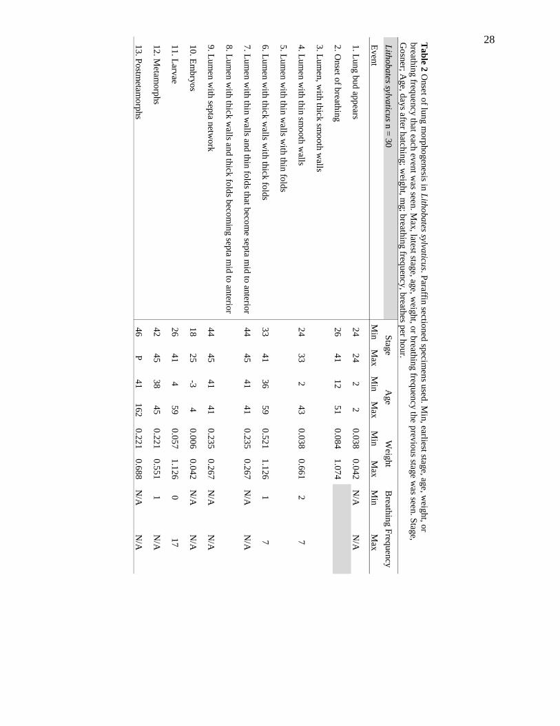
ble 2
Onse
t of lu
ng m
orp
ho
gen
esis in
Lith
ob
ates sylva
ticus. P
araffin sectio
ned
specim
en
s used
. Min
, earliest stage, ag
e, w
eight, o
r
breath
ing freq
uency th
at each
even
t was see
n. M
ax, late
st stage, ag
e, weig
ht, o
r breath
ing freq
uen
cy th
e prev
iou
s stage w
as see
n. S
tage,
Go
sner; A
ge, d
ays a
fter hatc
hin
g; w
eight, m
g; b
reathin
g freq
uen
cy, b
reathes p
er ho
ur.
Lith
ob
ates sylva
ticus n
= 3
0
Stag
e
Age
Weig
ht
Breath
ing F
requency
Even
t M
in
Max
M
in
Max
M
in
Max
M
in
Max
1. L
un
g b
ud
app
ears 2
4
24
2
2
0.0
38
0
.04
2
N/A
N
/A
2. O
nset o
f breath
ing
2
6
41
12
51
0.0
84
1
.07
4
3. L
um
en, w
ith th
ick sm
oo
th w
alls
4. L
um
en w
ith th
in sm
oo
th w
alls 2
4
33
2
43
0.0
38
0
.66
1
2
7
5. L
um
en w
ith th
in w
alls with
thin
fold
s
6. L
um
en w
ith th
ick w
alls with
thic
k fo
lds
33
41
36
59
0.5
21
1
.12
6
1
7
7. L
um
en w
ith th
in w
alls and
thin
fold
s that b
ecom
e septa m
id to
anterio
r 4
4
45
41
41
0.2
35
0
.26
7
N/A
N
/A
8. L
um
en w
ith th
ick w
alls an
d th
ick fo
lds b
ecom
ing sep
ta mid
to an
terior
9. L
um
en w
ith sep
ta netw
ork
4
4
45
41
41
0.2
35
0
.26
7
N/A
N
/A
10
. Em
bry
os
18
25
-3
4
0.0
06
0
.04
2
N/A
N
/A
11
. Larv
ae 2
6
41
4
59
0.0
57
1
.12
6
0
17
12
. Metam
orp
hs
42
45
38
45
0.2
21
0
.55
1
1
N/A
13
. Po
stmeta
mo
rphs
46
P
41
16
2
0.2
21
0.6
88
N/A
N
/A

29
Ta
ble 3
Onse
t of lu
ng m
orp
ho
gen
esis in
An
axyru
s am
erican
us. P
araffin sec
tioned
specim
ens u
sed. M
in, earliest sta
ge, ag
e, weig
ht, o
r breath
ing
frequen
cy th
at each e
vent w
as seen. M
ax, late
st stage, ag
e, weig
ht, o
r breath
ing freq
uency th
e prev
ious stag
e was see
n. S
tage, G
osn
er; Age, d
ays
after hatc
hin
g; w
eight, m
g; b
reathin
g freq
uency, b
reathes p
er ho
ur.
An
axyru
s am
erican
us n
= 1
7
Stag
e
Age
Weig
ht
Breath
ing F
requency
Even
t M
in
Max
M
in
Max
M
in
Max
M
in
Max
1. L
un
g b
ud
app
ears 3
1
37
32
60
0.0
93
0
.17
5
0
0
2. O
nset o
f breath
ing
*
46
P
10
0
15
7
0.1
03
0
.26
3. L
um
en, w
ith th
ick sm
oo
th w
alls 3
1
37
32
60
0.0
93
0
.17
5
0
0
4. L
um
en w
ith th
in sm
oo
th w
alls
5. L
um
en w
ith th
in w
alls with
thin
fold
s 4
6
46
10
0
10
0
0.1
31
0.1
31
0
0
6. L
um
en w
ith th
ick w
alls with
thic
k fo
lds
39
41
68
71
0.1
12
0
.13
0
0
7. L
um
en w
ith th
in w
alls and
thin
fold
s that b
ecom
e septa m
id to
anterio
r
8. L
um
en w
ith th
ick w
alls an
d th
ick fo
lds b
ecom
ing sep
ta mid
to an
terior
43
44
83
83
0.0
66
0
.11
8
0
0
9. L
um
en w
ith sep
ta netw
ork
4
3
P
83
15
7
0.0
66
0
.16
3
0
0
10
. Em
bry
os
20
25
0
3
0.0
06
0
.01
3
0
0
11
. Larv
ae 2
6
41
2
85
0.0
07
0.3
33
0
0
12
. Metam
orp
hs
42
45
68
10
0
0.0
48
0
.25
3
0
0
13
. Po
stmeta
mo
rphs
46
P
10
0
15
7
0.1
03
0
.26
N/A
N
/A

30
Ta
ble 4
Onse
t of lu
ng m
orp
ho
gen
esis in
Am
bysto
ma
ma
cula
tum
. Paraffin
sectioned
specim
ens u
sed. M
in, earliest stag
e, ag
e, weig
ht, o
r breath
ing fre
quen
cy
that each
event w
as seen. M
ax
, latest stage, ag
e, weig
ht, o
r breath
ing freq
uen
cy th
e prev
ious stag
e w
as see
n. S
tage, S
ala
man
der; A
ge, d
ays a
fter hatc
hin
g;
weig
ht, m
g; b
reathin
g freq
uen
cy, b
reathes p
er ho
ur.
Am
bysto
ma
ma
cula
tum
n =
15
S
tage
Age
Weig
ht
Breath
ing F
requency
Even
t M
in
Max
M
in
Max
M
in
Max
M
in
Max
1. L
un
g b
ud
app
ears 5
1
55
17
52
0.0
78
0
.32
9
2
3
2. O
nset o
f breath
ing
4
7
60
13
10
9
0.0
55
1
.27
3
3. L
um
en, w
ith th
ick sm
oo
th w
alls
4. L
um
en w
ith th
in sm
oo
th w
alls 5
1
57
17
93
0.0
78
1
.13
7
2
4
5. L
um
en w
ith th
in w
alls with
thin
fold
s 5
8
61
10
0
13
6
1.1
16
1
.52
3
0
7
6. L
um
en w
ith th
ick w
alls with
thic
k fo
lds
7. L
um
en w
ith th
in w
alls and
thin
fold
s that b
ecom
e septa m
id to
anterio
r
8. L
um
en w
ith th
ick w
alls an
d th
ick fo
lds b
ecom
ing sep
ta mid
to an
terior
9. L
um
en w
ith sep
ta netw
ork
10
. Em
bry
os
39
40
-1
0
0.0
25
0.0
28
11
. Larv
ae 4
1
55
5
67
0.0
25
0
.45
8
0
8
12
. Metam
orp
hs
56
60
60
11
6
0.3
1
1.4
29
0
12
13
. Po
stmeta
mo
rphs
61
P
11
6
13
6
1.2
47
2.0
13
N/A
N
/A

31
Table 5 Relationships between stage, age, weight, and breathing frequency to relative lung length of
each species. Stage, Gosner for L. sylvaticus and A. americanus, Salamander for A. maculatum; Age,
days after hatching; Weight, mg; Breathing frequency, breaths per hour. Whole mount specimens used.
Lithobates sylvaticus n = 54 Slope Intercept P-value
R squared
value
Average
relative lung
length
Stage 0.005368 0.558111 0.1016 0.0507
Age 0.001135 0.694486 0.2968 0.02092
Weight 0.0512 0.70793 0.3164 0.0004432
Breathing frequency -4.703x10⁻⁵ 0.7328 0.9897 3.219x10⁻⁶
Max relative
lung length
Stage 0.010458 0.444083 0.0007427 0.1982
Age 0.002711 0.693 0.00932 0.123
Weight 0.14225 0.71556 0.003397 0.1535
Breathing frequency -0.001691 0.791497 0.6381 0.004287
Anaxyrus americanus n = 48
Average
relative lung
length
Stage 0.26543 -0.751382 1.108x10^-6 0.4062
Age 0.006349 -0.155898 2.355x10^-8 0.4958
Weight -0.22896 0.35487 0.5972 0.006119
Max relative
lung length
Stage 0.027973 -0.78924 4.415x10^-7 0.4289
Age 0.0065509 -0.151398 1.777x10^-8 0.5018
Weight -0.30352 0.38872 0.4941 0.01022
Ambystoma maculatum n = 38
Average
relative lung
length
Stage 0.053651 -2.278091 3.245x10⁻⁶ 0.4565
Age 0.003676 0.4602582 6.495x10⁻⁵ 0.3618
Weight 0.2432 0.55187 0.000455 0.2925
Breathing frequency 0.024899 0.586995 0.00546 0.1954
Max relative
lung length
Stage 0.06058 -2.63511 6.585x10⁻⁷ 0.5015
Age 0.0042321 0.4523013 1.356x10⁻⁵ 0.4131
Weight 0.28985 0.55282 7.274x10⁻⁵ 0.3579
Breathing frequency 0.02651 0.605756 0.006109 0.1908

32
Figures
Figure 1 Cladogram of jawed vertebrates and the presence of lungs. Taxa in blue
indicate vertebrates with lungs.

33
Figure 2 Main difference between the gas bladder and the lung. A. Lungs occur ventral to the pharynx or digestive gut. B.
Gas bladders occur dorsal to the pharynx or digestive gut.
A
B

34
Figure 3 Lung Anatomy. A. First hypothesis of amphibian lung
septa anatomy. B. Second hypothesis of amphibian lung septa
anatomy. C. Mammal, alveolar lung anatomy. Blue, primary
septa; purple, secondary septa; green, tertiary septa.
A
C
B

35
Figure 4 General Anuran metamorphosis. Brackets
indicate presence of each respiratory surface. The
dashed bracket for lungs indicates possible presence
of organ depending on timing of development.
Cuta
neous
Lu
ng
s
Gills

36
Figure 5 Landmarked images used in measuring relative lung lengths. Pink dots
indicate points of measurements. A, the start of the bronchi; B, the caudal end of
the pleuroperitoneal cavity; C the caudal tip of right lung; D the caudal top of the
left lung. Not all specimens had lungs and some only had one lung present.
A
D
B
C

37
Figure 6 Lung morphogenesis of Lithobates sylvaticus. C and E at 4x magnification; A,D, and F-I at 10x
magnification; B at 20x magnification.

38
Figure 7 Lung morphogenesis of Anaxyrus americanus. D, F, and J at 4x magnification; B, H, and L at 10x
magnification; A, C, E, G, I, and K at 20x magnification.

39
Figure 8 Lung morphogenesis of Ambystoma maculatum. E at 4x magnification; A, B, C, and F at 10x
magnification; D at 20x magnification.

40
Figure 9 Relationships between average relative lung length and each independent variable. Blue,
Lithobates sylvaticus; Orange, Anaxyrus americanus; purple, Ambystoma maculatum. Anaxyrus
americanus were not observed to breathe air during larval and metamorphic development. Whole
mount specimens used.

41
Figure 10 Relationships between maximum relative lung length and each independent variable. Blue,
Lithobates sylvaticus; Orange, Anaxyrus americanus; purple, Ambystoma maculatum. Anaxyrus
americanus were not observed to breathe air during larval and metamorphic development. Whole
mount specimens used.

42
Figure 11 Stages present for each lung stage, onset of breathing, and developmental period. Blue, Lithobates sylvaticus;
Orange, Anaxyrus americanus; purple, Ambystoma maculatum. The y-axis corresponds to the events in Table 2-4. Paraffin
sectioned specimens and whole mount specimens used. The left side of each bar indicates the minimum organismal stage
that the lung stage occurred, and the right side of each bar indicates the maximum organismal stage that the previous lung
stage occurred. Each bar creates an underestimation of the actual range of each lung stage compared to organismal stage.

43
Figure 12 Weights present for each lung stage, onset of breathing, and developmental period. Blue, Lithobates sylvaticus;
Orange, Anaxyrus americanus; purple, Ambystoma maculatum. The y-axis corresponds to the events in Table 2-4. Paraffin
sectioned specimens and whole mount specimens used. The left side of each bar indicates the minimum weight that the
lung stage occurred, and the right side of each bar indicates the maximum weight that the previous lung stage occurred.
Each bar creates an underestimation of the actual range of each lung stage compared to weight.

44
Figure 13 Ages present for each lung stage, onset of breathing, and developmental period. Blue, Lithobates sylvaticus;
Orange, Anaxyrus americanus; purple, Ambystoma maculatum. The y-axis corresponds to the events in Table 2-4. Paraffin
sectioned specimens and whole mount specimens used. The left side of each bar indicates the minimum age that the lung
stage occurred, and the right side of each bar indicates the maximum age that the previous lung stage occurred. Each bar
creates an underestimation of the actual range of each lung stage compared to age.

45
Figure 14 Breaths per hour present for each lung stage and developmental period. Blue, Lithobates sylvaticus; Orange,
Anaxyrus americanus; purple, Ambystoma maculatum. The y-axis corresponds to the events in Table 2-4. Paraffin
sectioned specimens and whole mount specimens used. The left side of each bar indicates the minimum breaths per hour
that occurred at that lung stage, and the right side of each bar indicates the maximum breathes per hour that occurred at
the previous lung stage. Each bar creates an underestimation of the actual range of each lung stage compared to breathes
per hour.
46
LITERATURE CITED
Altig, R. and McDiarmid, R. W. (1999). Body Plan: Development and Morphology. In
Tadpoles: The Biology of Anuran Larvae (ed. McDiarmid, R. W.) and Altig, R.),
pp. 24–51. Chicago, IL: University of Chicago Press.
Bartel, H. and Lametschwandtner, A. (2000). Intussusceptive Microvascular Growth in
the Lung of Larval Xenopus laevis Daudin: A Light Microscope, Transmission
Electron Microscope and SEM study of Microvascular Corrosion Casts. Anat.
Embryol. (Berl.) 202, 55–65.
Beane, J. C., Braswell, A. L., Mitchell, J. C., Palmer, W. M. and Harrison III, J. R.
(2010). Amphibians and Reptiles of the Carolinas and Virginia. 2nd ed. Univ of
North Carolina Press.
Brainerd, E. L. (2015). Major Transformations in Vertebrate Breathing Mechanisms. In
Great Transformations in Vertebrate Evolution (ed. Dial, K. P.), Shubin, N.), and
Brainerd, E. L.), pp. 47–61. Chicago, IL: University of Chicago Press.
Broughton, R. E., Betancur-R., R., Li, C., Arratia, G. and Ortí, G. (2013). Multi-
locus Phylogenetic Analysis Reveals the Pattern and Tempo of Bony Fish
Evolution. PLoS Curr.
Brown, D. D. and Cai, L. (2007). Amphibian Metamorphosis. Dev. Biol. 306, 20–33.
Burggren, W. W. and Just, J. J. (1992). Developmental Changes in Physiological
Systems. In Environmental Physiology of the Amphibians (ed. Feder, M. E.) and
Burggren, W. W.), pp. 467–530. Chicago, IL: University of Chicago Press.
Daniels, C. B. and Orgeig, S. (2003). Pulmonary Surfactant: The Key to the Evolution
of Air Breathing. News Physiol Sci 18, 151–157.
Daniels, C. B., Orgeig, S., Sullivan, L. C., Ling, N., Bennett, M. B., Schürch, S., Val,
A. L. and Brauner, C. J. (2004). The Origin and Evolution of the Surfactant
System in Fish: Insights into the Evolution of Lungs and Swim Bladders. Physiol.
Biochem. Zool. 77, 732–749.
Duellman, W. E. and Trueb, L. (1994). Biology of Amphibians. Baltimore, MD: John
Hopkins University Press.
Duncker, H.-R. (2004). Vertebrate Lungs: Structure, Topography and Mechanics.
Respir. Physiol. Neurobiol. 144, 111–124.
Francis, E. T. B. (1934). The Anatomy of the Salamander. Oxford, UK: Clarendon Press.

47
Goniakowska-Witalińska, L. (1978). Ultrastructural and Morphometric Study of the
Lung of the European Salamander, Salamandra salamandra L. Cell Tissue Res.
191, 343–356.
Goniakowska-Witalińska, L. (1980). Ultrastructural and Morphometric Changes in the
Lung of Newt, Triturus cristatus carnifex Laur. during Ontogeny. J Anat 130,
571–583.
Goniakowska-Witalińska, L. (1982). Development of the Larval Lung of Salamandra
salamandra L. A Scanning and Transmission Electron Microscopic Study. Anat.
Embryol. (Berl.) 164, 113–137.
Gosner, K. L. (1960). A Simplified Table for Staging Anuran Embryos and Larvae with
Notes on Identification. Herpetologica 16, 183–190.
Hermida, G. N., Fiorito, L. E. and Farias, A. (1998). The Lung of the Common Toad,
Bufo arenarum (Anura: Bufonidae). A Light and Electron Microscopy Study.
Biocell 22, 19–26.
Kardong, K. V. (2015). Vertebrates: Comparative Anatomy, Function, Evolution. 7th ed.
New York. NY: McGraw-Hill.
Liem, K. F., Bemis, W. E., Walker Jr, W. F. and Grande, L. (2001). Functional
Anatomy of the Vertebrates: An Evolutionary Perspective. 3rd ed. Orlando, FL:
Harcourt College Publishers.
Longo, S., Riccio, M. and McCune, A. R. (2013). Homology of lungs and gas bladders:
Insights from arterial vasculature: Vasculature and Homology of Air-Filled
Organs. J. Morphol. 274, 687–703.
Maina, J. N. (2006). Development, Structure, and Function of a Novel Respiratory
Organ, the Lung-air Sac System of Birds: To Go Where No Other Vertebrate Has
Gone. Biol. Rev. 81, 545.
McDiarmid, R. W. and Altig, R. (1999). Research: Materials and Techniques. In
Tadpoles: The Biology of Anuran Larvae (ed. McDiarmid, R. W.) and Altig, R.),
pp. 7–23. Chicago, IL: University of Chicago Press.
Mekeel, A. G. (1930). Pulmonary Development in the Lungless Salamanders.
Metzger, R. J., Klein, O. D., Martin, G. R. and Krasnow, M. A. (2008). The
Branching Programme of Mouse Lung Development. Nature 453, 745–750.

48
Miyata, K. and Ose, K. (2012). Thyroid Hormone-Disrupting Effects and the
Amphibian Metamorphosis Assay. J. Toxicol. Pathol. 25, 1–9.
Nieuwkoop, P. D. and Faber, J. (1956). Normal Table of Xenopus laevis (Daudin). A
Systematical and Chronological Survey of the Development from the Fertilized
Egg till the End of Metamorphosis. Amsterdam: North-Holland Publishing
Company.
Noble, G. K. (1931). The Biology of the Amphibia. New York, NY: McGraw-Hill Inc.
Nodzenski, E., Wassersug, R. J. and Inger, R. F. (1989). Developmental Differences in
Visceral Morphology of Megophryine Pelobatid Tadpoles in Relation to their
Body Form and Mode of Life. Biol. J. Linn. Soc. 38, 369–388.
Okada, Y., Ishiko, S., Daido, S., Kim, J. and Ikeda, S. (1962). Comparative
Morphology of the Lung with Special Reference to the Alveolar Epithelial cells:
I. Lung of the Amphibia. Acta Tuberc. Jpn. 11, 63–72.
Perry, S. F. and Sander, M. (2004). Reconstructing the Evolution of the Respiratory
Apparatus in Tetrapods. Respir. Physiol. Neurobiol. 144, 125–139.
Perry, S. F., Wilson, R. J., Straus, C., Harris, M. B. and Remmers, J. E. (2001).
Which Came First, the Lung or the Breath? Comp. Biochem. Physiol. A. Mol.
Integr. Physiol. 129, 37–47.
Pyron, A. R. and Wiens, J. J. (2011). A Large-Scale Phylogeny of Amphibia Including
Over 2800 Species, and a Revised Classification of Extant Frogs, Salamanders,
and Caecilians. Mol. Phylogenet. Evol. 61, 543–583.
R Core Team (2016). R Lang. Environ. Stat. Comput. R Found. Stat. Comput. Vienna
Austria.
Rankin, S. A., Thi Tran, H., Wlizla, M., Mancini, P., Shifley, E. T., Bloor, S. D.,
Han, L., Vleminckx, K., Wert, S. E. and Zorn, A. M. (2015). A Molecular
Atlas of Xenopus Respiratory System Development: Molecular Atlas of Xenopus
Respiratory System Development. Dev. Dyn. 244, 69–85.
Rawlins, E. L. (2011). The Building Blocks of Mammalian Lung Development. Dev.
Dyn. 240, 463–476.
Romer, A. S. and Parsons, T. A. (1977). The Vertebrate Body. Philadelphia, PA:
W.B.Saunders.
49
Rose, C. S. (2014). Caging, but not Air Deprivation, Slows Tadpole Growth and
Development in the Amphibian Xenopus laevis. J. Exp. Zool. Part Ecol. Genet.
Physiol. 321, 365–375.
Rose, C. S. and James, B. (2013). Plasticity of Lung Development in the Amphibian,
Xenopus laevis. Biol. Open 2, 1324–1335.
Rugh, R. (1962). Experimental Embryology Techniques and Procedures. Minneapolis,
MN: Burgess Publishing Co.
Shi, Y.-B. (2000). Amphibian Metamorphosis: From Morphology to Molecular Biology.
Wiley.
Shoemaker, V. H., Hillman, S. S., Hillyard, S. D., Jackson, D. C., McClanahan, L.
L., Withers, P. C. and Wygoda, M. L. (1992). Exchange of Water, Ions, and
Respiratory Gases in Terrestrial Amphibians. In Environmental Physiology of the
Amphibians (ed. Feder, M. E.) and Burggren, W. W.), pp. 125–150. Chicago, IL:
University of Chicago Press.
Shoop, C. R. (1974). Yearly Variation in Larval Survival of Ambystoma maculatum.
Ecology 55, 440–444.
Smith, D. G. and Campbell, G. (1976). The Anatomy of the Pulmonary Vascular Bed in
the Toad Bufo marinus. Cell Tissue Res. 165, 199–213.
Ultsch, G. R., Bradford, D. F. and Freda, J. (1999). Physiology: Coping with the
Environment. In Tadpoles: The Biology of Anuran Larvae (ed. McDiarmid, R.
W.) and Altig, R.), pp. 189–214. Chicago, IL: University of Chicago Press.
Viertel, B. and Richter, S. (1999). Anatomy: Viscera and Endocrines. In Tadpoles: The
Biology of Anuran Larvae (ed. McDiarmid, R. W.) and Altig, R.), pp. 92–148.
Chicago, IL: University of Chicago Press.
Virginia Herpetological Society.
Wassersug, R. J. and Seibert, E. A. (1975). Behavioral Responses of Amphibian
Larvae to Variation in Dissolved Oxygen. Copeia 86–103.
Waterman, F. A. (1939). The Origin and Development of the Internal Musculature of
the Frog Lung (Rana pipiens).


![[ 135 ] ON INTRASPECIFIC AND INTERSPECIFIC COMPETITION IN ... · BY A. C. CROMBIE From the Zoological Laboratory, Cambridge (Received 20 October 1943) (With Five Text-figures) The](https://static.fdocuments.net/doc/165x107/5e2affd524b4ce03467fcc61/-135-on-intraspecific-and-interspecific-competition-in-by-a-c-crombie-from.jpg)