Introduction to the Central Nervous System Structure · Introduction to the Nervous System ......
48
Chapter 14 Introduction to the Central Nervous System Structure
Transcript of Introduction to the Central Nervous System Structure · Introduction to the Nervous System ......

Chapter 14
Introduction to the CentralNervous System Structure

Introduction to the Nervous System
• Aristotle thought brain was a ‘radiator’ to cool blood
• Hippocrates was more accurate
– “from the brain only, arises our pleasures, joys, laughter, and jests, as well as our sorrows, pains, griefs, and tears”
• The cessation of brain activity = clinical criterion of death

Evolution of the CNS
– spinal cord changed little throughout vertebrate evolution
– brain has changed a great deal over the last 20 million years.
• greatest growth in areas of vision, memory, and motor control of the prehensile hand.
• brain functions are “layered” // see Triune Brain
– “primitive functions” placed lower in the CNS
– more “advanced functions” placed higher in the CNS architecture

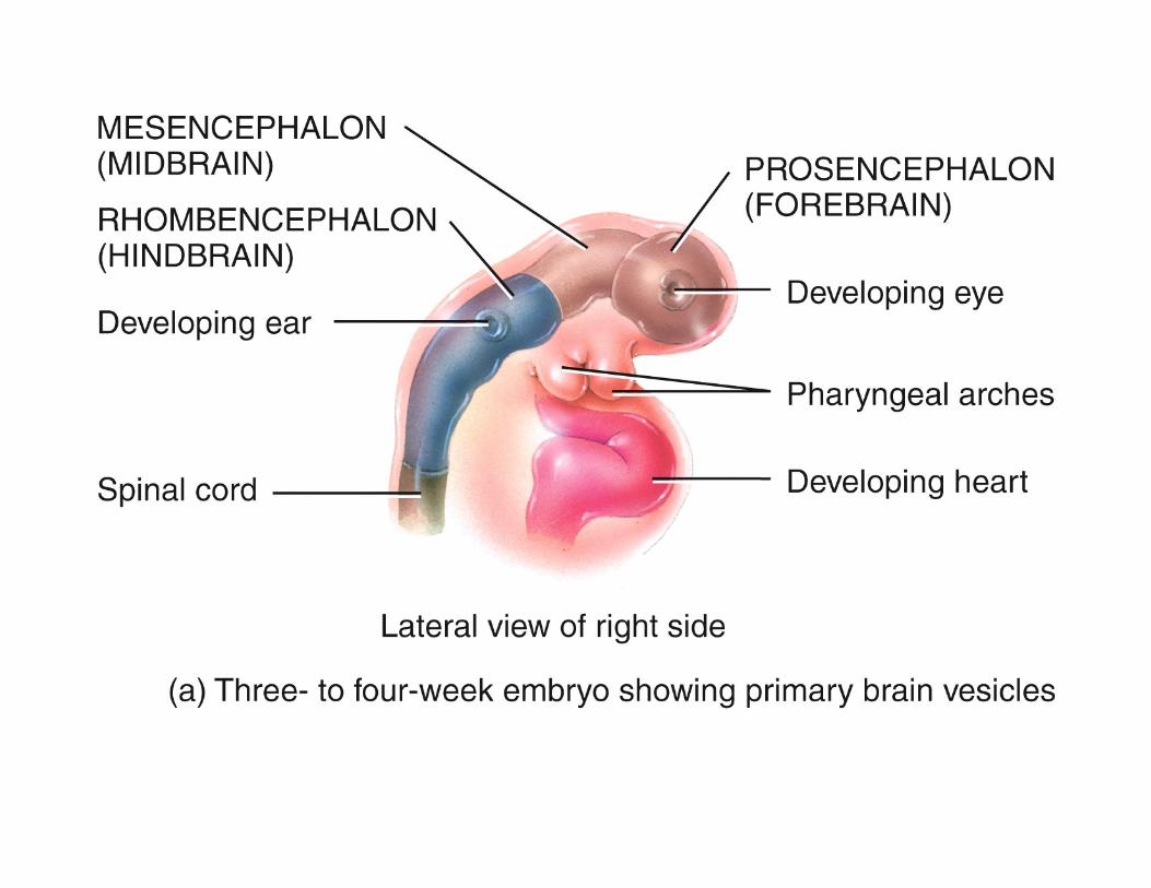
There Are Two Different Methods Used to Define the
Central Nervous System
• Forebrain– Diencephalon– Cerebrum
• Hindbrain– Brain Stem– Pons– Midbrain– Cerebellum
The first system is more commonly used in the study of embryonic growth and development. (see next slide)
To study the adult brain we will use the second method. (slide 2)

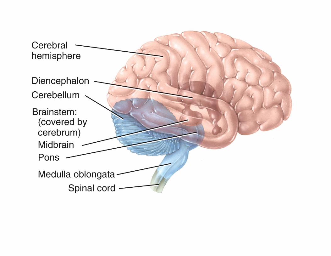
Three Major Divisions of the Adult Brain
– cerebrum is 83% of brain volume; cerebral hemispheres, gyri and sulci, longitudinal fissure, corpus callosum
– cerebellum contains 50% of the neurons; second largest brain region, located in posterior cranial fossa
– brainstem the portion of the brain that remains if the cerebrum and cerebellum are removed;
• Diencephalon• Midbrain• Pons• medulla oblongata
Brainstem
Cerebellum
Cerebrum
Spinal cord
Rostral Caudal
Central sulcus
Lateral sulcus
Gyri
(b) Lateral view
Temporal lobe
Copyright © The McGraw-Hill Companies, Inc. Permission required for reproduction or display.
Know This For Exam

Directional Terms, Landmarks, and Divisions
• rostral - toward the forehead
• caudal - toward the occipital lobe / spinal cord
• brain weighs about– 1600 g (3.5 lb) in men– 1450 g in women
Brainstem
Cerebellum
Cerebrum
Spinal cord
Rostral Caudal
Central sulcus
Lateral sulcus
Gyri
(b) Lateral view
Temporal lobe
Cerebrum
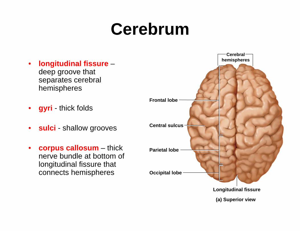
• longitudinal fissure –deep groove that separates cerebral hemispheres
• gyri - thick folds
• sulci - shallow grooves
• corpus callosum – thick nerve bundle at bottom of longitudinal fissure that connects hemispheres
Frontal lobe
Occipital lobe
Central sulcus
Longitudinal fissure
Parietal lobe
(a) Superior view
Cerebralhemispheres


Corpus callosum

Brainstem
• This is what remains of the brain after the cerebrum and cerebellum are removed from the whole brain.
• major components– diencephalon
– midbrain
– pons
– medulla oblongata
Brainstem
Cerebellum
Cerebrum
Spinal cord
Rostral Caudal
Central sulcus
Lateral sulcus
Gyri
(b) Lateral view
Temporal lobe
Copyright © The McGraw-Hill Companies, Inc. Permission required for reproduction or display.

Note: Diencephalon also includes hypothalamus and epithalamus (pineal gland & habenula)


Cerebellum
• occupies posterior cranial fossa
• marked by gyri, sulci, and fissures
• about 10% of brain volume
• contains over 50% of brain neurons


Median Section of the Brain
leaves
Thalamus
Hypothalamus
Frontal lobe
Corpus callosum
Cingulate gyrus
Optic chiasm
Pituitary gland
Mammillary body
Midbrain
Pons
Central sulcus
Parietal lobe
Parieto–occipital sulcus
Occipital lobe
Pineal glandHabenula
Posterior commissure
Cerebral aqueduct
Fourth ventricle
Cerebellum
(a)
EpithalamusAnteriorcommissure
Temporal lobe
Medullaoblongata

Median Section of Cadaver Brain
Corpus callosum
Cingulate gyrus
Lateral ventricle
Thalamus
Hypothalamus
Midbrain
Cerebellum
Fourth ventriclePons
(b)
Choroid plexusPineal gland
Occipital lobe
Parieto–occipitalsulcus
Posteriorcommissure
Medullaoblongata

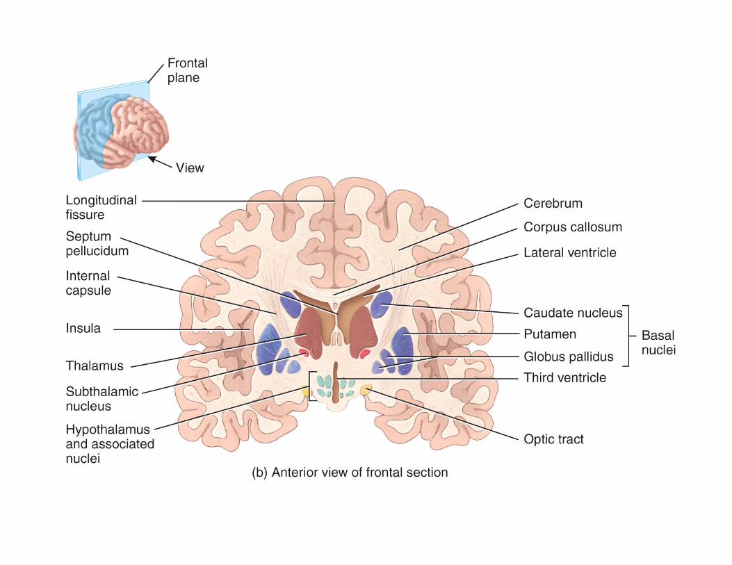
Gray and White Matter• gray matter – consists of the
neuron cell bodies, dendrites, and synapses
– dull white color when fresh
– due to little myelin on the surface of the cell bodies, dendrites and synapses
– Grey matter forms surface layer, ”the cortex”, over cerebrum and cerebellum
– forms nuclei deep within brain

Gray and White Matter
• white matter - bundles of axons
– lies deep to cortical gray matter, opposite relationship in the spinal cord
– pearly white color frommyelin around nerve fibers
– composed of tracts, bundles of axons,
– connect one part of the brain to another, and to the spinal cord


The Central Theme of This Class is Structure and Function.
We Have Just Reviewed the Structural Organization of the
Brain. Now We Will Explore the Functional Organization of the Brain.

Triune Brain Theory• The modern brain evolved in three evolutionary stages or
formations.
• Each formation existed for relatively long stable periods followed by the development of another formation. This pattern existed throughout the evolution of the vertebrate brain.
• The Three Formations
– Protoreptilian Brain Formation:– Paleomammalian Brain Formation (Limbic System):– Neomammalian Brain Formation (Neocortex):

Summary of the TriuneBrain Theory
Protoreptilian
Formation
Paleomammalian
FormationNeocortex
Formation

The most efficient model for understanding the brain in terms of its evolutionary history is the famous triune brain theory develop by Paul MacLean. According to this theory, the following three distinct brains emerged successively in the course of evolution. These three brains now co-inhabit the human skull.
Protoreptilian
Formation
Paleomammalian
FormationNeocortex
Formation

The reptilian brain (or Protoreptilian), the oldest of the three brain formations. It controls the body's vital functions such as heart rate, breathing, body temperature and balance.
Our reptilian brain includes the main structures found in a reptile's brain: the brainstem and the cerebellum (later in evolutionary development the reptilian brain included the basal nuclei and mid-brain).
The reptilian brain is reliable. It responds to stimuli with genetically encoded instinctual action plans for primitive survival behaviors, such as exploration, feeding, aggression, dominance, and sexuality. The RB tends to be somewhat rigid and compulsive.

The limbic brain processes innate emotions (fear, aggression, pleasure), records these experiences, and process these experiences with a motivational system (i.e. reward or pleasure pathways) which then shape behavioral responses to incoming stimuli based on instincts and past experience. This is how we mediate the social emotions, playfulness, and maternal nurturance.
The limbic brain (also called Paleomammalianformation) emerged in the first mammals. It gave the early mammal brains the ability to record memories of behaviors as agreeable and disagreeable experiences. It is responsible for what are called emotions in human beings .
The main structures of the limbic brain are the hippocampus, the amygdala, and the hypothalamus (includes other interconnected structures - the amygdala, hippocampus, hypothalamus and other structures which are sometimes called the limbic lobe or simply the limbic system). The limbic brain is the seat of our value judgments. These are decisions, made as part of our unconscious brainactivity that then exert a strong influence on our conscious behavior.

The neocortex (also called Neomammalian formation) first assumed importance in whales, primates and culminated in the human brain. Two large cerebral hemispheres are the dominate structures of the neocortex. This is the location responsible for the development of human language, abstract thought, imagination, and consciousness. The neocortex is flexible and has almost infinite learning abilities .
The neocortex is also the location for Declarative Knowledge. This is the knowledge we learn about from the world that we live in. It is knowledge derived especially from sight, sound and touch. The neocortex with its declarative knowledge is most developed in humans. Without a neocortex, we would not have a culture.

These three parts of the brain are still functional in today’s human brain and they do not operate independently of one another They have established numerous interconnections through which they influence one another.
So the uncounsciousemotional brain will make value judgments which precede our conscious thoughts. Neural pathways between the limbic system and the cortex compete to shape the outcome of the experience. Will the emotional brain cripple the neocortex? Will the experience leave you paralyzed in fear and anxiety? Will you be unable to use the neocortex to remember that you left your keys in your coat pocket?
Human brain showing nerve tracks that connect the three brain formations.

The Limbic System
(The Paleo-Mammalian Brain Formation)
If you want to learn more about the structure and function of the limbic system then go to the links for Dr. Robert Sapolsky’s videos on Web site’s Home Page.

• For more information about the Triune Brain Theory see www.mc3cb.com ///
– go to the Archival Articles Link / see brain section.

What is a PET Scan?
• A PET scan uses radiation, or nuclear medicine imaging, to produce 3-dimensional, color images of the functional processes within the human body. PET stands for positron emission tomography.
• The machine detects pairs of gamma rays that are emitted indirectly by a tracer (positron-emitting radionuclide), which is placed in the body on a biologically active molecule (e.g. glucose). The images are reconstructed by computer analysis.
• Modern machines often use a Computer Tomography X-ray scan which is performed on a patient at the same time in the same machine.
New non-invasive technologies allow neuroscientist to study both the structure and function of a living brain. CT,
PET, and fMRI are now the new images of the brain.

PET Image

Images of the Mind• Positron emission tomography (PET)
– visualize increases in blood flow when brain areas are active
– injection of radioactively labeled glucose
– Metabolic active areas of brain will “light up”
– So if you ask a patient to do something or say something during a PET Scan then the area of the brain processing the command will increase metabolism to process the request and it will “light up”
What is fMRI?
• Functional magnetic resonance imaging or functional MRI (fMRI) is a functional neuroimaging procedure using MRI technology that measures brain activity by detecting associated changes in blood flow. This technique relies on the fact that cerebral blood flow and neuronal activation are coupled. When an area of the brain is in use, blood flow to that region also increases.
• The primary form of fMRI uses th e blood-oxygen-level-dependent (BOLD) contrast, discovered by Seiji Ogawa. This is a type of specialized brain and body scan used to map neural activity in the brain or spinal cord of humans or other animals by imaging the change in blood flow (hemodynamic response) related to energy use by brain cells. Since the early 1990s, fMRI has come to dominate brain mapping research because it does not require people to undergo shots, surgery, or to ingest substances, or be exposed to radiation, etc. Other methods of obtaining contrast are arterial spin labeling and diffusion MRI.
• The procedure is similar to MRI but uses the change in magnetization between oxygen-rich and oxygen-poor blood as its basic measure. This measure is frequently corrupted by noise from various sources and hence statistical procedures are used to extract the underlying signal. The resulting brain activation can be presented graphically by color-coding the strength of activation across the brain or the specific region studied. The technique can localize activity to within millimeters but, using standard techniques, no better than within a window of a few seconds
Images of the Mind
• functional magnetic resonance imaging (fMRI)
– looks at increase in blood flow to an area (additional glucose is needed in active area)
– magnetic properties of hemoglobin dependent on difference between oxygenated and unoxygenated magnetic properties of hemoglobin / how much oxygen is bound to it
– quick, safe and accurate method to see brain function

fMRI

What is CT Imaging?
• X-ray computed tomography (x-ray CT) is a technology that uses computer-processed x-rays to produce tomographic images (virtual 'slices') of specific areas of the scanned object, allowing the user to see inside without cutting. Digital geometry processing is used to generate a three-dimensional image of the inside of an object from a large series of two-dimensional radiographic images taken around a single axis of rotation. Medical imaging is the most common application of x-ray CT. Its cross-sectional images are used for diagostic and therapeutic purposes in various medical disciplines. The rest of this article discusses medical-imaging x-ray CT.

CT Image

PET Scans and Language Task
Broca area
Primary motor cortexPremotor areaPrimary auditory cortex
1 2 3 4
Rostral Caudal
Visual cortex
The word car is seen inthe visual cortex.
Wernicke area conceivesof the verb drive to go with it.
Wernicke area
Broca area compiles amotor program to speakthe word drive.
The primary motor cortexexecutes the program andthe word is spoken.