
Introduction to Imaging - Kampmann Lab · Introduction to Imaging DeLaine Larsen, PhD Director,...
23
Introduction to Imaging DeLaine Larsen, PhD Director, Nikon Imaging Center Website: nic.ucsf.edu
Transcript of Introduction to Imaging - Kampmann Lab · Introduction to Imaging DeLaine Larsen, PhD Director,...
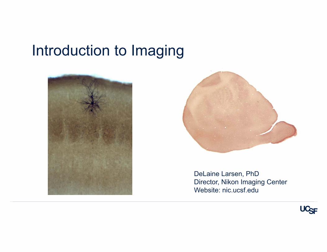
Introduction to Imaging
DeLaine Larsen, PhDDirector, Nikon Imaging CenterWebsite: nic.ucsf.edu
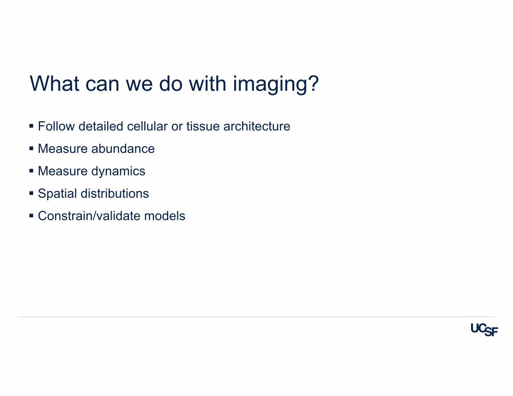
What can we do with imaging?
Follow detailed cellular or tissue architecture
Measure abundance
Measure dynamics
Spatial distributions
Constrain/validate models
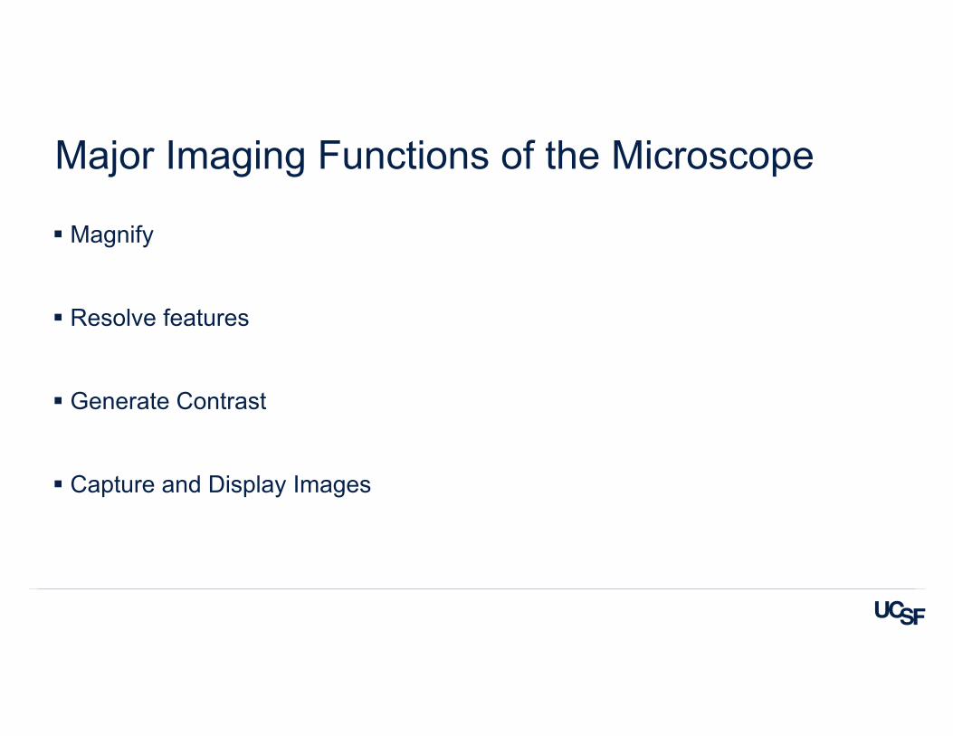
Major Imaging Functions of the Microscope
Magnify
Resolve features
Generate Contrast
Capture and Display Images
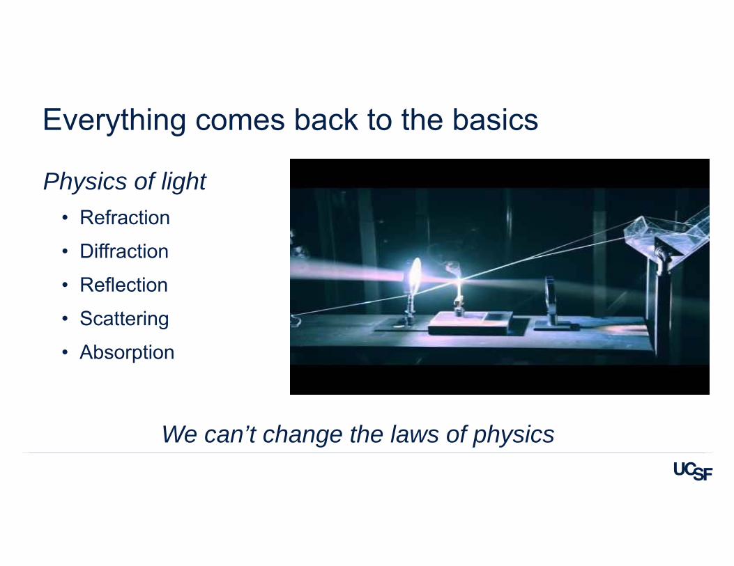
Everything comes back to the basics
Physics of light• Refraction
• Diffraction
• Reflection
• Scattering
• Absorption
We can’t change the laws of physics
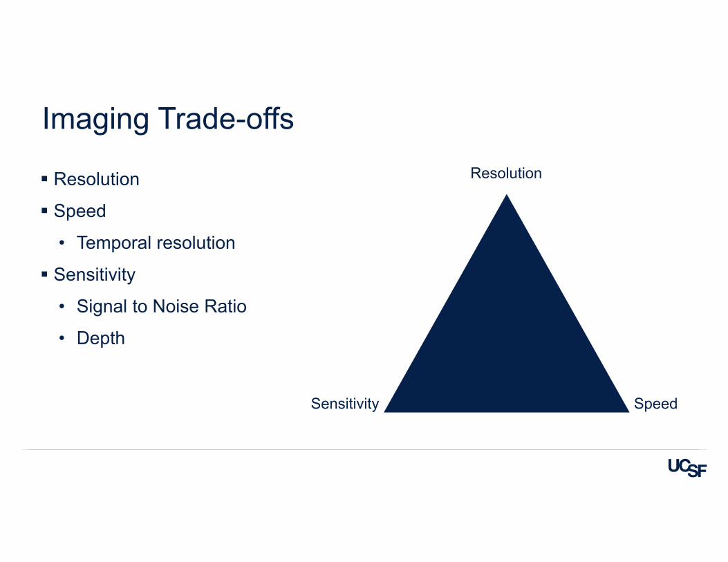
Imaging Trade-offs
Resolution
Speed
• Temporal resolution
Sensitivity
• Signal to Noise Ratio
• Depth
Resolution
SpeedSensitivity
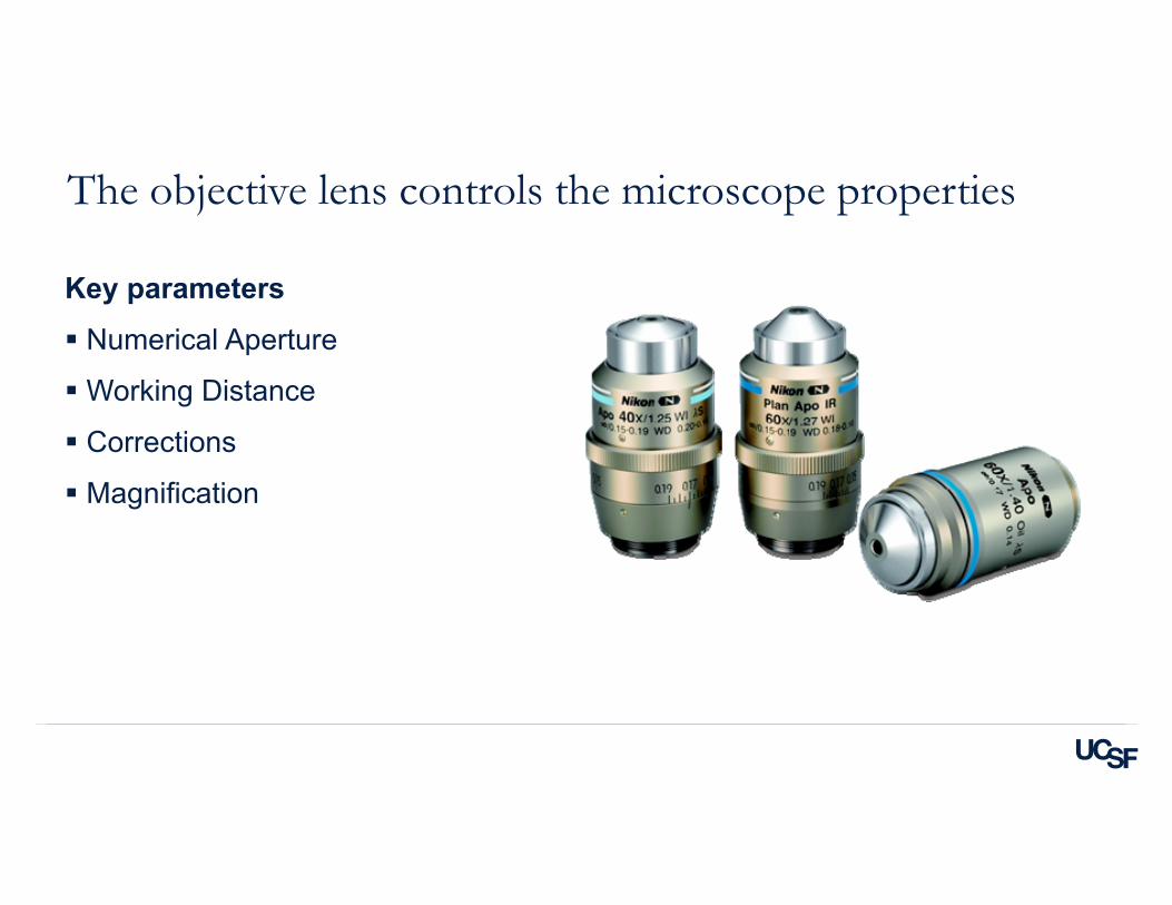
The objective lens controls the microscope properties
Key parameters
Numerical Aperture
Working Distance
Corrections
Magnification
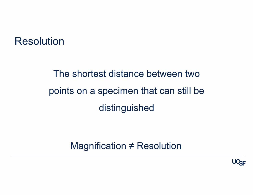
Resolution
The shortest distance between two
points on a specimen that can still be
distinguished
Magnification ≠ Resolution
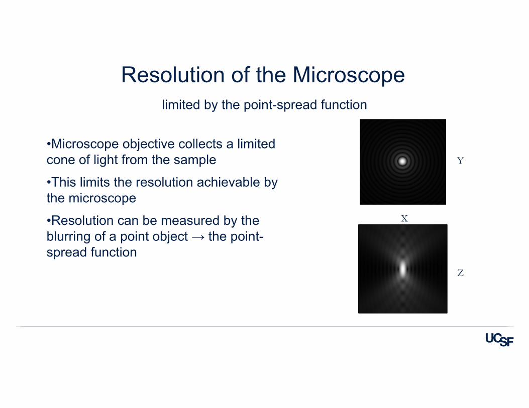
Y
Z
X
•Microscope objective collects a limited cone of light from the sample
•This limits the resolution achievable by the microscope
•Resolution can be measured by the blurring of a point object → the point-spread function
Resolution of the Microscopelimited by the point-spread function
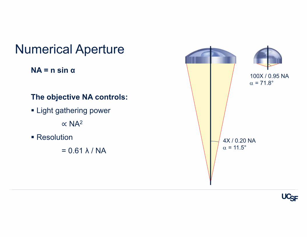
NA = n sin α
The objective NA controls:
Light gathering power
∝ NA2
Resolution
= 0.61 λ / NA
Numerical Aperture
4X / 0.20 NA = 11.5°
100X / 0.95 NA = 71.8°
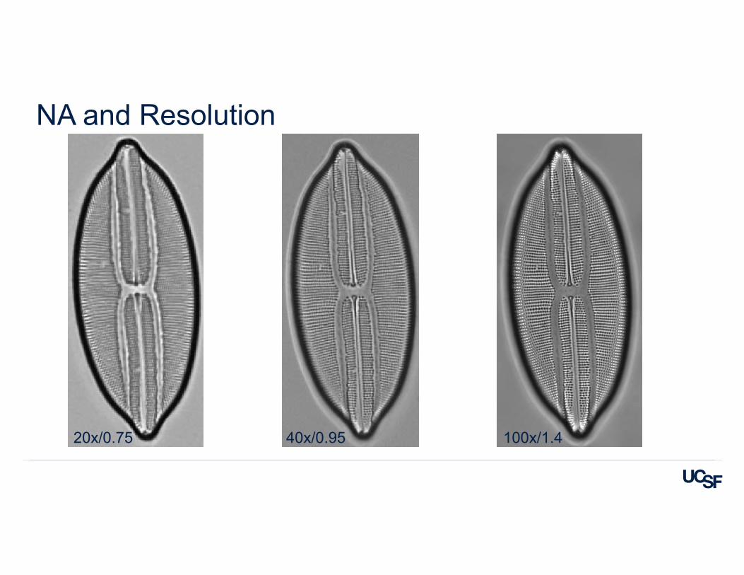
NA and Resolution
20x/0.75 40x/0.95 100x/1.4
Sensitivity
Sample side
Fluorophores
• Number
• Brightness
• Photostability
Phototoxicity
Signal to Noise Ratio
Hardware side
Camera or detector choice can determine
• Sensitivity
• Speed
• Sampling
Objective choice
Cameras
sCMOS
Large sensor size (2k x 2k)
Smaller pixel size
Fast
Fixed pattern noise
EMCCD
Smaller sensor size (512 x 512)
Larger pixels
Slower
More sensitive than sCMOS for dim samples
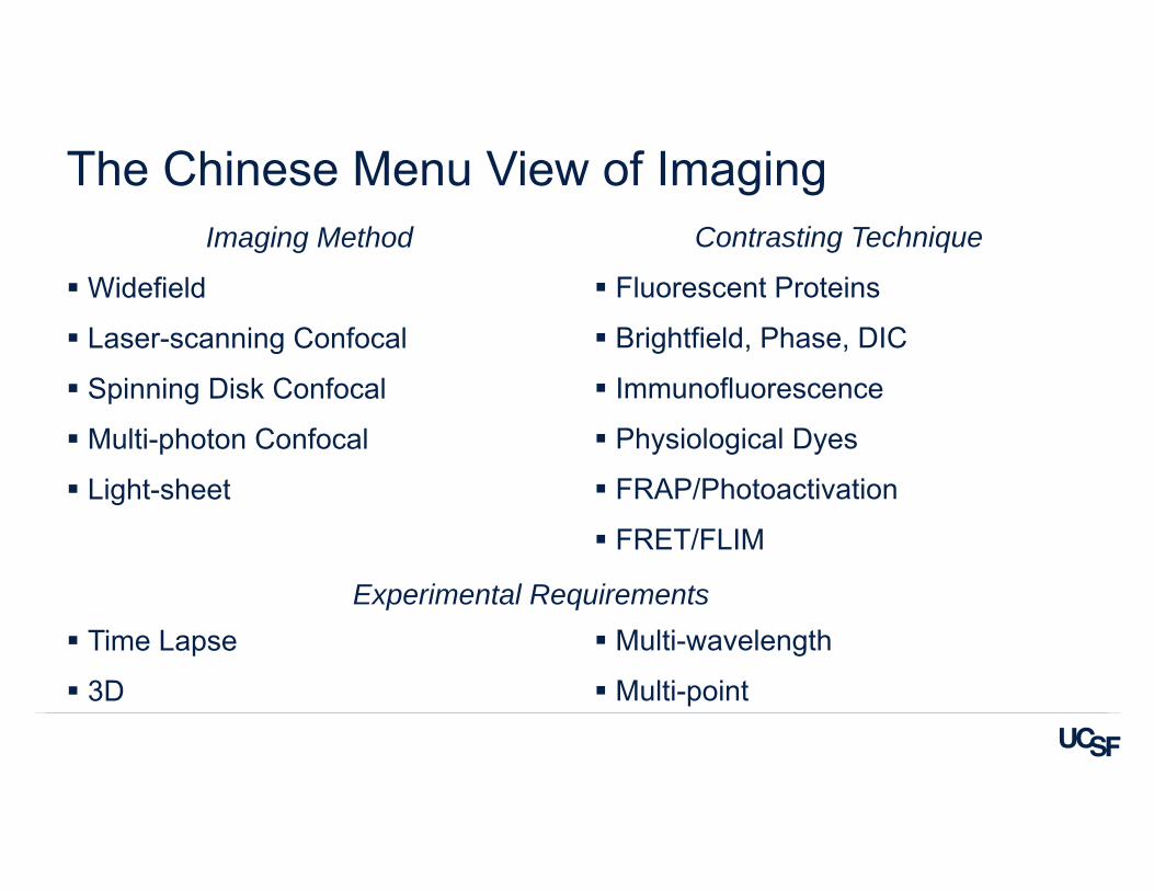
The Chinese Menu View of ImagingImaging Method
Widefield
Laser-scanning Confocal
Spinning Disk Confocal
Multi-photon Confocal
Light-sheet
Time Lapse
3D
Contrasting Technique
Fluorescent Proteins
Brightfield, Phase, DIC
Immunofluorescence
Physiological Dyes
FRAP/Photoactivation
FRET/FLIM
Multi-wavelength
Multi-point
Experimental Requirements
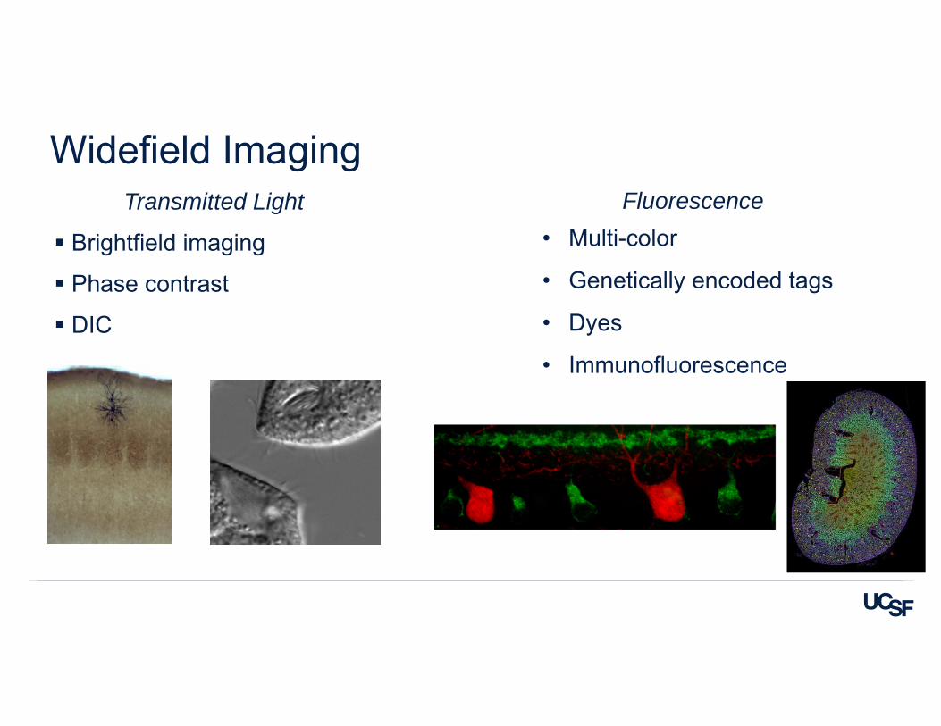
Widefield ImagingTransmitted Light
Brightfield imaging
Phase contrast
DIC
Fluorescence • Multi-color
• Genetically encoded tags
• Dyes
• Immunofluorescence
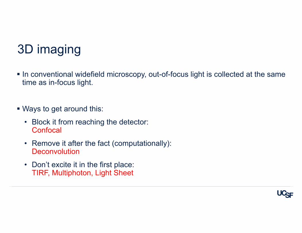
3D imaging
In conventional widefield microscopy, out-of-focus light is collected at the same time as in-focus light.
Ways to get around this:
• Block it from reaching the detector: Confocal
• Remove it after the fact (computationally):Deconvolution
• Don’t excite it in the first place:TIRF, Multiphoton, Light Sheet
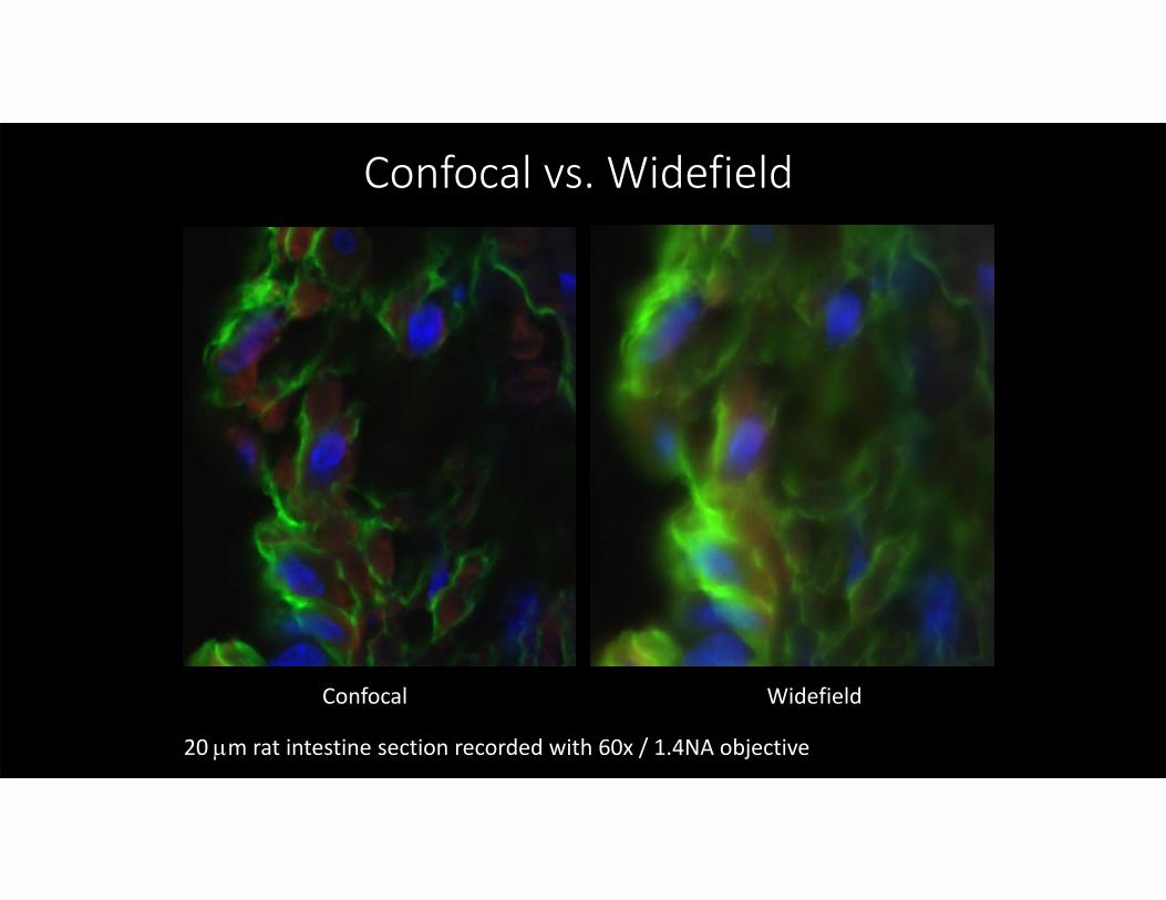
Confocal vs. Widefield
Confocal Widefield
20 m rat intestine section recorded with 60x / 1.4NA objective
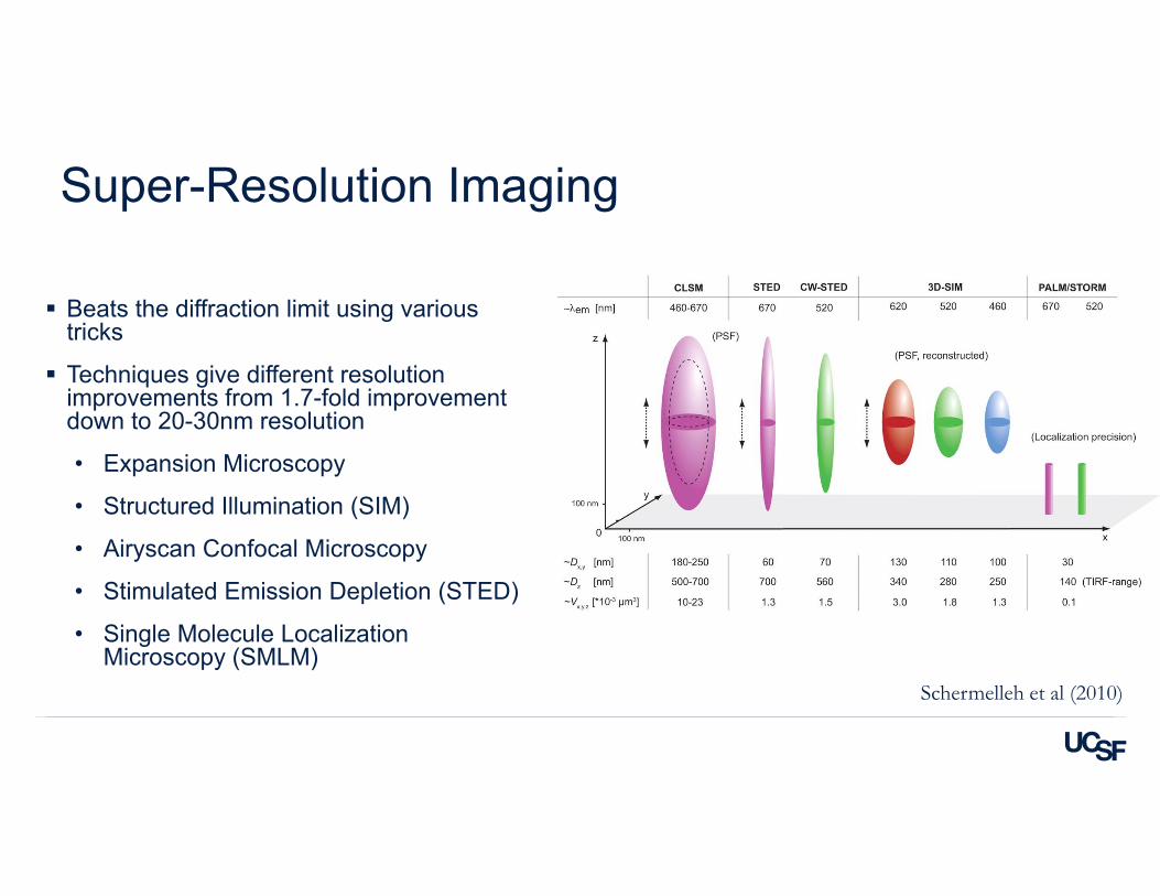
Super-Resolution Imaging
Beats the diffraction limit using various tricks
Techniques give different resolution improvements from 1.7-fold improvement down to 20-30nm resolution
• Expansion Microscopy
• Structured Illumination (SIM)
• Airyscan Confocal Microscopy
• Stimulated Emission Depletion (STED)
• Single Molecule Localization Microscopy (SMLM)
Schermelleh et al (2010)
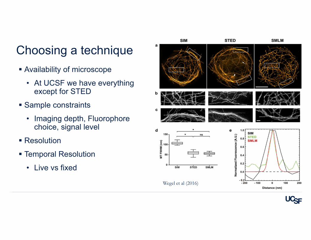
Choosing a technique Availability of microscope
• At UCSF we have everything except for STED
Sample constraints
• Imaging depth, Fluorophore choice, signal level
Resolution
Temporal Resolution
• Live vs fixed
Wegel et al (2016)
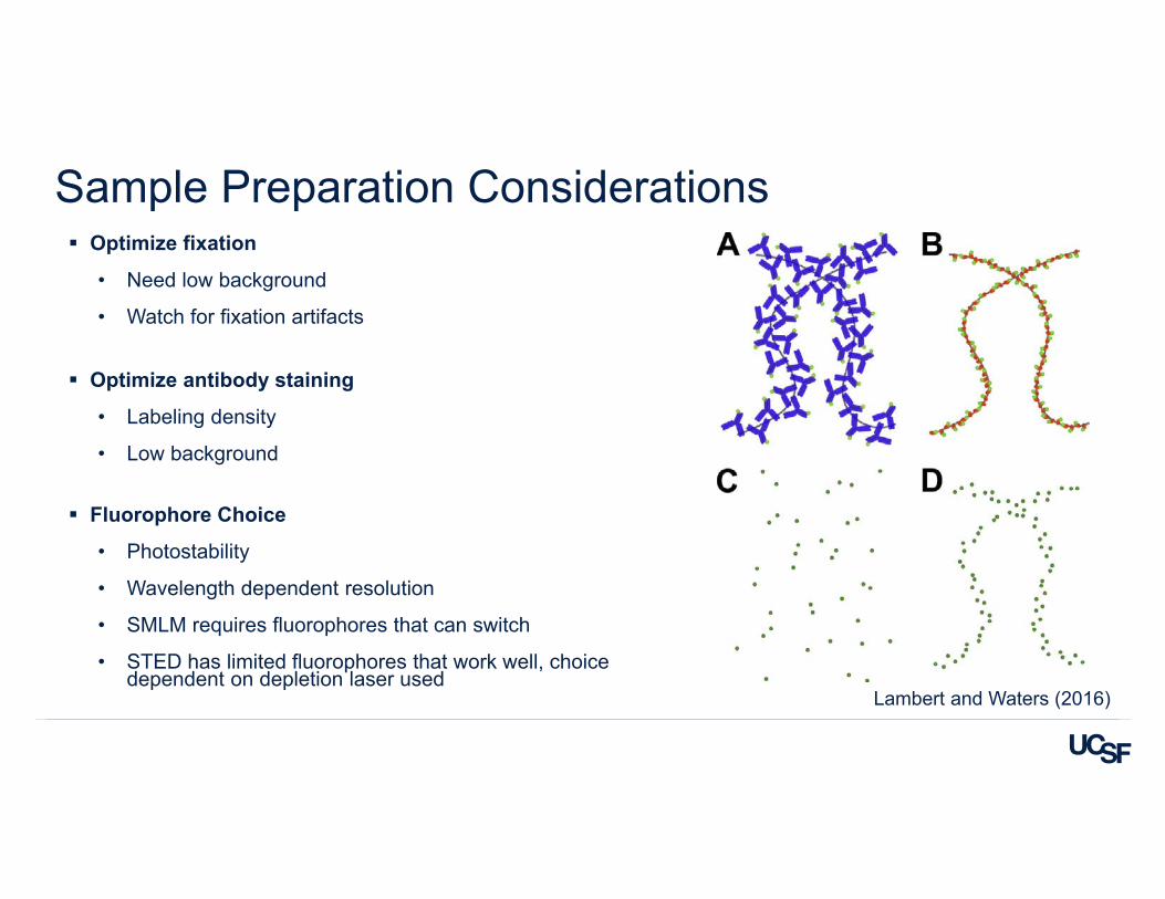
Sample Preparation Considerations Optimize fixation
• Need low background
• Watch for fixation artifacts
Optimize antibody staining
• Labeling density
• Low background
Fluorophore Choice
• Photostability
• Wavelength dependent resolution
• SMLM requires fluorophores that can switch
• STED has limited fluorophores that work well, choice dependent on depletion laser used
Lambert and Waters (2016)
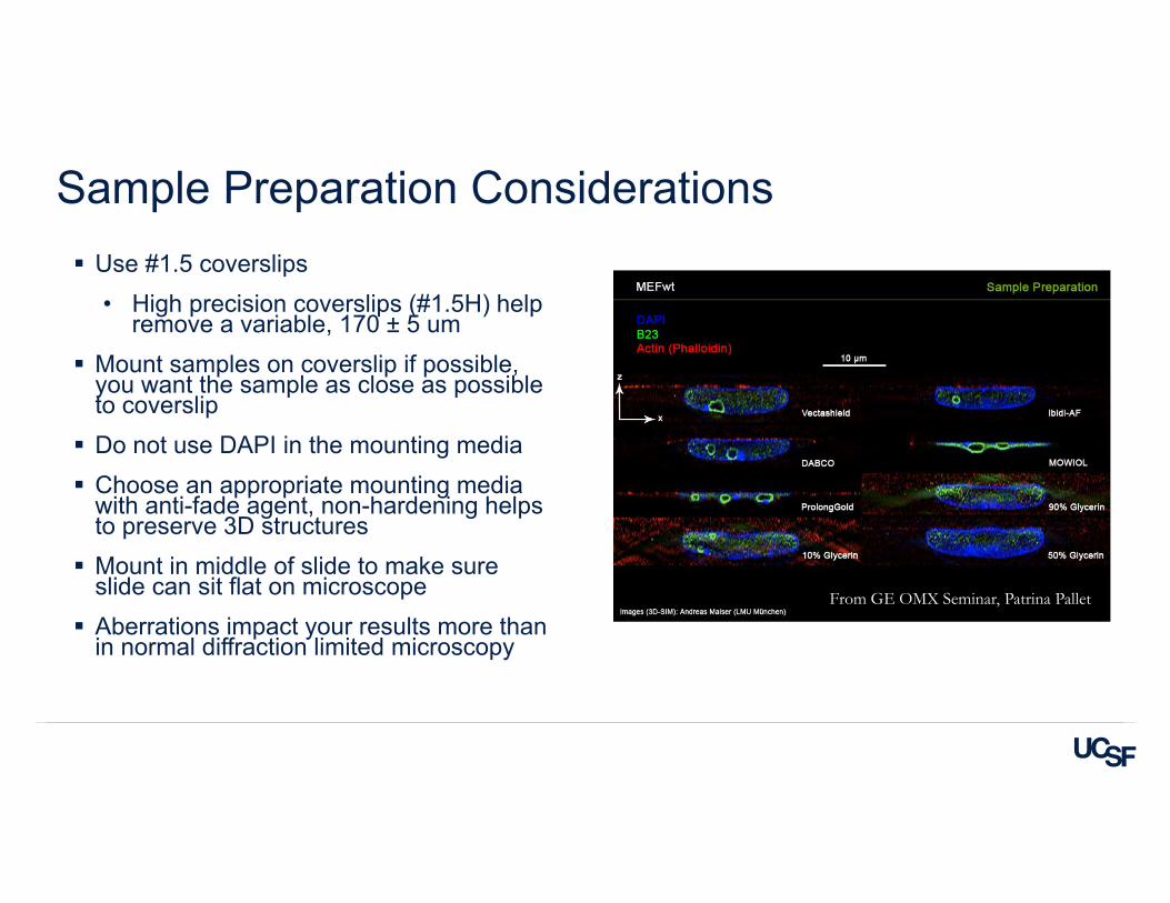
Sample Preparation Considerations Use #1.5 coverslips
• High precision coverslips (#1.5H) help remove a variable, 170 ± 5 um
Mount samples on coverslip if possible, you want the sample as close as possible to coverslip Do not use DAPI in the mounting media Choose an appropriate mounting media
with anti-fade agent, non-hardening helps to preserve 3D structures Mount in middle of slide to make sure
slide can sit flat on microscope Aberrations impact your results more than
in normal diffraction limited microscopy
From GE OMX Seminar, Patrina Pallet
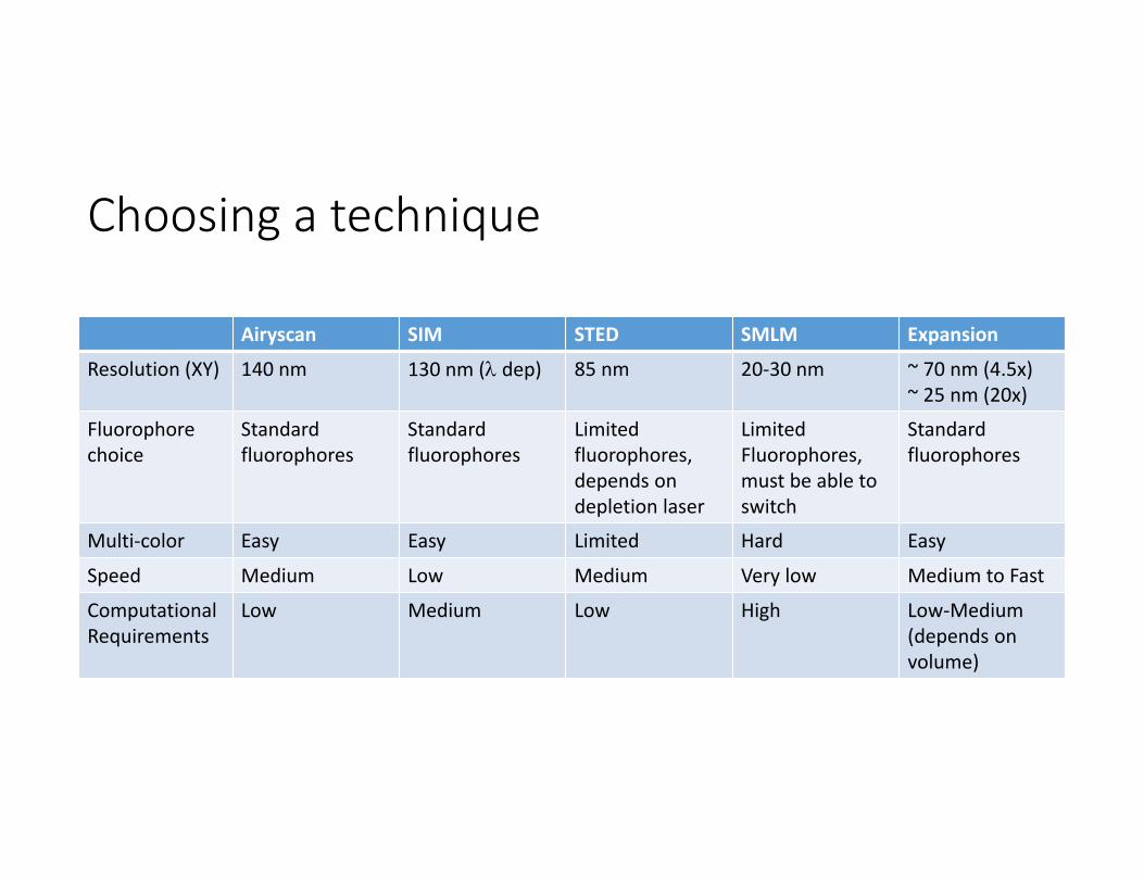
Choosing a technique
Airyscan SIM STED SMLM Expansion
Resolution (XY) 140 nm 130 nm ( dep) 85 nm 20‐30 nm ~ 70 nm (4.5x)~ 25 nm (20x)
Fluorophorechoice
Standard fluorophores
Standard fluorophores
Limitedfluorophores, depends on depletion laser
LimitedFluorophores, must be able to switch
Standard fluorophores
Multi‐color Easy Easy Limited Hard Easy
Speed Medium Low Medium Very low Medium to Fast
Computational Requirements
Low Medium Low High Low‐Medium(depends on volume)
Common Pitfalls
Garbage in – Garbage out
Details matter
You need pilot experiments
• Are your images giving you what you need?
• Do you need to optimize your sample prep, image collection, etc?
Watch your data size
• Current automation makes it trivial to capture large data sets
• Analysis of large data sets becomes the new bottleneck
Resources
http://www.ibiology.org/ibioeducation/taking‐courses/ibiology‐microscopy‐course.html
Nikon Imaging Center Wiki:http://nic.ucsf.edu/dokuwiki/doku.php?id=start
• Links to great resources under the heading Microscopy References and Education