
Amazon Web Services: Amazon Elastic Compute Cloud (Amazon EC2)


ConciseHistologyLESLIE P. GARTNER, PhDProfessor of Anatomy (Retired)Department of Biomedical SciencesBaltimore College of Dental SurgeryDental SchoolUniversity of MarylandBaltimore, Maryland
JAMES L. HIATT, PhDProfessor EmeritusDepartment of Biomedical SciencesBaltimore College of Dental SurgeryDental SchoolUniversity of MarylandBaltimore, Maryland

concise histology isBn: 978-0-7020-3114-4
Copyright © 2011 by Saunders, an imprint of Elsevier Inc. All rights reserved.
no part of this publication may be reproduced or transmitted in any form or by any means, electronic or mechanical, including photocopying, recording, or any information storage and retrieval system, without permission in writing from the publisher. Details on how to seek permission, further information about the Publisher’s permissions policies and our arrangements with organizations such as the copyright clearance center and the copyright licensing Agency, can be found at our website: www.elsevier.com/permissions.
this book and the individual contributions contained in it are protected under copyright by the Publisher (other than as may be noted herein).
Notices
Knowledge and best practice in this field are constantly changing. As new research and experience broaden our understanding, changes in research methods, professional practices, or medical treatment may become necessary.
Practitioners and researchers must always rely on their own experience and knowledge in evaluating and using any information, methods, compounds, or experiments described herein. in using such information or methods they should be mindful of their own safety and the safety of others, including parties for whom they have a professional responsibility.
With respect to any drug or pharmaceutical products identified, readers are advised to check the most current information provided (i) on procedures featured or (ii) by the manufacturer of each product to be administered, to verify the recommended dose or formula, the method and duration of administration, and contraindications. it is the responsibility of practitioners, relying on their own experience and knowledge of their patients, to make diagnoses, to determine dosages and the best treatment for each individual patient, and to take all appropriate safety precautions.
to the fullest extent of the law, neither the Publisher nor the authors, contributors, or editors, assume any liability for any injury and/or damage to persons or property as a matter of products liability, negligence or otherwise, or from any use or operation of any methods, products, instructions, or ideas contained in the material herein.
Library of Congress Cataloging-in-Publication Data
gartner, leslie P. concise histology / leslie P. gartner, James l. hiatt.—1st ed. p. ; cm. Based on: color textbook of histology / leslie P. gartner, James l. hiatt. 3rd ed. c2007. includes index. isBn 978-0-7020-3114-4 1. histology. i. hiatt, James l.,— ii. gartner, leslie P., 1943—color textbook of histology. iii. title. [DnlM: 1. histology—Atlases. Qs 517 g244c 2011] QM551.g366 2011 611’.018—dc22 2010013017
Printed in china
last digit is the print number: 9 8 7 6 5 4 3 2 1
Working together to grow libraries in developing countries
www.elsevier.com | www.bookaid.org | www.sabre.org
1600 John F. Kennedy Boulevardsuite 1800Philadelphia, PA 19103-2899
Acquisitions Editor: Kate DimockDevelopmental Editor: Barbara cicalese
Design Direction: lou ForgioneElectronic Media Manager: carol emery
To my wife, Roseann;
my daughter, Jennifer;
and my mother, Mary
LPG
To my grandchildren,
Nathan David,
James Mallary,
Hanna Elisabeth,
Alexandra Renate,
Eric James,
and Elise Victoria
JLH

v
once again, we are gratified to release a new histol-ogy textbook, one that is based on the third edition of our Color Textbook of Histology, a well-established textbook not only in its original language but also in several other languages.
in the past three decades, histology has evolved from the purely descriptive science of microscopic anatomy to a composite study integrating functional anatomy with both molecular and cell biology. this new textbook is designed in an unusual manner in that each even-numbered page tells the story in words and the facing odd-numbered page illustrates the textual story by beautiful four-color illustrations that are borrowed from the third edition of our Color Textbook of Histology. therefore, each set of facing pages may be thought of as individual learning units. to demonstrate the relevance of the information presented to the health professions, almost every learning unit is reinforced by clinical considerations pertinent to the topic. students and faculty alike will, no doubt, note the absence of photomicrographs and electron micrographs in Concise Histology. We made a deliberate decision to exclude that material from the hard copy and to place it, instead, on the student consult website that is associated with this book. We did that to reduce the size of the book, thereby making life easier for the student who has to learn material that a decade ago was taught in 16
Prefaceweeks and currently is done so in perhaps half that time. student consult houses not only all the illus-trations located on the right side of the facing pages of the book but also 150 photomicrographs and elec-tron micrographs, identified by chapter, with appro-priate examination questions and the answers to those questions so that the student can test his or her ability not only to recognize the organs/tissues/cells in question but also their functional characteristics. included on student consult are clinical scenarios with appropriate UsMle i-type questions that not only further demonstrate the relevance of histology to the health sciences but also prepare medical stu-dents for the histology component of the boards. the designs of the hard copy of this textbook, as well as that of the ancillary web-based material, intend to highlight the essential concepts underlying our pre-sentation of histology, namely that there is a close relationship between structure and function.
Although we have made every effort to present a complete and accurate account of the subject matter, we realize that there are omissions and errors in any undertaking of this magnitude. therefore, we con-tinue to encourage and welcome suggestions, advice, and criticism that will facilitate the improvement of future editions of this textbook.
leslie P. gartnerJames l. hiatt

vii
histology is a visual subject; therefore, excellent graphic illustrations are imperative. For that we are indebted to todd smith for his careful attention to detail in revising and creating new illustrations. We also thank our many colleagues from around the world and their publishers who generously permit-ted us to borrow illustrative materials.
AcknowledgmentsFinally, our thanks go to the project team at else-
vier for all their help, namely Kate Dimock, Barbara cicalese, lou Forgione, and carol emery. We also thank linnea hermanson for her painstaking effort in the production of this text book.
ix
Contents
1 Introduction to Histology . . . . . . . . . . . . . . . . . . . . . . . . . . . . . . . . . . . . . . . . . . . . . . . . . . . . . . 2
2 Cytoplasm . . . . . . . . . . . . . . . . . . . . . . . . . . . . . . . . . . . . . . . . . . . . . . . . . . . . . . . . . . . . . . . . . . . . . . . . . . . 8
3 Nucleus . . . . . . . . . . . . . . . . . . . . . . . . . . . . . . . . . . . . . . . . . . . . . . . . . . . . . . . . . . . . . . . . . . . . . . . . . . . . . .26
4 Extracellular Matrix . . . . . . . . . . . . . . . . . . . . . . . . . . . . . . . . . . . . . . . . . . . . . . . . . . . . . . . . . . . . .40
5 Epithelium and Glands . . . . . . . . . . . . . . . . . . . . . . . . . . . . . . . . . . . . . . . . . . . . . . . . . . . . . . . .48
6 Connective Tissue . . . . . . . . . . . . . . . . . . . . . . . . . . . . . . . . . . . . . . . . . . . . . . . . . . . . . . . . . . . . . .62
7 Cartilage and Bone . . . . . . . . . . . . . . . . . . . . . . . . . . . . . . . . . . . . . . . . . . . . . . . . . . . . . . . . . . . . .74
8 Muscle . . . . . . . . . . . . . . . . . . . . . . . . . . . . . . . . . . . . . . . . . . . . . . . . . . . . . . . . . . . . . . . . . . . . . . . . . . . . . . . .94
9 Nervous Tissue . . . . . . . . . . . . . . . . . . . . . . . . . . . . . . . . . . . . . . . . . . . . . . . . . . . . . . . . . . . . . . . . . 108
10 Blood and Hematopoiesis . . . . . . . . . . . . . . . . . . . . . . . . . . . . . . . . . . . . . . . . . . . . . . . . . 132
11 Circulatory System . . . . . . . . . . . . . . . . . . . . . . . . . . . . . . . . . . . . . . . . . . . . . . . . . . . . . . . . . . . . 152
12 Lymphoid (Immune) System . . . . . . . . . . . . . . . . . . . . . . . . . . . . . . . . . . . . . . . . . . . . . . 168
13 Endocrine System . . . . . . . . . . . . . . . . . . . . . . . . . . . . . . . . . . . . . . . . . . . . . . . . . . . . . . . . . . . . . 188
14 Integument . . . . . . . . . . . . . . . . . . . . . . . . . . . . . . . . . . . . . . . . . . . . . . . . . . . . . . . . . . . . . . . . . . . . . . . 204
15 Respiratory System . . . . . . . . . . . . . . . . . . . . . . . . . . . . . . . . . . . . . . . . . . . . . . . . . . . . . . . . . . . 218
16 Digestive System: Oral Cavity . . . . . . . . . . . . . . . . . . . . . . . . . . . . . . . . . . . . . . . . . . . 230
17 Digestive System: Alimentary Canal . . . . . . . . . . . . . . . . . . . . . . . . . . . . . . . . . . 238
18 Digestive System: Glands . . . . . . . . . . . . . . . . . . . . . . . . . . . . . . . . . . . . . . . . . . . . . . . . . 250
19 Urinary System . . . . . . . . . . . . . . . . . . . . . . . . . . . . . . . . . . . . . . . . . . . . . . . . . . . . . . . . . . . . . . . . . 260
20 Female Reproductive System . . . . . . . . . . . . . . . . . . . . . . . . . . . . . . . . . . . . . . . . . . . . 272
21 Male Reproductive System . . . . . . . . . . . . . . . . . . . . . . . . . . . . . . . . . . . . . . . . . . . . . . . 286
22 Special Senses . . . . . . . . . . . . . . . . . . . . . . . . . . . . . . . . . . . . . . . . . . . . . . . . . . . . . . . . . . . . . . . . . 304
Index . . . . . . . . . . . . . . . . . . . . . . . . . . . . . . . . . . . . . . . . . . . . . . . . . . . . . . . . . . . . . . . . . . . . . . . . . . . . . . . . . . . . . . . 325

1
Concise Histology
2
1 INTRoDucTIoN To HISToLoGy
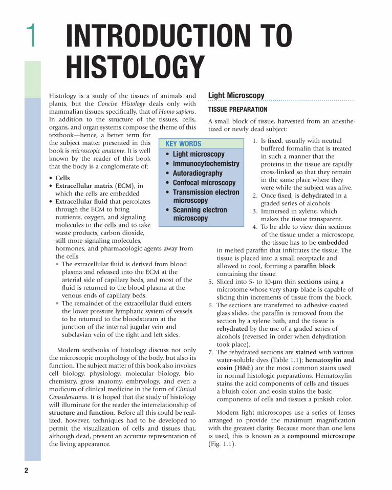
histology is a study of the tissues of animals and plants, but the Concise Histology deals only with mammalian tissues, specifically, that of Homo sapiens. in addition to the structure of the tissues, cells, organs, and organ systems compose the theme of this textbook—hence, a better term for the subject matter presented in this book is microscopic anatomy. it is well known by the reader of this book that the body is a conglomerate of:
• Cells• Extracellular matrix (ECM), in
which the cells are embedded• Extracellular fluid that percolates
through the ecM to bring nutrients, oxygen, and signaling molecules to the cells and to take waste products, carbon dioxide, still more signaling molecules, hormones, and pharmacologic agents away from the cells• the extracellular fluid is derived from blood
plasma and released into the ecM at the arterial side of capillary beds, and most of the fluid is returned to the blood plasma at the venous ends of capillary beds.
• the remainder of the extracellular fluid enters the lower pressure lymphatic system of vessels to be returned to the bloodstream at the junction of the internal jugular vein and subclavian vein of the right and left sides.
Modern textbooks of histology discuss not only the microscopic morphology of the body, but also its function. the subject matter of this book also invokes cell biology, physiology, molecular biology, bio-chemistry, gross anatomy, embryology, and even a modicum of clinical medicine in the form of Clinical Considerations. it is hoped that the study of histology will illuminate for the reader the interrelationship of structure and function. Before all this could be real-ized, however, techniques had to be developed to permit the visualization of cells and tissues that, although dead, present an accurate representation of the living appearance.
Light Microscopy
TISSuE PREPARATIoN
A small block of tissue, harvested from an anesthe-tized or newly dead subject:
1. is fixed, usually with neutral buffered formalin that is treated in such a manner that the proteins in the tissue are rapidly cross-linked so that they remain in the same place where they were while the subject was alive.
2. once fixed, is dehydrated in a graded series of alcohols
3. immersed in xylene, which makes the tissue transparent.
4. to be able to view thin sections of the tissue under a microscope, the tissue has to be embedded
in melted paraffin that infiltrates the tissue. the tissue is placed into a small receptacle and allowed to cool, forming a paraffin block containing the tissue.
5. sliced into 5- to 10-µm thin sections using a microtome whose very sharp blade is capable of slicing thin increments of tissue from the block.
6. the sections are transferred to adhesive-coated glass slides, the paraffin is removed from the section by a xylene bath, and the tissue is rehydrated by the use of a graded series of alcohols (reversed in order when dehydration took place).
7. the rehydrated sections are stained with various water-soluble dyes (table 1.1); hematoxylin and eosin (H&E) are the most common stains used in normal histologic preparations. hematoxylin stains the acid components of cells and tissues a bluish color, and eosin stains the basic components of cells and tissues a pinkish color.
Modern light microscopes use a series of lenses arranged to provide the maximum magnification with the greatest clarity. Because more than one lens is used, this is known as a compound microscope (Fig. 1.1).
KEy WoRDS• Light microscopy• Immunocytochemistry• Autoradiography• confocal microscopy• Transmission electron
microscopy• Scanning electron
microscopy

Chapter
IntroduCtIon to HIstology
1
3Table 1.1 COMMON HISTOLOGIC STAINS AND REACTIONS
Reagent Result
hematoxylin Blue—nucleus; acidic regions of the cytoplasm; cartilage matrixeosin Pink—basic regions of the cytoplasm; collagen fibersMasson’s trichrome Dark blue—nuclei
Red—muscle, keratin, cytoplasmLight blue—mucinogen, collagen
orcein elastic stain Brown—elastic fibersWeigert’s elastic stain Blue—elastic fiberssilver stain Black—reticular fibersiron hematoxylin Black—striations of muscle, nuclei, erythrocytesPeriodic acid–schiff Magenta—glycogen and carbohydrate-rich moleculesWright’s and giemsa* Pink—erythrocytes, eosinophil stains
Blue—cytoplasm of monocytes of blood cells and lymphocytes
*Used for granules differential staining of blood cells.
Figure 1.1 comparison of light, transmission electron, and scanning electron microscopes. (From Gartner LP, Hiatt JL: Color Textbook of Histology, 3rd ed. Philadelphia, Saunders, 2007, p 4.)
Image in eye
Light microscope Transmission electronmicroscope
Scanning electronmicroscope
Ocular lens
Anode
Electronicamplifier
Condenser lens
Lamp Mirror
Specimen
Specimen
Condenserlens
Condenser lens
Scanning beam
Electron detector
Specimen
Viewing window
Image on viewing screen
Image on viewing screen
Projectionlens
Objectivelens
Scanning coil
Anode
Cathode
Television screen

Chapter
IntroduCtIon to HIstology
1
4red color indicates that glycogen was present at that particular location.
• other histochemical and cytochemical techniques can localize enzymes; however, it is not the enzyme that is visualized, but the presence of the reaction product that precipitated as a colored compound at the site of the reaction.
• Immunocytochemistry provides a more accurate localization of a particular macromolecule than does histochemistry or cytochemistry.• this is a more complex method, however,
because it involves the development of an antibody against the macromolecule of interest in the direct method, or
• Development of an antibody against a primary antibody in the indirect method (Fig. 1.3) and labeling the developed antibody with a fluorescing label, such as rhodamine or fluorescein. the indirect method is more sensitive and more accurate than the direct method because more fluorescent labeled antibodies bind to the primary antibody than in the direct method. Additionally, most of the time, primary antibodies are more expensive and more limited in their availability.
• immunocytochemistry can also be applied to electron microscopy by attaching the heavy metal ferritin instead of a fluorescent label.
• the method of autoradiography uses a radioactive isotope (usually tritium, 3h), which is integrated into the molecule that is being investigated.• if one wishes to follow the synthesis of a
particular protein, tritiated amino acid is fed into the system, and specimens are harvested at defined periods.
• sections are processed in a normal fashion, but instead of a coverslip, photographic emulsion is placed on the section, and the slide is stored in the dark for many weeks.
• the emulsion is developed and fixed as if it were a photographic plate, and a coverslip is placed over the section.
• Microscopic examination displays the presence of silver grains over the regions where the isotope labeled molecule was located.
• A method of autoradiography has been developed for electron microscopy.
TISSuE PREPARATIoN (cont.)
A high-intensity lightbulb provides the light, which is focused on the specimen from below by a condenser lens. the light that passes through the specimen is gathered by one of the objective lenses that sits on a rotatable turret, allowing a change in magnification from low to medium to high, and an oil lens, which in conventional microscopes magni-fies the image 4, 10, 20, 40, and 100 times. the first three are dry lenses, whereas the oil lens uses immer-sion oil to act as an interface between the glass of the slide and the glass of the objective lens. the light from the objective lens is gathered by the ocular lens, usually 10 times, for final magnification of 40, 100, 200, 400, and 1000 times, and the image is focused on the retina.
INTERPRETATIoN of MIcRoScoPIc SEcTIoNS
histologic sections are two-dimensional planes cut from a three-dimensional structure. initially, it is difficult for the student to reconcile the image seen in the microscope with the tissue or organ from which it was harvested. A simple demonstration of a coiled tube sectioned at various angles (Fig. 1.2) is instructive in learning how to reconstruct the three-dimensional morphology from viewing a series of two-dimensional sections.
ADvANcED vISuALIzATIoN PRocEDuRES
Various techniques were developed to use the micro-scope in elucidating functional aspects of the cells, tissues, and organs being studied. the most com-monly used techniques are histochemistry (and cyto-chemistry), immunocytochemistry, and autoradiogra phy.
• Histochemistry and cytochemistry use chemical reactions, enzymatic processes, and physicochemical processes that not only stain the tissue, but also permit the localization of extracellular and intracellular macromolecules of interest.• one of the most used histochemical methods
is the periodic acid–schiff (PAs) reagent, which stains glycogen and molecules rich in carbohydrates a purplish-red color. By treating consecutive sections with the enzyme amylase, to digest glycogen, the absence of the purplish-
-

Chapter
IntroduCtIon to HIstology
1
5
Figure 1.2 two-dimensional views of a three-dimensional tube sectioned in various planes. (From Gartner LP, Hiatt JL: Color Textbook of Histology, 3rd ed. Philadelphia, Saunders, 2007, p 4.)
Cross section Longitudinal
section
Oblique section
Diagram showing the different appearances of sections cut through a curved tube at different levels
Figure 1.3 Direct and indirect methods of immunocytochemistry. Left, An antibody against an antigen was labeled with a fluorescent dye and viewed with a fluorescent microscope. Fluorescence occurs only over the location of the labeled antibody. Right, Fluorescent labeled antibodies were prepared against an antibody that reacts with a particular antigen. When viewed with a fluorescent microscope, the fluorescence represents the location of the antibody that reacts with the antigen. (From Gartner LP, Hiatt JL: Color Textbook of Histology, 3rd ed. Philadelphia, Saunders, 2007, p 5.)
Fluoresceinated antibody
Antigen
Tissue section
Wash
Direct Indirect
Add fluoresceinated anti-antibody
Antigen
Antibody
Chapter
IntroduCtIon to HIstology
1
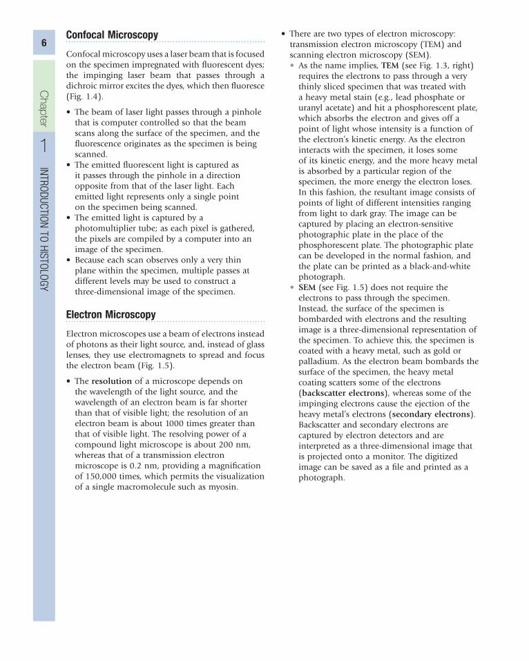
6confocal Microscopy
confocal microscopy uses a laser beam that is focused on the specimen impregnated with fluorescent dyes; the impinging laser beam that passes through a dichroic mirror excites the dyes, which then fluoresce (Fig. 1.4).
• the beam of laser light passes through a pinhole that is computer controlled so that the beam scans along the surface of the specimen, and the fluorescence originates as the specimen is being scanned.
• the emitted fluorescent light is captured as it passes through the pinhole in a direction opposite from that of the laser light. each emitted light represents only a single point on the specimen being scanned.
• the emitted light is captured by a photomultiplier tube; as each pixel is gathered, the pixels are compiled by a computer into an image of the specimen.
• Because each scan observes only a very thin plane within the specimen, multiple passes at different levels may be used to construct a three-dimensional image of the specimen.
Electron Microscopy
electron microscopes use a beam of electrons instead of photons as their light source, and, instead of glass lenses, they use electromagnets to spread and focus the electron beam (Fig. 1.5).
• the resolution of a microscope depends on the wavelength of the light source, and the wavelength of an electron beam is far shorter than that of visible light; the resolution of an electron beam is about 1000 times greater than that of visible light. the resolving power of a compound light microscope is about 200 nm, whereas that of a transmission electron microscope is 0.2 nm, providing a magnification of 150,000 times, which permits the visualization of a single macromolecule such as myosin.
• there are two types of electron microscopy: transmission electron microscopy (teM) and scanning electron microscopy (seM).• As the name implies, TEM (see Fig. 1.3, right)
requires the electrons to pass through a very thinly sliced specimen that was treated with a heavy metal stain (e.g., lead phosphate or uranyl acetate) and hit a phosphorescent plate, which absorbs the electron and gives off a point of light whose intensity is a function of the electron’s kinetic energy. As the electron interacts with the specimen, it loses some of its kinetic energy, and the more heavy metal is absorbed by a particular region of the specimen, the more energy the electron loses. in this fashion, the resultant image consists of points of light of different intensities ranging from light to dark gray. the image can be captured by placing an electron-sensitive photographic plate in the place of the phosphorescent plate. the photographic plate can be developed in the normal fashion, and the plate can be printed as a black-and-white photograph.
• SEM (see Fig. 1.5) does not require the electrons to pass through the specimen. instead, the surface of the specimen is bombarded with electrons and the resulting image is a three-dimensional representation of the specimen. to achieve this, the specimen is coated with a heavy metal, such as gold or palladium. As the electron beam bombards the surface of the specimen, the heavy metal coating scatters some of the electrons (backscatter electrons), whereas some of the impinging electrons cause the ejection of the heavy metal’s electrons (secondary electrons). Backscatter and secondary electrons are captured by electron detectors and are interpreted as a three-dimensional image that is projected onto a monitor. the digitized image can be saved as a file and printed as a photograph.

Chapter
IntroduCtIon to HIstology
1
7
Figure 1.4 confocal microscope displaying the pinhole through which the laser beam enters to scan the specimen and the path of the fluorescent light that subsequently is emitted by the specimen to be captured by the photomultiplier detector. (From Gartner LP, Hiatt JL: Color Textbook of Histology, 3rd ed. Philadelphia, Saunders, 2007, p 8.)
Specimen
Photomultiplierdetector
Pinhole aperture
Pinhole aperture
Laser withlaser light
Scanning mirror
Scanning mirror
Figure 1.5 comparison of light, transmission electron, and scanning electron microscopes. (From Gartner LP, Hiatt JL: Color Textbook of Histology, 3rd ed. Philadelphia, Saunders, 2007, p 4.)
Image in eye
Light microscope Transmission electronmicroscope
Scanning electronmicroscope
Ocular lens
Anode
Electronicamplifier
Condenser lens
Lamp Mirror
Specimen
Specimen
Condenserlens
Condenser lens
Scanning beam
Electron detector
Specimen
Viewing window
Image on viewing screen
Image on viewing screen
Projectionlens
Objectivelens
Scanning coil
Anode
Cathode
Television screen