Introduction: Superresolution fluorescence microscopy...Introduction: Superresolution fluorescence...
7
Introduction: Superresolution fluorescence microscopy CS/CME/Biophys/BMI 371 March 8, 2017 Ron Dror 1
Transcript of Introduction: Superresolution fluorescence microscopy...Introduction: Superresolution fluorescence...

Introduction:Superresolutionfluorescence
microscopy
CS/CME/Biophys/BMI371March8,2017
RonDror
1
A limit on focusing light• The physics of light—in particular, the fact that it is a wave—
imposes a fundamental limit on how well a lens can focus it • The light from a single point in space will not focus to a single point • Instead, it will focus to a disk-like pattern called an “Airy pattern”
– This means the observed image will be slightly blurred – In fact, we can think of the observed image as the true image convolved
with the Airy pattern. This constitutes a low-pass filter.
2
You’renotresponsiblefordetailsoftheunderlyingphysicshere
Airypattern
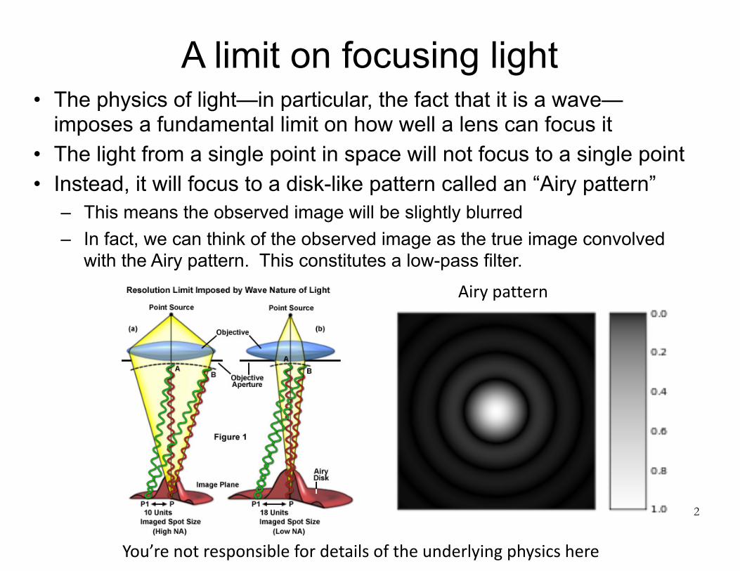
The diffraction limit
• This limit on how well one can focus light is known as “the diffraction limit” – It’s literally “written in stone” in Jena, Germany (on a memorial to
Ernst Abbe, who derived it) • The radius d of the Airy disk (the central spot of the Airy
pattern) is proportional to the wavelength λ of the light • Don’t worry about the other parameters (n sinθ, the
“numerical aperture,” is usually between 0.1 and 1)
http://en.wikipedia.org/wiki/STED_microscopy#mediaviewer/File:Ernst-Abbe-Denkmal_Jena_F%C3%BCrstengraben_-_20140802_125708.jpg

Super-resolution fluorescence microscopy
• A number of recently developed techniques achieve resolution well beyond the diffraction limit – This requires violating some of the assumptions of that limit—
e.g., assuming some prior knowledge of the true image • The most popular of these techniques is known as either
STORM (stochastic optical reconstruction microscopy) or PALM (photoactivation localization microscopy)
4
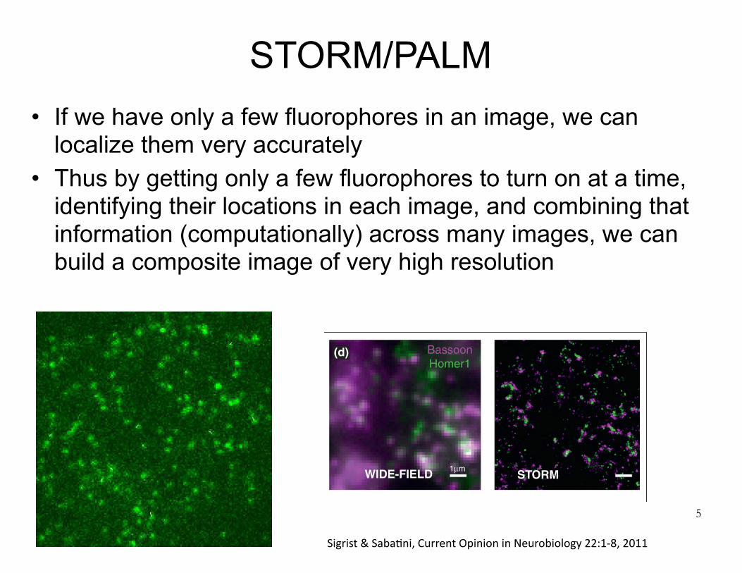
STORM/PALM• If we have only a few fluorophores in an image, we can
localize them very accurately • Thus by getting only a few fluorophores to turn on at a time,
identifying their locations in each image, and combining that information (computationally) across many images, we can build a composite image of very high resolution
5
and beyond. Nevertheless, there still is substantial roomfor improvement and optimization for both techniques.Particularly, both methods pose challenges when it comesto multichannel super-resolution imaging and to improv-ing imaging resolution in the z-axis. For STORM andPALM, multiple photo-switchable fluorophores of differ-ent colors have been described and recently used to
produce multicolor maps of, for example, PSD compo-sition [26!!]. In addition, incorporation of cylindricallenses and interferometric measurements allow for im-provement in z-axis (axial) resolution, possibly to levelseven beyond the xy place (transverse) resolution [27!,28!].However, these approaches have not yet seen widespreaduse and the need for high precision chromatic aberration
4 Neurotechnology
CONEUR-990; NO. OF PAGES 8
Please cite this article in press as: Sigrist SJ, Sabatini BL. Optical super-resolution microscopy in neurobiology, Curr Opin Neurobiol (2011), doi:10.1016/j.conb.2011.10.014
Figure 2
confocal
(a)
WIDE-FIELD STORM
Homer1[N] Shank1
200 nm
Homer1 [N]
Homer1[C]
BRPNc82
BRPNc82
BRPN-Term
Bassoon
BRPN-Term
N-Terminus
Basson[N]
Bassoon [N]
00 1000-50Position (nm)
50
Bassoon [C]
Piccolo [C]
RIM1
NR2B
PSD95
GluR1CaMKll
Shank
GARABR1
Piccolo [N]
(e)(b)
(c)
STED
C-Terminus
STED STED
BRP
vesicles
planar vertical
Ca2+-channels
DLiprin-α
600 300
00
Homer1
1µm
(d)
Homer1 [C]
Pre-synaptic Synaptic Cleft Post-synaptic(f)
Current Opinion in Neurobiology
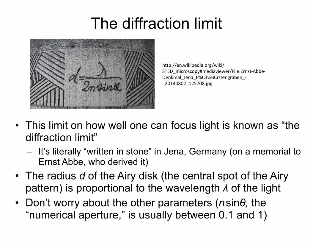
STED and STORM analysis of synaptic architecture. (a–c) STED imaging of Drosophila neuromuscular synapses. (a) STED microscopy reveals donut-shaped structures recognized by the monoclonal antibody Nc82 against the protein Bruchpilot that are not resolvable by confocal microscopy. (b)Upper panel: Boutons stained for BRP-N-Terminus (confocal; magenta) and BRP-Nc82 (STED; green) showing planar (arrow) and vertical (arrowhead)active zones. Lower panel: Magnifications of individual planar (left) and vertical (right) active zones stained for BRP-Nc82 (STED) and BRP-N-Terminus(confocal; b) and BRP-N-Terminus imaged with (STED). (c) Model of active zone organization at Drosophila neuromuscular synapses. (d–f) STORMimaging of presynaptic and postsynaptic scaffolding proteins. Presynaptic protein Bassoon and postsynaptic protein Homer1 in glomeruli of themouse olfactory bulb were identified by immunohistochemistry using Cy3-A647 and A405-A647 conjugated antibodies, respectively. The conventionalfluorescence image (d) shows punctate patterns that are partially overlapping, whereas the STORM image of the same area clearly resolves distinctsynaptic structures. Further enlargement of the conventional images (e) does not reveal detailed structure of the synapses whereas the correspondingSTORM images clearly distinguish the presynaptic Bassoon and postsynaptic Homer1 clusters. (f) Imaging of multiple proteins via STORM revealstheir differential localizations in the synapse.Taken with permission from [26!!].
Current Opinion in Neurobiology 2011, 22:1–8 www.sciencedirect.com
Sigrist&SabaXni,CurrentOpinioninNeurobiology22:1-8,2011
Tradeoff in spatial vs. temporal resolution
• STORM/PALM requires collection of many raw images for a single reconstructed image. This limits its temporal resolution, and also increases total light exposure, which damages cells.
• Monday’s papers present two methods to achieve better temporal resolution without sacrificing much spatial resolution – Improved computational reconstruction techniques using
compressed sensing (originally due to Emmanuel Candes, Stanford) • Basic idea: leverage the fact that the reconstructed image (in PALM/
STORM) is sparse, meaning there are few non-zero pixels – Lattice light sheet microscopy: a new super-resolution imaging
method, from the inventor of PALM. • Basic idea: use advanced optics to illuminate only a very thin plane.
Give us a bit of spatial resolution, but substantially increase temporal resolution and substantially decrease damaging light exposure
6

Background information
• Review on super-resolution microscopy (“Optical super-resolution microscopy in neurobiology”) – http://www.sciencedirect.com/science/article/pii/
S0959438811001802 • My introduction to fluorescence microscopy from CS/
CME/Biophys/BMI 279: – http://web.stanford.edu/class/cs279/lectures/lecture11.pdf
• If you’d like a more thorough coverage of compressed sensing (also called compressive sensing), see: – http://dsp.rice.edu/sites/dsp.rice.edu/files/cs/CSintro.pdf – This article is intended to be introductory, but it assumes
some knowledge of signal processing/information theory. It’s optional! 7