
INTRODUCTION - Engineeringby.genie.uottawa.ca/~mfenech/TEACH/MCG_3143/Entrees/2015/5/5... ·...
68
INTRODUCTION HUMAN BIOFLUID DYNAMIC SHOW Biofluid Mechanics MCG3143 Marianne Fenech Mechanical engineering [email protected] Room: A328 C.BY (mezzanine) Ext 1924
Transcript of INTRODUCTION - Engineeringby.genie.uottawa.ca/~mfenech/TEACH/MCG_3143/Entrees/2015/5/5... ·...

INTRODUCTION
HUMAN BIOFLUID DYNAMIC SHOW
Biofluid Mechanics
MCG3143
Marianne FenechMechanical engineering
[email protected]: A328 C.BY (mezzanine)Ext 1924

What is a Biofluid? Medical definition :A biological fluid. Biofluids can be excreted (such as urine or sweat), secreted (such as breast milk or bile), obtained with a needle (such as blood or cerebrospinal fluid), or develop as a result of a pathological process(such as blister or cyst fluid). The term biofluid is employed as both a noun and an adjective (as in biofluid dynamics and biofluid mechanics). (www.medecinenet.com)
‘There are numerous processes in the body under healthy and pathologic conditions in which biofluid mechanics play a central role. The delivery of various substances to and from tissues is accomplished through a combination of complex active and passive mass transfer processes. Biofluid mechanics also play a central role in locomotion, from water-bound amoeba and fish to the soaring of birds. ‘
Biofluid mechanics
Extracted from’ D. Bluestein and JE. Moore Jr. Biofluids Educational Issues: An Emerging Field Aims to Define Its Next Generation’, Annals of Biomedical Engineering 33 12 2005

Complex flows the cardiovascular system
Arteriovenous fistula is specific vessels created by a vascular operation in order to provide sufficient blood access for extra-corporeal circulation. (Karboutly et al.)
Modelisation of the turbulent flow through a femoral stenosis using Fluent sofware (Garcia et al.)
Complex unsteady velocity profile in the blood vessel: The Wormsley profile.
Pulsatile flow in a curved artery :fluid structure interactionNote. The displacements are exaggerated. Color scale represents the pressure. http://www.csc.fi/english/pages/elmer/examples/Coupled_problems/hemodyn
1
3
2

Microcirculation
Intravital microscopy of the microcirculation of the mouse’s cremaster , Chayer B, LBUM, CRCHUM & IRCM
Microcirculation in the human body, which is composed of numerous vessels with diameters less than 50 micrometers, is the main exchange site between blood and tissues.
Blood behavior in the microcirculation is very complex because it cannot be considered as a liquid flow, but rather as a collection of interacting particles.
Red blood cell deformation in a microvessel
4
5
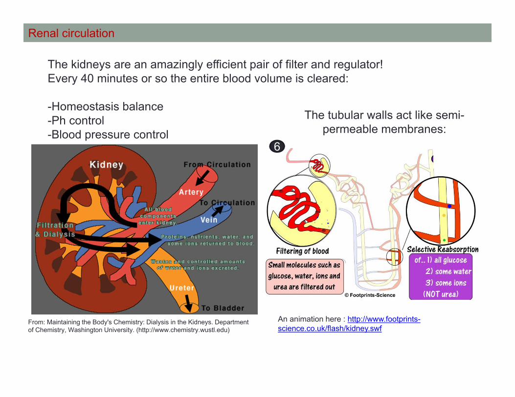
Renal circulation
The kidneys are an amazingly efficient pair of filter and regulator!Every 40 minutes or so the entire blood volume is cleared:
-Homeostasis balance-Ph control -Blood pressure control
An animation here : http://www.footprints-science.co.uk/flash/kidney.swf
The tubular walls act like semi-permeable membranes:
From: Maintaining the Body's Chemistry: Dialysis in the Kidneys. Department of Chemistry, Washington University. (http://www.chemistry.wustl.edu)
6

A exceptional swimmer: the Spermatozoa
It use just the viscous force to swim it is the particularity of this exceptional swimmer
What does it mean?Looks the difference between moving with inertial propulsion and viscous
propulsion:
2 self-propelled objects:
Inertial propulsion in the water
Inertial propulsion in a viscous fluid:It doesn’t work!
Viscouspropulsion
Inertial propulsion
viscous propulsion in a viscous fluid:It is working!
As spermatozoa!
From Multimedia Fluid mechanics
7 8
9 10
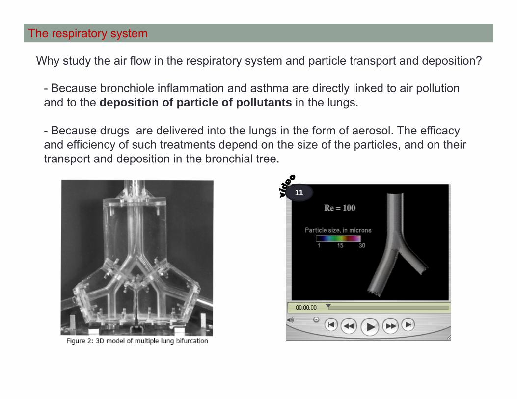
The respiratory system
- Because bronchiole inflammation and asthma are directly linked to air pollution and to the deposition of particle of pollutants in the lungs.
- Because drugs are delivered into the lungs in the form of aerosol. The efficacy and efficiency of such treatments depend on the size of the particles, and on their transport and deposition in the bronchial tree.
Why study the air flow in the respiratory system and particle transport and deposition?
11
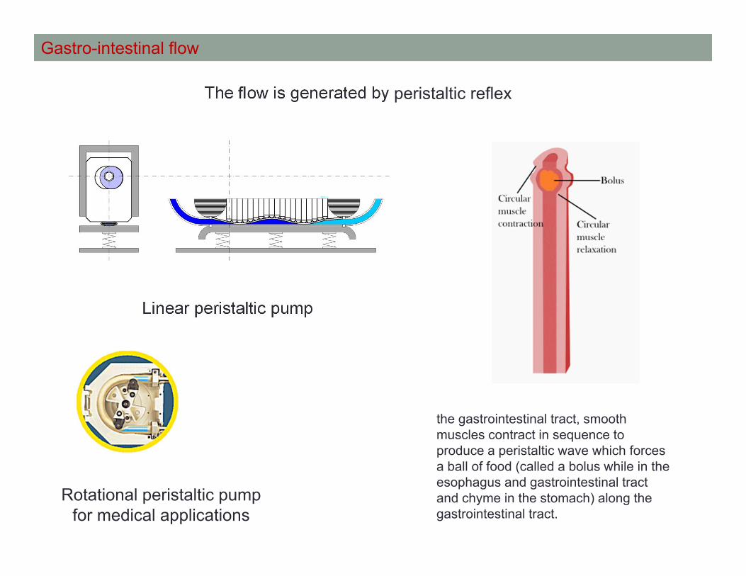
Gastro-intestinal flow
The flow is generated by peristaltic reflex
Linear peristaltic pump
Rotational peristaltic pump for medical applications
the gastrointestinal tract, smooth muscles contract in sequence to produce a peristaltic wave which forces a ball of food (called a bolus while in the esophagus and gastrointestinal tract and chyme in the stomach) along the gastrointestinal tract.

Total Artificial Heart
CardioWest12

CHAPTER 1
CARDIOVASCULAR SYSTEM
Biofluid Mechanics
MCG3143
Marianne FenechMechanical engineering
[email protected]: A328 C.BY (mezzanine)Ext 1924
11
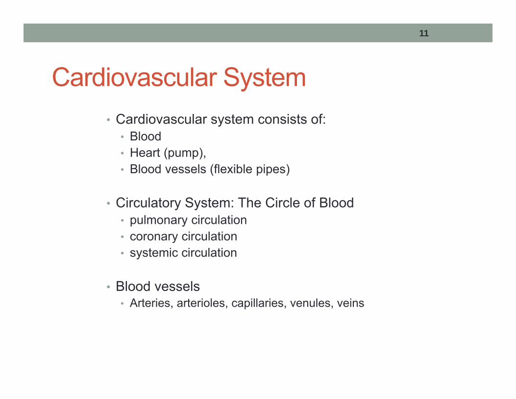
Cardiovascular System• Cardiovascular system consists of:
• Blood• Heart (pump),• Blood vessels (flexible pipes)
• Circulatory System: The Circle of Blood• pulmonary circulation• coronary circulation• systemic circulation
• Blood vessels • Arteries, arterioles, capillaries, venules, veins

12
THE HEART

13
Heart• Consists in two pumps in series circulating blood through the pulmonary and systemic circulation, respectivly.
• Is controlled by an internal electrical system
• Pumps about 5 liters of blood every minute, or 7000 liters of blood every day!

14
Heart Wall• The heart wall is made up of three layers:
• Inner layer – endocardium - thin layer of epithelial tissue
• Middle layer – myocardium – thick layer of muscle tissue
• Outer layer – epicardium – thin layer of epithelial tissue
• The heart is enclosed in a sac called the pericardium
• The inner layer of the pericardium is attached to the heart
• The outer layer of the pericardium is attached to the diaphragm
• These two layers are separated by pericardial fluid

15
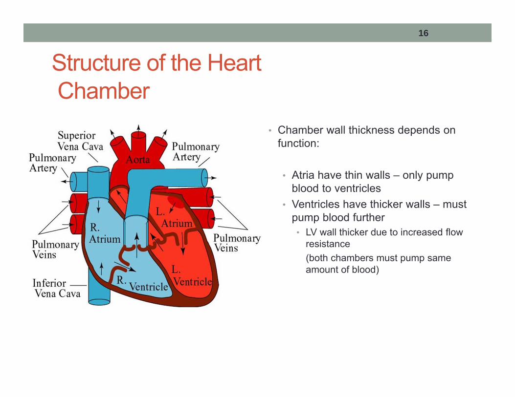
Structure of the HeartChamber
• The heart consists of 4 chambers:• Upper chambers:
- left and right atria (LA, RA)- separated by interatrial septum
• Lower chambers :- left and right ventricles (LV, RV)- separated by interventricular septum
• Left and right is denoted by how you view your own heart
16
Structure of the HeartChamber
• Chamber wall thickness depends on function:
• Atria have thin walls – only pump blood to ventricles
• Ventricles have thicker walls – must pump blood further• LV wall thicker due to increased flow
resistance (both chambers must pump same amount of blood)

17
Cardiac Cycle - Definitions
• Cardiac cycle = heartbeat
• One heartbeat takes approximately 0.8 s
• Systole – contraction of a chamber
• Diastole – relaxation of a chamber

18
Cardiac pressures

19
Cardiac Cycle - Steps1. Atrial contraction
• AV valves open, SL valves closed• Blood flows from atria to ventricles
2. Isovolumetric ventricular contraction• All four valves closed
3. Ventricular contraction (ejection)• SL valves open• Blood exits ventricles
4. Isovolumetric ventricular relaxation• All four valves closed
5. Passive ventricular filling• AV valves open• Blood flows from atria to ventricles
http://www.nhlbi.nih.gov/health/dci/Diseases/hhw/hhw_pumping.html
13

20
Structure of the Heart - Valves• The heart contains 4 one-way valves to control flow direction:
• The aortic valve separates the LV and the aorta• The pulmonary valve separates the RV and the pulmonary trunk• The mitral (bicuspid) valve separates the LA and the LV• The tricuspid valve separates the RA and the RV
Heart Valves (From Tortora, 2002).

21
Blood Circulation
• Blood flows through two loops• systemic circulation (body)• pulmonary circulation (lungs)
• Red denotes oxygenated blood
• Blue denotes deoxygenated blood
Systemic capillaries
Pulmonary capillaries
Blood Circulation.

22
Blood Circulation Path
1. Deoxygenated blood flows from RA to RV2. Deoxygenated blood is pumped from the RV to the lungs3. Gas exchange in the lungs (CO2 leaves blood, O2 enters)4. Oxygenated blood from the lungs enters the LA5. Oxygenated blood flows from LA to LV6. Oxygenated blood pumped from LV through body7. Gas exchange in capillaries (O2 leaves blood, CO2 enters)8. Deoxygenated blood returns from body to RA

23
Coronary Circulation
• The heart, like all organs, requires oxygen and nutrients from blood• These nutrients cannot diffuse through the
heart walls• Therefore, the heart requires its own blood
vessels (coronary vessels)
• Circulation of blood to and from the heart is known as the coronary circulation

24
Conduction – Action Potential
• Blood is pumped through the body due to heart muscle contractions• These contraction are caused by the propagation of a
voltage, called the action potential, through the heart muscle cells
• All cells have a membrane potential• Voltage difference across the cell membrane• When this potential exceeds a certain threshold value, it
is known as action potential• In the case of muscles, action potential causes the
muscle cells to contractGreat animation here:
http://www.nhlbi.nih.gov/health/dci/Diseases/hhw/hhw_electrical.html14
25
Conduction
• The conduction system describes the propagation of the action potential through the heart
• An electrocardiogram (ECG) measures the voltage in the heart during this propagation

26
Pacemakers• The SA node is the first natural pacemaker
• Generates action potential at a rate which controls the rate the heart beats
• If the SA node fails, the AV node becomes the pacemaker of the heart
• If both of these nodes fail, the heart will continue to beat, but at a rate too low for normal function• In this case, artificial pacemakers are implanted

27
Electrocardiogram (ECG)• P wave represents the propagation of the action potential from
the sinoatrial (SA) node to the atria• Atrial contraction occurs just after the P wave
• QRS wave represents the propagation of the action potential from the atria through the ventricles• Ventricular contraction occurs just after the QRS wave
• T wave corresponds to ventricular relaxation
ECG.

Velocity and pressures
Mean average pressure (MAP)
MAP= (ps+2pd)/3
As Systole last for about a 1/3 of the cardiac cycle.
Velocity at the end of the pressure pulse is about• 3-5m/s in aorta• 7-10m/s in large arteries• 35m/s in small arteriesIncreasing as wall stiffness increasing
Volume and flow• Diastole: cardiac muscle relaxing (blood flow in
chambers)• Systole: cardiac muscle contracting (flow flow out)
• End diastolic volume (ml) :EDV• End systolic volume (ml) :ESV
• Heart beat beat/minutes : HR
• Cardiac output CO(mL/min) = SV.HR
Stroke volume (ml/beat) : EDV-ESV

30
BLOOD VESSELS

31
Types of Blood Vessels
• Blood vessels can be divided into five groups:• Arteries – take blood away from the heart• Arterioles – small arteries• Capillaries – microscopic vessels gas
exchange• Venules – small veins• Veins – return blood to the heart

32
Blood Vessel Properties
Single Vessel Diameter
Cross-sectional Area
Volume
Aorta Arterioles VenulesCapillaries
Vena Cava
Comparison of vessel diameters, areas, and
volumes.

33
Definitions – Compliance & Resistance
• Compliance - a measure of the elastic properties of a blood vessel wall. The compliance measures how much the vessel wall will expand for a given pressure.C = dV/dP, where V is volume and P is pressure
• Resistance –
• From Hangen Poiseuille, hydraulic resistance can be defined as
• This equation shows that the resistance to flow in a vessel depends on the fluid viscosity, vessel radius, and length
4
8rLR
QPR
34
Definitions – Pressure
• Systolic pressure – Arterial pressure during systole
• Diastolic pressure – Arterial pressure during diastole
• Blood pressure can be measured by plethysmography (cuff attached to your arm)
• Blood pressure of 120/80 means Psys= 120 mm Hg,
Pdia = 80 mm Hg
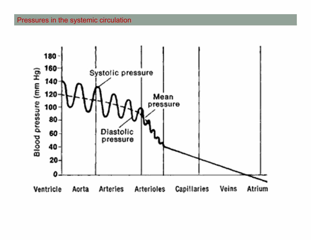
Pressures in the systemic circulation
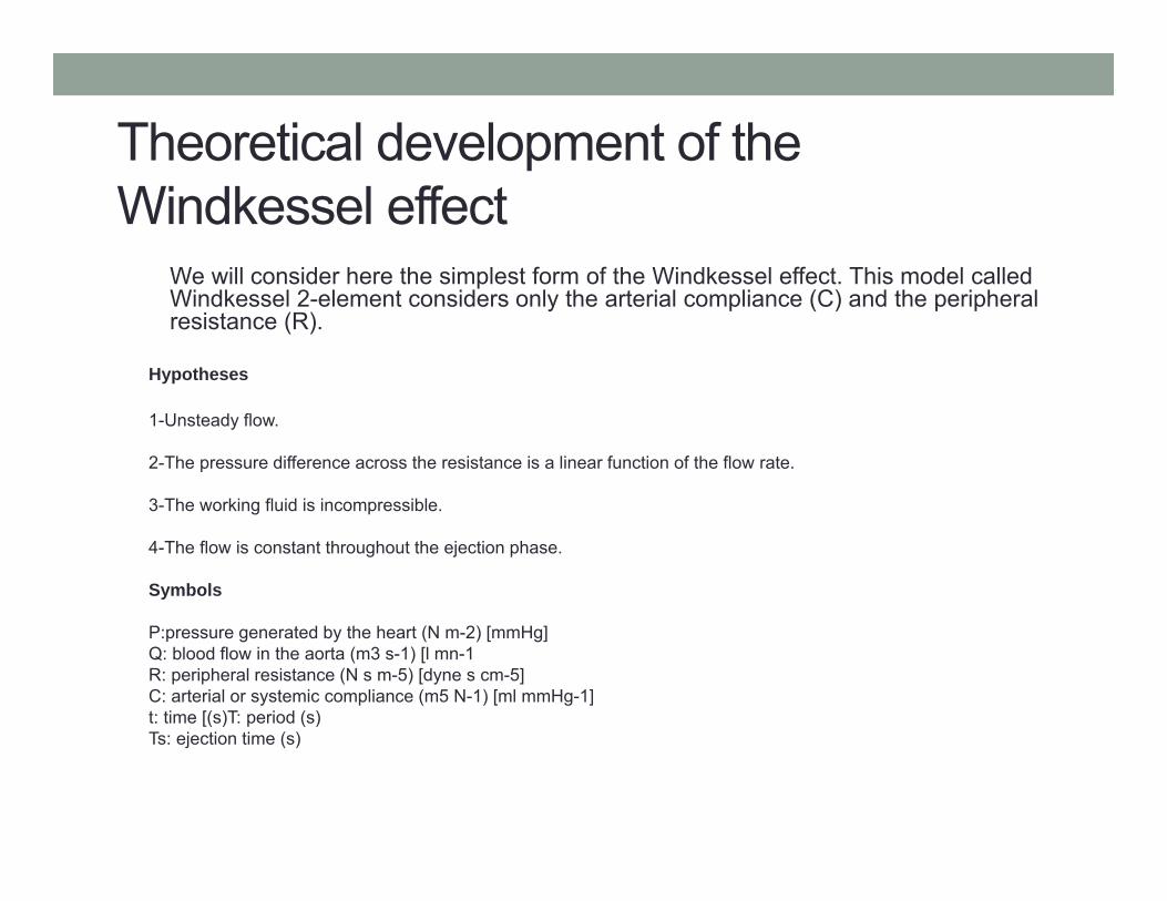
Theoretical development of the Windkessel effect
We will consider here the simplest form of the Windkessel effect. This model called Windkessel 2-element considers only the arterial compliance (C) and the peripheral resistance (R).
Hypotheses
1-Unsteady flow.
2-The pressure difference across the resistance is a linear function of the flow rate.
3-The working fluid is incompressible.
4-The flow is constant throughout the ejection phase.
Symbols
P:pressure generated by the heart (N m-2) [mmHg]Q: blood flow in the aorta (m3 s-1) [l mn-1R: peripheral resistance (N s m-5) [dyne s cm-5] C: arterial or systemic compliance (m5 N-1) [ml mmHg-1]t: time [(s)T: period (s) Ts: ejection time (s)

Theoretical development
Conservation of mass
Qcc is the flow to the compliance chamber.
Thus
Hyp: P-Pcv= R×Q1Pcv is the central veinus pressurePcv << P [Pcv≅5 mmHg vs. P≅100 mmHg ])
Hyp: Q=Cte throughout the systolic phase.
Therefore
Then
Systolic phase (valve in open position) 0≤t≤Ts

Finally the equation to be solved for the systolic phase is
Initial condition: P(t=0)=P0
Solving eq. I
a) Particular solution (Q=Cte=0)
Systolic phase (valve in open position) 0≤t≤Ts
Theoretical development
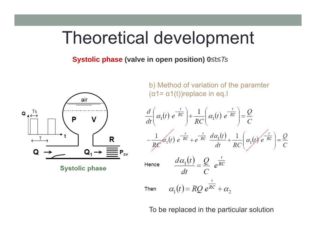
Systolic phase (valve in open position) 0≤t≤Ts
Theoretical development
b) Method of variation of the paramter(α1= α1(t))replace in eq.I
To be replaced in the particular solution

Systolic phase (valve in open position) 0≤t≤Ts
Theoretical development
c) The general solution for the systolic phase is therefore:
To determine the constant α2 we use the initial condition:
Finally, the pressure waveform for the systolic phase can be written as:

Theoretical developmentDiastolic phase (valve in closed position) Ts ≤ t ≤ T
It is same thing as for the systolic phase but with Q=Cte=0
Therefore;
The solution for this equation is under the following form:
Initial condition: P(t =Ts) = Ps(Ts) α3 is determined using the initial condition
Then,

Theoretical developmentDiastolic phase (valve in closed position) Ts ≤ t ≤ T
Finally, the pressure waveform for the diastolic phase can be written under the form:

Theoretical developmentSystolic / Diastolic phase
(valve in open position) / (valve in closed position) 0 ≤ t ≤ Ts / Ts ≤ t ≤ T
To compute the solution, we need to know: P0; Q; R; C; T; Ts.However, it is convenient to use a condition of recurrence to compute P0: P(0)=P(T)

Analysis of the solution
• We can notice from the analytical solution of the Windkessel2-element model the importance of the term (R×C) because it determines the “speed”of the exponential rise or decay. This product is called the characteristic time and is usually noted ().
No resistance Higth resistancecirculation in the veins, venules, capilary

Analysis of the solution
(Stiffer)
dPdVC

46
Blood Vessel Wall Layers
• Intima • a layer of epithelial
cells called the endothelium and a basement membrane
• Media• smooth muscle cells,
elastin and collagen fibres
• Adventitia• elastin and collagen
fibres
Wall Layers (From Tortora, 2002).
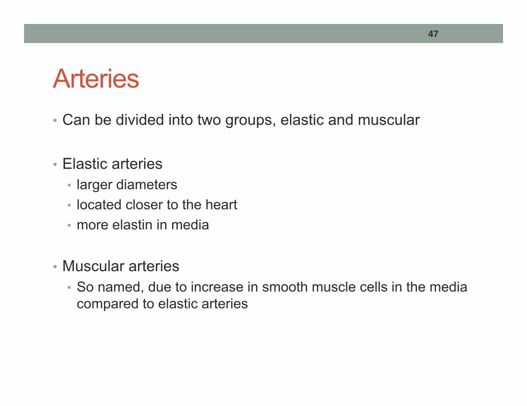
47
Arteries• Can be divided into two groups, elastic and muscular
• Elastic arteries • larger diameters• located closer to the heart• more elastin in media
• Muscular arteries• So named, due to increase in smooth muscle cells in the media
compared to elastic arteries

48
Arterioles• Small arteries
• Transition between arteries and capillaries
• Wall properties near arteries similar to muscular arteries, wall properties near capillaries similar to capillaries
• Important in determining flow resistance – recall
• Small change in vessel radius large change in resistance4
1r
R

49
Control of Blood Flow
• Passive control – compliant blood vessel walls expand and contract due to pressure differences
• Active control – nervous signals cause smooth muscle cells in vessel walls to contract, leading to a decrease in
vessel diameter –Vasoconstriction decrease in R
• Vasodilation – increase in vessel diameter due to relaxation of smooth muscle cells
increase in R

50
Capillaries
• Microscopic vessels responsible for exchange of gases, nutrients, and wastes
• Capillary walls have only the tunica intima• Allows for molecules to pass through vessel walls
• Small diameter, large number large surface area

51
Venules
• Small veins
• Transition between capillaries and veins
• Venules close to capillaries do not have a tunica adventitia• This layer forms in venules that are close to veins
52
Veins
• Tunica intima and tunica media are thinner in veins than in arteries
• Tunica adventitia is thicker in veins
• Elastic laminae is not found in veins
• Veins are more compliant than arteries and are capable of collapsing

53
Venous Valves• Veins, especially those in the lower limbs, contain one-way valves
• Venous pressure is much lower than arterial pressure
• Force needed for blood to flow back to the heart is only slightly larger than gravitational force
• Valves are needed to prevent backflow
• Skeletal muscle contraction helps move blood in veins


55
BLOOD

56
Blood
Blood cells 45%Red blood cellsWhite blood cells (leukocytes )Platelets (Thrombocytes)
Plasma 55%
Hematocrit (Ht or HCT) is the proportion of blood volume that is occupied by red blood cells45%
Blood volume in humans is 4 – 6 L
Blood makes up 8 % of total body weight

57
Blood Functions• Transportation
• oxygen, carbon dioxide, nutrients, wastes, hormones, enzymes
• Regulation• pH, body temperature, water content of cells
• Protection• clotting to prevent excessive blood loss• protect against diseases

58
Plasma• liquid component of blood, in which the blood cells in whole
blood would normally be suspended.
• plasma being the main medium for excretory product transportation
• Composition: 90 - 92 % water, 8-10 % solutes
• solutes include:• Proteins• Electrolytes• Metabolic wastes• Nutrients• Respiratory gases (O2, CO2, N2)• Regulatory substance (hormones, enzymes)

59
Red Blood Cells
• No nucleus in the cell
• Life cycle : 120 days
• 99 % of formed elements in blood
• 5-6×10⁶ red blood cells per mm3
• Responsible for oxygen transport
• Biconcave disc shape Red Blood Cells (From wikipedia)

60
Red blood cells
7 µm
2 µm
Also called erythrocytes
Properties:
Aggregation
Deformation
5 µm
5

61
•Physiological and reversible phenomena
•Increase the blood viscosity
•Hyper-aggregation is associated with the cardiovascular disease pathologies
Red blood cell aggregation
2
Time
Porcine blood
Human Blood
15

62
White Blood Cells (Leukocytes)
• Responsible for defense
• Contain nuclei
• 1% of blood
• 5-10×103 white blood cells per mm3
• 8-20 m diameter (depends on type)
• Life cycle – few days (depends on type)
• Rolling and extravasation / Phagocytosis 16 17

63

64
Platelets (Thrombocytes)
• Responsible for blood clotting
• Cell fragments - no nuclei
• 1.5-4×10⁵ platelets per mm3
• 2-4 m diameter
• Life cycle – 5-9 days
65
Blood Properties• Temperature: 38°C
• pH: 7.35 – 7.45
• Density: = 1057 kg/m3
• Viscosity: • blood is a non-Newtonian fluid• If assumed Newtonian, = 4×10⁻³ kg/m·s
• Recall, for a Newtonian fluid,
where is shear stress and du/dy is velocity gradientdydu
66
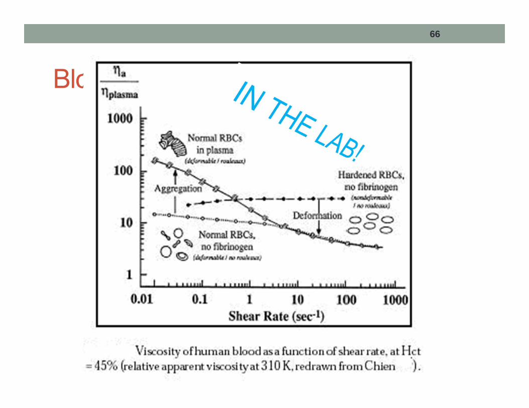
Blood Viscosity
67
Blood Viscosity
• Blood viscosity depends on three factors:• Shear rate• Percentage of cells in plasma• Vessel diameter

68
Factors Affecting Blood Viscosity
• Shear rate• At low shear rates, apparent viscosity increases. • Yield stress required to start fluid motion (Bingham Fluid)
• Cell concentration• Plasma is generally considered to be a Newtonian fluid at
physiological conditions ( = 1.6×10⁻³ kg/m·s)• Increasing RBC concentration increases viscosity linearly
(over a physiological range)
• Vessel diameter• Apparent viscosity is lower in small vessels (d < 0.3 mm)• Apparent viscosity approx. constant in vessels above 0.3
mm


![Diagnostic Uncertainties During Assessment of Serial ...milkyway.mie.uc.edu/rbanerje/publications/Biofluid dynamics/5.pdf · [24,26] and contractility [27,28] in recently conducted](https://static.fdocuments.net/doc/165x107/5e50c37080d1a7412b1ebac9/diagnostic-uncertainties-during-assessment-of-serial-dynamics5pdf-2426.jpg)


