
Introduction
12
Hystophatologic, Inmunohystochemic, and Ultrastructural Studies of Anterior Lens Capsule with Pseudoexfoliation Denisse Cornu -Melgoza, Erika Fernandez- Munoz, Jose Antonio Yamin -Lopez, Abelardo Rodriguez-Reyes Authors have no financial interest
-
Upload
keelie-bruce -
Category
Documents
-
view
21 -
download
0
description
Hystophatologic, Inmunohystochemic, and Ultrastructural Studies of Anterior Lens Capsule with Pseudoexfoliation. Denisse Cornu -Melgoza, Erika Fernandez- Munoz, Jose Antonio Yamin -Lopez, Abelardo Rodriguez-Reyes Authors have no financial interest. Introduction. Pseudoexfoliation syndrome. - PowerPoint PPT Presentation
Transcript of Introduction

Hystophatologic, Inmunohystochemic, and Ultrastructural Studies of
Anterior Lens Capsule with Pseudoexfoliation
Denisse Cornu -Melgoza, Erika Fernandez- Munoz, Jose Antonio Yamin -Lopez, Abelardo
Rodriguez-Reyes
Authors have no financial interest

Introduction Pseudoexfoliation syndrome.
Characterize for a deposit of an abnormal fibrillar material on the surface of some ocular and extraocular tissues
Described for the first time by Limberg 1917.
It is asociated with Glaucoma in a 40-60%
Suquin Guo, Matthew Gewirtz, Rajesh Thaker, Matthew Reed. Characterizing pseudoexfoliation syndrome through the use of ultrasound biomicroscopy. J Cataract Refract Surg. Apr 2006; 32(4): 614-7.

Propose
To determine by histophathology, inmunohystochemistry and ultraestructural studies the nature and origin of pseudoexfoliation material of the anterior lens capsule.

Methods
We obteined the product of the capsulorexis procedure realized during a conventional phacoemulsification and extracapsular cataract extraction in Asociacion para Evitar la Ceguera en Mexico (APEC).

RESULTS
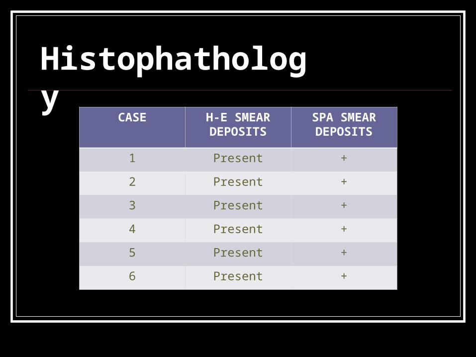
CASE H-E SMEAR DEPOSITS
SPA SMEAR DEPOSITS
1 Present +
2 Present +
3 Present +
4 Present +
5 Present +
6 Present +
Histophathology

Caso 1SPASPA
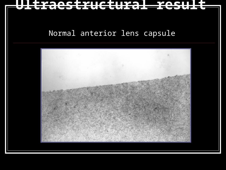
Ultraestructural result Normal anterior lens capsule

Pseudoexfoliation material.

Inmonohistochemical for colagen type IV
Conclusions.
Because of the affinity SPA (+), the electrodense deposits of a fibrillar material found on the anterior lens capsule and the positive smear of the cromogen for collagen type IV, we suggest that this deposits can be the product of degradation of some glycoprotein present in some structures of the anterior chamber (iris, anterior lens capsule, endothelium and zonule) like in the basal membrane.