INTRO TO THE MUSCULOSKELETAL SYSTEM / · PDF fileMicroscopic Anatomy (Compact Bone)...
17
INTRO TO THE MUSCULOSKELETAL SYSTEM / OVERVIEW OF BONES P2, 4, 6
Transcript of INTRO TO THE MUSCULOSKELETAL SYSTEM / · PDF fileMicroscopic Anatomy (Compact Bone)...

INTRO TO THE
MUSCULOSKELETAL SYSTEM /
OVERVIEW OF BONES
P2, 4, 6

Composition of the Human Body
• How many bones
are in our body?
• 270+ (at birth)
• 206 (adult)
• How many skeletal
muscles are in our
body?
• ~640 skeletal muscles

Major Functions
SKELETAL SYSTEM
• Support
• Protection
• Movement
• Storage
• Blood cell formation
MUSCULAR SYSTEM
• Movement
• Maintaining posture
• Stabilizing joints
• Generating Heat

Functions of the Bones
• Support • Internal framework
• Supports & anchors all soft organs
• Protection • Skull brain
• Vertebrae spinal cord
• Rib cage organs of thorax
• Movement • Works w/ skeletal muscles,
tendons, & ligaments
• Storage • Fat internal cavities of
bones
• Minerals Ca & P
• Neural transmission, muscle contraction, blood clotting
• Controlled by hormones
• Blood cell formation • AKA hematopoiesis
• Occurs in marrow cavities of certain bones

Divisions of the Skeleton
• 2 divisions:
• Axial skeleton – bones that form the longitudinal axis of the body
• Appendicular skeleton – bones of the limbs and girdles
• Skeletal system includes joints, cartilages, & ligaments

Classification of Bones
• 2 basic types of
osseous (bone)
tissue:
• Compact bone –
dense, smooth-
looking, &
homogenous
• Spongy bone –
composed of small
needlelike pieces of
bone; lots of open
space

Classification of Bones (cont’d)
SHAPES
• Long bones
• Longer than they are wide
• Typically includes shaft with
heads at both ends
• Mostly compact bone
• Short bones
• Typically cube-shaped
• Mostly spongy bone
• Sesamoid – special short
bone formed w/in tendons
• Flat bones
• Thin, flattened, & usually
curved
• 2 thin layers of compact
bone sandwiching 1 layer of
spongy bone
• Irregular bones
• Bones that don’t fit any of the
other groups

STRUCTURE OF A
LONG BONE P1-3

Gross Anatomy
• Diaphysis – shaft of long bone • Composed of compact bone
• Protected by fibrous CT membrane called periosteum
• Secured by CT fibers called perforating (Sharpey’s) fibers
• Epiphyses – ends of long bone • Thin layer of compact bone enclosing area filled w/
spongy bone
• Covered by glassy hyaline cartilage called articular cartilage
• Epiphyseal line – remnant of epiphyseal plate • Epiphyseal plate – flat plate of hyaline cartilage that
causes lengthwise growth of long bone
• Yellow marrow (medullary) cavity – storage area for adipose tissue
• Red marrow – area for blood cell formation
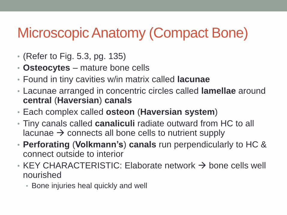
Microscopic Anatomy (Compact Bone)
• (Refer to Fig. 5.3, pg. 135)
• Osteocytes – mature bone cells
• Found in tiny cavities w/in matrix called lacunae
• Lacunae arranged in concentric circles called lamellae around central (Haversian) canals
• Each complex called osteon (Haversian system)
• Tiny canals called canaliculi radiate outward from HC to all lacunae connects all bone cells to nutrient supply
• Perforating (Volkmann’s) canals run perpendicularly to HC & connect outside to interior
• KEY CHARACTERISTIC: Elaborate network bone cells well nourished
• Bone injuries heal quickly and well

Bone as a Material
• One of the hardest in the body Ca salt deposits
• Relatively light
• Flexibility & remarkable ability to resist tension & other
forces collagen fibers

BONE FORMATION,
GROWTH, & REMODELING
P2, 4, 6

Bone Formation & Growth
• Cartilage & bone strongest & most supportive
• Embryos hyaline cartilage
• Young child to adult bone • Remaining cartilage in isolated areas
• Process of bone formation = ossification • Controlled by growth & sex hormones
• Ends when epiphyseal plates completely converted to bone
• Most use hyaline structures as “models”

Bone Formation & Growth (cont’d)
• 2 major phases: 1. Hyaline cartilage model covered w/ bone matrix by osteoblasts
(bone-forming cells)
2. Enclosed cartilage model digested medullary cavity formed
• 2 regions left: • Articular cartilages for life
• Epiphyseal plates during childhood
• Lengthening: • Epiphyseal end of plate new cartilage grows
• Diaphyseal end of plate old cartilage ossifies
• Widening: • Osteoblasts in periosteum ADD bone to diaphysis
• Osteoclasts in endosteum REMOVE bone from diaphysis

Bone Remodeling
• Remodeled continually in response to ∆s to 2 factors:
• Calcium levels in blood
• Hypocalcemia: Ca ↓ PTH ↑ osteoclasts activate break down bone matrix release Ca ions to blood
• Hypercalcemia: Ca ↑ deposited in bone as hard Ca salts
• Pull of gravity & muscles on skeleton
• Become thicker & form large projections to ↑ strength
• Osteoblasts lay down new matrix & trapped w/in become osteocytes
• PTH when (or if)
• Stresses of muscle pull & gravity where

BONE FRACTURES P2, 4, 6

Bone Fractures
• Fracture – break in a bone
• Simple (closed) – clean break; does NOT break skin
• Compound (open) – clean break; does break skin