
Intro to Protein Folding
53
arXiv:0705.1845v1 [physics.bio-ph] 13 May 2007 Introduction to protein folding for physicists Pablo Echenique ∗ Theoretical Physics Department, University of Zaragoza, Pedro Cerbuna 12, 50009, Zaragoza, Spain. Institute for Biocomputation and Physics of Complex Systems (BIFI), Edificio Cervantes, Corona de Arag´on 42, 50009, Zaragoza, Spain. February 1, 2008 Abstract The prediction of the three-dimensional native structure of proteins from the knowledge of their amino acid sequence, known as the protein folding problem, is one of the most important yet unsolved issues of modern science. Since the conformational behaviour of flexible molecules is noth- ing more than a complex physical problem, increasingly more physicists are moving into the study of protein systems, bringing with them power- ful mathematical and computational tools, as well as the sharp intuition and deep images inherent to the physics discipline. This work attempts to facilitate the first steps of such a transition. In order to achieve this goal, we provide an exhaustive account of the reasons underlying the protein folding problem enormous relevance and summarize the present-day sta- tus of the methods aimed to solving it. We also provide an introduction to the particular structure of these biological heteropolymers, and we phys- ically define the problem stating the assumptions behind this (commonly implicit) definition. Finally, we review the ‘special flavor’ of statistical mechanics that is typically used to study the astronomically large phase spaces of macromolecules. Throughout the whole work, much material that is found scattered in the literature has been put together here to improve comprehension and to serve as a handy reference. 1 Why study proteins? Virtually every scientific book or article starts with a paragraph in which the writer tries to persuade the readers that the topic discussed is very important for the future of humankind. We shall stick to that tradition in this work; but with the confidence that, in the case of proteins, the persuasion process will turn out to be rather easy and automatic. ∗ E-mail address: [email protected] — Web page: http://www.pabloechenique.com 1
-
Upload
rickey-castro -
Category
Documents
-
view
37 -
download
6
description
introduction to protein folding for physicists
Transcript of Intro to Protein Folding

arX
iv:0
705.
1845
v1 [
phys
ics.
bio-
ph]
13
May
200
7 Introduction to protein folding for physicists
Pablo Echenique∗
Theoretical Physics Department, University of Zaragoza,Pedro Cerbuna 12, 50009, Zaragoza, Spain.
Institute for Biocomputation and Physics of Complex Systems (BIFI),Edificio Cervantes, Corona de Aragon 42, 50009, Zaragoza, Spain.
February 1, 2008
Abstract
The prediction of the three-dimensional native structure of proteinsfrom the knowledge of their amino acid sequence, known as the proteinfolding problem, is one of the most important yet unsolved issues of modernscience. Since the conformational behaviour of flexible molecules is noth-ing more than a complex physical problem, increasingly more physicistsare moving into the study of protein systems, bringing with them power-ful mathematical and computational tools, as well as the sharp intuitionand deep images inherent to the physics discipline. This work attempts tofacilitate the first steps of such a transition. In order to achieve this goal,we provide an exhaustive account of the reasons underlying the proteinfolding problem enormous relevance and summarize the present-day sta-tus of the methods aimed to solving it. We also provide an introduction tothe particular structure of these biological heteropolymers, and we phys-ically define the problem stating the assumptions behind this (commonlyimplicit) definition. Finally, we review the ‘special flavor’ of statisticalmechanics that is typically used to study the astronomically large phasespaces of macromolecules. Throughout the whole work, much materialthat is found scattered in the literature has been put together here toimprove comprehension and to serve as a handy reference.
1 Why study proteins?
Virtually every scientific book or article starts with a paragraph in which thewriter tries to persuade the readers that the topic discussed is very importantfor the future of humankind. We shall stick to that tradition in this work; butwith the confidence that, in the case of proteins, the persuasion process willturn out to be rather easy and automatic.
∗E-mail address: [email protected] — Web page: http://www.pabloechenique.com
1

Proteins are a particular type of biological molecules that can be found inevery single living being on Earth. The characteristic that renders them es-sential for understanding life is simply their versatility. In contrast with therelatively limited structural variations present in other types of important bio-logical molecules, such as carbohydrates, lipids or nucleic acids, proteins displaya seemingly infinite capability for assuming different shapes and for producingvery specific catalytic regions on their surface. As a result, proteins constitutethe working force of the chemistry of living beings, performing almost everytask that is complicated. Quoting the first sentence of a section (which sharesthis section’s title) in Lesk’s book [1]:
In the drama of life on a molecular scale, proteins are where the
action is.
Just to state a few examples of what is meant by ‘action’, in living beings,proteins
• are passive building blocks of many biological structures, such as the coatsof viruses, the cellular cytoskeleton, the epidermal keratin or the collagenin bones and cartilages;
• transport and store other species, from electrons to macromolecules;
• as hormones, transmit information and signals between cells and organs;
• as antibodies, defend the organism against intruders;
• are the essential components of muscles, converting chemical energy intomechanical one, and allowing the animals to move and interact with theenvironment;
• control the passage of species through the membranes of cells and or-ganelles;
• control gene expression;
• are the essential agents in the transcription of the genetic information intomore proteins;
• together with some nucleic acids, form the ribosome, the large molecularorganelle where proteins themselves are synthesized;
• as chaperones, protect other proteins to help them to acquire their func-tional three-dimensional structure.
Due to this participation in almost every task that is essential for life, proteinscience constitutes a support of increasing importance for the development ofmodern Medicine. On one side, the lack or malfunction of particular proteins isbehind many pathologies; e.g., in most types of cancer, mutations are found in
2

Figure 1.1: Four molecular machines formed principally by proteins. Figures
taken from the Molecule of the month section of the RSCB Protein Data Bank
(http://www.pdb.org), we thank the RSCB PDB and David S. Goodsell, from the
Scripps Research Institute, for kind permission to use them. (a) ATP synthase: it
acts as an energy generator when it is traversed by protons that make its two cou-
pled engines rotate in reverse mode and the ATP molecule is produced. (b) RNA
polymerase: it slides along a thread of DNA reading the base pairs and synthesizing
a matching copy of RNA. (c) GroEL-GroES complex : it helps unfolded proteins to
fold by sheltering them from the overcrowded cellular cytoplasm. (d) Ribosome: it
polymerizes amino acids to form proteins following the instructions written in a thread
of mRNA.
3

the tumor suppressor p53 protein [2]. Also, abnormal protein aggregation char-acterizes many neurodegenerative disorders, including Huntington, Alzheimer,Creutzfeld-Jakob (‘mad cow’), or motor neuron diseases [3–5]. Finally, to at-tack the vital proteins of pathogens (HIV [6, 7], SARS [8], hepatitis [9], etc.),or to block the synthesis of proteins at the bacterial ribosome [10], are commonstrategies to battle infections in the frenetic field of rational drug design [11].
Apart from Medicine, the rest of human technology may also benefit fromthe solutions that Nature, after thousands of millions1 of years of ‘research’, hasfound to the typical practical problems. And that solutions are often proteins:New materials of extraordinary mechanical properties could be designed fromthe basis of the spider silk [12, 13], elastin [14] or collagen proteins [15]. Also,some attempts are being made to integrate these new biomaterials with livingorganic tissues and make them respond to stimuli [16]. Even further away onthe road that goes from passive structural functions to active tasks, no engineerwho has ever tried to solve a difficult chemical problem can avoid to experiencea feeling of almost religious inferiority when faced to the speed, efficiency andspecificity with which proteins cut, bend, repair, carry, link or modify otherchemical species. Hence, it is normal that we play with the idea of learning tocontrol that power and have, as a result, nanoengines, nanogenerators, nanoscis-sors, nanomachines in general [17]. The author of this work, in particular, felt asmall sting of awe when he learnt about the pump and the two coupled engines ofthe principal energy generator in the cell, the ATP synthase (figure 1.1a); aboutthe genetic Xerox machine, the RNA polymerase (figure 1.1b); about the hutwhere the proteins fold under shelter, the GroEL-GroES complex (figure 1.1c);or about the macromolecular factory where proteins are created, the ribosome
(figure 1.1d), to mention four specially impressive examples. Agreeing againwith Lesk [1]:
Proteins are fascinating molecular devices.
From a more academic standpoint, proteins are proving to be a powerfulcentre of interdisciplinary research, making many diverse fields and people withdifferent formations come in contact2. Proteins force biologists, biochemistsand chemists to learn more physics, mathematics and computation and forcemathematicians, physicists and computer technicians to learn more biology, bio-chemistry and chemistry. This, indeed, cannot be negative.
In 2005, in a special section of Science magazine entitled ‘What don’t weknow?’ [18], a selection of the hundred most interesting yet unanswered scientificquestions was presented. What indicates the role of proteins, and particularly ofthe protein folding problem (treated in section 3), as focuses of interdisciplinarycollaboration is not the inclusion of the question Can we predict how proteins
will fold?, which was a must, but the large number of other questions which were
1 Herein, we shall use the British convention for naming large numbers; in which 109=‘athousand million’, 1012=‘a billion’, 1015=‘a thousand billion’, 1018=‘a trillion’, and so on.
2 The Institute for Biocomputation and Physics of Complex Systems, which the author ispart of, constitutes an example of this rather new form of collaboration among scientists.
4

related to or even dependent on it, such as Why do humans have so few genes?,How much can human life span be extended?, What is the structure of water?,How does a single somatic cell become a whole plant?, How many proteins are
there in humans?, How do proteins find their partners?, How do prion diseases
work?, How will big pictures emerge from a sea of biological data?, How far can
we push chemical self-assembly? or Is an effective HIV vaccine feasible?.In this direction, probably the best example of the use that protein science
makes of the existing human expertise, and of the positive feedback that thisbrings up in terms of new developments and resources, can be found in the ma-chines that every one of us has on his/her desktops. In a first step, the enormousamount of biological data that emerges from the sequencing of the genomes ofdifferent living organisms requires computerized databases for its proper fil-tering. The NCBI GenBank database3, which is one of the most exhaustiverepositories of sequenced genetic material, has doubled the number of depositedDNA bases approximately every 18 months since 1982 (see figure 1.2a) and hasrecently (in August 2005) exceeded the milestone of 100 Gigabases (1011) fromover 165,000 species.
Among them, and according to the Entrez Genome Project database4, thesequencing of the complete genome of 366 organisms has been already achievedand there are 791 more to come in next few years. In the group of the completedones, most are bacteria, and there are only two mammals: the poor laboratorymouse, Mus Musculus, and, notably [19], the Homo Sapiens (with ∼ 3 · 109
bases and a mass-media-broadcast battle between the private firm Celera andthe public consortium IHGSC).
However, not all the DNA encodes proteins (not all the DNA is genes). Typ-ically, more than 95% of the genetic material in living beings is junk DNA, alsocalled non-coding DNA (a more neutral term which seems recommendable in thelight of some recent discoveries [20–22]). So, in a second step, the coding regionsmust be identified and each gene translated into the amino acid sequence of aparticular protein5. The UniProt database6 is, probably, the most comprehen-sive repository of these translated protein sequences and also of others comingfrom a variety of sources, including direct experimental determination [25, 26].UniProt is comprised by two different sub-databases: the Swiss-Prot ProteinKnowledgebase, which contains extensively human-annotated protein sequenceswith low redundancy; and TrEMBL, which contains computer-annotated se-quences extracted directly from the underlying nucleotide entries at databasessuch as GenBank and where only the most basic redundancies have been re-moved.
The UniProt/Swiss-Prot database contains, at the moment (on 30 May
3 http://www.ncbi.nlm.nih.gov/Genbank/4 http://www.ncbi.nlm.nih.gov/entrez/query.fcgi?db=genomeprj5 Note that many variations [23, 24] may occur before, during and after the process of
gene expression, so that the relation gene-to-protein is not one-to-one. The size of the humanproteome (the number of different proteins), for example, is estimated to be an order ofmagnitude or two larger than the size of the genome.
6 http://www.uniprot.org
5

106
107
108
109
1010
1011
1012
Apr
200
6
Dec
200
4
Dec
200
2
Dec
200
0
Dec
199
8
Dec
199
6
Dec
199
4
Dec
199
2
Dec
199
0
Dec
198
8
Nov
198
6
Nov
198
4
Dec
198
2
Num
ber
of d
epos
ited
DN
A b
ases
at G
enB
ank
100 Gigabases
103
104
105
106
107
Dec
200
6
Dec
200
5
Dec
200
4
Dec
200
3
Dec
200
2
Dec
200
1
Dec
200
0
Dec
199
9
Dec
199
8
Dec
199
7
Dec
199
6
Dec
199
5
Dec
199
4
Dec
199
3
Dec
199
2
Dec
199
1
Dec
199
0
Dec
198
9
Dec
198
8
Dec
198
7
Dec
198
6
Num
ber
of p
rote
in s
eque
nces
at U
niP
rot
Swiss-ProtTrEMBL
101
102
103
104
105
2005 2000 1995 1990 1985 1980 1975
Num
ber
of p
rote
in s
truc
ture
s at
PD
B
103
104
105
106
107
108
109
2005 2000 1995 1990 1985 1980 1975 1970
Num
ber
of tr
ansi
stor
s on
an
inte
grat
ed c
ircui
t
40048008
8080
8086
286
386
486
Pentium
Pentium II
Pentium III
Pentium IV
Itanium
Itanium 2
Itanium 2(9MB cache)
a b
dc
Figure 1.2: Recent exponential progress in genomics, proteomics and computer
technology. (a) Evolution of the number of DNA bases deposited at the GenBank
database. (b) Evolution of the number of protein sequences at the UniProt Swiss-Prot
and TrEMBL databases. (c) Evolution of the number of protein three-dimensional
structures at the Protein Data Bank. (d) Moore’s Law: evolution of the number of
transistors in the Intel CPUs.
2006), around 200,000 protein sequences from about 10,000 species, and it hasexperienced an exponential growth (since 1986), doubling the number of recordsapproximately every 41 months (see figure 1.2b). In turn, the UniProt/TrEMBLdatabase contains almost 3 million protein sequences from more than 100,000species, and its growth (from 1997) has also been exponential, doubling thenumber of records approximately every 16 months (see figure 1.2b).
After knowing the sequence of a protein, the next step towards the under-standing of biological processes is the characterization of its three-dimensionalstructure. Most proteins perform their function under a very specific native
shape which involves many twists, loops and bends of the linear chain of aminoacids (see section 3). This spatial structure is much more important than thesequence for biochemists to predict and understand the mechanisms of life and itcan be resolved, nowadays, by fundamentally two experimental techniques: forsmall proteins, nuclear magnetic resonance (NMR) [27,28] and, more commonly,for proteins of any size, x-ray crystallography [29–31]. The three-dimensionalstructures so obtained are deposited in a centralized public-access databasecalled Protein Data Bank (PDB)7 [32]. From the 13 structures deposited in1976 to the 33,782 (from more than a thousand species) stored in June 2006,the growth of the PDB has been (guess?) exponential, doubling the number ofrecords approximately every 3 years (see figure 1.2c).
7 http://www.rcsb.org/pdb/
6
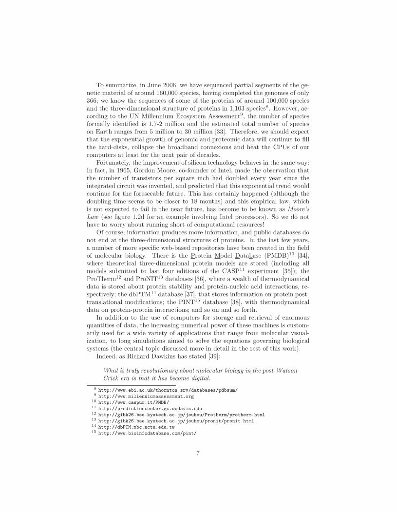
To summarize, in June 2006, we have sequenced partial segments of the ge-netic material of around 160,000 species, having completed the genomes of only366; we know the sequences of some of the proteins of around 100,000 speciesand the three-dimensional structure of proteins in 1,103 species8. However, ac-cording to the UN Millennium Ecosystem Assessment9, the number of speciesformally identified is 1.7-2 million and the estimated total number of specieson Earth ranges from 5 million to 30 million [33]. Therefore, we should expectthat the exponential growth of genomic and proteomic data will continue to fillthe hard-disks, collapse the broadband connexions and heat the CPUs of ourcomputers at least for the next pair of decades.
Fortunately, the improvement of silicon technology behaves in the same way:In fact, in 1965, Gordon Moore, co-founder of Intel, made the observation thatthe number of transistors per square inch had doubled every year since theintegrated circuit was invented, and predicted that this exponential trend wouldcontinue for the foreseeable future. This has certainly happened (although thedoubling time seems to be closer to 18 months) and this empirical law, whichis not expected to fail in the near future, has become to be known as Moore’s
Law (see figure 1.2d for an example involving Intel processors). So we do nothave to worry about running short of computational resources!
Of course, information produces more information, and public databases donot end at the three-dimensional structures of proteins. In the last few years,a number of more specific web-based repositories have been created in the fieldof molecular biology. There is the Protein Model Database (PMDB)10 [34],where theoretical three-dimensional protein models are stored (including allmodels submitted to last four editions of the CASP11 experiment [35]); theProTherm12 and ProNIT13 databases [36], where a wealth of thermodynamicaldata is stored about protein stability and protein-nucleic acid interactions, re-spectively; the dbPTM14 database [37], that stores information on protein post-translational modifications; the PINT15 database [38], with thermodynamicaldata on protein-protein interactions; and so on and so forth.
In addition to the use of computers for storage and retrieval of enormousquantities of data, the increasing numerical power of these machines is custom-arily used for a wide variety of applications that range from molecular visual-ization, to long simulations aimed to solve the equations governing biologicalsystems (the central topic discussed more in detail in the rest of this work).
Indeed, as Richard Dawkins has stated [39]:
What is truly revolutionary about molecular biology in the post-Watson-
Crick era is that it has become digital.
8 http://www.ebi.ac.uk/thornton-srv/databases/pdbsum/9 http://www.millenniumassessment.org
10 http://www.caspur.it/PMDB/11 http://predictioncenter.gc.ucdavis.edu12 http://gibk26.bse.kyutech.ac.jp/jouhou/Protherm/protherm.html13 http://gibk26.bse.kyutech.ac.jp/jouhou/pronit/pronit.html14 http://dbPTM.mbc.nctu.edu.tw15 http://www.bioinfodatabase.com/pint/
7

Finally, apart from all the convincing reasons and the appeals to authoritygiven above, what is crystal-clear is that proteins are an unsolved and difficultenigma. And those are two irresistible qualities for any flesh and blood scientist.
2 Summary of protein structure
In spite of their diverse biological functions, summarized in the previous section,proteins are a rather homogeneous class of molecules from the chemical pointof view. They are linear heteropolymers, i.e., unbranched chains of differentidentifiable monomeric units.
Before they are assembled into proteins, these building units are called amino
acids and can exist as stand alone stable molecules. All amino acids are made upof a central α-carbon with four groups attached to it: an amino group (—NH2),a carboxyl group (—COOH), a hydrogen atom and a fourth arbitrary group(—R) (see figure 2.2). In aqueous solvent and under physiological conditions,both the amino and carboxyl groups are charged, the first accepting one protonand getting a positive charge, and the second giving one proton away and gettinga negative charge (compare figures 2.2a and 2.2c).
When the group —R is not equal to one of the other three groups attachedto the α-carbon, the amino acid is chiral, i.e., like our hands, it may exist in twodifferent forms, which are mirror images of one another and cannot be super-imposed by rotating one of them in space (you cannot wear the left-hand gloveon your right hand). In chemical jargon, one says that the α-carbon constitutesan asymmetric centre and that the amino acid may exist as two different enan-
tiomers called L- (figure 2.2c) and D- (figure 2.2d) forms. It is common that,when used as prefixes, the L and D letters, which come from levorotatory anddextrorotatory, are written in small capitals, as in L- and D-. This nomenclatureis based on the possibility of associating the amino acids to the optically activeL- and D- enantiomers of glyceraldehyde, and could be related to the +/- or tothe Cahn, Ingold and Prelog’s R/S [41] notations. For us, it suffices to say that
Figure 2.1: Color and size code for the atom types used in most of the figures in this
section. All the figures have been made with the Gaussview graphical front-end of
Gaussian03 [40] and then modified with standard graphical applications.
8

Figure 2.2: Amino acids. (a) Uncharged L-enantiomer. (b) CORN mnemotechnic
rule to remember which one is the L-form. (c) Charged L-enantiomer (the predomi-
nant form found in living beings). (d) Charged D-enantiomer.
the D/L nomenclature is, by far, the most used one in protein science and theone that will be used in this work. For further details, take a look at the IUPACrecommendations at http://www.chem.qmul.ac.uk/iupac/AminoAcid/.
In principle, amino acids may be L- or D-, and the group —R may be any-thing provided that the resultant molecule is stable. However, for reasons thatare still unclear [42], the vast majority of proteins in all living beings are madeup of L-amino acids (as a rare exception, we may point out the fact that D-aminoacids can be found in some proteins produced by exotic sea-dwelling organisms,such as cone snails) and the groups —R (called side chains) that are codedin the genetic material comprise a set of only twenty possibilities (depicted infigure 2.5).
A frequently quoted mnemotechnic rule for remembering which one is theL-form of amino acids is the so-called CORN rule in figure 2.2b. Accordingto it, one must look from the hydrogen to the α-carbon and, if the three re-maining groups are labelled as in the figure, the word CORN must be read inthe clockwise sense of rotation. The author of this work does not find this rulevery useful, since normally he cannot recall if the sense is clockwise or coun-terclockwise. To know which form is the L- one, he draws the amino acid asin figure 2.2a or 2.2c, with the α-carbon in the centre, the amino group on theleft and the carboxyl group on the right, all of them in the plane of the paper(which is very natural and easy to remember because it matches the normalsense of writing with the fact that, conventionally, proteins start at —NH+
3 andend at —COO−). Finally, he must just remember that the side chain of theL-amino acid goes out of the paper approaching the reader (which is also natural
9

Figure 2.3: Peptide bond formation reaction. The peptide plane is indicated in green.
because the side chain is the relevant piece of information and we want to lookat it closely).
The process through which amino acids are assembled into proteins (calledgene expression or protein biosynthesis) is typically divided in two steps. In thefirst one, the transcription, the enzyme ARN polymerase (see figure 1.1b) bindsto the DNA in the cellular nucleus and makes a copy of a section –the gene–of the base sequence into a messenger RNA (mRNA) molecule. In the secondstep, called translation, the mRNA enters the ribosome (see figure 1.1d) and isread stopping at each base triplet (called codon). Now, a specific molecule oftransfer RNA (tRNA), which possesses the base triplet (called anticodon) thatis complementary to the codon, links to the mRNA bringing with her the aminoacid that is codified by the particular sequence of three bases. Each amino acidthat arrives to the ribosome in this way is covalently attached to the previousone and so added to the nascent protein. In this reaction, the peptide bond isformed and a water molecule is released (see figure 2.3). This process continuesuntil a stop codon is read and the transcription is complete.
The amino acid sequence of the resultant protein, read from the amino
terminus to the carboxyl terminus, is called primary structure; and the aminoacids included in such a polypeptide chain are normally termed amino acid
residues, or simply residues, in order to distinguish them from their isolatedform. The main chain formed by the repetition of α-carbons and the C’ and Natoms at the peptide bond is called backbone and the —R groups branching outfrom it are called side chains, as it has already been mentioned.
The specificity of each protein is provided by the different properties of thetwenty side chains in figure 2.5 and their particular positions in the sequence.In textbooks, it is customary to group them in small sets according to different
10

criteria in order to facilitate their learning. Classifications devised on the basisof the physical properties of these side chains may be sometimes overlapping(e.g., tryptophan contains polar regions as well as an aromatic ring, which, inturn, could be considered hydrophobic but is also capable of participating in,say, π-π interactions). Therefore, for a clearer presentation, we have chosen hereto classify the residues according to the chemical groups contained in each sidechain and discuss their physical properties individually.
Let us enumerate then the categories in figure 2.5 and point out any specialremark regarding the residues in them:
Special residues:
• Glycine is the smallest of all the amino acids: its side chain contains only ahydrogen atom. So, since its α-carbon has two hydrogens attached, glycineis the only achiral natural amino acid. Its affinity for water is mainlydetermined by the peptide groups in the backbone; therefore, glycine ishydrophilic.
• Proline is the only residue whose side chain is covalently linked to thebackbone (the backbone is indicated in purple in figure 2.5), giving prolineunique structural properties that will be discussed later. Since its sidechain is entirely aliphatic, proline is hydrophobic.
Sulfur-containing residues:
• Cysteine is a very important structural residue because, in a reaction cat-alyzed by protein disulfide isomerases (PDIs), it may form, with anothercysteine, a very stable covalent bond called disulfide bond (see figure 2.4).Curiously, all the L-amino acids are S-enantiomers according to the Cahn,Ingold and Prelog rules [41] except for cysteine, which is R-. This is proba-bly the reason that makes the D/L nomenclature favourite among proteinscientists [24]. Cysteine is a polar residue.
Figure 2.4: Disulfide bond between two cysteine residues.
11

Figure 2.5: Side chains of the twenty amino acid residues encoded in the genetic
material of living beings. They have been classified according to the chemical groups
they contain. The rotameric degrees of freedom χi are indicated with small arrows over
the bonds. The name of the heavy atoms and the numbering of the branches comply
with the IUPAC rules http://www.chem.qmul.ac.uk/iupac/AminoAcid/). Below the
molecular structure, the one letter code (green), the three letter code (red) and the
complete name (blue) of each amino acid may be found. In the case of proline, the
N and the α-carbon have been included in the scheme, and the backbone bonds have
been coloured in purple. The titratable residues Asp, Glu, Lys and Arg have been
represented in their charged forms, which is the most common one in aqueous solvent
under physiological conditions. Histidine is shown in its neutral ε2-tautomeric form.
12

• Methionine is mostly aliphatic and, henceforth, apolar.
Aliphatic residues:
• Alanine is the smallest chiral residue. This is the fundamental reason forusing alanine models, more than any other ones, in the computationallydemanding ab initio studies of peptides that are customarily performed inquantum chemistry [43–53]. It is hydrophobic, like all the residues in thisgroup.
• Valine is one of the three β-branched residues (i.e., those that have morethan one heavy atom attached to the β-carbon, apart from the α-carbon),together with isoleucine and threonine. It is hydrophobic.
• Leucine is hydrophobic.
• Isoleucine’s β-carbon constitutes an asymmetric centre and the only enan-tiomer that occurs naturally is the one depicted in the figure. Onlyisoleucine and threonine contain an asymmetric centre in their side chain.Isoleucine is β-branched and hydrophobic.
Acid residues:
• Aspartic acid is normally charged under physiological conditions. Hence,it is very hydrophilic.
• Glutamic acid is just one CH2 larger than aspartic acid. Their propertiesare very similar.
Amides:
• Asparagine contains a chemical group similar to the peptide bond. It ispolar and can act as a hydrogen bond donor or acceptor.
• Glutamine is just one CH2 larger than asparagine. Their physical prop-erties are very similar.
Figure 2.6: Three forms of histidine found in proteins. (a) Neutral ε2-tautomer. (b)
neutral δ1-tautomer. (c) Charged form.
13

Basic residues:
• Histidine is a special amino acid: in its neutral form, it may exist as twodifferent tautomers, called δ1 and ε2, depending on which nitrogen hasan hydrogen atom attached to it. The ε2-tautomer has been found tobe slightly more stable in model dipeptides [54], although both forms arefound in proteins. Histidine can readily accept a proton and get a positivecharge, in fact, it is the only side chain with a pKa in the physiologicalrange, so non-negligible proportions of both the charged and unchargedforms are typically present. Of course, histidine is hydrophilic.
• Lysine’s side chain is formed by a rather long chain of CH2 with an aminogroup at its end, which is nearly always positively charged. Therefore,lysine is very polar and hydrophilic.
• Arginine’s properties are similar to those of lysine, although its terminalguanidinium group is a stronger basis than the amino group and it mayalso participate in hydrogen bonds as a donor.
Alcohols:
• Serine is one of the smallest residues. It is polar due to the hydroxylgroup.
• Threonine’s β-carbon constitutes an asymmetric centre; the enantiomerthat occurs in living beings is the one shown in the figure. The physicalproperties of threonine are very similar to those of serine.
Aromatic residues:
• Phenylalanine is the smallest aromatic residue. Its benzyl side chain islargely apolar and interacts unfavourably with water. It may also partic-ipate in specific π-stacking interactions with other aromatic groups.
• Tyrosine’s properties are similar to those of phenylalanine, being onlyslightly more polar due to the presence of a hydroxyl group.
• Tryptophan, with 17 atoms in her side chain, is the largest residue. It ismainly hydrophobic, although it contains a small polar region and it canalso participate in π-π interactions, like all the residues in this category.
After having introduced the building blocks of proteins, some qualifyingremarks about them are worth to be done: On one side, why amino acidsencoded in DNA codons are the ones in the list or why there are exactly twenty ofthem are questions that are still subjects of controversy [55,56]. In fact, althoughthe side chains in figure 2.5 seem to confer enough versatility to proteins inmost cases, there are also rare exceptions in which other groups are needed toperform a particular function. For example, the amino acid selenocysteine maybe incorporated into some proteins at an UGA codon (which normally indicates
14

Figure 2.7: Typical definition of internal coordinates. r21 is the bond length between
atoms 2 and 1. θ321 is the bond angle formed by the bonds (2,1) and (3,2), it ranges
from 0 to 180 o. Finally ϕ4321 is the dihedral angle describing the rotation around the
bond (3,2); it is defined as the angle formed by the plane containing atoms 1, 2 and
3 and the plane containing atoms 2, 3 and 4; it ranges either from −180 o to 180 o
or from 0 o to 360 o, depending on the convention; the positive sense of rotation for
ϕ4321 is the one indicated in the figure. Also note that the definition is symmetric
under a complete change in the order of the atoms, in such a way that, quite trivially,
r21 = r12 and θ123 = θ321, but also, not so trivially, ϕ4321 = ϕ1234. (See reference [51]
for further information.)
a stop in the transcription), or the amino acid pyrrolysine at an UAG codon(which is also a stop indication in typical cases). In addition, the arginine sidechain may be post-translationally converted into citrulline by the action of afamily of enzymes called peptidylarginine deiminases (PADs).
On the other hand, the chemical (covalent) structure of the protein chainmay suffer from more complex modifications than just the inclusion of non-standard amino acid residues: A myriad of organic molecules may be covalentlylinked to specific points, the chain may be cleaved (cut), chemical groups maybe added or removed from the N- or C-termini, disulfide bonds may be formedbetween cysteines, and the side chains of the residues may undergo chemicalmodifications just like any other molecule [54]. The vast majority of thesechanges either depend on the existence of some chemical agent external to theprotein, or are catalyzed by an enzyme.
In this work, our interest is in the folding of proteins. This problem, whichwill be discussed in detail in the next section, is so huge and so difficult that,in the opinion of the author, there is no point in worrying about details, suchas the ones mentioned in the two preceding paragraphs, before the big pictureis at least preliminarily understood. Therefore, when we talk about the folding
15

of proteins in what follows, we will be thinking about single polypeptide chains,made up of L-amino acids, in water and without any other reagent present, withthe side chains chosen from the set in figure 2.5, and having underwent no post-translational modifications nor any chemical change on their groups. Finally,although some simple modifications, such as the formation of disulfide bondsor the trans → cis isomerization of Xaa-Pro peptide bonds (see what follows),could be more easily included in the first approach to the problem, we shall alsoleave them for a later stage.
Now, with this considerations, we have fixed the covalent structure of ourmolecule as well as the enantiomerism of the asymmetric centres it may contain.This information is enough to specify the three-dimensional arrangement of theatoms of small rigid molecules. However, long polymers and, particularly, pro-teins, possess degrees of freedom (termed soft) that require small amounts ofenergy to be changed while drastically altering the relative positions of groupsand atoms. In a first approximation, all bond lengths, bond angles and dihe-dral angles describing rotations around triple, double and partial double bonds(see figure 2.7) may be considered to be determined by the covalent struc-ture. Whereas dihedral angles describing rotations around single bonds may beconsidered to be variable and soft. The non-superimposable three-dimensionalarrangements of the molecule that correspond to different values of the softdegrees of freedom are called conformations.
In proteins, some of these soft dihedrals are located at the side chains; theyare the χi in figure 2.5 and, although they are important in the later stages of
a
b
Figure 2.8: Trans and cis conformations of the peptide plane. The bonds defining
the peptide bond dihedral angle ω are indicated in purple. (a) Trans conformation
(ω ≃ ±180 o). The most common one in proteins. (b) Cis conformation (ω ≃ 0 o).
Significantly found only in Xaa-Pro bonds.
16

Figure 2.9: Numeration of the heavy atoms and the dihedrals angles describing ro-
tations around backbone bonds. In agreement with IUPAC recommendations (see
http://www.chem.qmul.ac.uk/iupac/AminoAcid/). The peptide planes are indicated
as green rectangles.
the folding process and must be taken into account in any ambitious model ofthe system, their variation only alters the conformation locally. On the contrary,a small change in the dihedral angles located at the backbone of the polypeptidechain may drastically modify the relative position of many pairs of atoms andthey must be given special attention.
That is why, the special properties of the peptide bond, which is the basicbuilding block of the backbone, are very important to understand the conforma-tional behaviour of proteins. These properties arise from the fact that there isan electron pair delocalized between the C—N and C—O bonds (using the com-mon chemical image of resonance), which provokes that neither bond is singlenor double, but partial double bonds that have a mixed character. In particular,the partial double bond character of the peptide bond is the cause that thesix atoms in the green plane depicted in figures 2.3, 2.8 and 2.9 have a strongtendency to be coplanar, forming the so-called peptide plane. This coplanarityallows for only two different conformations: the one called trans (correspondingto ω ≃ ±180 o), in which the α-carbons lie at different sides of the line contain-ing the C—N bond; and the one called cis (corresponding to ω ≃ 0 o), in whichthey lie at the same side of that line (see figure 2.8).
Although the quantitative details are not completely elucidated yet and thevery protocol of protein structure determination by x-ray crystallography couldintroduce spurious effects in the structures deposited in the PDB [57], it seemsclear that a great majority of the peptide bonds in proteins are in the transconformation. Indeed, a superficial look at the two forms in figure 2.8 suggeststhat the steric clashes between substituents of consecutive α-carbons will bemore severe in the cis case. When the second residue is a Proline, however, thespecial structure of its side chain makes the probability of finding the cis con-
17

Figure 2.10: Original Ramachandran plot drawn by Ramachandran and Ramakrish-
nan in 1963 [60]. In dark-green, the fully allowed regions, calculated by letting the
atoms approach to the average clashing distance; in light-green, the partially allowed
regions, calculated by letting the atoms approach to the minimum clashing distance;
in white, the disallowed regions. Some points representing secondary structure ele-
ments are shown as red circles at the ideal (φ,ψ)-positions in table 1: (α) α-helix.
(π) π-helix. (310) 310-helix. (aβ) Antiparallel β-sheet. (pβ) Parallel β-sheet. (ppII)
Polyproline II.
former significantly higher: For Xaa-nonPro peptide bonds in native structures,the trans form is more common than the cis one with approximately a 3000:1proportion; while this ratio decreases to just 15:1 if the bond is Xaa-Pro [57].
In any case, due to the aforementioned partial double bond character of theC—N bond, the rotation barrier connecting the two states is estimated to be ofthe order of ∼ 20 kcal/mol [58], which is about 40 times larger than the ther-mal energy at physiological conditions, thus rendering the spontaneous trans →cis isomerization painfully slow. However, mother Nature makes use of everypossibility that she has at hand and, sometimes, there are a few peptide bondsthat must be cis in order for the protein to fold correctly or to function prop-erly. Since all peptide bonds are synthesized trans at the ribosome [59], thetrans → cis isomerization must be catalyzed by enzymes (called peptidylprolyl
isomerases (PPIs)) and, in the same spirit of the post-translational modifica-tions discussed before, this step may be taken into account in a later refinementof the theoretical models.
Therefore, we shall assume in what follows that all peptide bonds (even theXaa-Pro ones) are in the trans state and, henceforth, the conformation of theprotein will be essentially determined by the values of the φ and ψ angles, whichdescribe the rotation around the two single bonds next to each α-carbon (seefigure 2.9 for a definition of the dihedral angles associated to the backbone).
This assumption was introduced, as early as 1963, by Ramachandran and
18

φ ψα-helix −57 −47310-helix −49 −26π-helix −57 −70polyproline II −79 149parallel β-sheet −119 113antiparallel β-sheet −139 135
Table 1: Ramachandran angles (in degrees) of some important secondary structure
elements in polypeptides. Data taken from reference [1].
Ramakrishnan [60] and the φ and ψ coordinates are commonly named Ra-
machandran angles after the first one of them. In their famous paper [60], theyadditionally suppose that the bond lengths, bond angles and dihedral angles ondouble and partial double bonds are fixed and independent of φ and ψ, theydefine a typical distance up to which a specific pair of atoms may approach andalso a minimum one (taken from statistical studies of structures) and they drawthe first Ramachandran plot (see figure 2.10): A depiction of the regions in the(φ, ψ)-space that are energetically allowed or disallowed on the basis of the localsterical clashes between atoms that are close to the α-carbon.
One of the main advantages of this type of diagrams as ‘thinking tools’lies in the fact that (always in the approximation that the non-Ramachandranvariables are fixed) some very common repetitive structures found in proteinsmay be ideally depicted as a single point in the plot. In fact, these specialconformations, which are regarded as the next level of protein organizationafter the primary structure and are said to be elements of secondary structure,may be characterized exactly like that, i.e., by asking that a certain numberof consecutive residues present the same values of the φ and ψ angles. In thebook by Lesk [1], for example, one may found a table with the most common ofthese repetitive patterns, together with the corresponding (φ, ψ)-values takenfrom statistical investigations of experimentally resolved protein structures (seetable 1).
However, the non-Ramachandran variables are not really constant, and theelements of secondary structure do possess a certain degree of flexibility. More-over, the side chains may interact and exert different strains at different points ofthe chain, which provokes that, in the end, the secondary structure elements gainsome stability by slightly altering their ideal Ramachandran angles. Therefore,it is more appropriate to characterize them according to their hydrogen-bondingpattern, which, in fact, is the feature that makes these structures prevalent, pro-viding them with more energetic stability than other repetitive conformationswhich are close in the Ramachandran plot.
The first element of secondary structure that was found is the α-helix. Itis a coil-like16 structure, with ∼ 3.6 residues per turn, in which the carbonyl
16 Here, we use the word ‘coil’ to refer to the twisted shape of a telephone wire, a corkscrew
19

Figure 2.11: The three helices found in protein native structures. (a) 310-helix, (b)
α-helix, and (c) π-helix. In the three cases, the helices shown are 11-residues long. In
the standing views (above), the hydrogen bonds are depicted as green dotted lines and
the distance and number of turns spanned by 10 residues are indicated at the right of
the structures. Whereas in the standing views, the side chains and α-hydrogens have
been removed for visual convenience, in the zenithal views (below), they are included.
group (C=O) of each i-th residue forms a hydrogen bond with the amino group(N–H) of the residue i+ 4 (see figure 2.11b). According to a common notation,in which xy designates a helix with x residues per turn and y atoms in the ringclosed by the hydrogen bond [61], the α-helix is also called 3.613-helix.
She was theoretically proposed in 1951 by Pauling, Corey and Branson [62],who used precise information about the geometry of the non-Ramachandranvariables, taken from crystallographic studies of small molecules, to find thestructures compatible with the additional constraints that: (i) the peptide bondis planar, and (ii) every carbonyl and amino group participates in a hydrogenbond.
The experimental confirmation came from Max Perutz, who, together withKendrew and Bragg, had proposed in 1950 (one year before Pauling’s paper) aseries of helices with an integer number of residues per turn [61] that are notso commonly found in native structures of proteins (see however, the discussion
or the solenoid of an electromagnet. Although this is common English usage, the same wordoccurs frequently in protein science to designate different (and sometimes opposed) concepts.For example, a much used ideal model of the denatured state of proteins is termed random
coil, and a popular statistical description of helix formation is called helix-coil theory.
20

about the 310-helix below). Perutz read Pauling, Corey and Branson’s paperone Saturday morning [63] in spring 1951 and realized immediately that theirhelix looked very well: free of strain and with all donor and acceptor groupsparticipating in hydrogen bonds. So he rushed to the laboratory and put asample of horse hair (rich in keratin, a protein that contains α-helices) in thex-ray beam, knowing that, according to diffraction theory, the regular repeat ofthe ‘spiral staircase steps’ in Pauling’s structure should give rise to a strong x-ray reflection of 1.5 A spacing from planes perpendicular to the fiber axis. Theresult of the experiment was positive17 and, in the last years of the 50s, Perutzand Kendrew saw again the same signal in myoglobin and hemoglobin, whenthey resolved, for the first time in history, the structure of these proteins [64,65].
However, despite its being, by far, the most common, the α-helix is notthe only coil-like structure that can be found in native proteins [66–68]. Ifthe hydrogen bonds are formed between the carbonyl group (C=O) of eachi-th residue with the amino group (N–H) of the residue i + 3, one obtains a310-helix, which is more tightly wound and, therefore, longer than an α-helixof the same chain length (see figure 2.11a). The 310-helix is the fourth mostcommon conformation for a single residue after the α-helix, β-sheet and reverseturn18 [67] but, remarkably, due to its having an integer number of residuesper turn, it seemed more natural to scientists with crystallographic backgroundand was theoretically proposed before the α-helix [61, 69]. On the other hand,if the hydrogen bonds are formed between the carbonyl group (C=O) of eachi-th residue with the amino group (N–H) of the residue i + 5, one obtains aπ-helix (or 4.416-helix ), which is wider and shorter than an α-helix of the samelength (see figure 2.11c). It was originally proposed by Low and Baybutt in1952 [70], and, although the exact fraction of each type of helix in proteinnative structures depends up to a considerable extent on their definition (interms of Ramachandran angles, interatomic distances, energy of the hydrogenbonds, etc.), it seems clear that the π-helix is the less common of the three [66].Now, it is true that, in addition to these helices that have been experimentallyconfirmed, some others have been proposed. For example, in the same work inwhich Pauling, Corey and Branson introduce the α-helix [62], they also describeanother candidate: the γ-helix (or 5.117-helix ). Finally, Donohue performed, in1953, a systematic study of all possible helices and, in addition to the onesalready mentioned, he proposed a 2.27- and a 4.314-helix [71]. None of themhas been detected in resolved native proteins.
Among the secondary structure elements of proteins, not all regular localpatterns are helices: there exist also a variety of repetitive conformations thatdo not contain strong intra-chain hydrogen bonds and that are less curled thanthe structures in figure 2.11. For example, the polyproline II [72–74], which is
17 Linus Pauling was awarded the Nobel prize in chemistry in 1954 ‘for his research into thenature of the chemical bond and its application to the elucidation of the structure of complexsubstances’, and Max Perutz shared it with John Kendrew in 1962 ‘for their studies of thestructures of globular proteins’.
18 A conformation that some residues in proteins adopt when an acute turn in the chain isneeded.
21

Figure 2.12: β-sheets in the pure (a) antiparallel, and (b) parallel versions. On
the left, the top view is shown, with the side chains and the α-hydrogens omitted for
visual convenience and the directions of the strands indicated as yellow arrows. The
hydrogen bonds are represented as green dotted lines. On the right, the side view of
the sheets is depicted. In this case, the side chains and the α-hydrogens are included.
thought to be important in the unfolded state of proteins, and, principally, thefamily of the β-sheets, which are, together with the α-helices, the most recog-nizable secondary structure elements in native states of polypeptide chains19.
The β-sheets are rather plane structures that are typically formed by sev-eral individual β-strands , which align themselves to form stabilizing inter-chainhydrogen bonds with their neighbours. Two pure arrangements of these singlethreads may be found: the antiparallel β-sheets (see figure 2.12a), in which thestrands run in opposite directions (read from the amino to the carboxyl termi-nus); and the parallel β-sheets (see figure 2.12b), in which the strands run inthe same direction. In both cases, the side chains of neighbouring residues incontiguous strands branch out to the same side of the sheet and may interact.Of course, mixed parallel-antiparallel sheets can also be found.
The next level of protein organization, produced by the assembly of the ele-ments of secondary structure, and also of the chain segments that are devoid ofregularity, into a well defined three-dimensional shape, is called tertiary struc-
ture. The protein folding problem (omitting relevant qualifications that havebeen partially made and that will be recalled and made more explicit in whatfollows) may be said to be the attempt to predict the secondary and the tertiary
19 It is probably more correct to define the secondary structure as the conformationalrepetition in consecutive residues and, from this point of view, to consider the β-strand as theproper element of secondary structure. In this sense, the assembly of β-strands, the β-sheet,together with some other simple motifs such as the coiled coils made up of two helices, the silkfibroin (made up of stacked β-sheets) or collagen (three coiled threads of a repetitive structuresimilar to polyproline II), may be said to be elements of super-secondary structure, somewhatin between the local secondary structure and the global and more complex tertiary structure(see below).
22

structure from the primary structure, and it will be discussed in the next section.The quaternary structure, which refers to the way in which protein monomers
associate to form more complex systems made up of more than one individualchain (such as the ones in figure 1.1), will not be explored in this work.
3 The protein folding problem
As we have seen in the previous section and can visually check in figure 1.1, thebiologically functional native structure of a protein20 is highly complex. WhatKendrew saw in one of the first proteins ever resolved is essentially true for mostof them [75]:
The most striking features of the molecule were its irregularity and
its total lack of symmetry.
Now, since these polypeptide chains are synthesized linearly in the ribosome(i.e., they are not manufactured in the folded conformation), in principle, onemay imagine that some specific cellular machinery could be the responsible ofthe complicated process of folding and, in such a case, the prediction of thenative structure could be a daunting task. However, in a series of experimentsin the 50s, Christian B. Anfinsen ruled out this scenario and was awarded theNobel prize for it [76].
The most famous and illuminating experiment that he and his group per-formed is the refolding of bovine pancreatic ribonuclease (see the scheme infigure 3.1 for reference). They took this protein, which is 124 residues long andhas all her eight cysteines forming four disulfide bonds, and added, in a firststep, some reducing agent to cleave them. Then, they added urea up to a con-centration of 8 M. This substance is known for being a strong denaturing agent(an ‘unfolder’) and produced a ‘scrambled’ form of the protein which is muchless compact than the native structure and has no enzymatic activity. Fromthis scrambled state, they took two different experimental paths: in the positive
one, they removed the urea first and then added some oxidizing agent to reformthe disulfide bonds; whereas, in the negative path, they poured the oxidizingagent first and removed the urea in a second step.
The resultant species in the two paths are very different. If one removesthe urea first and then promotes the formation of disulfide bonds, an homoge-neous sample is obtained that is practically indistinguishable from the startingnative protein and that keeps full biological activity. The ribonuclease has been‘unscrambled’ ! However, if one takes the negative path and let the cysteinesform disulfide bonds before removing the denaturing agent, a mixture of prod-ucts is obtained containing many or all of the possible 105 isomeric disulfide
20 Most native states of proteins are flexible and are comprised not of only one conformationbut of a set of closely related structures. This flexibility is essential if they need to performany biological function. However, to economize words, we will use in what follows the termsnative state, native conformation and native structure as interchangeable.
23

Figure 3.1: Scheme of the refolding of the bovine pancreatic ribonuclease by Anfinsen.
The black arrows indicate fundamental steps of the experiment and the red labels next
to them designate: (R) addition of reducing agent (cleavage of the disulfide bonds),
(O) addition of oxidizing agent (reformation of the disulfide bonds), (↑U) and (↓U)
increase of the urea concentration up to 8 M and decrease to 0 M respectively. The
conformation of the backbone of the protein is schematically depicted by a black line,
the cysteines are shown as small yellow circles and the disulfide bonds as line segments
connecting them. The different states are labelled: (a) starting native enzyme with
full activity, (b) non-disulfide bonded, folded form, (c) representant of the ensemble
of inactive ‘scrambled’ ribonuclease, (d.1) non-disulfide bonded, folded form, (e.1)
refolded ribonuclease indistinguishable from (a), (d.2) representant of the ensemble of
the scrambled, disulfide bonded form, and, finally, (e.2) representant of the mixture
of the 105 isomeric disulfide bonded forms.
bonded forms21. This mixture is essentially inactive, having approximately 1%the activity of the native enzyme.
One of the most clear conclusions that are commonly drawn from this ex-periment is that all the information needed to reach the native state is encoded
in the sequence of amino acids. This important statement, which has stood thetest of time [24, 77], allows to isolate the system under study (both theoreti-cally and experimentally) and sharply defines the protein folding problem, i.e.,the prediction of the three-dimensional native structure of proteins from theiramino acid sequence (and the laws of physics).
It is true that we nowadays know of the existence of the so-called molecular
chaperones (see, for example, the GroEL-GroES complex in figure 1.1c), whichhelp the proteins fold in the cellular milieu [78–82]. However, according to themost accepted view [24,77], these molecular assistants do not add any structuralinformation to the process. Some of them simply prevent accidents related tothe cellular crowding from happening. Indeed, in the cytoplasm there is notmuch room: inside a typical bacterium, for example, the total macromolecular
21 Take an arbitrary cysteine: she can bond to any one of the other seven. From theremaining six, take another one at random: she can bond to five different partners. Take thereasoning to its final and we have 7 × 5 × 3 = 105 different possibilities.
24

concentration is approximately 350 mg/ml, whereas a typical protein crystalmay contain about 600 mg/ml [77]. This crowding may hinder the correct fold-ing of proteins, since partially folded states (of chains that are either free inthe cytoplasm or being synthesized in proximate ribosomes) have more ‘sticky’hydrophobic surface exposed than the native state, opening the door to aggre-gation. In order to avoid it, some chaperonins22 are in charge of providing ashelter in which the proteins can fold alone. Yet another pitfall is that, whenthe polypeptide chain is being synthesized in the ribosome, it may start to foldincorrectly and get trapped in a non-functional conformation separated by ahigh energetic barrier from the native state. Again, there exist some chaper-ones that bind to the nascent chain to prevent this from happening. As we havealready pointed out, all this assistance to fold is seen as lacking new structuralinformation and meant only to avoid traps which are not present in vitro. Itseems as if molecular chaperones’ aim is to make proteins believe that they arenot in a messy cell but in Anfinsen’s test tube!
The possibility that this state of affairs opens, the prediction of the three-dimensional native structure of proteins from the only knowledge of the aminoacid sequence, is often referred to as ‘the second half of the genetic code’ [83,84].The reason for such a vehement statement lays in the fact that not all proteinsare accessible to the experimental methods of structure resolution (mostly x-ray crystallography and NMR [54, 85]) and, for those that can be studied, theprocess is long and expensive, thus making the databases of known structuresgrow much more slowly than the databases of known sequences (see figure 1.2and the related discussion in section 1). To solve ‘the second half of the geneticcode’ and bridge this gap is the main objective of the hot scientific field ofprotein structure prediction [24, 86, 87].
The path that takes to this goal may be walked in two different ways [88,89]:Either at a fast pragmatic pace, using whatever information we have avail-able, increasingly refining the everything-goes prediction procedures by exten-sive trial-and-error tests and without any need of knowing the details of thephysical processes that take place; or at a slow thoughtful pace, starting fromfirst principles and seeking to arrive to the native structure using the samemeans that Nature uses: the laws of physics.
The different protocols belonging to the fast pragmatic way are commonlytermed knowledge-based, since they take profit from the already resolved struc-tures that are deposited in the PDB [32] or any other empirical information thatmay be statistically extracted from databases of experimental data. There arebasically three pure forms of knowledge-based strategies [90]:
• Homology modeling (also called comparative modeling) [85, 91] is basedon the observation that proteins with similar sequences frequently sharesimilar structures [92]. Following this approach, either the whole sequenceof the protein that we want to model (the target) or some segments ofit are aligned to a sequence of known structure (the template). Then, ifsome reasonable measure of the sequence similarity [93,94] is high enough,
22 A particular subset of the set of molecular chaperones.
25

the structure of the template is proposed to be the one adopted by thetarget in the region analysed. Using this strategy, one typically needsmore than 50% sequence identity between target and template to achievehigh accuracy, and the errors increase rapidly below 30% [87]. Therefore,comparative modeling cannot be used with all sequences, since some recentestimates indicate that ∼ 40% of genes in newly sequenced genomes do nothave significant sequence homology to proteins of known structure [95].
• Fold recognition (or threading) [86, 96] is based on the fact that, increas-ingly, new structures deposited in the PDB turn out to fold in shapes thathave been seen before, even though conventional sequence searches fail todetect the relationship [97]. Hence, when faced to a sequence that shareslow identity with the ones in the PDB, the threading user tries to fit itin each one of the structures in the databases of known folds, selectingthe best choices with the help of some scoring function (which may bephysics-based or not). Again, fold recognition methods are not flawlessand, according to various benchmarks, they fail to select the correct foldfrom the databases for ∼ 50% of the cases [86]. Moreover, the fold spaceis not completely known so, if faced with a novel fold, threading strate-gies are useless and they may even give false positives. Modern studiesestimate that approximately one third of known protein sequences mustpresent folds that have never been seen [98].
• New fold (or de novo prediction) methods [99, 100] must be used whenthe protein under study has low sequence identity with known structuresand fold recognition strategies fail to fit it in a known fold (because ofany of the two reasons discussed in the previous point). The specificstrategies used in new fold methods are very heterogeneous, ranging fromwell-established secondary structure prediction tools or sequence-basedidentification of sets of possible conformations for short fragments of chainsto numerical search methods, such as molecular dynamics, Monte Carloor genetic algorithms [97].
These knowledge-based strategies may be arbitrarily combined into mixedprotocols, and, although the frontiers between them may be sometimes blurry[35], it is clear that the more information available the easier to predict thenative structure (see figure 3.2). So that the three types of methods describedabove turn out to be written in increasing order of difficulty and they essentiallycoincide with the competing categories of the CASP experiment23 [35, 97]. Inthis important meeting, held every two years and whose initials loosely standfor Critical Assessment of techniques for protein Structure Prediction, exper-imental structural biologists are asked to release the amino acid sequences ofproteins (the CASP targets) whose structures are likely to be resolved beforethe contest starts. Then, the ‘prediction community’ gets on stage and their
23 Since CASP1, people has drifted towards knowledge-based methods and, nowadays, veryfew groups use pure ab initio approaches [101].
26

Figure 3.2: Schematic classification of protein structure prediction methods.
members submit the proposed structures (the models), which may be foundusing any chosen method. Finally, a committee of assessors, critically evaluatethe predictions, and the results are published, together with some contributionsby the best predictors, in a special issue of the journal Proteins.
Precisely, in the latest CASP meetings, the expected ordering (based on theavailable experimental information) of the three aforementioned categories ofprotein structure prediction has been observed to translate into different quali-ties of the proposed models (see figure 3.2). Hence, while comparative modelingwith high sequence similarity has proved to be the most reliable method topredict the native conformation of proteins (with an accuracy comparable tolow-resolution, experimentally determined structures) [87, 102], de novo model-ing has been shown to remain still unreliable [35,88] (although a special remarkshould be made about the increasingly good results that David Baker and hisgroup are achieving in this field with their program Rosetta [103,104]).
Opposed to these knowledge-based approaches, the computer simulation ofthe real physical process of protein folding24 without using any empirical in-formation and starting from first principles could be termed ab initio protein
folding or ab initio protein structure prediction depending whether the emphasisis laid on the process or on the goal.
Again, the frontier between de novo modeling and ab initio protein folding isnot sharply defined and some confusion might arise between the two terms. Forexample, the potential energy functions included in most empirical force fieldssuch as CHARMM [106, 107] contain parameters extracted from experimentaldata, while molecular dynamics attempts to fold proteins using these force fieldswill be considered by most people (including the author) to belong to the abinitio category. As always, the limit cases are clearer, and Baker’s Rosetta[103,104], which uses statistical data taken from the PDB to bias the secondary
24 Not a new idea [105].
27

structure conformational search, may be classified, without any doubt, as ade novo protocol; while, say, a (nowadays unfeasible) simulation of the foldingprocess using quantum mechanics, would be deep in the ab initio region. Thesituation is further complicated due to the fact that score functions which arebased (up to different degrees) on physical principles, are commonly used inconjunction with knowledge-based strategies to prune or refine the candidatemodels [87, 108]. In the end, the classification of the strategies for finding thenative structure of proteins is rather continuous with wriggly, blurry frontiers(see figure 3.2).
It is clear that, despite their obvious practical advantages and the superiorresults when compared to pure ab initio approaches [86], any knowledge-basedfeatures included in the prediction protocols render the assembly mechanismsphysically meaningless [109]. If we want to know the real details of proteinfolding as it happens, for example, to properly study and attack diseases thatare related to protein misfolding and aggregation [3], we must resort to pure abinitio strategies. In addition, ab initio folding does not require any experimentalinformation about the protein, apart from its amino acid sequence. Therefore, asnew fold strategies, it has a wider range of applicability than homology modelingand fold recognition, and, in contrast with the largely system-oriented protocolsdeveloped in the context of knowledge-based methods, most theoretical andcomputational improvements made while trying to ab initio fold proteins willbe perfectly applicable to other macromolecules.
The feedback between strategies is also an important point to stress. Apartfrom the obvious fact that the knowledge of the whole folding process includesthe capability of predicting the native conformation, and the problem of pro-tein structure prediction would be automatically solved if ab initio folding wereachieved, the design of accurate energy functions, which is a central part of abinitio strategies (see the next section), would also be very helpful to improveknowledge-based methods that make use of them (such as Rosetta [108]) or toprune and refine the candidate models on a second stage [87]. Additionally,to assign the correct conformation to those chain segments that are devoid ofsecondary structure (the problem known as loop modeling), may be consideredas a ‘mini protein folding problem’ [87], and the understanding of the physicalbehaviour of polypeptide chains would also include a solution to this issue. Inthe light of all these sweet promises, the long ab initio path to study proteinfolding constitutes an exciting field of present research.
Before we delve deeper in the details, let us define clearly the playfield inwhich the match shall take place: Although some details of the protein foldingprocess in vivo are under discussion [110] and many cellular processes are in-volved in helping and checking the arrival to the correct native structure [81];although some proteins have been shown to fold cotranslationally [111] (i.e.,during their synthesis in the ribosome) and many of them are known to be as-sisted by molecular chaperones (see the discussion above and references therein);although some proteins contain cis proline peptide bonds or disulfide bondedcysteines in their native structure, and must be in the presence of the respec-tive isomerases in order to fold in a reasonable time (see section 2); although
28

some residues may be post-translationally changed into side chains that are notincluded in the standard twenty that are depicted in figure 2.5; and, althoughsome non-peptide molecules may be covalently attached to the protein chain orsome cofactor or ion may be needed to reach the native structure, we agree withthe words by Alan Fersht [112]:
We can assume that what we learn about the mechanism of folding
of small, fast-folding proteins in vitro will apply to their folding in
vivo and, to a large extent, to the folding of individual domains in
larger proteins.
and decided to study those processes that do not include any of the afore-mentioned complications but that may be rightfully considered as intimatelyrelated to the process of folding in the cellular milieu and regarded as a firststep on top of which to build a more detailed theory.
Henceforth, we define the restricted protein folding problem, as the full de-scription of the physical behaviour, in aqueous solvent and physiological condi-tions, and (consequently) the prediction of the native structure, of completelysynthesized proteins, made up just of the twenty genetically encoded amino acidsin figure 2.5, without any molecule covalently attached to them, and needlessof molecular chaperones, cofactors, ions, disulfide bonds or cis proline peptidebonds in order to fold properly.
Explicitly mentioned or tacitly assumed, it is this restricted version of theproblem the one that is most amenable to physics-based methods and the onethat is more commonly tackled in the literature.
4 Folding mechanisms and energy functions
After having drawn the boundaries of the problem, we should ask the million-dollar question associated to it: How does a protein fold into its functional
native structure? In fact, since this feat is typically achieved in a very shorttime, we must add: How does a protein fold so fast? This is the question aboutthe mechanisms of protein folding, and, ever since Anfinsen’s experiments, ithas been asked once and again and only partially answered [76,89,109,113,114].
In order to define the theoretical framework that is relevant for the descrip-tion of the folding process and also to introduce the language that is typicallyused in the discussions about its mechanisms, let us start with a brief reminderof some important statistical mechanics relations. To do this, we will follow themain ideas in reference [115], although the notation and the assumptions regard-ing the form of the potential energy, as well as some other minor details, willbe different. The presentation will be axiomatic and we will restrict ourselvesto the situation in which the macroscopic parameters, such as the temperatureT or, say, the number of water molecules Nw, do not change. In these condi-tions, that allow us to drop any multiplicative terms in the partition functionsor the probabilities, and also to forget any additive terms to the energies, we
29

can only focus on the conformational preferences of the system (if, for exam-ple, the temperature changed, the neglected terms would be relevant and theexpressions that one would need to use would be different). For further detailsor for the more typical point of view in physics, in which the stress is placed inthe variation of the macroscopic thermodynamical parameters, see, for example,reference [116].
The system which we will talk about is the one defined by the restricted
protein folding problem in the previous section, i.e., one protein surrounded by
Nw water molecules25; however, one must have in mind that all the subsequentreasoning and the derived expressions are exactly the same for a dilute aqueoussolution of a macroscopic number of non-interacting proteins.
Now, if classical mechanics is assumed to be obeyed by our system26, theneach microscopic state is completely specified by the Euclidean27 coordinatesand momenta of the atoms that belong to the protein (denoted by xµ and πµ,respectively, with µ = 1, . . . , N) and those belonging to the water molecules(denoted by X m and Πm, with m = N + 1, . . . , N + Nw). The whole set ofmicroscopic states shall be called phase space and denoted by Γ×Γw, explicitlyindicating that it is formed as the direct product of the protein phase space Γand the water molecules one Γw.
The central physical object that determines the time behaviour of the systemis the Hamiltonian (or energy) function
H(xµ, Xm, πµ,Πm) =∑
µ
π 2µ
2Mµ+
∑
m
Π 2m
2Mm+ V (xµ, Xm) , (4.1)
where Mµ and Mm denote the atomic masses and V (xµ, X m) is the potential
energy.After equilibrium has been attained at temperature T , the microscopic de-
tails about the time trajectories can be forgot and the average behaviour can bedescribed by the laws of statistical mechanics. In the canonical ensemble, thepartition function [116] of the system, which is the basic object from which therest of relevant thermodynamical quantities may be extracted, is given by
Z =1
hN+NwNw!
∫
Γ×Γw
exp[
− βH(xµ, X m, πµ,Πm)]
dxµdX mdπµdΠm ,
(4.2)
25 At this point of the discussion, the possible presence of non-zero ionic strength is consid-ered to be a secondary issue.
26 Although non-relativistic quantum mechanics may be considered to be a much moreprecise theory to study the problem, the computer simulation of the dynamics of a system withso many particles using a quantum mechanical description lies far in the future. Nevertheless,this more fundamental theory can be used to design better classical potential energy functions(which is one of the main long-term goals of the research performed in our group).
27 Sometimes, the term Cartesian is used instead of Euclidean. Here, we prefer to use thelatter since it additionally implies the existence of a mass metric tensor that is proportional tothe identity matrix, whereas the Cartesian label only asks the n-tuples in the set of coordinatesto be bijective with the abstract points of the space [117].
30

where h is Planck’s constant, we adhere to the standard notation β := 1/RT(per-mole energy units are used all throughout this work, so R is preferred overkB) and Nw! is a combinatorial number that accounts for the quantum indistin-guishability of the Nw water molecules. Additionally, as we have anticipated,the multiplicative factor outside the integral sign is a constant that divides outfor any observable averages and represents just a change of reference in theHelmholtz free energy. Therefore, we will drop it from the previous expressionand the notation Z will be kept for convenience.
Next, since the principal interest lies on the conformational behaviour ofthe polypeptide chain, seeking to develop clearer images and, if possible, re-duce the computational demands, water coordinates and momenta are custom-arily averaged (or integrated) out [115, 118], leaving an effective Hamiltonian
Heff(xµ, πµ;T ) that depends only on the protein degrees of freedom and on thetemperature T , and whose potential energy (denoted by W (xµ;T )) is calledpotential of mean force or effective potential energy.
This effective Hamiltonian may be either empirically designed from scratch(which is the common practice in the classical force fields typically used to per-form molecular dynamics simulations [106, 107, 119–128]) or obtained from themore fundamental, original Hamiltonian H(xµ, X m, πµ,Πm) actually perform-ing the averaging out process. In statistical mechanics, the theoretical stepsthat must be followed if one chooses this second option are very straightforward(at least formally):
The integration over the water momenta Πm in equation (4.2) yields a T -dependent factor that includes the masses Mm and that shall be dropped bythe same considerations stated above. On the other hand, the integration of thewater coordinates Xm is not so trivial, and, except in the case of very simplepotentials, it can only be performed formally. To do this, we define the potential
of mean force or effective potential energy by
W (xµ;T ) := −RT ln
(∫
exp[
− βV (xµ, Xm)]
dX m
)
, (4.3)
and simply rewrite Z as
Z =
∫
Γ
exp[
− βHeff(xµ, πµ;T )]
dxµdπµ , (4.4)
with the effective Hamiltonian being
Heff(xµ, πµ;T ) =∑
µ
π 2µ
2Mµ+W (xµ;T ) . (4.5)
At this point, the protein momenta πµ may also be averaged out from theexpressions. This choice, which is very commonly taken in the literature, largelysimplifies the discussion about the mechanisms of protein folding and the im-ages and metaphors typically used in the field. However, to perform this av-erage is not completely harmless, since it brings up a number of technical and
31

interpretation-related difficulties mostly due to the fact that the marginal prob-ability density in the xµ-space in equation (4.7) is not invariant under a changeof coordinates28 (see appendix A and reference [52] for further details).
Bearing this in mind, the integration over πµ produces a new T -dependentfactor, which is dropped as usual, and yields a new form of the partition function,which is the one that will be used from now on in this section:
Z =
∫
Ω
exp[
− βW (xµ;T )]
dxµ , (4.6)
where Ω now denotes the positions part of the protein phase space Γ.Some remarks may be done at this point: On the one hand, if one further
assumes that the original potential energy V (xµ, X m) separates as a sum ofintra-protein, intra-water and water-protein interaction terms, the effective po-tential energy W (xµ;T ) in the equations above may be written as a sum of twoparts: a vacuum intra-protein energy and an effective solvation energy [115].Nevertheless, this simplification is neither justified a priori, nor necessary forthe subsequent reasoning about the mechanisms of protein folding; so it will notbe assumed herein.
On the other hand, the (in general, non-trivial) dependence of W (xµ;T ) onthe temperature T (see equation (4.3)) and the associated fact that it containsthe entropy of the water molecules, justifies its alternative denomination ofinternal or effective free energy, and also the suggestive notation F (xµ) :=W (xµ;T ) used in some works [130]. Here, however, we prefer to save the namefree energy for the one that contains some amount of protein conformationalentropy and that may be assigned to finite subsets (states) of the conformationalspace of the chain (see equation (4.10) and the discussion below).
Finally, we will stick to the notational practice of dropping (but remember-ing) the temperature T fromW andHeff . This is consistent with the situation ofconstant T that we wish to investigate and also very natural and common in theliterature. In fact, most Hamiltonian functions (and their respective potentials)that are considered to be ‘fundamental’ actually come from the averaging out ofdegrees of freedom more microscopical than the ones regarded as relevant, and,as a result, the coupling ‘constants’ contained in them are not really constant,but dependent on the temperature T .
Now, from the probability density function (PDF) in the protein conforma-tional space Ω, given by,
p(xµ) =exp
[
− βW (xµ)]
Z, (4.7)
we can tell that W (xµ) completely determines the conformational preferencesof the polypeptide chain in the thermodynamic equilibrium as a function ofeach point of Ω. On the opposite extreme of the details scale, we may chooseto describe the macroscopic state of the system as a whole (like it is normally
28 Note that, if the momenta πµ are kept in the integration measure, any canonical transfor-mation leaves the probability density invariant, since its Jacobian determinant is unity [129].
32

done in physics [116]) and define, for example, the Helmholtz free energy asF := −RT lnZ, where no trace of the microscopic details of the system remains.
In protein science, it is also common practice to take a point of view some-what in the middle of these two limit descriptions, and define states that areneither single points of Ω nor the whole set, but finite subsets Ωi ⊂ Ω compris-ing many different conformations that are related in some sense. These statesmust be precisely specified in order to be of any use, and they must fulfill somereasonable conditions, the most important of which is that they must be mutu-ally exclusive, so that Ωi ∩ Ωj = ∅, ∀i 6= j (i.e., no point can lie in two differentstates at the same time).
Since the two most relevant conceptual constructions used to think aboutprotein folding, the native (N ) and the unfolded (U) states, as well as a greatpart of the language used to talk about protein stability, fit in this formalism,we will now introduce the basic equations associated to it.
To begin with, one can define the partition function of a certain state Ωi as
Zi :=
∫
Ωi
exp[
− βW (xµ;T )]
dxµ , (4.8)
so that the probability of Ωi be given by
Pi :=Zi
Z. (4.9)
The Helmholtz free energy Fi of this state is
Fi := −RT lnZi , (4.10)
and the following relation for the free energy differences is satisfied:
∆Fij = Fj − Fi = −RT lnZj
Zi= −RT ln
Pj
Pi= −RT ln
[j]
[i]= −RT lnKij ,
(4.11)where [i] denotes the concentration (in chemical jargon) of the species i, andKij is the reaction constant (using again images borrowed from chemistry) ofthe i ↔ j equilibrium. It is precisely this dependence on the concentrations,together with the approximate equivalence between ∆F and ∆G at physio-logical conditions (where the term P∆V is negligible [115]), that renders equa-tion (4.11) very useful and ultimately justifies this point of view based on states,since it relates the quantity that describes protein stability and may be estimatedtheoretically (the folding free energy at constant temperature and constant pres-sure ∆Gfold := GN − GU ) with the observables that are commonly measuredin the laboratory (the concentrations [N ] and [U ] of the native and unfoldedstates) [24, 54, 131].
The next step to develop this state-centred formalism is to define the mi-
croscopic PDF in Ωi as the original one in equation (4.7) conditioned to theknowledge that the conformation xµ lies in Ωi:
33

pi(xµ) := p(xµ |xµ ∈ Ωi) =
p(xµ)
Pi=
exp[
− βW (xµ)]
Zi. (4.12)
Now, using this probability measure in Ωi, we may calculate the internal
energy Ui as the average potential energy in this state:
Ui := 〈W 〉i =
∫
Ωi
W (xµ)pi(xµ) dxµ , (4.13)
and also define the entropy of Ωi as
Si := −R
∫
Ωi
pi(xµ) ln pi(x
µ) dxµ . (4.14)
Finally, ending our statistical mechanics reminder, one can show that thenatural thermodynamic relation among the different state functions is recovered:
∆Fij = ∆Uij − T∆Sij ≃ ∆Gij = ∆Hij − T∆Sij , (4.15)
where H is the enthalpy, whose differences ∆Hij may be approximated by ∆Uij
neglecting the term P∆V again.Retaking the discussion about the mechanisms of protein folding, we see
(again) in equation (4.7) that the potential of mean force W (xµ) completelydetermines the conformational preferences of the polypeptide chain in the ther-modynamic equilibrium. Nevertheless, it is often useful to investigate alsothe underlying microscopic dynamics. The effective potential energy W (xµ)in equation (4.3) has been simply obtained in the previous paragraphs usingthe tools of statistical mechanics; the ‘dynamical averaging out’ of the solventdegrees of freedom in order to describe the time evolution of the protein subsys-tem, on the other hand, is a much more complicated (and certainly different)task [132–136]. However, if the relaxation of the solvent is fast compared to themotion of the polypeptide chain, the function W (xµ) turns out to be preciselythe effective ‘dynamical’ potential energy that determines the microscopic timeevolution of the protein degrees of freedom [133]. Although this condition couldbe very difficult to check for real cases and it has only been studied in simpli-fied model systems [132,134,136], molecular dynamics simulations with classicalforce fields and explicit water molecules suggest that it may be approximatelyfulfilled [133,137,138]. For the sake of brevity, in the discussion that follows, wewill assume that this fast-relaxation actually occurs, so that, when reasoningabout the graphical representations (commonly termed energy landscapes) ofthe effective potential energy W (xµ), we are entitled to switch back and forthfrom dynamical to statistical concepts.
Now, just after noting that F (xµ) is the central physical object needed totackle the elucidation of the folding mechanisms, we realize that the number ofdegrees of freedom N in an average-length polypeptide chain is large enoughfor the size of the conformational space (which is exponential on N) to beastronomically astronomical. This fact was, for years, regarded as a problem,and is normally called Levinthal’s paradox [139]. Although it belongs to the set of
34
paradoxes that (like Zeno’s or Epimenides’) are called so without actually beingproblematic29, thinking about it and using the language and the images relatedto it have dominated the views on folding mechanisms for a long time [89]. Theparadox itself was first stated in a talk entitled ‘How to fold graciously’ givenby Cyrus Levinthal in 1969 [140] and it essentially says that, if, in the course offolding, a protein is required to sample all possible conformations (a hypothesisthat ignores completely the laws of dynamics and statistical mechanics) and theconformation of a given residue is independent of the conformations of the rest(which is also false), then the protein will never fold to its native structure.
For example, let us assume that each one of the 124 residues in Anfinsen’sribonuclease (see section 3) can take up any of the six different discrete backboneconformations in table 1 (side chain degrees of freedom are not relevant inthis qualitative discussion, since they only affect the structure locally). Thismakes a total of 6124 ≃ 1096 different conformations for the chain. If theywere visited in the shortest possible time (say, ∼ 10−12 s, approximately thetime required for a single molecular vibration [141]), the protein would needabout 1076 years to sample the whole conformational space. Of course, thisargument is just a reductio ad absurdum proof (since proteins do fold!) of thea priori evident statement that protein folding cannot be a completely randomtrial-and-error process (i.e., a random walk in conformational space). The golf-
course energy landscape in figure 4.1a represents this non-realistic, paradoxicalsituation: the point describing the conformation of the chain wanders aimlesslyon the enormous denatured plateau until it suddenly finds the native well bypure chance.
Levinthal himself argued that a solution to his paradox could be that thefolding process occurs along well-defined pathways that take every protein, likean ordered column of ants, from the unfolded state to the native structure,visiting partially folded intermediates en route [114, 142]. The ant-trail energylandscape in figure 4.1b is a graphical depiction of the pathway image.
This view, which is typically referred to as the old view of folding [133,139,143], is largely influenced by the situation in simple chemical reactions, wherethe barriers surrounding the minimum energy paths that connect the differentlocal minima are very steep compared to RT , and the dynamical trajectoriesare, consequently, well defined. In protein folding, however, due to the factthat the principal driving forces are much weaker than those relevant for chem-ical reactions and comparable to RT , short-lived transient interactions mayform randomly among different residues in the chain and the system describesstochastic trajectories that are never the same. Henceforth, since the nativestate may be reached in many ways, it is unlikely that a single minimum energypath dominates over the rest of them [133].
In the late 80s, a new view of folding mechanisms began to emerge based onthese facts and inspired on the statistical mechanics of spin glasses [133,139,141,144–146]. According to it, when a large number of identical proteins (from 1015
29 In fact, Levinthal did not use the word ‘paradox’ and, just after stating the problem, heproposed a possible solution to it.
35

Figure 4.1: Possible energy landscapes of a protein. The conformational space is
assumed to be two-dimensional, the degrees of freedom being q1 and q2. The degrees
of freedom of the solvent have been integrated out (see the text), and the effective
potential energy W (q1, q2) is a function of these two variables, which are internal
degrees of freedom of the molecule. N stands for native state and it is assumed here
to be the global minimum. (a) Flat golf course: the energy landscape as it would
be if Levinthal’s paradox were a real problem. (b) Ant trail : the old-view pathway
solution to Levinthal’s paradox. (c) New-view smooth funnel. (d) More realistic
partially rugged funnel. (Figures taken from reference [130] with kind permission and
somewhat modified.)
to 1018 [147]) are introduced in a test tube in the conditions of the restricted
protein folding problem defined in section 3, a conformational equilibrium isattained between the native ensemble of states N and the ensemble made up ofthe rest of (non-functional) conformations (the unfolded state U). At the sametime, what is happening at the microscopic level is that each single molecule isfollowing a partially stochastic trajectory determined by the intrinsic energeticsof the system (given by W (xµ)) and subject to random fluctuations due to thethermal noise. Of course, all trajectories are different, some towards the nativestate and some towards the unfolded state, but, if we focus on a single moleculeat an arbitrary time, the probability that she is wandering in the native basinis very high (typically more than 99%) and, in the rare case that we happen tochoose a protein that is presently unfolded, we will most certainly watch a veryfast race towards the native state.
In order for this to happen, we need that the energy landscape be funneled
towards the native state, like in figures 4.1c and 4.1d, so that any microscopictrajectory has more probability to evolve in the native direction than in theopposite one at every point of the conformational space (the ‘ruggedness’ of thefunnel must also be small in order to avoid getting trapped in deep local minimaduring the course of folding). In this way, the solution to Levinthal’s paradoxcould be said to be ‘funnels, not tunnels’ [148], and the deterministic pathwayimage is changed by a statistical treatment in which folding is a heterogeneousreaction involving broad ensembles of structures [149], the kinetic intermediates
36

that are sometimes observed experimentally being simply more or less deep wellsin the walls of the funnel. Anyway, although this new view has been validatedboth experimentally [150] and theoretically [147], and it is widely accepted ascorrect by the scientific community, one must note that it is not contradictorywith the old view, since the latter is only a particular case of the former inwhich the funnel presents a deep canyon through which most of the individualproteins roll downwards. In fact, in some studied cases, one may find a singlepathway that dominates statistically [138,147].
A marginal issue that arises both in the old and new views, is whether thenative state is the global minimum of the effective potential energy W (xµ) ofthe protein (in which case the folding process is said to be thermodynamically
controlled) or it is just the lowest-lying kinetically-accessible local minimum (inwhich case we talk about kinetic control) [115]. This question was raised byAnfinsen [76], who assumed the first case to be the correct answer and calledthe assumption the thermodynamic hypothesis. Although Levinthal pointed outa few years later that this was not necessary and that kinetic control was per-fectly possible [140], and also despite some indications against it [151,152], it isnow widely accepted that the thermodynamic hypothesis is fulfilled most of thetimes, and almost always for small single-domain proteins [24, 77, 81, 115]. Ofcourse, nothing fundamental changes in the overall picture if the energy land-scape is funneled towards a local minimum of W (xµ) instead of being funneledto a global one, however, from the computational point of view there is a dif-ference: In the latter case, the prediction of the native state may be tackledboth dynamically and by simple minimization30 of the function W (xµ) (forexample, using simulated annealing [153, 154] or similar schemes), whereas, ifthe thermodynamic hypothesis is broken, the native structure may still be foundperforming molecular dynamics simulations, but minimization procedures couldbe misleading and technically problematic. This is so because, although localminima may also be found and described, the knowledge about towards whichone of them the protein trajectories converge depends on kinetic information,which is absent from the typical minimization algorithms.
Now, even though a funneled energy function provides the only consistentimage that accounts for all the experimental facts about protein folding, onemust still explain the fact that the landscape is just like that. If one looksat a protein as if it were the first time, one sees that it is a heteropolymermade up of twenty different types of amino acid monomers (see section 2).Such a system, due to its many degrees of freedom, the constraints imposedby chain connectivity and the different affinities that the monomers show fortheir neighbours and for the environment, presents a large degree of frustration,that is, there is not a single conformation of the chain which optimizes all theinteractions at the same time31. For the vast majority of the sequences, this
30 See appendix A for some technical but relevant remarks about the minimization of theeffective potential energy function.
31 In order to be entitled to give such a simple definition, we need that the effective potentialenergy of the system separates as a sum of terms with the minima at different points (eitherbecause it is split in few-body terms, or because it is split in different ‘types of interactions’,
37

would lead to a rugged energy landscape with many low-energy states, highbarriers, strong traps, etc.; up to a certain degree, a landscape similar to thatof spin glasses. A landscape in which fast-folding to a unique three-dimensionalstructure is impossible!
However, a protein is not a random heteropolymer. Its sequence has beenselected and improved along thousands of millions of years by natural selec-tion32, and the score function that decided the contest, the fitness that drovethe process, is just its ability to fold into a well-defined native structure in abiologically reasonable time33. Henceforth, the energy landscape of a protein isnot like the majority of them, proteins are a selected minority of heteropoly-mers for which there exists a privileged structure (the native one) so that, inevery point of the conformational space, it is more stabilizing, on average, toform ‘native contacts’ than to form ‘non-native’ ones (an image radically imple-mented by Go-type models [156]). Bryngelson and Wolynes [146] have termedthis fewer conflicting interactions than typically expected the principle of min-
imal frustration, and this takes us to a natural definition of a protein (opposedto a general polypeptide): a protein is a polypeptide chain whose sequence hasbeen naturally selected to satisfy the principle of minimal frustration.
Now, we should note that this funneled shape emerges from a very delicatebalance. Proteins are only marginally stable in solution, with an unfolding freeenergy ∆Gunfold typically in the 5 – 15 kcal/mol range. However, if we splitthis relatively small value into its enthalphic and entropic contributions, usingequation (4.15) and the already mentioned fact that the term P∆V is negligibleat physiological conditions [115],
∆Gunfold = ∆Hunfold − T∆Sunfold , (4.16)
we find that it is made up of the difference between two quantities (∆Hunfold
and T∆Sunfold) that are typically an order of magnitude larger than ∆Gunfold
itself [115,157], i.e., the native state is enthalpically favoured by hundreds of kilo-calories per mole and entropically penalized by approximately the same amount.
In addition, both quantities are strongly dependent on the details of the ef-fective potential energyW (xµ) (see equations (4.13) and (4.14)), which could beimagined to be made up of the sum of thousands of non-covalent terms each oneof a size comparable to ∆Gunfold. This very fine tuning that has been achievedafter thousands of millions of years of natural selection is easily destroyed by a
such as van der Waals, Coulomb, hydrogen-bonds, etc.). This is a classical image which isrigorously wrong but approximately true (and very useful to think). If one does not want toassume the existence of ‘interactions’ or few-body terms that may conflict with one another,one may jump directly to the conclusion, noting that the energy landscape of a randomheteropolymer is glassy but without introducing the concept of frustration.
32 The problem of finding the protein needle in the astronomical haystack of all possiblesequences and its solution are presented as another paradox, the blind watchmaker paradox,and inspiringly discussed by Richard Dawkins in reference [155].
33 One may argue that the ability to perform a catalytic function also enters the fitnesscriterium. While this is true, it is probably a less important factor than the folding skill, sincethe active site of enzymes is generally localized in a small region of the surface of the proteinand it could be, in principle, assembled on top of many different folds.
38

single-residue mutation or by slightly altering the temperature, the pH or theconcentration of certain substances in the environment (parameters on whichW (xµ) implicitly depends).
For the same reasons, if the folding process is intended to be simulatedtheoretically, the chances of missing the native state and (what is even worse)of producing a non-funneled landscape, which is very difficult to explore us-ing conventional molecular dynamics or minimization algorithms, are very highif poor energy functions are used [144, 158, 159]. Therefore, it is not surpris-ing that current force fields [106, 107, 119–128], which include a number ofstrong assumptions (additivity of the ‘interactions’, mostly pairwise terms, sim-ple functional forms, etc.), are widely recognized to be incapable of foldingproteins [24, 86, 100,102,160–163].
The improvement of the effective potential energy functions describing poly-peptides, with the long-term goal of reliable ab initio folding, is one of the mainobjectives pursued in our group, and probably one of the central issues thatmust be solved before the wider framework of the protein folding problem canbe tackled. The enormous mathematical and computational complexity thatthe study of these topics entails, renders the incorporation of the physicistscommunity essential for the future advances in molecular biology. That theboundaries of what is normally considered ‘physics’ are expanding is obvious,and so it is that the investigation of the behaviour of biological macromoleculesis a very appealing part of the new territory to explore.
Acknowledgments
I wish to thank J. L. Alonso, J. Sancho and I. Calvo for illuminating discussionsand for the invaluable help to perform the transition mentioned in the title ofthis work.
This work has been supported by the research projects E24/3 and PM048(Aragon Government), MEC (Spain) FIS2006-12781-C02-01 and MCyT (Spain)FIS2004-05073-C04-01. P. Echenique and is supported by a BIFI research con-tract.
A Probability density functions
Let us define a stochastic or random variable34 as a pair (X, p), with X a subsetof R
n for some n and p a function that takes n-tuples x ≡ (x1, . . . , xn) ∈ X topositive real numbers,
p : X −→ [0,∞)x 7−→ p(x)
Then, X is called range, sample space or phase space, and p is termed proba-
bility distribution or probability density function (PDF). The phase space can be
34 See Van Kampen [164] for a more complete introduction to probability theory.
39

discrete, a case with which we shall not deal here, or continuous, so that p(x) dx(with dx := dx1 · · · dxn) represents the probability of occurrence of some n-tuple in the set defined by (x, x+ dx) := (x1, x1 + dx1) × · · · × (xn, xn + dxn),and the following normalization condition is satisfied:
∫
X
p(x) dx = 1 . (A.1)
It is precisely in the continuous case where the interpretation of the functionp(x) alone is a bit problematic, and playing intuitively with the concepts derivedfrom it becomes dangerous. On one side, it is obvious that p(x) is not theprobability of the value x happening, since the probability of any specific pointin a continuous space must be zero (what is the probability of selecting a randomnumber between 3 and 4 and obtaining exactly π?). In fact, the correct wayof using p(x) to assign probabilities to the n-tuples in X is ‘to multiply it bydifferentials’ and say that it is the probability that any point in a differentiallysmall interval occurs (as we have done in the paragraph above equation (A.1)).The reason for this may be expressed in many ways: one may say that p(x) is anobject that only makes sense under an integral sign (like a Dirac delta), or onemay realize that only probabilities of finite subsets of X can have any meaning.In fact, it is this last statement the one that focuses the attention on the factthat, if we decide to reparameterize X and perform a change of variables x ′(x),what should not change are the integrals over finite subsets of X , and, therefore,p(x) cannot transform as a scalar quantity (i.e., satisfying p ′(x ′) = p(x (x ′))),but according to a different rule.
If we denote the Jacobian matrix of the change of variables by ∂x/∂x ′, wemust have that
p ′(x ′) =
∣
∣
∣
∣
det
(
∂x
∂x ′
)∣
∣
∣
∣
p(x (x ′)) , (A.2)
so that, for any finite set Y ⊂ X (with its image by the transformation denotedby Y ′), and indicating the probability of a set with a capital P , we have thenecessary property
P (Y ) :=
∫
Y
p(x) dx =
∫
Y ′
p ′(x ′) dx ′ =: P ′(Y ′) . (A.3)
All in all, the object that has meaning content is P and not p. If one needsto talk about things such as the most probable regions, or the most probable
states, or the most probable points, or if one needs to compare in any other waythe relative probabilities of different parts of the phase space X , an arbitrary
partition of X into finite subsets (X1, . . . , Xi, . . .) must be defined35. TheseXi should be considered more useful states than the individual points x ∈ Xand their probabilities P (Xi), which, contrarily to p(x), do not depend on the
35 Two additional reasonable properties should be asked to such a partition: (i) the setsin it must be exclusive, i.e., Xi ∩ Xj = ∅,∀i 6= j, and (ii) they must fill the phase space,S
i Xi = X
40

5a/2
2a
3a/2
a
a/2
0a3a/5a/20
p(x)p’(x’(x))
Figure A.1: Probability density functions p(x) and p ′(x ′(x)) in equations (A.4) and
(A.5) respectively. In the axes, the quantities x and p(x) are shown for convenience.
Note that the area enclosed by the two curves is different; this is because p ′(x ′(x)) is
normalized with the measure dx ′ and not with dx, which is the one implicitly assumed
in this representation.
coordinates chosen, should be used as the meaningful quantities about which tomake well-defined probabilistic statements.
To illustrate this, let us see an example: suppose we have a 1-dimensionalPDF
p(x) =6
a3x (a− x) . (A.4)
The maximum of p(x) is at x = a/2, however, it would not be very cleverto declare that x = a/2 is the most probable value of x, since one may chooseto describe the problem with a different but perfectly legitimate variable x ′,whose relation to x is, say, x = x ′2, and find the PDF in terms of x ′ usingequation (A.2):
p ′(x ′) =12
a3x ′3 (a− x ′2) . (A.5)
Now, insisting on the mistake, we may find the maximum of p ′(x ′), whichlies at x ′ = (3a/5)1/2 (see figure A.1), and declare it the most probable value
of x ′. But, according to the change of variables given by x = x ′2, the pointx ′ = (3a/5)1/2 corresponds to x = 3a/5 and, certainly, it is not possible thatx = a/2 and x = 3a/5 are the most probable values of x at the same time!
To sum up, only finite regions of continuous phase spaces can be termedstates and meaningfully assigned a probability that do not depend on the co-ordinates chosen. In order to do that, an arbitrary partition of the phase spacemust be defined.
41

Far for being an academic remark, this is relevant in the study of the equilib-rium of proteins, where, very commonly, Anfinsen’s thermodynamic hypothesis
is invoked (see section 4). Loosely speaking, it says that the functional native
state of proteins lies at the minimum of the effective potential energy (i.e., themaximum of the associated Boltzmann PDF, proportional to e−βW , in equa-tion (4.7)), but, according to the properties of PDFs described in the previousparagraphs, much more qualifying is needed.
First, one must note that all complications arise from the choice of integrat-ing out the momenta (for example, in equation (4.6)) to describe the equilibriumdistribution of the system with a PDF dependent only on the potential energy.If the momenta were kept and the PDF expressed in terms of the completeHamiltonian as p(qµ, πµ) = e−βH/Z, then, it would be invariant under canon-ical changes of coordinates (which are the physically allowed ones), since theJacobian determinant that appears in equation (A.2) equals unity in such acase. If we now look, using this complete description in terms of H , for themost probable point (qµ, πµ) in the whole dynamical phase space, the answerdoes not depend on the coordinates chosen: It is the point with all momenta πµ
set to zero (since the kinetic energy is a positive defined quadratic form onthe πµ), and the positions qµ set to those that minimize the potential energyV (qµ), denoted by qµ
min. If we now perform a point transformation, which is a
particular case of the larger group of canonical transformations [165],
qµ → q ′µ(qµ) and πµ → π ′µ =
∂qν
∂q ′µπν , (A.6)
the most probable point in the new coordinates turns out to be ‘the same one’,i.e., the point (q ′µ, π ′
µ) = (q ′µ(qµmin), 0), and all the insights about the problem
are consistent.However, if one decides to integrate out the momenta, the marginal PDF
on the positions that remains has a more complicated meaning than the jointone on the whole phase space and lacks the reasonable properties discussedabove. The central issue is that the marginal p(qµ) (for example, the one inequation (4.7)) quantifies the probability that the positions of the system bein the interval (qµ, qµ + dqµ) without any knowledge about the momenta, or,otherwise stated, for any value of the momenta.
In Euclidean coordinates, the volume in momenta space does not dependon the positions, however, in general curvilinear coordinates, the accessible mo-menta volume is different from point to point, and one can say the same aboutthe kinetic entropy (see reference [52]) associated with the removed πµ, which,apart from the potential energy, also enters the coordinate PDF.
If, despite these inconveniences, the description in terms of only the positionsqµ is chosen to be kept (which is typically recommendable from the computa-tional point of view), two different approaches may be followed to assure themeaningfulness of the statements made: Either some partition of the conforma-tional space into finite subsets must be defined, as it is described in the beginningof this appendix and as it is done in reference [118], or the position-dependentkinetic entropies that appear when curvilinear coordinates are used and that are
42
introduced in reference [52] must be included in the effective potential energyfunction.
References
[1] A. M. Lesk, Introduction to Protein Architecture, Oxford University Press,Oxford, 2001.
[2] Y. Cho, S. Gorina, P. D. Jeffrey, and N. P. Pavletich, Crystal structure ofa p53 tumor suppressor-DNA complex: Understanding tumorigenic mutations,Science 265 (1994) 346–355.
[3] C. M. Dobson, Protein-misfolding diseases: Getting out of shape, Nature 729(2002) 729.
[4] J. W. Kelly, Towards and understanding of amyloidgenesis, Nat. Struct. Biol.9 (2002) 323.
[5] E. H. Koo, P. T. Lansbury Jr., and J. W. Kelly, Amyloid diseases: Ab-normal protein aggregation in neurodegeneration, Proc. Natl. Acad. Sci. USA96 (1999) 9989–9990.
[6] M. H. Nielsen, F. S. Pedersen, and J. Kjems, Molecular strategies to inhibitHIV-1 replication, Retrovirology 2 (2005) 10.
[7] H. Ohtaka and E. Freire, Adaptive inhibitors of the HIV-1 protease, Prog.Biophys. Mol. Biol. 88 (2005) 193–208.
[8] U. Bacha, J. Barrila, A. Velazquez-Campoy, S. Leavitt, and E. Freire,Identification of novel inhibitors of the SARS associated coronavirus main pro-teinase 3CLpro, Biochemistry 43 (2004) 4906–4912.
[9] S. Venkatraman, F. G. Njoroge, V. M. Girijavallabhan, V. S. Madison,N. H. Yao, A. J. Prongay, N. Butkiewicz, and J. Pichardo, Design andsynthesis of depeptidized macrocyclic inhibitors of Hepatitis C NS3-4A proteaseusing structure-based drug design, J. Med. Chem. 48 (2005) 5088–5091.
[10] J. Poehlsgaard and S. Douthwaite, The bacterial ribosome as a target forantibiotics, Nat. Rev. Microbiol. 3 (2005) 870–881.
[11] C. Smith, Drug target validation: Hitting the target, Nature 422 (2003) 341–347.
[12] F. Vollrath and D. Porter, Spider silk as archetypal protein elastomer, SoftMatter 2 (2006) 377-385.
[13] J. Scheller, K.-H. Guhrs, F. Grosse, and U. Conrad, Production of spidersilk proteins in tobacco and potato, Nat. Biotech. 19 (2001) 573–577.
[14] C. M. Bellingham and F. W. Keeley, Self-ordered polymerization of elastin-based biomaterials, Curr. Opin. Sol. State Mat. Sci. 8 (2004) 135–139.
43

[15] A. Y. Wang, X. Mo, C. S. Chen, and S. M. Yu, Facile modification ofcollagen directed by collagen mimetic peptides, J. Am. Chem. Soc. 127 (2005)4130–4131.
[16] S. A. Maskarinec and D. A. Tirrell, Protein engineering approaches tobiomaterials design, Curr. Opin. Biotech. 16 (2005) 422–426.
[17] M. Strong, Protein nanomachines, PLoS Biology 2 (2004) 0305.
[18] Many authors, What don’t we know?, Science 309 (2005) 78–102.
[19] L. D. Stein, Human genome: End of the beginning, Nature 431 (2004) 915–916.
[20] A. Woolfe, M. Goodson, D. K. Goode, P. Snell, G. K. McEwen,T. Vavouri, S. F. Smith, P. North, H. Callaway, K. Kelly, K. Walter,I. Abnizova, W. Gilks, Y. J. K. Edwards, J. E. Cooke, and G. Elgar,Highly conserved non-coding sequences are associated with vertebrate develop-ment, PLoS Biology 3 (2005) 0116.
[21] M. A. Nobrega, I. Ovcharenko, V. Afzal, and E. M. Rubin, Scanninghuman gene deserts for long-range enhancers, Science 302 (2003) 413.
[22] W. W. Gibbs, The unseen genome: Gems among the junk, Scientific American289 (2003) 46–53.
[23] G. Gibson and S. V. Muse, A Primer of Genome Science, Sinauer, Sunderland,2nd edition, 2004.
[24] C. Gomez-Moreno Calera and J. Sancho Sanz, editors, Estructura deProteınas, Ariel ciencia, Barcelona, 2003.
[25] B. Boeckmann, A. Bairoch, R. Apweiler, M.-C. Blatter, A. Estreicher,E. Gasteiger, M. J. Martin, K. Michoud, C. 0’Donovan, I. Phan, S. Pil-
bout, and M. Schneider, The SWISS-PROT protein knowledgebase and itssupplement TrEMBL in 2003, Nucleic Acids Research 31 (2003) 365–370.
[26] S. Orchard, H. Hermjakob, and R. Apweiler, Annotating the human pro-teome, Mol. Cell. Proteomics 4 (2005) 435–440.
[27] J. T. Pelton and L. R. McLean, Spectroscopic methods for analysis of proteinsecondary structure, Anal. Biochem. 277 (2000) 167–176.
[28] D. A. Case, H. J. Dyson, and P. E. Wright, Use of chemical shifts andcoupling constants in nuclear magnetic resonance structural studies on peptidesand proteins, Methods Enzymol. 239 (1994) 392–416.
[29] G. J. Kleywegt, Validation of protein crystal structures, Acta Crystallogr. D56 (2000) 249–265.
[30] J. Drenth, Principles of Protein X-Ray Crystallography, Springer-Verlag, NewYork, 1999.
[31] J. P. Glusker, X-ray crystallography of proteins, Methods Biochem. Anal. 37(1994) 1–72.
44

[32] H. M. Berman, J. Westbrook, Z. Feng, G. Gilliland, T. N. Bhat,H. Weissig, I. N. Shindyalov, and P. E. Bourne, The Protein Data Bank,Nucleic Acids Research 28 (2000) 235–242.
[33] Millennium Ecosystem Assessment. Ecosystems and Human Well-being: Biodi-versity Synthesis, Washington, DC, 2005, http://www.millenniumassessment.org/en/Products.Synthesis.aspx.
[34] T. Castrigiano, P. D’Onorio De Meo, D. Cozzetto, I. G. Talamo, andA. Tramontano, The PMDB Protein Model Database, Nucleic Acids Research34 (2006) 306–309.
[35] A. Tramontano, Of men and machines, Nat. Struct. Biol. 10 (2003) 87–90.
[36] M. D. S. Kumar, K. A. Bava, M. M. Gromiha, P. Prabakaran, K. Ki-
tajima, H. Uedaira, and A. Sarai, ProTherm and ProNIT: Thermodynamicdatabases for proteins and protein-nucleic acid interactions, Nucleic Acids Re-search 34 (2006) 204–206.
[37] T.-Y. Lee, H.-D. Huang, J.-H. Hung, H.-Y. Huang, Y.-S. Yang, and T.-H.
Wang, dbPTM: An information repository of protein post-translational modifi-cation, Nucleic Acids Research 34 (2006) 622–627.
[38] M. D. S. Kumar and M. M. Gromiha, PINT: Protein-protein InteractionsThermodynamic Database, Nucleic Acids Research 34 (2006) 195-198.
[39] R. Dawkins, River Out of Eden: A Darwinian View of Life, BasicBooks, NewYork, 1995.
[40] M. J. Frisch, G. W. Trucks, H. B. Schlegel, G. E. Scuseria, M. A. Robb,J. R. Cheeseman, J. A. Montgomery, Jr., T. Vreven, K. N. Kudin, J. C.
Burant, J. M. Millam, S. S. Iyengar, J. Tomasi, V. Barone, B. Men-
nucci, M. Cossi, G. Scalmani, N. Rega, G. A. Petersson, H. Nakatsuji,M. Hada, M. Ehara, K. Toyota, R. Fukuda, J. Hasegawa, M. Ishida,T. Nakajima, Y. Honda, O. Kitao, H. Nakai, M. Klene, X. Li, J. E.
Knox, H. P. Hratchian, J. B. Cross, V. Bakken, C. Adamo, J. Jaramillo,R. Gomperts, R. E. Stratmann, O. Yazyev, A. J. Austin, R. Cammi,C. Pomelli, J. W. Ochterski, P. Y. Ayala, K. Morokuma, G. A.
Voth, P. Salvador, J. J. Dannenberg, V. G. Zakrzewski, S. Dapprich,A. D. Daniels, M. C. Strain, O. Farkas, D. K. Malick, A. D. Rabuck,K. Raghavachari, J. B. Foresman, J. V. Ortiz, Q. Cui, A. G. Baboul,S. Clifford, J. Cioslowski, B. B. Stefanov, G. Liu, A. Liashenko,P. Piskorz, I. Komaromi, R. L. Martin, D. J. Fox, T. Keith, M. A.
Al-Laham, C. Y. Peng, A. Nanayakkara, M. Challacombe, P. M. W.
Gill, B. Johnson, W. Chen, M. W. Wong, C. Gonzalez, and J. A. Pople,Gaussian 03, Revision C.02, 2004, Gaussian, Inc., Wallingford, CT.
[41] R. S. Cahn, S. C. Ingold, and V. Prelog, Specification of molecular chirality,Angew. Chem. Int. Ed. 5 (1966) 385–415.
[42] M. Klussmann, H. Iwamura, S. P. Mathew, D. H. Wells Jr., U. Pandya,A. Armstrong, and D. G. Blackmond, Thermodynamic control of asymmet-ric amplification in amino acid catalysis, Nature 441 (2006) 621–623.
45

[43] T. Head-Gordon, M. Head-Gordon, M. J. Frisch, C. Brooks III, andJ. Pople, A theoretical study of alanine dipeptide and analogs, Intl. J. Quant.Chem. 16 (1989) 311-322.
[44] R. F. Frey, J. Coffin, S. Q. Newton, M. Ramek, V. K. W. Cheng, F. A.
Momany, and L. Schafer, Importance of correlation-gradient geometry opti-mization for molecular conformational analyses, J. Am. Chem. Soc. 114 (1992)5369–5377.
[45] I. R. Gould, W. D. Cornell, and I. H. Hillier, A quantum mechanicalinvestigation of the conformational energetics of the alanine and glycine dipep-tides in the gas phase and in aqueous solution, J. Am. Chem. Soc. 116 (1994)9250–9256.
[46] M. Beachy, D. Chasman, R. Murphy, T. Halgren, and R. Friesner, Ac-curate ab initio quantum chemical determination of the relative energetics ofpeptide conformations and assessment of empirical force fields, J. Am. Chem.Soc. 119 (1997) 5908–5920.
[47] C.-H. Yu, M. A. Norman, L. Schafer, M. Ramek, A. Peeters, and C. van
Alsenoy, Ab initio conformational analysis of N-formyl L-alanine amide in-cluding electron correlation, J. Mol. Struct. 567–568 (2001) 361–374.
[48] R. Vargas, J. Garza, B. P. Hay, and D. A. Dixon, Conformational study ofthe alanine dipeptide at the MP2 and DFT levels, J. Phys. Chem. A 106 (2002)3213–3218.
[49] A. Perczel, O. Farkas, I. Jakli, I. A. Topol, and I. G. Csizmadia, Pep-tide models. XXXIII. Extrapolation of low-level Hartree-Fock data of peptideconformation to large basis set SCF, MP2, DFT and CCSD(T) results. The Ra-machandran surface of alanine dipeptide computed at various levels of theory,J. Comp. Chem. 24 (2003) 1026–1042.
[50] Z.-X. Wang and Y. Duan, Solvation effects on alanine dipeptide: A MP2/cc-pVTZ//MP2/6-31G** study of (Φ,Ψ) energy maps and conformers in the gasphase, ether and water, J. Comp. Chem. 25 (2004) 1699–1716.
[51] P. Echenique and J. L. Alonso, Definition of Systematic, Approximately Sep-arable and Modular Internal Coordinates (SASMIC) for macromolecular simu-lation, J. Comp. Chem. 27 (2006) 1076–1087.
[52] P. Echenique, I. Calvo, and J. L. Alonso, Quantum mechanical calculationof the effects of stiff and rigid constraints in the conformational equilibrium ofthe Alanine dipeptide, J. Comp. Chem. 27 (2006) 1748–1755.
[53] P. Echenique and J. L. Alonso, Efficient model chemistries for peptides. I.Split-valence Gaussian basis sets and the heterolevel approximation in RHF andMP2, Submitted, 2007.
[54] T. E. Creighton, Proteins: Structures and Molecular Properties, Freeman,W. H., New York, 2nd edition, 1992.
[55] M. Di Giulio, The origin of the genetic code: Theories and their relationships,a review, Biosystems 80 (2005) 175–184.
46

[56] R. D. Knight, S. J. Freeland, and L. F. Landweber, Selection, history andchemistry: The three faces of the genetic code, Trends Biochem. Sci. 24 (1999)241–247.
[57] M. S. Weiss, A. Jabs, and R. Hilgenfeld, Peptide bonds revisited, Nat.Struct. Biol. 5 (1998) 676.
[58] J. F. Swain and L. M. Gierash, A new twist for an Hsp70 chaperone, Nat.Struct. Biol. 9 (2002) 406–408.
[59] V. I. Lim and A. S. Spirin, Stereochemical analysis of ribosomal transpeptida-tion conformation of nascent peptide, J. Mol. Biol. 188 (1986) 565–574.
[60] G. N. Ramachandran and C. Ramakrishnan, Stereochemistry of polypeptidechain configurations, J. Mol. Biol. 7 (1963) 95–99.
[61] L. Bragg, J. C. Kendrew, and M. F. Perutz, Polypeptide chain config-urations in crystalline proteins, Proc. Roy. Soc. London Ser. A 203 (1950)321–357.
[62] L. Pauling, R. B. Corey, and H. R. Branson, The structure of proteins:Two hydrogen-bonded helical configurations of the polypeptide chain, Proc. Natl.Acad. Sci. USA 37 (1951) 205–211.
[63] D. Eisenberg, The discovery of the α-helix and β-sheet, the principal structuralfeatures of proteins, Proc. Natl. Acad. Sci. USA 100 (2003) 11207–11210.
[64] J. C. Kendrew, G. Bodo, H. M. Dintzis, R. G. Parrish, H. Wyckoff, andD. C. Phillips, A three-dimensional model of the myoglobin molecule obtainedby x-ray analysis, Nature 181 (1958) 662–666.
[65] M. F. Perutz, M. G. Rossman, A. F. Cullis, H. Muirhead, G. Will,and A. C. T. North, Structure of haemoglobin: A three-dimensional Fouriersynthesis at 5.5 A resolution, obtained by x-ray analysis, Nature 185 (1960)416–422.
[66] M. N. Fodje and S. Al-Karadaghi, Occurrence, conformational features andamino acid propensities for the π-helix, Protein Eng. 15 (2002) 353–358.
[67] M. E. Karpen, P. L. De-Haseth, and K. E. Neet, Differences in the aminoacid distributions of 310-helices and α-helices, Prot. Sci. 1 (1992) 1333–1342.
[68] E. N. Baker and R. E. Hubbard, Hydrogen bonding in globular proteins,Prog. Biophys. Mol. Biol. 44 (1984) 97–179.
[69] M. L. Higgins, The structure of fibrous proteins, Chem. Rev. 32 (1943) 195–218.
[70] B. W. Low and R. B. Baybutt, The π helix – A hydrogen bonded configurationof the polypeptide chain, J. Am. Chem. Soc. 74 (1952) 5806–5807.
[71] J. Donohue, Hydrogen bonded helical configurations of the polypeptide chain,Proc. Natl. Acad. Sci. USA 39 (1953) 470–478.
47

[72] B. Zagrovic, J. Lipfert, E. J. Sorin, I. S. Millett, W. F. van Gunsteren,S. Doniach, and V. S. Pande, Unusual compactness of a polyproline type IIstructure, Proc. Natl. Acad. Sci. USA 102 (2005) 11698–11703.
[73] R. V. Pappu and G. D. Rose, A simple model for polyproline II structure inunfolded states of alanine-based peptides, Prot. Sci. 11 (2002) 2437–2455.
[74] B. J. Stapley and T. P. Creamer, A survey of left-handed polyproline IIhelices, Prot. Sci. 8 (1999) 587–595.
[75] J. C. Kendrew, Myoglobin and the structure of proteins, Nobel Lecture, 1962,http://nobelprize.org/nobel prizes/chemistry/laureates/1962/.
[76] C. B. Anfinsen, Principles that govern the folding of protein chains, Science181 (1973) 223–230.
[77] C. M. Dobson, The nature and significance of protein folding, in Mechanismof Protein Folding, edited by R. H. Pain, pp. 1–33, Oxford University Press,New York, 2000.
[78] F. U. Hartl and M. Hayer-Hartl, Molecular chaperones in the cytosol: fromnascent chain to folded protein, Science 295 (2002) 1852–1858.
[79] F. U. Hartl, Molecular chaperones in cellular protein folding, Nature 381(2002) 571–580.
[80] J. Ellis, Proteins as molecular chaperones, Nature 328 (1987) 378–379.
[81] C. M. Dobson, Protein folding and misfolding, Nature 426 (2003) 884–890.
[82] A. L. Horwich, E. U. Weber-Ban, and D. Finley, Chaperone rings in proteinfolding and degradation, Proc. Natl. Acad. Sci. USA 96 (1999) 11033–11040.
[83] C. Hardin, T. V. Pogorelov, and Z. Luthey-Schulten, Ab initio proteinstructure prediction, Curr. Opin. Struct. Biol. 12 (2002) 176–181.
[84] Y. Duan and P. A. Kollman, Computational protein folding: From lattice toall-atom, IBM Systems Journal 40 (2001) 297–309.
[85] E. Krieger, S. B. Nabuurs, and G. Vriend, Homology modeling, in StructuralBioinformatics, edited by P. E. Bourne and H. Weissig, pp. 507–521, Wiley-Liss, 2003.
[86] K. Ginalski, N. V. Grishin, A. Godzik, and L. Rychlewski, Practicallessons from protein structure prediction, Nucleic Acids Research 33 (2005)1874–1891.
[87] M. Jacobson and A. Sali, Comparative protein structure modeling and itsapplications to drug discovery, Ann. Rep. Med. Chem. 39 (2004) 259–276.
[88] V. Daggett and A. Fersht, The present view of the mechanism of proteinfolding, Nat. Rev. Mol. Cell Biol. 4 (2003) 497.
[89] B. Honig, Protein folding: From the Levinthal paradox to structure prediction,J. Mol. Biol. 293 (1999) 283–293.
48

[90] D. Baker and A. Sali, Protein structure prediction and structural genomics,Science 294 (2001) 93–96.
[91] M. A. Marti-Renom, A. C. Stuart, A. Fiser, R. Sanchez, F. Melo, andA. Sali, Comparative protein structure modeling of genes and genomes, Annu.Rev. Biophys. Biomol. Struct. 29 (2000) 291–325.
[92] C. Chothia and A. M. Lesk, The relation between the divergence of sequenceand structure in proteins, EMBO J. 5 (1986) 823–826.
[93] S. F. Altschul, T. L. Madden, A. A. Schaffer, J. Zhang, Z. Zhang,W. Miller, and L. D. J., Gapped BLAST and PSI-BLAST: A new generationof protein database search programs, Nucleic Acids Research 25 (1997) 3389–33402.
[94] S. Henikoff, Scores for sequence searches and alignments, Curr. Opin. Struct.Biol. 6 (1996) 352–360.
[95] U. Pieper, N. Eswar, F. P. Davis, H. Braberg, M. S. Madhusudhan,A. Rossi, M. Marti-Renom, R. Karchin, B. M. Webb, D. Eramian, M.-Y.
Shen, L. Kelly, F. Melo, and A. Sali, MODBASE: A database of annotatedcomparative protein structure models and associated resources, Nucleic AcidsResearch 34 (2006) 291–295.
[96] J. U. Bowie, R. Luthy, and D. Eisenberg, A method to identify proteinsequences that fold into a known three-dimensional structure, Science 253 (1991)164–170.
[97] J. Moult, K. Fidelis, B. Rost, T. Hubbard, and A. Tramontano, Criti-cal Assessment of Methods of Protein Structure Prediction (CASP)—Round 6,PROTEINS: Struct. Funct. Bioinf. 7 (2005) 3–7.
[98] C. A. Orengo and J. M. Thornton, Protein families and their evolution—Astructural perspective, Annu. Rev. Biochem. 74 (2005) 867–900.
[99] P. Bradley, K. M. S. Misura, and D. Baker, Toward high-resolution denovo structure prediction for small proteins, Science 309 (2005) 1868–1871.
[100] O. Schueler-Furman, C. Wang, P. Bradley, K. Misura, and D. Baker,Progress in modeling of protein structures and interactions, Science 310 (2005)638–642.
[101] J. Moult, A decade of CASP: Progress, bottlenecks and prognosis in proteinstructure prediction, Curr. Opin. Struct. Biol. 15 (2005) 285–289.
[102] R. Bonneau and D. Baker, Ab initio protein structure prediction: Progressand prospects, Annu. Rev. Biophys. Biomol. Struct. 30 (2001) 173–189.
[103] C. A. Rohl, C. E. Strauss, K. M. Misura, and D. Baker, Protein structureprediction using Rossetta, Methods Enzymol. 383 (2004) 66–93.
[104] C. A. Rohl, C. E. Strauss, D. Chivian, and D. Baker, Modeling structurallyvariable regions in homologous proteins with Rossetta, PROTEINS: Struct.Funct. Bioinf. 55 (2004) 656–677.
49

[105] M. Levitt and A. Warshel, Computer simulation of protein folding, Nature253 (1975) 694–698.
[106] A. D. MacKerell Jr., B. Brooks, C. L. Brooks III, L. Nilsson, B. Roux,Y. Won, and M. Karplus, CHARMM: The energy function and its parameter-ization with an overview of the program, in The Encyclopedia of ComputationalChemistry, edited by P. v. R. Schleyer et al., pp. 217–277, John Wiley &Sons, Chichester, 1998.
[107] B. R. Brooks, R. E. Bruccoleri, B. D. Olafson, D. J. States, S. Swami-
nathan, and M. Karplus, CHARMM: A program for macromolecular energy,minimization, and dynamics calculations, J. Comp. Chem. 4 (1983) 187–217.
[108] D. Baker, Prediction and design of macromolecular structures and interactions,Phil. Trans. R. Soc. London B Biol. Sci. 361 (2006) 459–463.
[109] J. Skolnick, Putting the pathway back into protein folding, Proc. Natl. Acad.Sci. USA 102 (2005) 2265–2266.
[110] M. A. Basharov, Protein folding, J. Cell. Mol. Med. 7 (2003) 223–237.
[111] B. Hardesty and G. Kramer, Folding of a nascent peptide on the ribosome,Prog. Nucleic Acid Res. Mol. Biol. 66 (2001) 41–66.
[112] A. R. Fersht and V. Daggett, Protein folding and unfolding at atomicresolution, Cell 108 (2002) 573–582.
[113] V. Daggett and A. R. Fersht, Is there a unifying mechanism for proteinfolding?, Trends Biochem. Sci. 28 (2003) 18–25.
[114] C. Levinthal, Are there pathways for protein folding?, J. Chim. Phys. 65(1968) 44–45.
[115] T. Lazaridis and M. Karplus, Thermodynamics of protein folding: a micro-scopic view, Biophys. Chem. 100 (2003) 367–395.
[116] W. Greiner, H. Stocker, and L. Neise, Thermodynamics and StatisticalMechanics, Classical Theoretical Physics, Springer, New York, 2004.
[117] B. A. Dubrovin, A. T. Fomenko, and S. P. Novikov, Modern Geometry —Methods and Applications, volume I. The Geometry of Surfaces, TransformationGroups and Fields, Springer, New York, 1984.
[118] T. Lazaridis and M. Karplus, Effective energy function for proteins in solu-tion, PROTEINS: Struct. Funct. Gen. 35 (1999) 133–152.
[119] W. F. Van Gunsteren and M. Karplus, Effects of constraints on the dy-namics of macromolecules, Macromolecules 15 (1982) 1528–1544.
[120] W. D. Cornell, P. Cieplak, C. I. Bayly, I. R. Gould, J. Merz, K. M.,D. M. Ferguson, D. C. Spellmeyer, T. Fox, J. W. Caldwell, and P. A.
Kollman, A second generation force field for the simulation of proteins, nucleicacids, and organic molecules, J. Am. Chem. Soc. 117 (1995) 5179–5197.
50

[121] D. A. Pearlman, D. A. Case, J. W. Caldwell, W. R. Ross, T. E.
Cheatham III, S. DeBolt, D. Ferguson, G. Seibel, and P. Kollman, AM-BER, a computer program for applying molecular mechanics, normal mode anal-ysis, molecular dynamics and free energy calculations to elucidate the structuresand energies of molecules, Comp. Phys. Commun. 91 (1995) 1–41.
[122] W. L. Jorgensen and J. Tirado-Rives, The OPLS potential functions forproteins. Energy minimization for crystals of cyclic peptides and Crambin, J.Am. Chem. Soc. 110 (1988) 1657–1666.
[123] W. L. Jorgensen, D. S. Maxwell, and J. Tirado-Rives, Developmentand testing of the OPLS all-atom force field on conformational energetics andproperties of organic liquids, J. Am. Chem. Soc. 118 (1996) 11225–11236.
[124] T. A. Halgren, Merck Molecular Force Field. I. Basis, form, scope,parametrization, and performance of MMFF94, J. Comp. Chem. 17 (1996)490–519.
[125] T. A. Halgren, Merck Molecular Force Field. II. MMFF94 van der Waals andelectrostatica parameters for intermolecular interactions, J. Comp. Chem. 17(1996) 520–552.
[126] T. A. Halgren, Merck Molecular Force Field. III. Molecular geometrics andvibrational frequencies for MMFF94, J. Comp. Chem. 17 (1996) 553–586.
[127] T. A. Halgren, Merck Molecular Force Field. IV. Conformational Energiesand Geometries for MMFF94, J. Comp. Chem. 17 (1996) 587–615.
[128] T. A. Halgren, Merck Molecular Force Field. V. Extension of MMFF94 usingexperimental data, additional computational data, and empirical rules, J. Comp.Chem. 17 (1996) 616–641.
[129] V. I. Arnold, Mathematical Methods of Classical Mechanics, Graduate Textsin Mathematics, Springer, New York, 2nd edition, 1989.
[130] K. A. Dill and H. S. Chan, From Levinthal to pathways to funnels: The “newview” of protein folding kinetics, Nat. Struct. Biol. 4 (1997) 10–19.
[131] A. Fersht, Structure and Mechanism in Protein Science: A Guide to EnzymeCatalysis and Protein Folding, Freeman, W. H., New York, 1998.
[132] S. Reich, Smoothed Langevin dynamics of highly oscillatory systems, PhysicaD 118 (2000) 210–224.
[133] C. M. Dobson, A. Sali, and M. Karplus, Protein folding: A perspective fromtheory and experiment, Angew. Chem. Int. Ed. 37 (1998) 868–893.
[134] D. Perchak, J. Skolnick, and R. Yaris, Dynamics of rigid and flexibleconstraints for polymers. Effect of the Fixman potential, Macromolecules 18(1985) 519–525.
[135] M. R. Pear and J. H. Weiner, Brownian dynamics study of a polymer chainof linked rigid bodies, J. Chem. Phys. 71 (1979) 212.
51

[136] E. Helfand, Flexible vs. rigid constraints in Statistical Mechanics, J. Chem.Phys. 71 (1979) 5000.
[137] T. Lazaridis and M. Karplus, Discrimination of the native from misfoldedprotein models with an energy function including implicit solvation, J. Mol. Biol.288 (1999) 477–487.
[138] T. Lazaridis and M. Karplus, “New view” of protein folding reconciled withthe old through multiple unfolding simulations, Science 278 (1997) 1928–1931.
[139] K. A. Dill, Polymer principles and protein folding, Prot. Sci. 8 (1999) 1166–1180.
[140] C. Levinthal, How to fold graciously, in Mossbauer Spectroscopy in BiologicalSystems, edited by J. T. P. DeBrunner and E. Munck, pp. 22–24, AllertonHouse, Monticello, Illinois, 1969, University of Illinois Press.
[141] J. D. Bryngelson, J. N. Onuchic, N. D. Socci, and P. G. Wolynes, Fun-nels, pathways, and the energy landscape of protein folding: A synthesis, Pro-teins 21 (1995) 167–195.
[142] H. S. Chan and K. A. Dill, Protein folding in the landscape perspective:Chevron plots and non-Arrhenius kinetics, PROTEINS: Struct. Funct. Gen. 30(1998) 2–33.
[143] R. L. Baldwin, The nature of protein folding pathways: The classical versusthe new view, J. Biomol. NMR 5 (1995) 103–109.
[144] J. N. Onuchic and P. G. Wolynes, Theory of protein folding, Curr. Opin.Struct. Biol. 14 (2004) 70–75.
[145] S. S. Plotkin and J. Onuchic, Understanding protein folding with energylandscape theory. Part I: Basic concepts, Quart. Rev. Biophys. 35 (2002) 111–167.
[146] J. D. Bryngelson and P. G. Wolynes, Spin-glasses and the statistical-mechanics of protein folding, Proc. Natl. Acad. Sci. USA 84 (1987) 7524–7528.
[147] R. Day and V. Daggett, Ensemble versus single-molecule protein unfolding,Proc. Natl. Acad. Sci. USA 102 (2005) 13445–1450.
[148] S. E. Radford and C. M. Dobson, Insights into protein folding using physicaltechniques: Studies of lysozyme and alpha-lactalbumin, Phil. Trans. R. Soc.London B Biol. Sci. 348 (1995) 17–25.
[149] C. D. Snow, H. Nguyen, V. S. Pande, and M. Gruebele, Absolute compari-son of simulated and experimental protein-folding dynamics, Nature 420 (2002)102–106.
[150] A. A. Deniz, T. A. Laurence, G. S. Beligere, M. Dahan, A. B. Martin,D. S. Chemla, P. E. Dawson, P. G. Schultz, and S. Weiss, Single-moleculeprotein folding: Diffusion fluorescence resonance energy transfer studies of thedenaturation of chymotrypsin inhibitor 2, Proc. Natl. Acad. Sci. USA 97 (2000)5179–5184.
52

[151] D. Baker, Metastable states and folding free energy barriers, Nat. Struct. Biol.5 (1998) 1021–1034.
[152] J. L. Sohl, S. S. Jaswal, and D. A. Agard, Unfolded conformations of α-lyticprotease are more stable than its native state, Nature 395 (1998) 817–819.
[153] S. Kirkpatrick, C. D. Gelatt, and M. P. Vecchi, Optimization by simulatedannealing, Science 220 (1983) 671–680.
[154] V. Cerny, A thermodynamical approach to the travelling salesman problem:An efficient simulation algorithm, J. Optimiz. Theory App. 45 (1985) 41–51.
[155] R. Dawkins, The Blind Watchmaker: Why the Evidence of Evolution Revealsa Universe without Design, W. W. Norton & Company, New York, 1987.
[156] N. Go and H. Taketomi, Respective roles of short- and long-range interactionsin protein folding, Proc. Natl. Acad. Sci. USA 75 (1978) 559–563.
[157] G. I. Makhatadze and P. L. Privalov, Energetics of protein structure, Adv.Prot. Chem. 47 (1995) 307–425.
[158] P. Derreumaux, Ab initio polypeptide structure prediction, Theo. Chem. Acc.104 (2000) 1–6.
[159] R. A. Abagyan, Protein structure prediction by global energy optimization, inComputer Simulations of Biomolecular Systems, edited by W. F. Van Gun-
steren, volume 3, Kluwer academic publishing, Dordrecht, 1997.
[160] C. D. Snow, E. J. Sorin, Y. M. Rhee, and V. S. Pande, How well cansimulation predict protein folding kinetics and thermodynamics?, Annu. Rev.Biophys. Biomol. Struct. 34 (2005) 43–69.
[161] A. R. MacKerell Jr., M. Feig, and C. L. Brooks III, Extending thetreatment of backbone energetics in protein force fields: Limitations of gas-phase quantum mechanics in reproducing protein conformational distributionsin molecular dynamics simulations, J. Comp. Chem. 25 (2004) 1400–1415.
[162] A. V. Morozov, T. Kortemme, K. Tsemekhman, and D. Baker, Closeagreement between the orientation dependence of hydrogen bonds observed inprotein structures and quantum mechanical calculations, Proc. Natl. Acad. Sci.USA 101 (2004) 6946–6951.
[163] M. Karplus and J. A. McCammon, Molecular dynamics simulations ofbiomolecules, Nat. Struct. Biol. 9 (2002) 646–652.
[164] N. G. van Kampen, Stochastic processes in Physics and Chemistry, North-Holland, Amsterdam, 1981.
[165] H. Goldstein, C. Poole, and J. Safko, Classical Mechanics, Addison-Wesley,3rd edition, 2002.
53


![Predicting Experimental Quantities in Protein Folding Kinetics ...ai.stanford.edu/~apaydin/recomb06.pdfplied to ligand-protein docking [17], protein folding [3,2], and RNA folding](https://static.fdocuments.net/doc/165x107/60d6bde9a1a7162f153e3cd1/predicting-experimental-quantities-in-protein-folding-kinetics-ai-apaydinrecomb06pdf.jpg)