Intrinsic Tryptophan Fluorescence in the Detection and Analysis of ...
21
Int. J. Mol. Sci. 2014, 15, 22518-22538; doi:10.3390/ijms151222518 OPEN ACCESS International Journal of Molecular Sciences ISSN 1422-0067 www.mdpi.com/journal/ijms Review Intrinsic Tryptophan Fluorescence in the Detection and Analysis of Proteins: A Focus on Förster Resonance Energy Transfer Techniques Amar B. T. Ghisaidoobe and Sang J. Chung * Department of Chemistry, Dongguk University, Seoul 100-715, Korea; E-Mail: [email protected] * Author to whom correspondence should be addressed; E-Mail: [email protected]; Tel.: +82-2-2260-8907; Fax: +82-2-2290-1523. External Editor: Herbert Schneckenburger Received: 8 October 2014; in revised form: 8 November 2014 / Accepted: 18 November 2014 / Published: 5 December 2014 Abstract: Förster resonance energy transfer (FRET) occurs when the distance between a donor fluorophore and an acceptor is within 10 nm, and its application often necessitates fluorescent labeling of biological targets. However, covalent modification of biomolecules can inadvertently give rise to conformational and/or functional changes. This review describes the application of intrinsic protein fluorescence, predominantly derived from tryptophan (λ EX ∼ 280 nm, λ EM ∼ 350 nm), in protein-related research and mainly focuses on label-free FRET techniques. In terms of wavelength and intensity, tryptophan fluorescence is strongly influenced by its (or the protein’s) local environment, which, in addition to fluorescence quenching, has been applied to study protein conformational changes. Intrinsic Förster resonance energy transfer (iFRET), a recently developed technique, utilizes the intrinsic fluorescence of tryptophan in conjunction with target-specific fluorescent probes as FRET donors and acceptors, respectively, for real time detection of native proteins. Keywords: FRET; label free detection; tryptophan fluorescence; intrinsic fluorescence; protein imaging; biosensors; immunoassay
Transcript of Intrinsic Tryptophan Fluorescence in the Detection and Analysis of ...
Int. J. Mol. Sci. 2014, 15, 22518-22538; doi:10.3390/ijms151222518OPEN ACCESS
International Journal of
Molecular SciencesISSN 1422-0067
www.mdpi.com/journal/ijms
Review
Intrinsic Tryptophan Fluorescence in the Detection andAnalysis of Proteins: A Focus on Förster ResonanceEnergy Transfer TechniquesAmar B. T. Ghisaidoobe and Sang J. Chung *
Department of Chemistry, Dongguk University, Seoul 100-715, Korea; E-Mail: [email protected]
* Author to whom correspondence should be addressed; E-Mail: [email protected];Tel.: +82-2-2260-8907; Fax: +82-2-2290-1523.
External Editor: Herbert Schneckenburger
Received: 8 October 2014; in revised form: 8 November 2014 / Accepted: 18 November 2014 /Published: 5 December 2014
Abstract: Förster resonance energy transfer (FRET) occurs when the distance between adonor fluorophore and an acceptor is within 10 nm, and its application often necessitatesfluorescent labeling of biological targets. However, covalent modification of biomoleculescan inadvertently give rise to conformational and/or functional changes. This reviewdescribes the application of intrinsic protein fluorescence, predominantly derived fromtryptophan (λEX ∼ 280 nm, λEM ∼ 350 nm), in protein-related research and mainlyfocuses on label-free FRET techniques. In terms of wavelength and intensity, tryptophanfluorescence is strongly influenced by its (or the protein’s) local environment, which,in addition to fluorescence quenching, has been applied to study protein conformationalchanges. Intrinsic Förster resonance energy transfer (iFRET), a recently developed technique,utilizes the intrinsic fluorescence of tryptophan in conjunction with target-specificfluorescent probes as FRET donors and acceptors, respectively, for real time detection ofnative proteins.
Keywords: FRET; label free detection; tryptophan fluorescence; intrinsic fluorescence;protein imaging; biosensors; immunoassay
Int. J. Mol. Sci. 2014, 15 22519
1. Introduction
Fluorescence spectroscopy has become a crucial tool in biochemical research by virtue of itsrobustness, high sensitivity and non-invasiveness [1]. The continuous development of sophisticatedoptics and electronics has stimulated the use of fluorescent moieties (fluorophores) for most biochemicalanalysis in preference to expensive and difficult to handle radio active tracers [2]. Fluorophores absorb lightof a specific wavelength (λEX), and after a brief interval, termed the fluorescence lifetime (τ), energy isemitted at a longer and specific wavelength (λEM) [3]. In general, the fluorescence study of biomolecules,e.g., lipids, (oligo)saccharides, oligonucleotides (DNA and RNA), proteins and membranes, requireselaborate fluorescent labeling processes. A large variety of fluorescent molecules are currently(commercially) available, which include biological fluorophores (e.g., the green fluorescent protein),organic dyes (e.g., fluorescein) and fluorescent nanoparticles (e.g., quantum dots). The choice of afluorophore mainly depends on the photophysical properties (e.g., absorption and emission wavelength,Stokes shift and quantum yield) applicable for specific research purposes and detection techniques.Another aspect which may influence the selection of a fluorescent moiety is the ease and selectivityat which it can be integrated in a specific target without impeding the natural function thereof. In thisvein, especially the green fluorescent protein (GFP) and its derivatives have found broad applicationin biochemistry and cell biology. GFP, a natural protein, was first isolated by Shimomura et al. fromAequorea jellyfish, and its encoding gene can be expressed in other organisms to give functional(chimeric) GFP [4,5]. Numerous approaches have been developed to selectively introduce the moreversatile organic dyes into (purified) proteins via chemical recognition-based labeling [6]. Furthermore,the use of biomimetics, such as fluorescent nucleotide triphosphates and fluorescent amino acidderivatives as monomers in the biosynthesis of their respective polymers, represents a viable approachto incorporate fluorescent tags.
Once the appropriate fluorescence source is installed, the photophysical properties of the target can beanalyzed with four different types of instruments, which individually provide distinct information. First,spectrofluorometers and microplate readers are used to measure the average photophysical properties ofbulk samples ranging from µL to mL quantities. Recently, it was demonstrated that the fluorescencereadout of biological assays can be accomplished with a smartphone-based fluorimeter, which offersa new approach towards portable biomolecular fluorescence assays [7]. Secondly, fluorescencemicroscopy can be used to resolve fluorescence as a function of spatial coordinates in two or threedimensions for objects less than ∼100 nm in size, and this gave rise to the development of singlemolecule fluorescence spectroscopy [8]. Next, fluorescence scanners (including microarray readers)are employed for macroscopic objects, such as electrophoresis gels and chromatograms, and can resolvefluorescence as a function of spatial coordinates in two dimensions. Finally, flow cytometers allowfluorescence analysis of individual particles (usually cells) as they flow in a fluid stream.
One of the major shortcomings of fluorescent molecules is their photolability, resulting in theirirreversible degradation or photobleaching. The rate of photobleaching depends on several factors,such as the fluorophore environment and the excitation wavelength. The development of new excitationmethods using two-photon and even three-photon excitation (e.g., in the case of two-photon excitation,fluorophores that have an absorption maximum of 300 nm are excited with two photons of light

Int. J. Mol. Sci. 2014, 15 22520
with a wavelength of 600 nm) can limit the rate of photobleaching [9,10]. Utilizing the differentfluorescence instruments, a plethora of fluorescence analysis methods have been developed to imagecomplex biomolecular assemblies, such as protein-protein and protein-ligand interactions, to studyprotein conformations and for (cell-based) assays. Förster resonance energy transfer (FRET), oftenused as a molecular ruler to determine intra-/inter-molecular distances between a donor fluorophore anda acceptor chromophore, has emerged as a versatile and powerful fluorescence technique [11].
Although significant progress has been achieved in the fluorescent labeling of biomolecules andthe application thereof, several major drawbacks still remain. The often tedious and time-consumingprocedures encountered during labeling of the targets may lead to unforeseen complications. It is wellknown that the covalent modification of biological targets can result in structural and functional changesthereof. To avoid this drawback, many approaches utilize the intrinsic fluorescence exhibited bymost cells, i.e., auto-fluorescence, originating from a large collection of species, including structuralcomponents e.g., proteins, flavins, NAD(P)H and metabolites [12]. The autofluorescence spectra ofcells are usually broad, encompass most of the visible spectral range and can interfere with fluorescencemeasurements [13]. In particular, the intrinsic fluorescence of proteins, originating from the aromaticamino acid constituents, have been extensively explored to study protein dynamics and conformationalchanges. A myriad of reports deals with changes in fluorescence intensity, absorption and emissionmaximum (λmax), band shape, anisotropy and fluorescence lifetimes of the fluorescent amino acidresidues in native proteins [14].
This review aims to provide insight into the utilization of tryptophan (Trp) fluorescence, the dominantsource of intrinsic protein fluorescence. Here, a brief description of the prevailing methods that utilizethe intrinsic fluorescence of Trp residues in (native) target proteins is presented. These methods includeanalysis of Trp fluorescence properties, such as changes in emission wavelength and intensity, absorptionmaxima and anisotropy, as a result of protein conformational changes. Due to the intricate nature of Trpfluorescence, data regarding the aforementioned fluorescence properties are frequently used in unionto corroborate their individual results. Next, quenching of Trp fluorescence by exposure to internal orexternal quenchers is described with a discussion of the various mechanisms leading to fluorescencequenching. Resonance energy transfer is one of the mechanisms by which fluorescence quenchingcan occur, and the frequently used equations to derive crucial information from FRET experiments arepresented. Here, we aim to give an overview of a recently-developed homogeneous assay that is basedon FRET. Albeit that Trp has been used as an internal fluorophore in numerous FRET experiments,in this approach, coined as intrinsic Förster resonance energy transfer (iFRET), high affinity ligandsconjugated to appropriate FRET acceptors are used in the detection of specific Trp expressing proteins.Here, FRET between active site Trp residues of native proteins and target-specific acceptor probes allowthe label-free detection of these native proteins, circumventing cumbersome and expensive fluorescentlabeling procedures. The iFRET technique holds high potential to detect specific target proteins incomplex mixtures and even in non-engineered cells.
Int. J. Mol. Sci. 2014, 15 22521
2. Tryptophan Fluorescence
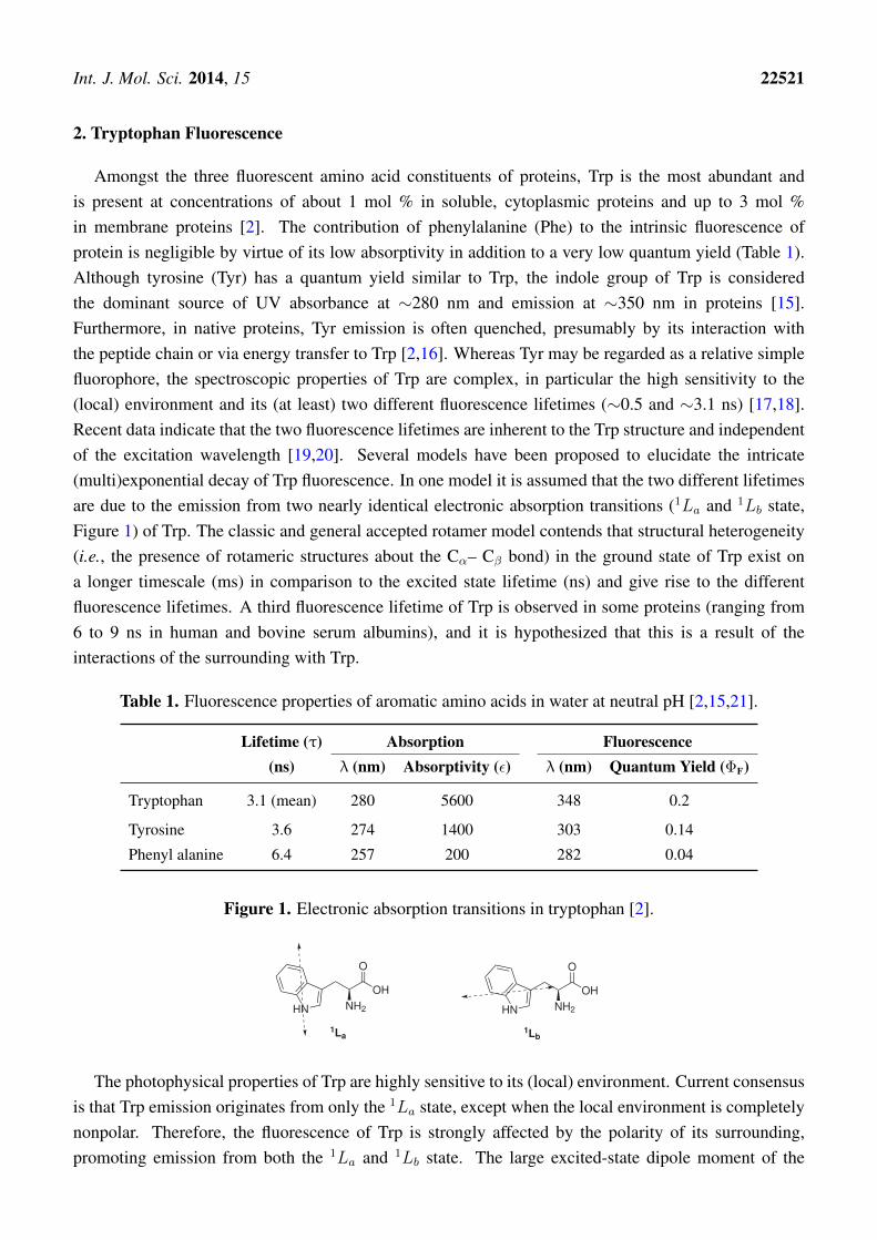
Amongst the three fluorescent amino acid constituents of proteins, Trp is the most abundant andis present at concentrations of about 1 mol % in soluble, cytoplasmic proteins and up to 3 mol %in membrane proteins [2]. The contribution of phenylalanine (Phe) to the intrinsic fluorescence ofprotein is negligible by virtue of its low absorptivity in addition to a very low quantum yield (Table 1).Although tyrosine (Tyr) has a quantum yield similar to Trp, the indole group of Trp is consideredthe dominant source of UV absorbance at ∼280 nm and emission at ∼350 nm in proteins [15].Furthermore, in native proteins, Tyr emission is often quenched, presumably by its interaction withthe peptide chain or via energy transfer to Trp [2,16]. Whereas Tyr may be regarded as a relative simplefluorophore, the spectroscopic properties of Trp are complex, in particular the high sensitivity to the(local) environment and its (at least) two different fluorescence lifetimes (∼0.5 and ∼3.1 ns) [17,18].Recent data indicate that the two fluorescence lifetimes are inherent to the Trp structure and independentof the excitation wavelength [19,20]. Several models have been proposed to elucidate the intricate(multi)exponential decay of Trp fluorescence. In one model it is assumed that the two different lifetimesare due to the emission from two nearly identical electronic absorption transitions (1La and 1Lb state,Figure 1) of Trp. The classic and general accepted rotamer model contends that structural heterogeneity(i.e., the presence of rotameric structures about the Cα– Cβ bond) in the ground state of Trp exist ona longer timescale (ms) in comparison to the excited state lifetime (ns) and give rise to the differentfluorescence lifetimes. A third fluorescence lifetime of Trp is observed in some proteins (ranging from6 to 9 ns in human and bovine serum albumins), and it is hypothesized that this is a result of theinteractions of the surrounding with Trp.
Table 1. Fluorescence properties of aromatic amino acids in water at neutral pH [2,15,21].
Lifetime (τ) Absorption Fluorescence(ns) λ (nm) Absorptivity (ε) λ (nm) Quantum Yield (ΦF)
Tryptophan 3.1 (mean) 280 5600 348 0.2
Tyrosine 3.6 274 1400 303 0.14
Phenyl alanine 6.4 257 200 282 0.04
Figure 1. Electronic absorption transitions in tryptophan [2].
OH
O
NH2HN
OH
O
NH2HN
1La1Lb
The photophysical properties of Trp are highly sensitive to its (local) environment. Current consensusis that Trp emission originates from only the 1La state, except when the local environment is completelynonpolar. Therefore, the fluorescence of Trp is strongly affected by the polarity of its surrounding,promoting emission from both the 1La and 1Lb state. The large excited-state dipole moment of the
Int. J. Mol. Sci. 2014, 15 22522
1La state (∼6 Debye), in addition to its higher sensitivity towards hydrogen bonding, with respect to theexcited 1Lb state, results in its transition shift to lower energy [2,22]. As a consequence, Trp fluorescencemaximum (λEM) and intensity are highly influenced by the polarity of its micro-environment, hydrogenbonding and other non-covalent interactions, displaying a red shift in increasing polarity due to theemission of the 1La state [23–25]. In hydrophobic environments, the 1Lb state may have the lowestenergy with respect to the 1La state and dominate the emission of Trp, which would explain the blueshift of Trp fluorescence to increasing non-polar environments. Azurin, a native folded protein, whereTrp-45 is embedded deep into a hydrophobic pocket, is a representable example of the least red-shiftedTrp-containing proteins (λEM ∼ 308 nm), comparable to skatole (3-methylindole, λEM ∼ 310 nm)in cyclohexane [26,27]. In contrast, (denatured) proteins displaying solvent-exposed Trp residue(s),e.g., glucagon and melittin (λEM ∼ 352 and 346 nm, respectively) are amongst the most red shifted.
In addition to the high sensitivity of Trp emission maximum and intensity to its local environment,Trp quantum yield (ΦF) can vary from 0.35 to 0.01 in different protein environments [28]. Interestingly,the quantum yield of skatole is ∼0.3, regardless of the polarity of the solvent, being pure hydrocarbonsor water [29]. The frequently proposed mechanism to explain the decrease in fluorescence quantumyield has been the electron transfer quenching by, for instance, the local peptide carbonyl group orby neighboring amino acid side chains [30–32]. The high dependency of Trp fluorescence on itssurroundings have been exploited in numerous studies to elucidate, e.g., protein conformational changesand interactions with ligands. However, the interpretation of Trp spectroscopic data can be challengingwhen all of the different factors that can lead to the observed changes are taken into consideration.
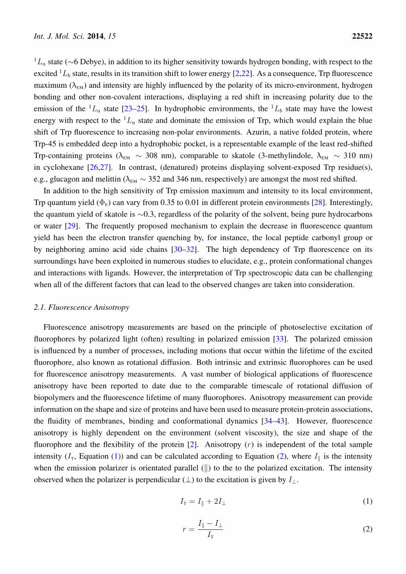
2.1. Fluorescence Anisotropy
Fluorescence anisotropy measurements are based on the principle of photoselective excitation offluorophores by polarized light (often) resulting in polarized emission [33]. The polarized emissionis influenced by a number of processes, including motions that occur within the lifetime of the excitedfluorophore, also known as rotational diffusion. Both intrinsic and extrinsic fluorophores can be usedfor fluorescence anisotropy measurements. A vast number of biological applications of fluorescenceanisotropy have been reported to date due to the comparable timescale of rotational diffusion ofbiopolymers and the fluorescence lifetime of many fluorophores. Anisotropy measurement can provideinformation on the shape and size of proteins and have been used to measure protein-protein associations,the fluidity of membranes, binding and conformational dynamics [34–43]. However, fluorescenceanisotropy is highly dependent on the environment (solvent viscosity), the size and shape of thefluorophore and the flexibility of the protein [2]. Anisotropy (r) is independent of the total sampleintensity (IT, Equation (1)) and can be calculated according to Equation (2), where I‖ is the intensitywhen the emission polarizer is orientated parallel (‖) to the to the polarized excitation. The intensityobserved when the polarizer is perpendicular (⊥) to the excitation is given by I⊥.
IT = I‖ + 2I⊥ (1)
r =I‖ − I⊥IT
(2)

Int. J. Mol. Sci. 2014, 15 22523
2.2. Fluorescence Quenching
Fluorescence quenching is an indispensable tool in protein research, since these studies are, ingeneral, easy to perform, require only a small sample and are non-destructive. Quenching studiesutilizing intrinsic Trp fluorescence of proteins can give a wealth of information regarding the locationof the fluorophore within its macromolecular structure, thus providing structural information of themacro-molecule [44–47]. A variety of mechanisms, e.g., proton- and electron transfer, long-range energytransfer, induced conformational changes and various intramolecular reactions lay at the basis of Trpfluorescence quenching by external or internal ligands. The different mechanisms of quenching canbe classified as dynamic quenching (collisional encounters) or static quenching (ground-state complexformation) between fluorophores and quenchers. In the case of collisional quenching, the dependenceof the emission intensity on the quencher concentration (Q) is given by the well-known Stern–Volmerequation (Equation (3)). In this equation, F0 and F represent the fluorescence intensity, and τ0 and τ
are the lifetime in the absence (subscript 0) and presence of the quencher, respectively; and Ksv is theStern–Volmer constant.
F0
F=
τ0
τ= 1 +Ksv · Q (3)
Dynamic and static quenching can be distinguished by their different dependence on temperatureand viscosity or, preferably, by lifetime measurements. Van de Weert et al. highlighted some pitfalls inthe application of fluorescence quenching by external ligands [48]. It was proposed that the observedchanges in fluorescence may arise due to an inner-filter effect and collisional (dynamic) quenching inaddition to ligand binding (static quenching). Absorbance (or optical dispersion) of light at the excitationor emission wavelength is referred to as the inner-filter effect. Collisional quenching involves thecontact of the excited fluorophore with ions or molecules that facilitate non-radiative transition to theground state. External quenchers, such as molecular oxygen and paramagnetic moieties, are believedto induce rapid inter-system crossing of excited aromatic fluorophores by an electron spin exchangeprocess, facilitating the conversion to the ground state. In the case of halogen ions and heavy atoms, theinter-system crossing via a spin-orbital coupling mechanism is promoted. Different types of quencherscan induce electron transfer to the excited state of the fluorophore. Amides, such as acrylamide, actas electron acceptors of the singlet state (of the excited aromatic fluorophore) [33]. Osysko et al.studied the electron transfer from the excited indole ring of Trp to one of the amides in the proteinbackbone, which is believed to be the major cause of fluorescence quenching by internal ligands [49].Utilizing two model dipeptides, it was found that high pH results in a high quantum yield due to thelow efficiency of the electron transfer event induced by the carboxylate ion, whereas low pH reduces thequantum yield by increasing the rate of electron transfer to the amide. Additionally, Chen et al. foundthat lysine and tyrosine side chains quench 3-methylindole (a representable model for Trp) fluorescenceby excited-state proton transfer, and glutamine, asparagine, glutamic- and aspartic acid, cysteine andhistidine side chains operate by an excited-state electron transfer mechanism [16]. Goldberg et al.
Int. J. Mol. Sci. 2014, 15 22524
reported the quenching of Trp and Tyr intrinsic fluorescence by thioamides in a distance-dependentmanner [50]. The mechanism by which thioamides operate in fluorescence quenching is still uncertain;however, experimental data suggest an electron transfer mechanism. The probability of fluorescencequenching with external quenchers depends on the rate of collision of the quencher and the excitedfluorophore (dynamic quenching). Therefore, it is expected that the fluorescence of Trp residues locatedon the surface of the protein will be influenced to a greater degree by the quencher, with respectto Trp residues embedded deep in the protein matrix. Fluorescence quenching can also arise via aresonance energy transfer mechanism, with a suitable acceptor molecule or metal ion. Cupredoxins,which possess a Trp residue located within 1 nm of the copper ion, e.g., azurin [51], stellacyanin [52]and amicyanin [53], exhibit the phenomenon of Trp fluorescence quenching upon metal ion binding viaeither Förster resonance energy transfer or an electron transfer mechanism [54,55].
2.3. Selected Examples of Tryptophan Fluorescence in the Elucidation of Protein Structures
The dependency of Trp fluorescence properties (e.g., absorption and emission maxima,fluorescence intensity, anisotropy and quantum yield) on its micro environment has been exploitedin numerous studies to derive inference with respect to protein conformational changes and/or proteindenaturation [14,56–61]. Gorinstein et al. utilized the change in intrinsic Trp fluorescence and surfacehydrophobicity of human serum proteins to evaluate the effect of beer consumption on protein integrityin patients suffering from coronary artery disease [62]. A slightly red-shifted fluorescence (λEM),a decrease in fluorescence intensity and surface hydrophobicity were observed in human serum albuminand human serum globulin after 30 days of exposure of patients to moderate alcohol consumption.The altered fluorescence properties of these human serum proteins were attributed to structural disruptionof the proteins in addition to a change in their compactability, as a result of ethanol consumption.
The denaturation of bovine serum albumin (BSA) under the influence of sodium dodecylsulfate (SDS) at various pH values was investigated based on fluorescence anisotropy measurementof tryptophan, in addition to its fluorescence quenching [63]. BSA, a globular protein with a molecularweight of 64 kDa and an isoelectric point (pI) of 4.9, is involved in the transportation of physiologicalmetabolites. BSA contains two tryptophan residues, and their fluorescence is quenched in the presence ofionic detergents, such as SDS, which is associated with the denaturation of BSA. The observed stepwisequenching of BSA intrinsic fluorescence in addition to a gradual increase in the degree of anisotropy(derived from Equation (2)) indicated a two-stage denaturation process. The first stage involved theloosening of protein globules, and the second stage is complete unfolding of the protein. It was foundthat at pH values lower than the pI, the generally positive-charged BSA (5 µM) binds strongly to theSDS anions, resulting in strong BSA fluorescence quenching. In contrast to the aforementioned, at pHvalues higher than the pI, only weak quenching of BSA fluorescence was observed. These findings werecorroborated by a higher increase in the degree of BSA fluorescence anisotropy at pH values lower thanthe pI in comparison to pH values higher than the pI.
Besides native Trp containing proteins, e.g., azurin, bovine and human serum albumin, site-directedmutagenesis enables the introduction (or deletion) of Trp residues into proteins. However, the alterationof Trp residues in proteins can influence its shape and function. This approach was employed by
Int. J. Mol. Sci. 2014, 15 22525
Kozachkov et al., who observed two essential conformational changes in the Na+ /H+ antiporter NhaAbased on Trp fluorescence [64]. NhaA plays a crucial role in maintaining pH and Na+ homeostasisin Escherichia coli and other enterobacteria and, possibly, Homo sapiens. Native NhaA contains eightTrp residues, which result in high background fluorescence. Therefore, site-directed mutagenesis wasemployed to generate single Trp mutants in addition to a Trp-less variant of NhaA. Two single tryptophanvariants, Trp/F136W and Trp/F339W, grown on selective media, were found to have antiport activitiesthat are similar to the Trp-less NhaA. Fluorescence studies with the single Trp/F136W mutant revealedthat a pH shift from pH 6.0 to 8.5 induces a red shift in Trp emission. Furthermore, a dramatic increasein fluorescence, which was reversible, was observed, and the addition of either Na+ or Li+ had no effect.In contrast, the single Trp/F339W mutant remained inert to pH changes; however, the addition of eitherNa+ or Li+ drastically resulted in fluorescence quenching at alkaline pH.
3. Förster Resonance Energy Transfer (FRET)
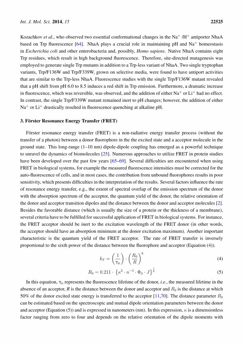
Förster resonance energy transfer (FRET) is a non-radiative energy transfer process (without thetransfer of a photon) between a donor fluorophore in the the excited state and a acceptor molecule in theground state. This long-range (1–10 nm) dipole-dipole coupling has emerged as a powerful techniqueto unravel the dynamics of biomolecules [25]. Numerous approaches to utilize FRET in protein studieshave been developed over the past few years [65–69]. Several difficulties are encountered when usingFRET in biological systems, for example the measured fluorescence intensities must be corrected for theauto-fluorescence of cells, and in most cases, the contribution from unbound fluorophores results in poorsensitivity, which presents difficulties in the interpretation of the results. Several factors influence the rateof resonance energy transfer, e.g., the extent of spectral overlap of the emission spectrum of the donorwith the absorption spectrum of the acceptor, the quantum yield of the donor, the relative orientation ofthe donor and acceptor transition dipoles and the distance between the donor and acceptor molecules [2].Besides the favorable distance (which is usually the size of a protein or the thickness of a membrane),several criteria have to be fulfilled for successful application of FRET in biological systems. For instance,the FRET acceptor should be inert to the excitation wavelength of the FRET donor (in other words,the acceptor should have an absorption minimum at the donor excitation maximum). Another importantcharacteristic is the quantum yield of the FRET acceptor. The rate of FRET transfer is inverselyproportional to the sixth power of the distance between the fluorophore and acceptor (Equation (4)).
kT =
(1
τD
)·(R0
R
)6
(4)
R0 = 0.211 ·{κ2 · n−4 · ΦD · J
} 16 (5)
In this equation, τD represents the fluorescence lifetime of the donor, i.e., the measured lifetime in theabsence of an acceptor, R is the distance between the donor and acceptor and R0 is the distance at which50% of the donor excited state energy is transferred to the acceptor [11,70]. The distance parameter R0
can be estimated based on the spectroscopic and mutual dipole orientation parameters between the donorand acceptor (Equation (5)) and is expressed in nanometers (nm). In this expression, κ is a dimensionlessfactor ranging from zero to four and depends on the relative orientation of the dipole moments with
Int. J. Mol. Sci. 2014, 15 22526
respect to the donor emission and the acceptor absorption and with respect to the axis connecting donorand acceptor. Head to tail parallel transition dipoles (→ ·· →) give a value of four for κ2, and for paralleloriented dipoles (↑ ·· ↑) κ2 is one. However, when the orientation of the dipoles are perpendicular(↑ ·· ↙), κ2 equals zero, causing serious calculation errors. In general, κ2 is assumed to be 2/3 in thecalculation of R0, which holds true for randomized orientation of donor and acceptor dipoles as a resultof rotational diffusion proceeding energy transfer. In the case of a range of static orientations of donorand acceptor during the fluorescent lifetime, κ2 may be assumed to be 0.476 [2]. To minimize calculationerrors in determining R0, the limit of κ2 can be obtained from fluorescence anisotropy measurements ofthe donor and acceptor. At present, X-ray crystallography and NMR spectroscopy are the only methodsavailable to measure κ2; however, these methods render distance determination by FRET redundant. Therefractive index of the corresponding medium is given by n, and ΦD is the quantum yield of the donor.For most FRET donor/acceptor pairs, the distance R0 is in the 1–7 nm range.
J = εA
∞∫0
fD(λ)fA(λ)λ4dλ
∞∫0
fD(λ)dλ
(6)
E = 1− τDA
τD
= 1− IDA
ID
=1
1 + (R/R0)6(7)
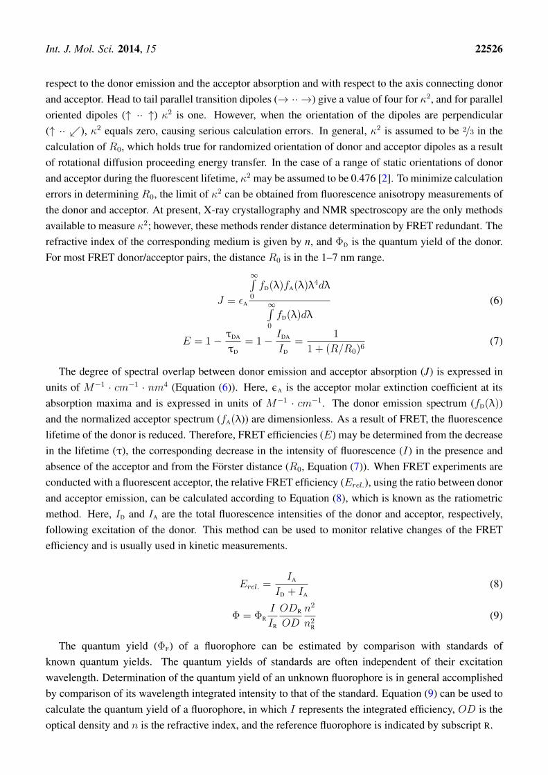
The degree of spectral overlap between donor emission and acceptor absorption (J) is expressed inunits of M−1 · cm−1 · nm4 (Equation (6)). Here, εA is the acceptor molar extinction coefficient at itsabsorption maxima and is expressed in units of M−1 · cm−1. The donor emission spectrum (fD(λ))and the normalized acceptor spectrum (fA(λ)) are dimensionless. As a result of FRET, the fluorescencelifetime of the donor is reduced. Therefore, FRET efficiencies (E) may be determined from the decreasein the lifetime (τ), the corresponding decrease in the intensity of fluorescence (I) in the presence andabsence of the acceptor and from the Förster distance (R0, Equation (7)). When FRET experiments areconducted with a fluorescent acceptor, the relative FRET efficiency (Erel.), using the ratio between donorand acceptor emission, can be calculated according to Equation (8), which is known as the ratiometricmethod. Here, ID and IA are the total fluorescence intensities of the donor and acceptor, respectively,following excitation of the donor. This method can be used to monitor relative changes of the FRETefficiency and is usually used in kinetic measurements.
Erel. =IA
ID + IA
(8)
Φ = ΦR
I
IR
ODR
OD
n2
n2R
(9)
The quantum yield (ΦF) of a fluorophore can be estimated by comparison with standards ofknown quantum yields. The quantum yields of standards are often independent of their excitationwavelength. Determination of the quantum yield of an unknown fluorophore is in general accomplishedby comparison of its wavelength integrated intensity to that of the standard. Equation (9) can be used tocalculate the quantum yield of a fluorophore, in which I represents the integrated efficiency, OD is theoptical density and n is the refractive index, and the reference fluorophore is indicated by subscript R.
Int. J. Mol. Sci. 2014, 15 22527
One approach to utilize FRET is in the development of fluorescence immunoassay (FIA). In FIA,the amino acid residue(s) of target proteins are labeled with either a FRET acceptor or donor, withpreference for the latter, in combination with a target specific antibody (or high affinity ligand), which islabeled with the complementary FRET donor or acceptor. Alternatively, two antibodies with selectivityfor different epitopes on the protein surface can be used in sandwich-type fluorescence immunoassay.Labeling of both the antibodies with either a FRET donor or acceptor would result in FRET fluorescenceupon binding of both antibodies to the protein [71]. However, the labeling of biopolymers, e.g., proteinsand antibodies, with either a FRET donor or acceptor can be cumbersome and expensive and couldinduce conformational and functional changes of the targets, resulting in aberrancy.
4. Intrinsic Förster Resonance Energy Transfer
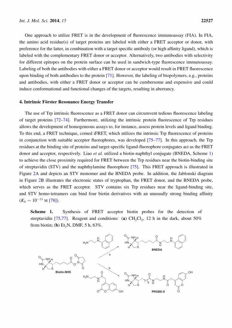
The use of Trp intrinsic fluorescence as a FRET donor can circumvent tedious fluorescence labelingof target proteins [72–74]. Furthermore, utilizing the intrinsic protein fluorescence of Trp residuesallows the development of homogeneous assays to, for instance, assess protein levels and ligand binding.To this end, a FRET technique, coined iFRET, which utilizes the intrinsic Trp fluorescence of proteinsin conjunction with suitable acceptor fluorophores, was developed [75–77]. In this approach, the Trpresidues at the binding site of proteins and target-specific ligand-fluorophore conjugates act as the FRETdonor and acceptor, respectively. Liao et al. utilized a biotin-naphthyl conjugate (BNEDA, Scheme 1)to achieve the close proximity required for FRET between the Trp residues near the biotin-binding siteof streptavidin (STV) and the naphthylamine fluorophore [75]. This FRET approach is illustrated inFigure 2A and depicts an STV monomer and the BNEDA probe. In addition, the Jablonski diagramin Figure 2B illustrates the electronic states of tryptophan, the FRET donor, and the BNEDA probe,which serves as the FRET acceptor. STV contains six Trp residues near the ligand-binding site,and STV homo-tetramers can bind four biotin derivatives with an unusually strong binding affinity(KD ∼ 10−14 M [78]).
Scheme 1. Synthesis of FRET acceptor biotin probes for the detection ofstreptavidin [75,77]. Reagent and conditions: (a) CH2Cl2, 12 h in the dark, about 50%from biotin; (b) Et3N, DMF, 5 h, 63%.
H2N
HN
NH
HN
SH
H
NH
HN
O
O
O
3O
O
OH
PROBE-II
NH
HN
SH
HOO
O
Biotin-NHS
NH
HN
SH
H
NH
OHN
BNEDA
O
N
O
O a
b
O
OO OH
NH
H2N
Int. J. Mol. Sci. 2014, 15 22528
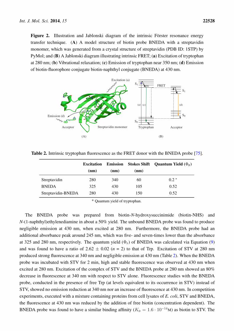
Figure 2. Illustration and Jablonski diagram of the intrinsic Förster resonance energytransfer technique. (A) A model structure of biotin probe BNEDA with a streptavidinmonomer, which was generated from a crystal structure of streptavidin (PDB ID: 1STP) byPyMol; and (B) A Jablonski diagram illustrating intrinsic FRET; (a) Excitation of tryptophanat 280 nm; (b) Vibrational relaxation; (c) Emission of tryptophan near 350 nm; (d) Emissionof biotin-fluorophore conjugate biotin-naphthyl conjugate (BNEDA) at 430 nm.
S0
S1
S1
FRET
(a) (c)
(b)
(d)
Tryptophan AcceptorAcceptor Streptavidin monomer
Excitation (a)
Emission (d) S0
(A) (B)
Table 2. Intrinsic tryptophan fluorescence as the FRET donor with the BNEDA probe [75].
Excitation Emission Stokes Shift Quantum Yield (ΦF)(nm) (nm) (nm)
Streptavidin 280 340 60 0.2 ∗
BNEDA 325 430 105 0.52
Streptavidin-BNEDA 280 430 150 0.52
* Quantum yield of tryptophan.
The BNEDA probe was prepared from biotin-N-hydroxysuccinimide (biotin-NHS) andN-(1-naphthyl)ethylenediamine in about a 50% yield. The unbound BNEDA probe was found to producenegligible emission at 430 nm, when excited at 280 nm. Furthermore, the BNEDA probe had anadditional absorbance peak around 245 nm, which was five- and seven-times lower than the absorbanceat 325 and 280 nm, respectively. The quantum yield (ΦF) of BNEDA was calculated via Equation (9)and was found to have a ratio of 2.62 ± 0.02 (n = 2) to that of Trp. Excitation of STV at 280 nmproduced strong fluorescence at 340 nm and negligible emission at 430 nm (Table 2). When the BNEDAprobe was incubated with STV for 2 min, high and stable fluorescence was observed at 430 nm whenexcited at 280 nm. Excitation of the complex of STV and the BNEDA probe at 280 nm showed an 80%decrease in fluorescence at 340 nm with respect to STV alone. Fluorescence studies with the BNEDAprobe, conducted in the presence of free Trp (at levels equivalent to its occurrence in STV) instead ofSTV, showed no emission reduction at 340 nm nor an increase of fluorescence at 430 nm. In competitionexperiments, executed with a mixture containing proteins from cell lysates of E. coli, STV and BNEDA,the fluorescence at 430 nm was reduced by the addition of free biotin (concentration dependent). TheBNEDA probe was found to have a similar binding affinity (KD = 1.6 · 10−14M) as biotin to STV. The
Int. J. Mol. Sci. 2014, 15 22529
limit of detection of an STV subunit with the BNEDA probe, in the presence of proteins from E. colicell lysates (2.0 mg/mL), was found to be ∼ 0.15 nM.
The iFRET fluorescence from the BNEDA probe (λmax = 430 nm) is very close to theauto-fluorescence of cells when irradiated at 280 nm, which may hamper in vivo application thereof.It was shown that the background fluorescence remained consistent in the presence of substances suchas MeOH, DMSO, MgCl2, NaCl, NaN3, glycerol and proteins from E. coli cell lysates (2.0 mg/mL).In addition, the relative short Stokes shift of the BNEDA probe during FRET experiments (90 nm)may induce self-absorption, which in turn would decrease the accuracy of the measurement. With thisrationale in mind, Kim and co-workers developed coumarin derivatives as efficient FRET acceptors forTrp emission [77]. The most promising iFRET probe, PROBE-II, comprises biotin (i.e., as a ligand forSTV) conjugated via a linker to a fluorescent coumarin derivative and was obtained in 63% yield frombiotin-NHS (Scheme 1).
When PROBE-II was irradiated with UV-light (280 nm), corresponding to STV excitationwavelength, low fluorescence was detected at 460 nm. Illumination of PROBE-II with near-UV-light(360 nm), corresponding to its absorption maxima, resulted in strong fluorescence at 460 nm (Table 3).The quantum efficiency (ΦF) of PROBE-II, with umbelliferone as the standard (ΦF = 0.7), was foundto be 0.47. iFRET experiments were conducted in the presence of STV, and bovine serum albumin(BSA) was used as a negative control to gain insight into the selectivity of the iFRET probe. BSA wasselected as the negative control due to the presence of two Trp residues in its sequence; however, BSAhas no affinity towards biotin. Spectroscopic data revealed the characteristic FRET phenomenon whenPROBE-II was used in the presence of native STV. The FRET efficiency was calculated according toEquation (7) and was found to be 0.14. The Förster distance was derived from Equations (4) and (5) andwas calculated to be 2.04 nm. On the contrary, FRET signals were absent when PROBE-II was evaluatedin the presence of either BSA or denatured STV.
Table 3. Intrinsic tryptophan fluorescence as the FRET donor with coumarin-basedPROBE-II [77].
Excitation (nm) Emission (nm) Stokes Shift (nm)
Streptavidin 280 340 60
PROBE-II 340 460 120
Streptavidin-PROBE-II 280 460 180
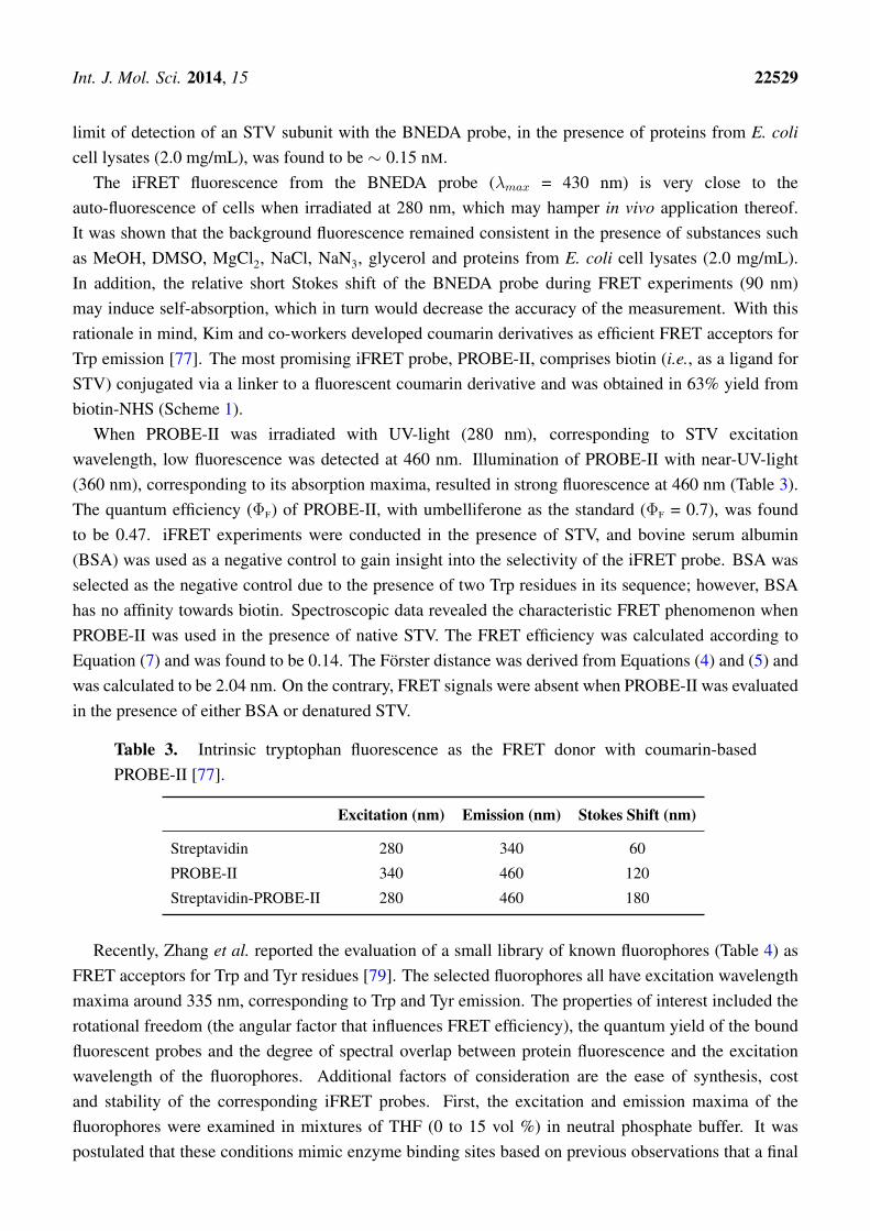
Recently, Zhang et al. reported the evaluation of a small library of known fluorophores (Table 4) asFRET acceptors for Trp and Tyr residues [79]. The selected fluorophores all have excitation wavelengthmaxima around 335 nm, corresponding to Trp and Tyr emission. The properties of interest included therotational freedom (the angular factor that influences FRET efficiency), the quantum yield of the boundfluorescent probes and the degree of spectral overlap between protein fluorescence and the excitationwavelength of the fluorophores. Additional factors of consideration are the ease of synthesis, costand stability of the corresponding iFRET probes. First, the excitation and emission maxima of thefluorophores were examined in mixtures of THF (0 to 15 vol %) in neutral phosphate buffer. It waspostulated that these conditions mimic enzyme binding sites based on previous observations that a final
Int. J. Mol. Sci. 2014, 15 22530
concentration of 15 vol % THF in neutral phosphate buffer caused a blue-shift in the emission maximaof the free BNEDA probe (Scheme 1), comparable to the blue shift resulting from the binding of theprobe to STV. A selection of the reported findings is presented in Table 4, showing the emission andexcitation maxima, the quantum yield (ΦF) and Stokes shift of the tested library of fluorophores in thepresence (15 vol %) and absence of THF. The dansyl fluorophore (D) exhibits the most blue-shiftedemission under the influence of THF, whereas 8-hydroxyquinine (B2) and the coumarin derivatives, C1and C2, revealed only minor spectral changes. The naphthyl derivatives, A1 and A2, gave the highestquantum yield, which was shown to increase by the addition of THF. The difference in excitation around280 and 335 nm was analyzed and based on those ratios; in addition to the other fluorescent properties,1-naphthylamine (A1) was deemed most suitable as the FRET acceptor and the coumarin derivative C1the second best. Considering the synthetic challenges associated with the derivatization of C1, it wasdecided to exclude coumarin derivatives from further assessment. Conjugates of 1-naphthyl amine (A1),dansyl amide (D) and acridine-9-carboxyl acid (E2) with target-specific ligands were further evaluatedas iFRET probes.
Table 4. Small library of fluorophores as the FRET acceptors in conjunction with tryptophanas the FRET donor and their fluorescence properties at different levels of THF in neutralphosphate buffer [79].
HNNH2
HNOH
O
OH
OH
A1 A2 B1 B2
OHO O
ON O
O
OH
N
SO
HN
O
OH
O
N
O OH
N
N
O
O
OH
C1
C2 D E1 E2 F
Excitation (nm) Emission (nm) Stokes Shift (nm) Quantum Yield (ΦF)% THF 0 15 0 15 0 15 0 15
A1 325 330 443 431 118 101 0.71 0.89
A2 324 325 447 439 123 114 0.68 0.81
B1 293 296 468 466 175 173 0.12 0.15
B2 324 324 411 407 87 83 0.24 0.25
C1 323 323 451 451 128 128 0.45 0.37
C2 384 385 481 473 97 88 0.10 0.16
D 327 329 562 551 235 222 0.04 0.15
E1 357 357 436 433 79 76 0.17 0.25
E2 354 356 448 430 94 74 0.35 0.23
F 309 306 491 488 182 182 0.04 0.08
Int. J. Mol. Sci. 2014, 15 22531
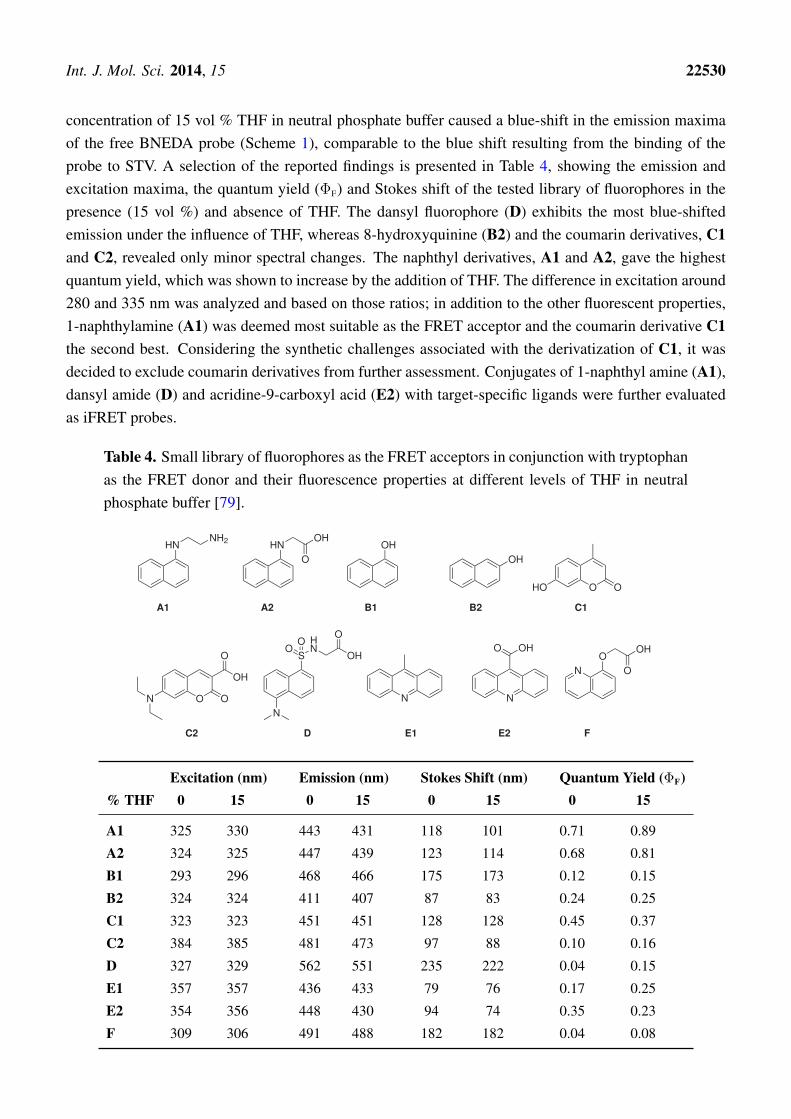
The study of RNA binding proteins, which often displays Trp residues at the RNA binding site, via theiFRET technique was reported by the group of Xie [76]. To minimize the disturbance of RNA-proteininteractions by the use of classic fluorophores, a fluorescent nucleoside derivative (AN, Scheme 2) wasutilized. The absorptivity of nucleoside AN at 280 nm (i.e., the λmax of Trp) was found to be minimal,whereas its absorption at 350 nm overlaps with the emission of Trp, producing fluorescence at 440 nm.A FRET efficiency of 0.42± 0.04 suggested excellent FRET pairing of AN with Trp. The critical Försterdistance was experimentally determined to be 2.2 nm, which is a proper distance to monitor RNA-proteinbinding events.
Scheme 2. Synthesis of a fluorescent (oligo)nucleotide as the FRET acceptor withtryptophan as the FRET donor [76]. Reagent and conditions: (a) Six steps, 9% overallyield; (b) Standard solid-phase RNA synthesis.
O
OO
DMTrO N
NH
PacHN O
O
OSi(iPr)3P
N
CEO
PA-U66
5' - G G U C U G C G C A G C C
3' - C C G G A C G C A G U66C G A
G G
AU
G
A
A
O
OHOH
HO N
NH
NH2 O
O
AN Fluoresecent RRE
a b
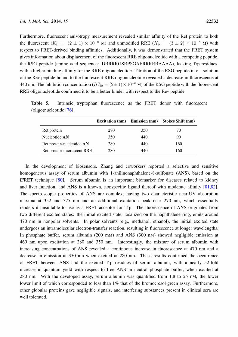
Artificial nucleoside AN was converted in six steps with an overall yield of 9% to the3’-phosphoramidite nucleotide PA-U66, rendering it suitable for usage in standard solid-phase RNAsynthesis. The artificial fluorescent nucleotide was utilized to investigate the binding of the Rev protein,a key HIV-1 regulatory protein, with the Rev responsive element (RRE), its cognate RNA target. TheRev protein is involved in the transport of immature viral mRNA from the nucleus to the cytoplasm ofthe host cell. The Rev protein specifically binds RRE with high affinity, which has been attributed to thearginine-rich domain of the Rev and Stem-loop IIB of the RRE. The Rev peptide (amino acid sequence:DTRQARRNRRRRW45RERQRAAAAR) contains a single Trp (W45) residue, which is embeddedwithin the RNA binding domain. Uridine nucleotide (U66) of the RRE, positioned in the vicinity ofthe Rev binding site, was selected to incorporate the developed fluorescent nucleotide PA-U66. Themodified 34-mer RRE oligonucleotide was prepared via standard solid-phase oligonucleotide synthesis,utilizing phosphoramidite chemistry. After purification (by PAGE) and analysis (by MALDI-TOFmass spectrometry), the modified, folded RRE oligonucleotide containing the fluorescent nucleobasewas found to have a similar melting temperature (Tm = 66 ± 1 ◦C) with respect to unmodified RRE(Tm = 68 ± 1 ◦C). The similar melting temperature indicates that the two oligomers are of comparablestability, which suggests that the incorporation of the fluorescent nucleobase resulted in minimalperturbation. A lower quantum yield was observed (emission at 440 nm) for the fluorescent RREconstruct in comparison to parent nucleoside AN, when excited at 350 nm.
When the Ret protein was titrated into the modified RRE oligonucleotide, excitation at 280 showeda continues decrease of emission around 350 nm and an increase of fluorescence at 440 nm (Table 5).The binding constant (KD = (7± 5)×10−8 M) of the fluorescent RRE ribonucleotide to the Ret proteinwas derived from FRET efficiencies and is in agreement with literature values using unmodified RRE.
Int. J. Mol. Sci. 2014, 15 22532
Furthermore, fluorescent anisotropy measurement revealed similar affinity of the Ret protein to boththe fluorescent (KD = (2 ± 1) × 10−8 M) and unmodified RRE (KD = (3 ± 2) × 10−8 M) withrespect to FRET-derived binding affinities. Additionally, it was demonstrated that the FRET systemgives information about displacement of the fluorescent RRE oligonucleotide with a competing peptide,the RSG peptide (amino acid sequence: DRRRRGSRPSGAERRRRRAAAA), lacking Trp residues,with a higher binding affinity for the RRE oligonucleotide. Titration of the RSG peptide into a solutionof the Rev peptide bound to the fluorescent RRE oligonucleotide revealed a decrease in fluorescence at440 nm. The inhibition concentration (IC50 = (2±1)×10−6 M) of the RSG peptide with the fluorescentRRE oligonucleotide confirmed it to be a better binder with respect to the Rev peptide.
Table 5. Intrinsic tryptophan fluorescence as the FRET donor with fluorescent(oligo)nucleotide [76].
Excitation (nm) Emission (nm) Stokes Shift (nm)
Ret protein 280 350 70
Nucleotide AN 350 440 90
Ret protein-nucleotide AN 280 440 160
Ret protein-fluorescent RRE 280 440 160
In the development of biosensors, Zhang and coworkers reported a selective and sensitivehomogeneous assay of serum albumin with 1-anilinonaphthalene-8-sulfonate (ANS), based on theiFRET technique [80]. Serum albumin is an important biomarker for diseases related to kidneyand liver function, and ANS is a known, nonspecific ligand thereof with moderate affinity [81,82].The spectroscopic properties of ANS are complex, having two characteristic near-UV absorptionmaxima at 352 and 375 nm and an additional excitation peak near 270 nm, which essentiallyrenders it unsuitable to use as a FRET acceptor for Trp. The fluorescence of ANS originates fromtwo different excited states: the initial excited state, localized on the naphthalene ring, emits around470 nm in nonpolar solvents. In polar solvents (e.g., methanol, ethanol), the initial excited stateundergoes an intramolecular electron-transfer reaction, resulting in fluorescence at longer wavelengths.In phosphate buffer, serum albumin (200 mM) and ANS (300 nM) showed negligible emission at460 nm upon excitation at 280 and 350 nm. Interestingly, the mixture of serum albumin withincreasing concentrations of ANS revealed a continuous increase in fluorescence at 470 nm and adecrease in emission at 350 nm when excited at 280 nm. These results confirmed the occurrenceof FRET between ANS and the excited Trp residues of serum albumin, with a nearly 52-foldincrease in quantum yield with respect to free ANS in neutral phosphate buffer, when excited at280 nm. With the developed assay, serum albumin was quantified from 1.8 to 25 nM, the lowerlower limit of which corresponded to less than 1% that of the bromocresol green assay. Furthermore,other globular proteins gave negligible signals, and interfering substances present in clinical sera arewell tolerated.
Int. J. Mol. Sci. 2014, 15 22533
5. Concluding Remarks
Fluorescence spectrometry has become a standard tool in many areas of research and represents aversatile alternative for classic studies employing radioactive labels. A plethora of fluorescent techniquesand methods have been developed to study biological events. The complex nature of biologicalsystems has facilitated the development and exploration of several label-free approaches. Especially theutilization of natural fluorophores, such as the fluorescent amino acid residues, are gaining widespreadapplications. The dependence of Trp fluorescent properties on its micro-environment has enabled thestudy of different facets of proteins. Methods based on the changes in Trp absorption and emissionmaxima, fluorescence intensity and anisotropy have proven invaluable in protein research. FRET isa powerful technique to gain specific and real-time information of biological processes. The use of theintrinsic fluorescence of target proteins, originating from Trp residues, as the FRET donor in conjunctionwith target-specific fluorescent probes as the complementary FRET acceptor is a valuable addition to thetoolbox. This technique holds true value, e.g., in drug discovery approaches (high throughput screening),(bio)imaging and in the development of biosensors.
Acknowledgments
This work was supported by the Bio-Synergy Research Project (NRF-2012M3A9C4048775), thePioneer Research Center Program (NRF-2012-0009543) of the National Research Foundation of Korea(NRF) funded by the Korean Ministry of Science, ICT and Future Planning and by the DonggukUniversity Research Fund of 2013.
Author Contributions
Amar B. T. Ghisaidoobe and Sang J. Chung conceived of the article structure; Amar B. T. Ghisaidoobewrote and assembled the manuscript. Sang J. Chung edited the manuscript.
Conflicts of Interest
The authors declare no conflict of interest.
References
1. Ladokhin, A.S.; Jayasinghe, S.; White, S.H. How to measure and analyze tryptophan fluorescencein membranes properly, and why bother? Anal. Biochem. 2000, 285, 235–245.
2. Lakowicz, J.R. Principles of Fluorescence Spectroscopy, 3rd ed.; Spinger: Berlin/Heidelberg,Germany, 2006.
3. Sauer, M.; Hofkens, J.; Enderlein, J. Handbook of Fluorescence Spectroscopy and Imaging;WILEY-VCH Verlag GmbH & Co. KGaA: Weinheim, Germany, 2011.
4. Shimomura, O.; Johnson, F.; Saiga, Y. Extraction, purification and properties of aequorin,a bioluminescent protein from the luminous hydromedusan, Aequorea. J. Cell. Comp. Physiol.1962, 59, 223–239.
5. Tsien, R.Y. The green fluorescence protein. Annu. Rev. Biochem. 1998, 67, 509–544.
Int. J. Mol. Sci. 2014, 15 22534
6. Jung, D.; Min, K.; Jung, J.; Jang, W.; Kwon, Y. Chemical biology-based approaches on fluorescentlabeling of proteins in live cells. Mol. BioSyst. 2013, 9, 862–872.
7. Yu, H.; Tan, Y.; Cunningham, B.T. Smartphone fluorescence spectroscopy. Anal. Chem. 2014,86, 8805–8813.
8. Basak, S.; Chattopadhyay, K. Studies of protein folding and dynamics using single moleculefluorescence spectroscopy. Phys. Chem. Chem. Phys. 2014, 16, 11139–11149.
9. Lippitz, M.; Erker, W.; Decker, H.; van Holde, K.E.; Basché, T. Two-photon excitation microscopyof tryptophan-containing proteins. PNAS 2002, 99, 2772–2777.
10. Hell, S.W.; Bahlmann, K.; Schrader, M.; Soini, A.; Malak, H.; Gryczynski, I.; Lakowicz, J.R.Three-photon excitation in fluorescence microscopy. J. Biomed. Opt. 1996, 1, 71–74.
11. Sun, Y.; Wallrabe, H.; Seo, S.A.; Periasamy, A. FRET microscopy in 2010: The legacy of TheodorFörster-Resonance-Energy-Transfer on the 100th anniversary of his birth. Chemphyschem 2011,12, 462–474.
12. Monici, M. Cell and tissue autofluorescence research and diagnostic applications.Biotechnol. Annu. Rev. 2005, 11, 227–256.
13. Billinton, N.; Knight, A.W. Seeing the wood through the trees: A review of techniques fordistinguishing green fluorescent protein from endogenous autofluorescence. Anal. Biochem. 2001,291, 175–197.
14. Royer, C.A. Probing protein folding and conformational transitions with fluorescence. Chem. Rev.2006, 106, 1769–1784.
15. Teale, F.W.J.; Weber, G. Ultraviolet fluorescence of the aromatic amino acids. Biochem. J. 1957,65, 476–482.
16. Chen, Y.; Barkley, M.D. Toward understanding tryptophan fluorescence in proteins. Biochemistry1998, 37, 9976–9982.
17. Swaminathan, R.; Krishnamoorthy, G.; Periasamy, N. Similarity of fluorescence lifetimedistributions for single tryptophan proteins in the random coil state. Biophys. J. 1994,67, 2013–2023.
18. Gudgin, E.; Lopez-Delgado, R.; Ware, W.R. The tryptophan fluorescence lifetime puzzle. A studyof decay times in aqueous solution as a function of pH and buffer composition. Can. J. Chem.1981, 59, 1037–1044.
19. Albani, J. Origin of tryptophan fluorescence lifetimes part 1. Fluorescence lifetimes origin oftryptophan free in solution. J. Fluoresc. 2014, 24, 93–104.
20. Albani, J. Origin of tryptophan fluorescence lifetimes. Part 2: Fluorescence lifetimes origin oftryptophan in proteins. J. Fluoresc. 2014, 24, 105–117.
21. Schmid, F.X. Biological macromolecules: UV-visible spectrophotometry. In eLS; John Wiley &Sons, Ltd.: Hoboken, NJ, USA, 2001.
22. Pierce, D.; Boxer, S. Stark effect spectroscopy of tryptophan. Biophys. J. 1995, 68, 1583–1591.23. Teale, F.W.J. The ultraviolet fluorescence of proteins in neutral solution. Biochem. J. 1960,
72, 381–388.
Int. J. Mol. Sci. 2014, 15 22535
24. Gryczynski, I.; Wiczk, W.; Johnson, M.L.; Lakowicz, J.R. Lifetime distributions and anisotropydecays of indole fluorescence in cyclohexane/ethanol mixtures by frequency-domain fluorometry.Biophys. Chem. 1988, 32, 173–185.
25. Piston, D.W.; Kremers, G.J. Fluorescent protein FRET: The good, the bad and the ugly.Trends Biochem. Sci. 2007, 32, 407–414.
26. Eftink, M.R. Biophysical and Biochemical Aspects of Fluorescence Spectroscopy; Springer:New York, NY, USA, 1991.
27. Szabo, A.; Stepanik, T.; Wayner, D.; Young, N. Conformational heterogeneity of the copper bindingsite in azurin. A time-resolved fluorescence study. Biophys. J. 1983, 41, 233–244.
28. Eftink, M.R. Fluorescence techniques for studying protein structure. In Methods of BiochemicalAnalysis; John Wiley & Sons, Inc.: Hoboken, NJ, USA, 2006; pp. 127–205.
29. Callis, P.R.; Vivian, J.T. Understanding the variable fluorescence quantum yield of tryptophanin proteins using QM-MM simulations. Quenching by charge transfer to the peptide backbone.Chem. Phys. Lett. 2003, 369, 409–414.
30. Steiner, R.; Kirby, E. The interaction of the ground and excited states of indole derivatives withelectron scavengers. J. Phys. Chem. 1969, 73, 4130–4135.
31. Chen, Y.; Liu, B.; Yu, H.T.; Barkley, M. The peptide bond quenches indole fluorescence. J. Am.Chem. Soc. 1996, 118, 9271–9278.
32. Adams, P.; Chen, Y.; Ma, K.; Zagorski, M.; Sönnichsen, F.; McLaughlin, M.; Barkley, M.Intramolecular quenching of tryptophan fluorescence by the peptide bond in cyclic hexapeptides.J. Am. Chem. Soc. 2002, 124, 9278–9286.
33. Lakowicz, J.R. Topics in Fluorescence Spectroscopy, Volume 2, Protein Fluorescence;KIuwer Academic Publishers: Dordrecht, The Netherlands, 2000; Volume 6.
34. Chen, Y.W.; Lee, C.H.; Huang, Y.T.; Pan, Y.J.; Lin, S.M.; Lo, Y.Y.; Lee, C.H.;Huang, L.K.; Huang, Y.F.; Hsu, Y.D. Functional and fluorescence analyses of tryptophan residuesin H+-pyrophosphatase of Clostridium tetani. J. Bioenerg. Biomembr. 2014, 46, 127–134.
35. Gasymov, O.K.; Abduragimov, A.R.; Glasgow, B.J. Tryptophan rotamer distribution revealed forthe alpha-helix in tear lipocalin by site-directed tryptophan fluorescence. J. Phys. Chem. B 2012,116, 13381–13388.
36. Ghosh, S.; Paul, B.K.; Chattopadhyay, N. Interaction of cyclodextrins with human and bovineserum albumins: A combined spectroscopic and computational investigation. J. Chem. Sci. 2014,126, 931–944.
37. Halder, U.C.; Chakraborty, J.; Das, N.; Bose, S. Tryptophan dynamics in the exploration ofmicro-conformational changes of refolded beta-lactoglobulin after thermal exposure: A steady stateand time-resolved fluorescence approach. J. Photochem. Photobiol. B 2012, 109, 50–57.
38. Kierdaszuk, B. Fluorescence anisotropy of tyrosinate anion using one-, two- and three-photonexcitation. J. Fluoresc. 2013, 23, 339–347.
39. Li, J.; Henry, E.; Wang, L.; Delelis, O.; Wang, H.; Simon, F.; Tauc, P.; Brochon, J.C.;Zhao, Y.; Deprez, E. Comparative study of the fatty acid binding process of a new FABPfrom cherax quadricarinatus by fluorescence intensity, lifetime and anisotropy. PLoS One 2012,7, e51079.
Int. J. Mol. Sci. 2014, 15 22536
40. Mukherjee, M.; Sardar, P.S.; Ghorai, S.K.; Samanta, S.K.; Roy, A.S.; Dasgupta, S.;Ghosh, S. A comparative study of interaction of tetracycline with several proteins using timeresolved anisotropy, phosphorescence, docking and FRET. PLoS One 2013, 8, e60940.
41. Roy, A.S.; Dinda, A.K.; Chaudhury, S.; Dasgupta, S. Binding of antioxidant flavonol morin to thenative state of bovine serum albumin: Effects of urea and metal ions on the binding. J. Lumin.2014, 145, 741–751.
42. Sarkar, S.S.; Udgaonkar, J.B.; Krishnamoorthy, G. Unfolding of a small protein proceeds via dryand wet globules and a solvated transition state. Biophys. J. 2013, 105, 2392–2402.
43. Sergeeva, I.A.; Shirshin, E.A.; Zhdanova, N.G.; Gibizova, V.V.; Petrova, G.P.; Kurguzenkov, S.A.;Fadeev, V.V. The effect of lead cations on the fluorescence characteristics of bovine serum albuminin aqueous solution. Opt. Spectrosc. 2013, 115, 171–176.
44. Padayachee, E.; Whiteley, C. Etiology of Alzheimer’s disease: Kinetic, thermodynamic andfluorimetric analyses of interactions of pseudo Aβ-peptides with neuronal nitric oxide synthase.Neuropeptides 2013, 47, 321–327.
45. Yang, X.; Hu, X.; Xu, B.; Wang, X.; Qin, J.; He, C.; Xie, Y.; Li, Y.; Liu, L.; Liao, F. Fluorometrictitration approach for calibration of quantity of binding site of purified monoclonal antibodyrecognizing epitope/hapten nonfluorescent at 340 nm. Anal. Chem. 2014, 86, 5667–5672.
46. Guo, X.; Li, X.; Jiang, Y.; Wu, Q.; Chang, H.; Diao, X.; Sun, Y.; Pan, X.; Zhou, N.A spectroscopic study on the interaction between p-nitrophenol and bovine serum albumin.J. Lumin. 2014, 149, 353–360.
47. Xu, C.; Gu, J.; Ma, X.; Dong, T.; Meng, X. Investigation on the interaction of pyrene with bovineserum albumin using spectroscopic methods. Spectrochim. Acta A 2014, 125, 391–395.
48. Van de Weert, M.; Stella, L. Fluorescence quenching and ligand binding: A critical discussion of apopular methodology. J. Mol. Struct. 2011, 998, 144–150.
49. Osysko, A.P.; Muíño, P.L. Fluorescence quenching of tryptophan and tryptophanyl dipeptides insolution. J. Biophys. Chem. 2011, 2, 316–321.
50. Goldberg, J.M.; Wissner, R.F.; Klein, A.M.; Petersson, E.J. Thioamide quenching of intrinsicprotein fluorescence. Chem. Commun. 2012, 48, 1550–1552.
51. Baker, E.N. Structure of azurin from Alcaligenes denitrificans refinement at 1.8 Å resolutionand comparison of the two crystallographically independent molecules. J. Mol. Biol. 1988,203, 1071–1095.
52. Hart, P.J.; Eisenberg, D.; Nersissian, A.M.; Valentine, J.S.; Herrmann, R.G.; Nalbandyan, R.M.A missing link in cupredoxins: Crystal structure of cucumber stellacyanin at 1.6 Å resolution.Protein Sci. 1996, 5, 2175–2183.
53. Husain, M.; Davidson, V.L.; Smith, A.J. Properties of paracoccus denitrificans amicyanin.Biochemistry 1986, 25, 2431–2436.
54. Delfino, I.; Cannistraro, S. Optical investigation of the electron transfer protein azurin-goldnanoparticle system. Biophys. Chem. 2009, 139, 1–7.
55. Dow, B.A.; Sukumar, N.; Matos, J.O.; Choi, M.; Schulte, A.; Tatulian, S.A.; Davidson, V.L.The sole tryptophan of amicyanin enhances its thermal stability but does not influence the electronicproperties of the type 1 copper site. Arch. Biochem. Biophys. 2014, 550, 20–27.
Int. J. Mol. Sci. 2014, 15 22537
56. Asadi, M.; Asadi, Z.; Zarei, L.; Sadi, S.B.; Amirghofran, Z. Affinity to bovine serum albumin andanticancer activity of some new water-soluble metal Schiff base complexes. Spectrochim. Acta A2014, 133, 697–706.
57. Clerici, M.; Colombo, G.; Secundo, F.; Gagliano, N.; Colombo, R.; Portinaro, N.; Giustarini, D.;Milzani, A.; Rossi, R.; Dalle-Donne, I. Cigarette smoke induces alterations in the drug bindingproperties of human serum albumin. Blood Cell Mol. Dis. 2014, 53, 149–156.
58. Pospisil, P.; Luxem, K.E.; Ener, M.; Sykora, J.; Kocabova, J.; Gray, H.B.; Vlcek, A., Jr.; Hof, M.Fluorescence quenching of (dimethylamino)naphthalene dyes badan and prodan by tryptophan incytochromes P450 and micelles. J. Phys. Chem. B 2014, 118, 10085–10091.
59. Biswas, A.; Swarnkar, R.K.; Hussain, B.; Sahoo, S.K.; Pradeepkumar, P.I.; Patwari, G.N.;Anand, R. Fluorescence quenching studies of gamma-butyrolactone binding protein (CprB) fromstreptomyces coelicolor A3(2). J. Phys. Chem. B 2014, 118, 10035–10042.
60. Michalek, M.; Aisenbrey, C.; Bechinger, B. Investigation of membrane penetration depthand interactions of the amino-terminal domain of huntingtin: Refined analysis by tryptophanfluorescence measurement. Eur. Biophys. J. Biophy. 2014, 43, 347–360.
61. Rub, M.A.; Khan, J.M.; Asiri, A.M.; Khan, R.H.; ud Din, K. Study on the interaction betweenamphiphilic drug and bovine serum albumin: A thermodynamic and spectroscopic description.J. Lumin. 2014, 155, 39–46.
62. Gorinstein, S.; Goshev, I.; Moncheva, S.; Zemser, M.; Weisz, M.; Caspi, A.; Libman, I.;Lerner, H.T.; Trakhtenberg, S.; Martín-Belloso, O. Intrinsic tryptophan fluorescence of humanserum proteins and related conformational changes. J. Protein Chem. 2000, 19, 637–642.
63. Vlasova, I.M.; Zhuravleva, V.V.; Saletsky, A.M. Denaturation of bovine serum albumininitiated by sodium dodecyl sulfate as monitored via the intrinsic fluorescence of the protein.Russ. J. Phys. Chem. B 2014, 8, 385–390.
64. Kozachkov, L.; Padan, E. Site-directed tryptophan fluorescence reveals two essentialconformational changes in the Na+/H+ antiporter NhaA. PNAS 2011, 108, 15769–15774.
65. Gingras, A.; Sarette, J.; Shawler, E.; Lee, T.; Freund, S.; Holwitt, E.; Hicks, B.W. Fluorescentproteins as biosensors by quenching resonance energy transfer from endogenous tryptophan:Detection of nitroaromatic explosives. Biosens. Bioelectron. 2013, 48, 251–257.
66. Jyothikumar, V.; Sun, Y.; Periasamy, A. Investigation of tryptophan-NADH interactions in livehuman cells using three-photon fluorescence lifetime imaging and Forster resonance energy transfermicroscopy. J. Biomed. Opt. 2013, 18, 060501.
67. Li, T.; Byun, J.Y.; Kim, B.B.; Shin, Y.B.; Kim, M.G. Label-free homogeneous FRET immunoassayfor the detection of mycotoxins that utilizes quenching of the intrinsic fluorescence of antibodies.Biosens. Bioelectron. 2013, 42, 403–408.
68. Sarkar, A.; Bhattacharya, S.C. Selective fluorescence resonance energy transfer from serumalbumins to a bio-active 3-pyrazolyl-2-pyrazoline derivative: A spectroscopic analysis. J. Lumin.2012, 132, 2612–2618.
69. Tirado-Guizar, A.; Pina-Luis, G.; Paraguay-Delgado, F.; Ramirez-Herrera, D. Size-dependentenhanced energy transfer from tryptophan to CdSe/mercaptopropionic acid quantum dots: A newfluorescence resonance energy transfer nanosensor. Sci. Adv. Mater. 2014, 6, 492–499.
Int. J. Mol. Sci. 2014, 15 22538
70. Clegg, R.M. Fluorescence resonance energy transfer. Curr. Opin. Biotechnol. 1995, 6, 103–110.71. Blomberg, K.; Hurskainen, P.; Hemmilä, I. Terbium and rhodamine as labels in a homogeneous
time-resolved fluorometric energy transfer assay of the β subunit of human chorionic gonadotropinin gerum. Clin. Chem. 1999, 45, 855–861.
72. Sahoo, H.; Roccatano, D.; Zacharias, M.; Nau, W.M. Distance distributions of short polypeptidesrecovered by fluorescence resonance energy transfer in the 10 Å domain. J. Am. Chem. Soc. 2006,128, 8118–8119.
73. Zauner, G.; Lonardi, E.; Bubacco, L.; Aartsma, T.J.; Canters, G.W.; Tepper, A.W.J.W.Tryptophan-to-dye fluorescence energy transfer applied to oxygen sensing by using type-3 copperproteins. Chem-Eur. J. 2007, 13, 7085–7090.
74. Li, Q.; Seeger, S. Multidonor deep-UV FRET study of protein-ligand binding and its potential toobtain structure information. J. Phys. Chem. B 2011, 115, 13643–13649.
75. Liao, F.; Xie, Y.; Yang, X.; Deng, P.; Chen, Y.; Xie, G.; Zhu, S.; Liu, B.; Yuan, H.;Liao, J. Homogeneous noncompetitive assay of protein via Förster-resonance-energy-transfer withtryptophan residue(s) as intrinsic donor(s) and fluorescent ligand as acceptor. Biosens. Bioelectron.2009, 25, 112–117.
76. Xie, Y.; Maxson, T.; Tor, Y. Fluorescent ribonucleoside as a FRET acceptor for tryptophan in nativeproteins. J. Am. Chem. Soc. 2010, 132, 11896–11897.
77. Kim, J.H.; Sumranjit, J.; Kang, H.J.; Chung, S.J. Discovery of coumarin derivatives as fluorescenceacceptors for intrinsic fluorescence resonance energy transfer of proteins. Mol. BioSyst. 2014,10, 30–33.
78. Gitlin, G.; Bayer, E.A.; Wilchek, M. Studies on the biotin-binding sites of avidin and streptavidintyrosine residues are involved in the binding site. Biochem. J. 1990, 269, 517–530.
79. Zhang, Y.; Yang, X.; Liu, L.; Huang, Z.; Pu, J.; Long, G.; Zhang, L.; Liu, D.; Xu, B.;Liao, J. Comparison of Förster-resonance-energy-transfer acceptors for tryptophan and tyrosineresidues in native proteins as donors. J. Fluoresc. 2013, 23, 147–157.
80. Qin, J.; Li, Y.; He, C.; Yang, X.; Xie, Y.; Hu, X.; Chen, C.; Wang, L.; Pu, J.; Liao, F. Selectiveand sensitive homogenous assay of serum albumin with 1-anilinonaphthalene-8-sulphonate as abiosensor. Anal. Chim. Acta 2014, 829, 60–67.
81. Essassi, D.; Zini, R.; Tillement, J.P. Use of 1-anilino-8-naphthalene sulfonate as a fluorescent probein the investigation of drug interactions with human alpha-1-acid glycoprotein and serum albumin.J. Pharm. Sci. 1990, 79, 9–13.
82. Cattoni, D.I.; Kaufman, S.B.; Flecha, F.L.G. Kinetics and thermodynamics of the interaction of1-anilino-naphthalene-8-sulfonate with proteins. Biochim. Biophys. Acta Proteins Proteomics2009, 1794, 1700–1708.
c© 2014 by the authors; licensee MDPI, Basel, Switzerland. This article is an open access articledistributed under the terms and conditions of the Creative Commons Attribution license(http://creativecommons.org/licenses/by/4.0/).