Intravenously administered nanoparticles increase survival ... · Intravenously administered...
6
Intravenously administered nanoparticles increase survival following blast trauma Margaret M. Lashof-Sullivan a , Erin Shoffstall a , Kristyn T. Atkins a , Nickolas Keane b , Cynthia Bir b , Pamela VandeVord c , and Erin B. Lavik a,1 a Department of Biomedical Engineering, Case Western Reserve University, Cleveland, OH 44106; b Department of Biomedical Engineering, Wayne State University, Detroit, MI 48201; and c School of Biomedical Engineering and Sciences, Virginia Polytechnic Institute and State University, Blacksburg, VA 24061 Edited by Robert Langer, Massachusetts Institute of Technology, Cambridge, MA, and approved May 30, 2014 (received for review April 16, 2014) Explosions account for 79% of combat-related injuries, leading to multiorgan hemorrhage and uncontrolled bleeding. Uncontrolled bleeding is the leading cause of death in battlefield traumas as well as in civilian life. We need to stop the bleeding quickly to save lives, but, shockingly, there are no treatments to stop internal bleeding. A therapy that halts bleeding in a site-specific manner and is safe, stable at room temperature, and easily administered is critical for the advancement of trauma care. To address this need, we have developed hemostatic nanoparticles that are adminis- tered intravenously. When tested in a model of blast trauma with multiorgan hemorrhaging, i.v. administration of the hemostatic nanoparticles led to a significant improvement in survival over the short term (1 h postblast). No complications from this treatment were apparent out to 3 wk. This work demonstrates that these particles have the potential to save lives and fundamentally change trauma care. polytrauma | synthetic platelets | lung | clot | medic O n the battlefield, hemorrhage is a leading cause of pre- ventable death (1). Blast injuries account for 79% of com- bat-related injuries and the majority of cases of traumatic brain injury (2). There are three classifications of blast injury: primary, secondary, and tertiary. Primary blast injuries refer to the direct effects of the overpressure wave, whereas secondary and tertiary insults result from objects propelled by the blast wind and the individual being thrown against other objects, respectively. Ow- ing to the rapid change in pressure, blast traumas can involve hemorrhage in multiple organs, particularly the air-filled organs, brain, and spinal cord. Blast trauma is unique and difficult to treat because it can damage multiple organs and cause significant hemorrhaging. The shocking reality is that there are no treatments for internal bleeding, although early intervention is essential to minimizing the mortality associated with severe trauma (3). Uncontrolled bleeding is no less lethal beyond the battlefield, being the leading cause of death for civilians age 5–44 y (4, 5). We need a therapy that can be administered in the field to stop internal bleeding. This therapy must be extremely safe, stable at room temperature, and easily administered. Various therapies, ranging from platelets to recombinant factors to mi- croparticles and nanoparticles, have been considered to date. Administration of allogeneic platelets confers a significant survival advantage in patients with massive trauma, but these platelets have a short shelf life, and administration can cause graft-versus-host disease, alloimmunization, and transfusion- associated lung injuries (6–8). These problems motivated the de- velopment of platelet substitutes. Typically, these nanoparticles and microparticles take advantage of the clotting cascade through peptide binding to receptors on activated platelets such as the glycoprotein IIb/IIIa receptor, which can bind fibrinogen, Arg- Gly-Asp (RGD), and dodecapeptide-H12 (HHLGGAKQAGDV). Early designs included RGD-conjugated red blood cells, which were effective in vitro and fibrinogen-coated albumin micro- particles, which significantly reduced bleeding time and volume in thrombocytopenic rabbits (9, 10). Platelet-derived particles showed promising results in vitro and in thrombocytopenic rabbits, but did not significantly reduce prolonged bleeding times in throm- bocytopenic primates (11, 12). Liposomal nanoparticles are also a potential synthetic core for particles, and particles decorated with RGD and the von Willebrand factor-binding peptide VBP promoted platelet aggregation in vivo and reduced bleeding time in a mouse tail bleeding model (13). Similarly, liposomes carrying the fibrinogen γ chain dodecapeptide (HHLGGAK- QAGDV) (14, 15) were effective in thrombocytopenic rats, but might not be effective in healthy models. In addition to the platelet mimics, recombinant factor 7 (rFVIIa; NovoSeven) has been used to augment hemostasis by supple- menting the coagulation cascade. Although rFVIIa can control or reduce massive bleeding in trauma patients, immunogenic and thromboembolic complications are unavoidable risks (16, 17). Nevertheless, rFVIIa is used in the clinic in trauma and surgical situations when bleeding cannot be controlled through other means (16). The data on rFVIIa’s efficacy is variable, and it is very expensive; a single dose costs approximately $10,000, and multiple doses are typically needed to impact hemostasis (16). We need an effective, safe therapy for the field. To address this need, we have developed hemostatic nano- particles that can halt bleeding when delivered intravenously (18– 20). We hypothesized that administration of these hemostatic nanoparticles could increase short-term and long-term survival following blast trauma. We developed a full-body blast model that replicates the injuries seen in personnel exposed to explosions, and tested the effects of administration of hemostatic nanoparticles, control nanoparticles, saline, and rFVIIa on survival, hemorrhaging, and behavioral outcomes following blast trauma. Results Development of the Blast Trauma Model. We investigated an 8-msec duration of blast overpressures of 15, 20, and 25 psi to determine the most appropriate blast pressure for modeling blast damage and lethality in mice. Pairs of animals were secured in the prone Significance We have developed hemostatic nanoparticles that reduce bleeding and increase survival in both the short term and long term following the complex injuries sustained during blast trauma. This treatment has the potential to be deployed by first responders to save lives. Author contributions: M.M.L.-S., C.B., P.V., and E.B.L. designed research; M.M.L.-S., E.S., K.T.A., N.K., and P.V. performed research; M.M.L.-S., K.T.A., P.V., and E.B.L. analyzed data; and M.M.L.-S., P.V., and E.B.L. wrote the paper. Conflict of interest statement: E.B.L. is an inventor listed on patents related to this technology. This article is a PNAS Direct Submission. 1 To whom correspondence should be addressed. E-mail: [email protected]. This article contains supporting information online at www.pnas.org/lookup/suppl/doi:10. 1073/pnas.1406979111/-/DCSupplemental. www.pnas.org/cgi/doi/10.1073/pnas.1406979111 PNAS | July 15, 2014 | vol. 111 | no. 28 | 10293–10298 MEDICAL SCIENCES Downloaded by guest on May 30, 2020
Transcript of Intravenously administered nanoparticles increase survival ... · Intravenously administered...
Intravenously administered nanoparticles increasesurvival following blast traumaMargaret M. Lashof-Sullivana, Erin Shoffstalla, Kristyn T. Atkinsa, Nickolas Keaneb, Cynthia Birb, Pamela VandeVordc,and Erin B. Lavika,1
aDepartment of Biomedical Engineering, Case Western Reserve University, Cleveland, OH 44106; bDepartment of Biomedical Engineering, Wayne StateUniversity, Detroit, MI 48201; and cSchool of Biomedical Engineering and Sciences, Virginia Polytechnic Institute and State University, Blacksburg, VA 24061
Edited by Robert Langer, Massachusetts Institute of Technology, Cambridge, MA, and approved May 30, 2014 (received for review April 16, 2014)
Explosions account for 79% of combat-related injuries, leading tomultiorgan hemorrhage and uncontrolled bleeding. Uncontrolledbleeding is the leading cause of death in battlefield traumas aswell as in civilian life. We need to stop the bleeding quickly to savelives, but, shockingly, there are no treatments to stop internalbleeding. A therapy that halts bleeding in a site-specific mannerand is safe, stable at room temperature, and easily administered iscritical for the advancement of trauma care. To address this need,we have developed hemostatic nanoparticles that are adminis-tered intravenously. When tested in a model of blast trauma withmultiorgan hemorrhaging, i.v. administration of the hemostaticnanoparticles led to a significant improvement in survival over theshort term (1 h postblast). No complications from this treatmentwere apparent out to 3 wk. This work demonstrates that theseparticles have the potential to save lives and fundamentallychange trauma care.
polytrauma | synthetic platelets | lung | clot | medic
On the battlefield, hemorrhage is a leading cause of pre-ventable death (1). Blast injuries account for 79% of com-
bat-related injuries and the majority of cases of traumatic braininjury (2). There are three classifications of blast injury: primary,secondary, and tertiary. Primary blast injuries refer to the directeffects of the overpressure wave, whereas secondary and tertiaryinsults result from objects propelled by the blast wind and theindividual being thrown against other objects, respectively. Ow-ing to the rapid change in pressure, blast traumas can involvehemorrhage in multiple organs, particularly the air-filled organs,brain, and spinal cord.Blast trauma is unique and difficult to treat because it can
damage multiple organs and cause significant hemorrhaging.The shocking reality is that there are no treatments for internalbleeding, although early intervention is essential to minimizingthe mortality associated with severe trauma (3). Uncontrolledbleeding is no less lethal beyond the battlefield, being theleading cause of death for civilians age 5–44 y (4, 5).We need a therapy that can be administered in the field
to stop internal bleeding. This therapy must be extremely safe,stable at room temperature, and easily administered. Varioustherapies, ranging from platelets to recombinant factors to mi-croparticles and nanoparticles, have been considered to date.Administration of allogeneic platelets confers a significantsurvival advantage in patients with massive trauma, but theseplatelets have a short shelf life, and administration can causegraft-versus-host disease, alloimmunization, and transfusion-associated lung injuries (6–8). These problems motivated the de-velopment of platelet substitutes. Typically, these nanoparticlesand microparticles take advantage of the clotting cascade throughpeptide binding to receptors on activated platelets such as theglycoprotein IIb/IIIa receptor, which can bind fibrinogen, Arg-Gly-Asp (RGD), and dodecapeptide-H12 (HHLGGAKQAGDV).Early designs included RGD-conjugated red blood cells, whichwere effective in vitro and fibrinogen-coated albumin micro-particles, which significantly reduced bleeding time and volume in
thrombocytopenic rabbits (9, 10). Platelet-derived particles showedpromising results in vitro and in thrombocytopenic rabbits, butdid not significantly reduce prolonged bleeding times in throm-bocytopenic primates (11, 12). Liposomal nanoparticles are alsoa potential synthetic core for particles, and particles decoratedwith RGD and the von Willebrand factor-binding peptide VBPpromoted platelet aggregation in vivo and reduced bleedingtime in a mouse tail bleeding model (13). Similarly, liposomescarrying the fibrinogen γ chain dodecapeptide (HHLGGAK-QAGDV) (14, 15) were effective in thrombocytopenic rats, butmight not be effective in healthy models.In addition to the platelet mimics, recombinant factor 7 (rFVIIa;
NovoSeven) has been used to augment hemostasis by supple-menting the coagulation cascade. Although rFVIIa can controlor reduce massive bleeding in trauma patients, immunogenic andthromboembolic complications are unavoidable risks (16, 17).Nevertheless, rFVIIa is used in the clinic in trauma and surgicalsituations when bleeding cannot be controlled through othermeans (16). The data on rFVIIa’s efficacy is variable, and it isvery expensive; a single dose costs approximately $10,000, andmultiple doses are typically needed to impact hemostasis (16).We need an effective, safe therapy for the field.To address this need, we have developed hemostatic nano-
particles that can halt bleeding when delivered intravenously (18–20). We hypothesized that administration of these hemostaticnanoparticles could increase short-term and long-term survivalfollowing blast trauma. We developed a full-body blast model thatreplicates the injuries seen in personnel exposed to explosions, andtested the effects of administration of hemostatic nanoparticles,control nanoparticles, saline, and rFVIIa on survival, hemorrhaging,and behavioral outcomes following blast trauma.
ResultsDevelopment of the Blast Trauma Model.We investigated an 8-msecduration of blast overpressures of 15, 20, and 25 psi to determinethe most appropriate blast pressure for modeling blast damageand lethality in mice. Pairs of animals were secured in the prone
Significance
We have developed hemostatic nanoparticles that reducebleeding and increase survival in both the short term and longterm following the complex injuries sustained during blasttrauma. This treatment has the potential to be deployed byfirst responders to save lives.
Author contributions: M.M.L.-S., C.B., P.V., and E.B.L. designed research; M.M.L.-S., E.S.,K.T.A., N.K., and P.V. performed research; M.M.L.-S., K.T.A., P.V., and E.B.L. analyzed data;and M.M.L.-S., P.V., and E.B.L. wrote the paper.
Conflict of interest statement: E.B.L. is an inventor listed on patents related to thistechnology.
This article is a PNAS Direct Submission.1To whom correspondence should be addressed. E-mail: [email protected].
This article contains supporting information online at www.pnas.org/lookup/suppl/doi:10.1073/pnas.1406979111/-/DCSupplemental.
www.pnas.org/cgi/doi/10.1073/pnas.1406979111 PNAS | July 15, 2014 | vol. 111 | no. 28 | 10293–10298
MED
ICALSC
IENCE
S
Dow
nloa
ded
by g
uest
on
May
30,
202
0

position in a mesh harness attached to a movable frame directlyin line with the main blasting chamber and then exposed to blastoverpressure (Fig. 1 A and B). After exposure, animals weremonitored for 1 h to determine the lethality at different pres-sures. As expected, increasing the pressure of the blast increasedthe lethality. In this preliminary study, 100% of the untreatedmice blasted at 15 psi survived, compared with 60% of thoseblasted at 20 psi and only 10% of those blasted at 25 psi (Fig. 1C).After the mice were exposed to the primary blast wave, sec-
ondary winds caused movement of the harness, resulting in ter-tiary blast trauma. Examination of the gross anatomy of theorgans following injury revealed extensive injury to the thoracicand abdominal regions. Organs including the liver, kidneys, andlungs exhibited damage ranging from slight tears in the tissue todiffuse and severe hemorrhaging and contusions. Lung and liver
contusions were observed most often, whereas the gastrointes-tinal (GI) tract was intact with no abnormalities (Fig. 1 D and E).The injuries to the lungs were quantified histologically using
eosin staining for red blood cells (Figs. S1 and S2). The degree oflung injury was significantly increased at 20 and 25 psi. Thisclosely correlated with the oxygen saturation levels in theseanimals, which were significantly lower than those in the animalsin the sham-treated and 15-psi groups (Fig. S1D). The extensivelung injuries and lower oxygen saturation are consistent with theclinical presentation following blast trauma.Our model is a complicated model that is sensitive to both pri-
mary and tertiary events, making the interactions in the harnesscritical to the extent of injury. This can be challenging from a sci-entific standpoint, but these injuries closely model what is seen inpatients following blast trauma. Based on the CONWEP software,
Fig. 1. Development of the blast model and testing paradigm. (A) Schematic of the blast tube setup. (B) Mice are held in a harness that is on a mobile frameto reduce the degree of tertiary blast injury from the animals striking the harness. (C) The fraction of animals that survive at each pressure tested showingthat 20 psi led to 60% survival, and 25 psi led to 10% survival. (D) Gross examination of organs from 20 psi indicates significant lung injury along with smallhemorrhages in the other major organs. (E) Gross examination of the organs at 25 psi shows far more extensive hemorrhaging in all of the organs. (F)Schematic of the blast experiment. Immediately following the blast trauma, the particles are administered i.v. by retro-orbital injection.
10294 | www.pnas.org/cgi/doi/10.1073/pnas.1406979111 Lashof-Sullivan et al.
Dow
nloa
ded
by g
uest
on
May
30,
202
0
20 psi would be the equivalent of standing 5 m away from a 10-kgTNT equivalence or 10 m away from a 80-kg TNT equivalence.However, no mathematical models available take into account thedifferences in structure between mice and humans; informationfrom these models must be considered in context, and the clinicalpresentations in the animal model can provide a more applicableunderstanding.
Investigating the Role of Hemostatic Nanoparticles on LethalityFollowing Blast Trauma. Based on the extensive hemorrhagingand 40% lethality, we assessed the capability of the hemostatic
nanoparticles to halt internal bleeding at 20 psi. We fabricatedand characterized our hemostatic nanoparticles (Fig. 2) andadministered them i.v. via retro-orbital injections following the20-psi blast trauma (Fig. 1F). We began our study by examiningdosing and investigating the addition of poly(acrylic acid) (PAA)as a flocculating agent on the survival after injury (Fig. S3). PAAis used during nanoparticle synthesis to enable easy collectionand subsequent resuspension of particles, and also has beenimplicated as an anticoagulant (21). In our small-scale study, wefound that particles with PAA were at least as effective as par-ticles made without PAA at reducing lethality, and thus we used
Fig. 2. Characterization of hemostatic nanoparticles. (A) Schematic of particle design. (B) Scanning electron micrograph of hemostatic nanoparticles showingsize range and spherical geometry. (C) DLS histogram of particles shown in the scanning electron micrograph. (D) Table summarizing DLS data for hemostaticnanoparticles and control nanoparticles loaded with coumarin 6 (C6) with an average hydrodynamic diameter of 500–550 nm. (E) NMR of hemostaticnanoparticles showing the PEG content in both deuterated chloroform and deuterated water. The enrichment of PEG in the deuterated water demonstratesthat the PEG in the hemostatic nanoparticles have PEG at the surfaces of the particles. (F) Release curve for C6 from the hemostatic nanoparticles showingthat <1% of the C6 is released over the first week after administration. Error bars denote SEM. (G) Formulations of treatments containing PAA.
Lashof-Sullivan et al. PNAS | July 15, 2014 | vol. 111 | no. 28 | 10295
MED
ICALSC
IENCE
S
Dow
nloa
ded
by g
uest
on
May
30,
202
0
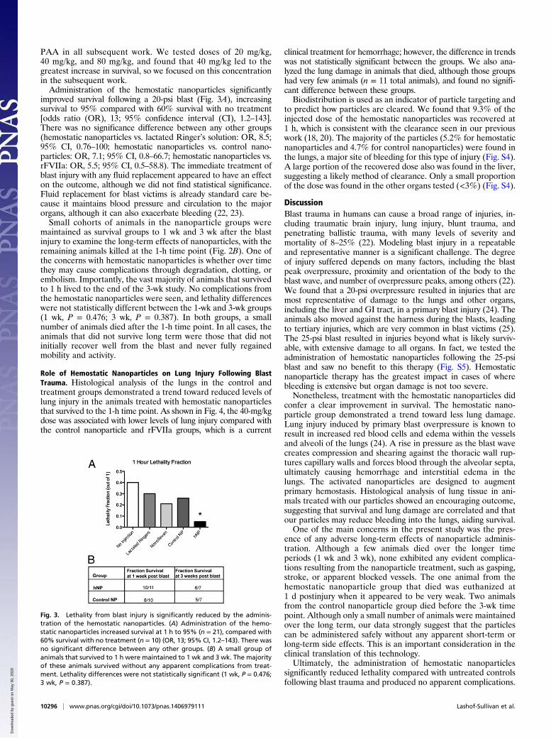
PAA in all subsequent work. We tested doses of 20 mg/kg,40 mg/kg, and 80 mg/kg, and found that 40 mg/kg led to thegreatest increase in survival, so we focused on this concentrationin the subsequent work.Administration of the hemostatic nanoparticles significantly
improved survival following a 20-psi blast (Fig. 3A), increasingsurvival to 95% compared with 60% survival with no treatment[odds ratio (OR), 13; 95% confidence interval (CI), 1.2–143].There was no significance difference between any other groups(hemostatic nanoparticles vs. lactated Ringer’s solution: OR, 8.5;95% CI, 0.76–100; hemostatic nanoparticles vs. control nano-particles: OR, 7.1; 95% CI, 0.8–66.7; hemostatic nanoparticles vs.rFVIIa: OR, 5.5; 95% CI, 0.5–58.8). The immediate treatment ofblast injury with any fluid replacement appeared to have an effecton the outcome, although we did not find statistical significance.Fluid replacement for blast victims is already standard care be-cause it maintains blood pressure and circulation to the majororgans, although it can also exacerbate bleeding (22, 23).Small cohorts of animals in the nanoparticle groups were
maintained as survival groups to 1 wk and 3 wk after the blastinjury to examine the long-term effects of nanoparticles, with theremaining animals killed at the 1-h time point (Fig. 2B). One ofthe concerns with hemostatic nanoparticles is whether over timethey may cause complications through degradation, clotting, orembolism. Importantly, the vast majority of animals that survivedto 1 h lived to the end of the 3-wk study. No complications fromthe hemostatic nanoparticles were seen, and lethality differenceswere not statistically different between the 1-wk and 3-wk groups(1 wk, P = 0.476; 3 wk, P = 0.387). In both groups, a smallnumber of animals died after the 1-h time point. In all cases, theanimals that did not survive long term were those that did notinitially recover well from the blast and never fully regainedmobility and activity.
Role of Hemostatic Nanoparticles on Lung Injury Following BlastTrauma. Histological analysis of the lungs in the control andtreatment groups demonstrated a trend toward reduced levels oflung injury in the animals treated with hemostatic nanoparticlesthat survived to the 1-h time point. As shown in Fig. 4, the 40-mg/kgdose was associated with lower levels of lung injury compared withthe control nanoparticle and rFVIIa groups, which is a current
clinical treatment for hemorrhage; however, the difference in trendswas not statistically significant between the groups. We also ana-lyzed the lung damage in animals that died, although those groupshad very few animals (n = 11 total animals), and found no signifi-cant difference between these groups.Biodistribution is used as an indicator of particle targeting and
to predict how particles are cleared. We found that 9.3% of theinjected dose of the hemostatic nanoparticles was recovered at1 h, which is consistent with the clearance seen in our previouswork (18, 20). The majority of the particles (5.2% for hemostaticnanoparticles and 4.7% for control nanoparticles) were found inthe lungs, a major site of bleeding for this type of injury (Fig. S4).A large portion of the recovered dose also was found in the liver,suggesting a likely method of clearance. Only a small proportionof the dose was found in the other organs tested (<3%) (Fig. S4).
DiscussionBlast trauma in humans can cause a broad range of injuries, in-cluding traumatic brain injury, lung injury, blunt trauma, andpenetrating ballistic trauma, with many levels of severity andmortality of 8–25% (22). Modeling blast injury in a repeatableand representative manner is a significant challenge. The degreeof injury suffered depends on many factors, including the blastpeak overpressure, proximity and orientation of the body to theblast wave, and number of overpressure peaks, among others (22).We found that a 20-psi overpressure resulted in injuries that aremost representative of damage to the lungs and other organs,including the liver and GI tract, in a primary blast injury (24). Theanimals also moved against the harness during the blasts, leadingto tertiary injuries, which are very common in blast victims (25).The 25-psi blast resulted in injuries beyond what is likely surviv-able, with extensive damage to all organs. In fact, we tested theadministration of hemostatic nanoparticles following the 25-psiblast and saw no benefit to this therapy (Fig. S5). Hemostaticnanoparticle therapy has the greatest impact in cases of wherebleeding is extensive but organ damage is not too severe.Nonetheless, treatment with the hemostatic nanoparticles did
confer a clear improvement in survival. The hemostatic nano-particle group demonstrated a trend toward less lung damage.Lung injury induced by primary blast overpressure is known toresult in increased red blood cells and edema within the vesselsand alveoli of the lungs (24). A rise in pressure as the blast wavecreates compression and shearing against the thoracic wall rup-tures capillary walls and forces blood through the alveolar septa,ultimately causing hemorrhage and interstitial edema in thelungs. The activated nanoparticles are designed to augmentprimary hemostasis. Histological analysis of lung tissue in ani-mals treated with our particles showed an encouraging outcome,suggesting that survival and lung damage are correlated and thatour particles may reduce bleeding into the lungs, aiding survival.One of the main concerns in the present study was the pres-
ence of any adverse long-term effects of nanoparticle adminis-tration. Although a few animals died over the longer timeperiods (1 wk and 3 wk), none exhibited any evident complica-tions resulting from the nanoparticle treatment, such as gasping,stroke, or apparent blocked vessels. The one animal from thehemostatic nanoparticle group that died was euthanized at1 d postinjury when it appeared to be very weak. Two animalsfrom the control nanoparticle group died before the 3-wk timepoint. Although only a small number of animals were maintainedover the long term, our data strongly suggest that the particlescan be administered safely without any apparent short-term orlong-term side effects. This is an important consideration in theclinical translation of this technology.Ultimately, the administration of hemostatic nanoparticles
significantly reduced lethality compared with untreated controlsfollowing blast trauma and produced no apparent complications.
Fig. 3. Lethality from blast injury is significantly reduced by the adminis-tration of the hemostatic nanoparticles. (A) Administration of the hemo-static nanoparticles increased survival at 1 h to 95% (n = 21), compared with60% survival with no treatment (n = 10) (OR, 13; 95% CI, 1.2–143). There wasno significant difference between any other groups. (B) A small group ofanimals that survived to 1 h were maintained to 1 wk and 3 wk. The majorityof these animals survived without any apparent complications from treat-ment. Lethality differences were not statistically significant (1 wk, P = 0.476;3 wk, P = 0.387).
10296 | www.pnas.org/cgi/doi/10.1073/pnas.1406979111 Lashof-Sullivan et al.
Dow
nloa
ded
by g
uest
on
May
30,
202
0

Our model suggests that these nanoparticles may be a powerfulfront- line therapy for blast trauma.
Materials and MethodsExperimental Design. Our first step in the experimental design was to de-termine the degree of lethality with respect to overpressure in our blastmodel. Based on previous experience with blast trauma, we performed a pilotstudy with eight animals per group (only six animals were used for the 15-psiblast, because all survived) to determine the pressure that led to hemor-rhaging and 50% lethality. We found that 20 psi was optimal based onthese considerations.
We blocked each round of testing and performed all of the treatmentsduring each round of testing. The person performing the blast testing andmonitoring the animals was blinded to the treatment, as were all of the teammembers who maintained animals after the experiment and performedhistological assessments.
An animal was excluded from the analysis if the video of the blast showedthat the harness was loose or the animal hit its head on the screws holdingthe apparatus. Animals were not excluded for any other reason. We antic-ipated needing 15–20 animals per group based on a power analysis. Theexperiments were halted when significance was determined.
Materials. Poly(lactic-co-glycolic) acid (PLGA) (Resomer 503H) was purchasedfrom Evonik Industries. Poly-l-lysine (PLL) and poly(ethylene glycol) (PEG)(∼4,600 Da) were purchased from Sigma-Aldrich. All reagents were ACSgrade and were purchased from Thermo Fisher Scientific. PLGA-PLL-PEGblock copolymer was created using standard bioconjugation techniques asdescribed previously (26).
Particle Synthesis. PLGA-PLL-PEG-GRGDS block copolymer for the hemostaticnanoparticles and PLGA-PLL-PEG-GRADSP were synthesized using protocolsdescribed previously (18–20). In brief, the triblock polymer was synthesizedusing stepwise conjugate reactions. The PLGA was coupled to poly(e-cbz-L-lysine) (PLL-cbz; PLL with carbobenzoxy-protected amine side groups). Theconjugation was confirmed by UV-vis spectroscopy. After the PGA-PLL-cbzwas deprotected with HBr, the free amines of the PLL-NH3 were reactedwith CDI-activated PEG in a 5:1 molar excess (27).
The GRGDSwas conjugated to PEG-PLGA (or the conservatively substitutedGRADSP) as described previously (20). In brief, the peptide was conjugated bydissolving the PLGA-PLL-PEG (1 g) in anhydrous DMSO to a concentration of100 mg/mL and the oligopeptide (25 mg) was dissolved in 1 mL of DMSO andadded to the stirring polymer solution. The free amine of the oligopeptidethen reacted with free end of the CDI-activated PEG. The mixture reacted to3 h, and was then transferred to dialysis tubing (SpectraPor, 2 kFa molecularweight cutoff) and dialyzed for 4 h before being snap-frozen in liquidnitrogen and lyophilized (20).
To formparticles, the polymerwas dissolved at a concentration of 20mg/mLin acetonitrile containing coumarin-6 (C6), a fluorescent dye used to trackthe particles after injection (loaded at 1% wt/wt). This dye has been pre-viously shown to release <0.5% of the initial loading by 24 h and 1.5% by 7 d.This solution was added dropwise to a volume of stirring PBS twice that ofthe acetonitrile (28). Precipitated nanoparticles form as the water-misciblesolvent is displaced.
Coacervate Precipitation. Themethod for nanoparticle collectionwas adaptedfrom D’Addio et al. (29). In brief, one mass equivalent of dry PAA (Sigma-Aldrich; molecular weight 1,800) was added to the stirring particle suspen-sion. Then 1% (wt/vol) PAA was added to the stirring suspension untilflocculation occurred, at ∼10 mL. The flocculated particles were collected bycentrifugation at 500 × g and rinsed three times with 1% PAA (with cen-trifugation at 250 × g at 4 °C for 2 min between rinses). After the final rinse,particles were resuspended to ∼10 mg/mL with deionized water, snap-frozen in liquid nitrogen, and lyophilized. Particles were resuspended inlactated Ringer’s solution before use.
Characterization. Nanoparticles were characterized for size and polydispersityusing dynamic light scattering (DLS) (90Plus; Brookhaven Instruments) andscanning electron microscopy (Hitachi S4500). DLS data were represented asthe effective diameter as calculated using the 90Plus software. The PEG coronaof the nanoparticles was characterized by NMR (600-MHz Varian Inova NMRspectrometer). Data were collected with particles suspended in deuteratedwater and again with particles dissolved in deuterated chloroform.
Blast Trauma Injury. Male C57BL/6 mice (9-10 wk old; Harlan Labs) were usedin this study. The mice were allowed to acclimate for a period of three daysbefore testing. They were handled by researchers to help diminish fear, andwere given food and water ad libitumwhile being cycled on a 12-h light/darkschedule. Approval for all experiments was obtained from the Wayne StateUniversity’s Institutional Animal Care and Use Committee before testing. Acustom-built 0.3 m diameter shock tube located at Wayne State UniversityBioengineering Center was used to induce blast overpressure. Mylar sheets(GE Richards Graphic Supplies) were placed between the compressionchamber (driver) and the testing chamber (driven) to attain peak pressures.Pressure sensors (Free-Field ICP Blast Pressure Sensor 137A22; PCB Piezo-tronics) were placed within the driver and driven sections to accuratelymeasure the static overpressure within the tube. An additional pressuresensor was placed on the platform holding the mice to most accuratelydetermine the level of static overpressure the mice were exposed to. Aportable analog-to-digital data acquisition system (DASH 8HF; Astro-Med)collected the data from all pressure transducers at 250 kHz per channel.
Before blast exposure, two mice were anesthetized with a ketamine/xylazine solution and their weights recorded. After being monitored for10 min postinjection of anesthetics they were placed in a custom-built re-straint harness and exposed to a whole-body blast. Animals were exposed toa single shock wave with an intensity of 0, 15, 20, or 25 psi for an 8-msecpositive phase duration.
Administration of Particles Following Blast Trauma. After the blast exposure,animals were immediately removed, placed on a heating pad, and monitoredfor a 1-h evaluation period. Within 5 min of the blast, the treatments (he-mostatic nanoparticles, 50 μL of a 20 mg/mL solution in lactated Ringer’s;control nanoparticles, 50 μL of a 20 mg/mL solution in lactated Ringer’s;rFVIIa, 50 μL; lactated Ringer’s, 50 μL; or no treatment) were administeredretro-orbitally.
If the animal died before the 1-h assessment, the organs (lungs, kidneys,spleen, liver, GI, and brain) were quickly harvested for histological analysis.On survival to the 1-h time point, the animal was killed by transcardialperfusion of cold saline (0.9% sodium chloride) followed by fixative solutioncontaining 4% paraformaldehyde. Brains were dehydrated in a solutioncontaining 15% sucrose for 24 h before being fixed in paraformaldehydesolution. Tissues were kept for histological assessment, and some organs wereacquired for biodistribution. A small cohort of animals was sustained for up to
Fig. 4. Lung injury following blast trauma. (A) Representative image oflungs damaged at 20 psi showing the large quantity of red blood cells in thelungs. (B) Quantification of lung injury in the groups at 20 psi. There is atrend toward reduced injury in the hemostatic nanoparticle group, but thedifferences are not significant. Error bars denote SEM.
Lashof-Sullivan et al. PNAS | July 15, 2014 | vol. 111 | no. 28 | 10297
MED
ICALSC
IENCE
S
Dow
nloa
ded
by g
uest
on
May
30,
202
0

3 wk postinjury to evaluate both a correlation between survival in the acutephase and long-term survival, as well as any complications associated withadministration of hemostatic nanoparticles or control nanoparticles.
Statistics for Lethality Study. The persons performing the blast trauma andadministering the treatment in this study were blinded to the treatments.Another individual also blinded to the treatment independently recordeddeath. Survival was analyzed by binomial logistic regression with Wald χ2
tests between ORs (SAS).
Characterization of Lung Injury Following Blast Trauma. Forty-eight hoursafter fixation, the lungs were placed in OCT embedding medium andallowed to freeze on dry ice. The tissues were then cut and stained withhematoxylin and eosin (H&E) or eosin only. Eosin-only sections were usedto quantify lung injury. Images were taken of three regions of interest ineach lung tissue section. Using ImageJ software, the images were con-verted to grayscale, and optical density readings were collected to de-termine the level of hemorrhaging in the lung tissue. Fig. S3 shows howeach section was analyzed. After the percent of injured area was calcu-lated, significance was determined and error was reported as mean ± SD.Histological statistical analysis was calculated with two-way ANOVA, fol-lowed by a post hoc LSD test. Significance was indicated by P < 0.05.
Biodistribution of Particles Following Blast Trauma. Biodistribution was per-formed using protocols described previously (19). In brief, major organs(liver, kidneys, heart, spleen, lungs, and brain) were harvested and lyophi-lized for the biodistribution assay performed on the HPLC. The dry weight ofthe whole organ was recorded and 100–200 mg of dry tissue was homoge-nized (Precellys 24) and incubated overnight in acetonitrile at 37 °C. Thisdissolved any nanoparticles present in the tissue and left the C6 in the or-ganic solvent solution. Tubes were then centrifuged at 15,000 × g for 10 minto remove solid matter and supernatant was tested on the HPLC. Mobilephase was 80% acetonitrile and 20% aqueous (8% acetic acid). Stationaryphase was a Waters Symmetry C18 column (100 Å, 5 μm, 3.9 mm × 150 mm).Samples that oversaturated on the fluorescence detector (450/490 nmex/em) were diluted and rerun. Based on the known C6 loading and in-jection volume of particles, data are represented as the percentage of par-ticles injected in each organ.
ACKNOWLEDGMENTS. This work was funded by US Department of DefenseGrant W81XWH-11-2-0014 and National Institutes of Health Director’s NewInnovator Award DP20D007338. The content is solely the responsibility ofthe authors and does not necessarily represent the official views of theOffice of the Director, National Institutes of Health, or the NationalInstitutes of Health.
1. Champion HR, Bellamy RF, Roberts CP, Leppaniemi A (2003) A profile of combat in-jury. J Trauma 54(5, Suppl):S13–S19.
2. Krug EG, Sharma GK, Lozano R (2000) The global burden of injuries. Am J PublicHealth 90(4):523–526.
3. Regel G, Stalp M, Lehmann U, Seekamp A (1997) Prehospital care, importance of earlyintervention on outcome. Acta Anaesthesiol Scand Suppl 110:71–76.
4. Eastridge BJ, et al. (2012) Death on the battlefield (2001-2011): Implications for thefuture of combat casualty care. J Trauma Acute Care Surg 73(6, Suppl 5):S431–S437.
5. Warden D (2006) Military TBI during the Iraq and Afghanistan wars. J Head TraumaRehabil 21(5):398–402.
6. Kauvar DS, Lefering R, Wade CE (2006) Impact of hemorrhage on trauma outcome:An overview of epidemiology, clinical presentations, and therapeutic considerations.J Trauma 60(6, Suppl):S3–S11.
7. Malone DL, et al. (2003) Blood transfusion, independent of shock severity, is associ-ated with worse outcome in trauma. J Trauma 54(5):898–905.
8. Ketchum L, Hess JR, Hiippala S (2006) Indications for early fresh-frozen plasma,cryoprecipitate, and platelet transfusion in trauma. J Trauma 60(6, Suppl):S51–S58.
9. Coller BS, et al. (1992) Thromboerythrocytes: In vitro studies of a potential autolo-gous, semi-artificial alternative to platelet transfusions. J Clin Invest 89(2):546–555.
10. Levi M, et al. (1999) Fibrinogen-coated albumin microcapsules reduce bleeding inseverely thrombocytopenic rabbits. Nat Med 5(1):107–111.
11. Fitzpatrick G, Vibhudatta A, Agashe H, Dee J (2010) Trehalose-stabilized freeze- driedhuman platelets, Thrombosomes, reduce blood loss in thrombocytopenic rabbit earbleed model by as much as 89.5%. Vox Sang 99(Suppl 1):261.
12. Fitzpatrick GM, Cliff R, Tandon N (2013) Thrombosomes: A platelet-derived hemo-static agent for control of noncompressible hemorrhage. Transfusion 53(Suppl 1):100S–106S.
13. Modery-Pawlowski CL, Tian LL, Ravikumar M, Wong TL, Sen Gupta A (2013) In vitroand in vivo hemostatic capabilities of a functionally integrated platelet-mimetic li-posomal nanoconstruct. Biomaterials 34(12):3031–3041.
14. Okamura Y, et al. (2005) Hemostatic effects of phospholipid vesicles carrying fibrin-ogen gamma chain dodecapeptide in vitro and in vivo. Bioconjug Chem 16(6):1589–1596.
15. Okamura Y, et al. (2010) Visualization of liposomes carrying fibrinogen gamma-chaindodecapeptide accumulated to sites of vascular injury using computed tomography.Nanomedicine (Lond Print) 6(2):391–396.
16. Benharash P, Bongard F, Putnam B (2005) Use of recombinant factor VIIa for ad-junctive hemorrhage control in trauma and surgical patients. Am Surg 71(9):776–780.
17. Martinowitz U, Zaarur M, Yaron BL, Blumenfeld A, Martonovits G (2004) Treatingtraumatic bleeding in a combat setting: Possible role of recombinant activated factorVII. Mil Med 169(12, Suppl):16-8, 4.
18. Bertram JP, et al. (2009) Intravenous hemostat: Nanotechnology to halt bleeding. SciTransl Med 1(11):11ra22.
19. Shoffstall AJ, et al. (2012) Intravenous hemostatic nanoparticles increase survivalfollowing blunt trauma injury. Biomacromolecules 13(11):3850–3857.
20. Shoffstall AJ, et al. (2013) Tuning ligand density on intravenous hemostatic nano-particles dramatically increases survival following blunt trauma. Biomacromolecules14(8):2790–2797.
21. Monien BH, Cheang KI, Desai UR (2005) Mechanism of poly(acrylic acid) accelerationof antithrombin inhibition of thrombin: Implications for the design of novel heparinmimics. J Med Chem 48(16):5360–5368.
22. Mayorga MA (1997) The pathology of primary blast overpressure injury. Toxicology121(1):17–28.
23. DePalma RG, Burris DG, Champion HR, Hodgson MJ (2005) Blast injuries. N Engl J Med352(13):1335–1342.
24. Elsayed NM (1997) Toxicology of blast overpressure. Toxicology 121(1):1–15.25. Wightman JM, Gladish SL (2001) Explosions and blast injuries. Ann Emerg Med 37(6):
664–678.26. Bertram JP, et al. (2009) Functionalized poly(lactic-co-glycolic acid) enhances drug
delivery and provides chemical moieties for surface engineering while preservingbiocompatibility. Acta Biomater 5(8):2860–2871.
27. Hermanson G (1996) Bioconjugate Techniques (Academic Press, San Diego), pp xxv, 785.28. Cheng J, et al. (2007) Formulation of functionalized PLGA-PEG nanoparticles for in
vivo targeted drug delivery. Biomaterials 28(5):869–876.29. D’Addio SM, et al. (2010) Novel method for concentrating and drying polymeric
nanoparticles: Hydrogen bonding coacervate precipitation. Mol Pharm 7(2):557–564.
10298 | www.pnas.org/cgi/doi/10.1073/pnas.1406979111 Lashof-Sullivan et al.
Dow
nloa
ded
by g
uest
on
May
30,
202
0