Intramuscular Innervation of Primate Extraocular Muscles ... Intramuscular Innervation of Primate...
7
Intramuscular Innervation of Primate Extraocular Muscles: Unique Compartmentalization in Horizontal Recti Roberta Martins da Silva Costa, 1 Jennifer Kung, 1 Vadims Poukens, 1 Lawrence Yoo, 1 Lawrence Tychsen, 2,3,4 and Joseph L. Demer 1,5,6,7 PURPOSE. It has been proposed that the lateral rectus (LR), like many skeletal and craniofacial muscles, comprises multiple neuromuscular compartments subserving different physiologi- cal functions. To explore the anatomic potential of compart- mentalization in all four rectus extraocular muscles (EOMs), evidence was sought of possible regional selectivity in intra- muscular innervation of all rectus EOMs. METHODS. Whole orbits of two humans and one macaque mon- key were serially sectioned at 10 m thickness and stained with Masson’s trichrome. Three-dimensional reconstruction was performed of the intramuscular courses of motor nerves from the deep orbit to the anterior extents of their arboriza- tions within all four rectus EOMs in each orbit. RESULTS. Findings concorded in monkey and human orbits. Externally to the global surface of the lateral (LR) and medial rectus (MR) EOMs, motor nerve trunks bifurcated into approx- imately equal-sized branches before entering the global layer and observing a segregation of subsequent arborization into superior zones that exhibited minimal overlap along the length of the LR and only modest overlap for MR. In contrast, intra- muscular branches of the superior and the nasal portion of the inferior rectus were highly mixed. CONCLUSIONS. Consistent segregation of intramuscular motor nerve arborization suggests functionally distinct superior and inferior zones within the horizontal rectus EOMs in both hu- mans and monkeys. Reduced or absent compartmentalization in vertical rectus EOMs supports a potential functional role for differential innervation in horizontal rectus zones that could mediate previously unrecognized vertical oculorotary actions. ( Invest Ophthalmol Vis Sci. 2011;52:2830 –2836) DOI:10.1167/ iovs.10-6651 S everal skeletal muscles are composed of multiple neuro- muscular compartments that can be controlled individually by corresponding motor neuron pools. 1–3 The transversus ab- dominis is composed of distinct regions that contract differen- tially. 3 Similarly, the human cricothyroid muscle has three bellies with distinct functions innervated by separate motor nerve branches. 3 The triceps brachii is composed of multiple fascicles that may be considered as distinct muscles with com- pletely independent motoneuron subnucleus innervation. 4 There exist more extraocular muscle (EOM) fibers and mo- tor neurons than apparently required by conventionally recog- nized mechanisms of ocular motility. 5,6 Could this be because individual EOMs are compartmentalized to implement multiple functions? One such example is the active pulley hypothesis that proposes that orbital layers of EOMs insert in connective tissue rings, called pulleys, through which pass the global layer fibers that in turn insert on the sclera to rotate the eye. Con- sequently, during EOM contraction, shifts in pulley positions influence EOM pulling directions. 7–10 This laminar aspect of compartmentalization is evident in all oculorotary EOMs. Peng et al. 11 traced the intramuscular arborization of the abducens nerve (CN6) within the lateral rectus (LR) muscles of two macaque monkeys and two humans, demonstrating that CN6 bifurcates externally to the EOM into two major trunks whose arborizations remain segregated into superior versus inferior zones throughout the EOM’s length. Based on this finding, Peng et al. 11 proposed that selective neural control of the two LR zones could execute significant torsional and ver- tical actions that are not classically recognized. This neuroana- tomical finding of Peng et al. 11 is consistent with the LR’s dual-headed origin in the deep orbit. 12,13 Some aspects of the LR are unique among EOMs, however, so that compartmental- ization of LR innervation might be a developmental artifact rather than neural control strategy. Older studies of EOM em- bryology claimed that the LR originates from two different myotomes. 14,15 More recent studies in birds demonstrate that the LR arises from somitomeres 4 and 5, 16,17 and that CN6 arises from both rhombomeres 5 and 6. 16 Separation of CN6 into two parallel nerve trunks is not rare. Indeed, 8 –15% of CN6 in humans are split in this way. 18 –21 The LR can also exhibit longitudinal splitting, most prominently in congenital disorders of cranial nerve development such as processes in- volving the EOM or cranial nerve development such as con- genital fibrosis of the extraocular muscles type 1, 22 congenital oculomotor palsy, 23 congenital trochlear palsy, 23 and Duane syndrome. 24 –26 The possibility of selective activation of compartments re- quires topographical projection of motor nerves within segre- gated EOM regions. If selective control of rectus EOM com- partments is a general neural strategy, evidence of compartmentalization should be evident in the intramuscular innervation of other rectus EOMs besides the LR. This study aimed at confirming, in additional specimens, Peng et al.’s report of compartmentalized LR innervation 11 and extending study by tridimensional reconstruction of nerve arborizations, to the inferior (IR), medial (MR), and superior rectus (SR) muscles of humans and monkeys. From the Departments of 1 Ophthalmology, 5 Neuroscience, 7 Neu- rology, and 6 Bioengineering Interdepartmental Programs, University of California, Los Angeles, California; and Departments of 2 Ophthalmol- ogy and Visual Sciences, 3 Anatomy and Neurobiology, and 4 Pediatrics, Washington University School of Medicine, St. Louis, Missouri. Supported by the U.S. Public Health Service; National Eye Institute Grants No. EY08313 and EY00331; and Research to Prevent Blindness. Submitted for publication September 29, 2010; revised November 5, 2010; accepted November 6, 2010. Disclosure: R.M. Costa, None; J. Kung, None; V. Poukens, None; L. Yoo, None; L. Tychsen, None; J.L. Demer, None Corresponding author: Joseph L. Demer, Jules Stein Eye Institute, 100 Stein Plaza, UCLA, Los Angeles, CA 90095-7002; [email protected]. Eye Movements, Strabismus, Amblyopia, and Neuro-Ophthalmology Investigative Ophthalmology & Visual Science, April 2011, Vol. 52, No. 5 2830 Copyright 2011 The Association for Research in Vision and Ophthalmology, Inc. Downloaded from iovs.arvojournals.org on 05/07/2019
Transcript of Intramuscular Innervation of Primate Extraocular Muscles ... Intramuscular Innervation of Primate...

Intramuscular Innervation of Primate ExtraocularMuscles: Unique Compartmentalization inHorizontal Recti
Roberta Martins da Silva Costa,1 Jennifer Kung,1 Vadims Poukens,1 Lawrence Yoo,1
Lawrence Tychsen,2,3,4 and Joseph L. Demer1,5,6,7
PURPOSE. It has been proposed that the lateral rectus (LR), likemany skeletal and craniofacial muscles, comprises multipleneuromuscular compartments subserving different physiologi-cal functions. To explore the anatomic potential of compart-mentalization in all four rectus extraocular muscles (EOMs),evidence was sought of possible regional selectivity in intra-muscular innervation of all rectus EOMs.
METHODS. Whole orbits of two humans and one macaque mon-key were serially sectioned at 10 �m thickness and stainedwith Masson’s trichrome. Three-dimensional reconstructionwas performed of the intramuscular courses of motor nervesfrom the deep orbit to the anterior extents of their arboriza-tions within all four rectus EOMs in each orbit.
RESULTS. Findings concorded in monkey and human orbits.Externally to the global surface of the lateral (LR) and medialrectus (MR) EOMs, motor nerve trunks bifurcated into approx-imately equal-sized branches before entering the global layerand observing a segregation of subsequent arborization intosuperior zones that exhibited minimal overlap along the lengthof the LR and only modest overlap for MR. In contrast, intra-muscular branches of the superior and the nasal portion of theinferior rectus were highly mixed.
CONCLUSIONS. Consistent segregation of intramuscular motornerve arborization suggests functionally distinct superior andinferior zones within the horizontal rectus EOMs in both hu-mans and monkeys. Reduced or absent compartmentalizationin vertical rectus EOMs supports a potential functional role fordifferential innervation in horizontal rectus zones that couldmediate previously unrecognized vertical oculorotary actions.(Invest Ophthalmol Vis Sci. 2011;52:2830–2836) DOI:10.1167/iovs.10-6651
Several skeletal muscles are composed of multiple neuro-muscular compartments that can be controlled individually
by corresponding motor neuron pools.1–3 The transversus ab-dominis is composed of distinct regions that contract differen-tially.3 Similarly, the human cricothyroid muscle has three
bellies with distinct functions innervated by separate motornerve branches.3 The triceps brachii is composed of multiplefascicles that may be considered as distinct muscles with com-pletely independent motoneuron subnucleus innervation.4
There exist more extraocular muscle (EOM) fibers and mo-tor neurons than apparently required by conventionally recog-nized mechanisms of ocular motility.5,6 Could this be becauseindividual EOMs are compartmentalized to implement multiplefunctions? One such example is the active pulley hypothesisthat proposes that orbital layers of EOMs insert in connectivetissue rings, called pulleys, through which pass the global layerfibers that in turn insert on the sclera to rotate the eye. Con-sequently, during EOM contraction, shifts in pulley positionsinfluence EOM pulling directions.7–10 This laminar aspect ofcompartmentalization is evident in all oculorotary EOMs.
Peng et al.11 traced the intramuscular arborization of theabducens nerve (CN6) within the lateral rectus (LR) muscles oftwo macaque monkeys and two humans, demonstrating thatCN6 bifurcates externally to the EOM into two major trunkswhose arborizations remain segregated into superior versusinferior zones throughout the EOM’s length. Based on thisfinding, Peng et al.11 proposed that selective neural control ofthe two LR zones could execute significant torsional and ver-tical actions that are not classically recognized. This neuroana-tomical finding of Peng et al.11 is consistent with the LR’sdual-headed origin in the deep orbit.12,13 Some aspects of theLR are unique among EOMs, however, so that compartmental-ization of LR innervation might be a developmental artifactrather than neural control strategy. Older studies of EOM em-bryology claimed that the LR originates from two differentmyotomes.14,15 More recent studies in birds demonstrate thatthe LR arises from somitomeres 4 and 5,16,17 and that CN6arises from both rhombomeres 5 and 6.16 Separation of CN6into two parallel nerve trunks is not rare. Indeed, 8–15% ofCN6 in humans are split in this way.18–21 The LR can alsoexhibit longitudinal splitting, most prominently in congenitaldisorders of cranial nerve development such as processes in-volving the EOM or cranial nerve development such as con-genital fibrosis of the extraocular muscles type 1,22 congenitaloculomotor palsy,23 congenital trochlear palsy,23 and Duanesyndrome.24–26
The possibility of selective activation of compartments re-quires topographical projection of motor nerves within segre-gated EOM regions. If selective control of rectus EOM com-partments is a general neural strategy, evidence ofcompartmentalization should be evident in the intramuscularinnervation of other rectus EOMs besides the LR. This studyaimed at confirming, in additional specimens, Peng et al.’sreport of compartmentalized LR innervation11 and extendingstudy by tridimensional reconstruction of nerve arborizations,to the inferior (IR), medial (MR), and superior rectus (SR)muscles of humans and monkeys.
From the Departments of 1Ophthalmology, 5Neuroscience, 7Neu-rology, and 6Bioengineering Interdepartmental Programs, University ofCalifornia, Los Angeles, California; and Departments of 2Ophthalmol-ogy and Visual Sciences, 3Anatomy and Neurobiology, and 4Pediatrics,Washington University School of Medicine, St. Louis, Missouri.
Supported by the U.S. Public Health Service; National Eye InstituteGrants No. EY08313 and EY00331; and Research to Prevent Blindness.
Submitted for publication September 29, 2010; revised November5, 2010; accepted November 6, 2010.
Disclosure: R.M. Costa, None; J. Kung, None; V. Poukens,None; L. Yoo, None; L. Tychsen, None; J.L. Demer, None
Corresponding author: Joseph L. Demer, Jules Stein Eye Institute,100 Stein Plaza, UCLA, Los Angeles, CA 90095-7002; [email protected].
Eye Movements, Strabismus, Amblyopia, and Neuro-Ophthalmology
Investigative Ophthalmology & Visual Science, April 2011, Vol. 52, No. 52830 Copyright 2011 The Association for Research in Vision and Ophthalmology, Inc.
Downloaded from iovs.arvojournals.org on 05/07/2019

MATERIALS AND METHODS
Source and Preparation of Specimen
A whole orbit was obtained through tissue sharing from a 7-year-old,nonstrabismic, male macaque monkey (M3) that had participated inbehavioral studies of eye movements by means of surgically implantedscleral magnetic search coils. All experiments had been performed incompliance with the ARVO resolution on the use of animals in re-search. At the conclusion of behavioral studies, the monkey had beenterminally anesthetized and perfused via the left ventricle with 10%neutral buffered formalin. The brain was removed, and the head wasimmersed in 10% neutral phosphate buffered formalin. Magneticsearch coils were removed by minimal dissection from very anteriorlocations on the sclera that would have had no effect on EOM inner-vation.
Orbits were obtained from 4-year-old (H6) and 17-month-old (H7)male human cadavers that had been donated to a tissue bank (IIAM,Scranton, PA). All procedures were performed in conformity with legalrequirements and had the approval of the UCLA Ethics Committee.Heads were frozen by the tissue bank to �78°C within 24 hours ofdeath and then slowly thawed in 10% neutral buffered formalin for 1week before orbit extraction.
Monkey and human orbits were removed en bloc with periorbita.After a 24-hour decalcification in 0.003 M EDTA and 1.35 N HCl, orbitswere dehydrated in alcohol and xylenes and placed in a vacuumchamber for paraffin embedding.
Histologic Processing
After the formalin fixation and paraffin embedding, orbits were sec-tioned in the quasi-coronal plane (perpendicular to the long axis of theorbit) at 10 �m thickness, mounted on 50 � 75 mm glass slides,stained with Masson’s trichrome (MT),27 and examined microscopi-cally. Masson’s trichrome stains EOM fibers red and nerves purple.Typical human orbits required approximately 4000 sections, whilemonkey orbits required approximately 3000 sections; approximatelyevery tenth section was stained and examined. Staining of sections wasperformed at smaller intervals in regions of motor nerve divisions andjunctions.
Microscopy
Digital photographs were taken using a light microscope (EclipseE800, Nikon, Tokyo, Japan) fitted with a digital camera (Nikon D1X)using �0.5–40 objectives. Images were processed using commercialimaging software (Photoshop CS4, Adobe Systems, San Jose, CA).
Reconstruction
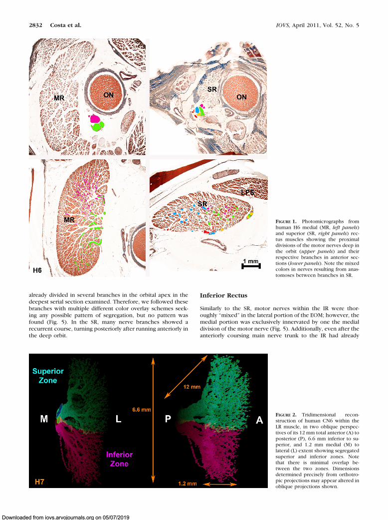
Histologic sections were analyzed sequentially as previously pub-lished.11 Nerves were traced from the posterior point, external to eachEOM, in which the main nerve trunk directed to that muscle firstbifurcated. Although this method potentially permits tracing of intra-muscular nerves both anterior and posteriorly, intramuscular motornerve branches large enough to trace did not travel posteriorly fromthe entry point. The main trunk of CN6 and CN3 was identified deepin the orbits. From this point, the two primary bifurcations of CN6were overlayed in separate image layers with two different colors inserial sections (using Photoshop; Adobe Systems). The same processwas performed with CN3 from the point that the branch to eachindividual EOM separated from the main CN3 trunk. If a given nervedeveloped more than two main branches at its initial division, thesebranches were color overlayed with the maximum effort to segregatethem by similarity using two main colors. In one human superiorrectus (SR), the CN3 trunk directed to the EOM had already dividedinto several branches in the initial section obtained at the orbital apex.In this case, multiple different colors were used to overlay each branchuniquely (Fig. 1). Using serial sections from this location, these sec-ondary nerve trunks and all their daughter branches were highlighted
to maintain colors corresponding to their lineage as far anteriorly asidentifiable nerve bundles were present. When an anastomosis oc-curred between two different nerve divisions that had been overlaidwith different colors, subsequent branches were colored with the twocolors mixed proportionally (Fig. 1). After color overlay had beenobtained in each serial section in the orbit, the colored nerve sectionsalone were reconstructed in three dimensions using the programImageJ (developed by Wayne Rasband, National Institutes of Health,Bethesda, MD; available at http://rsb.info.nih.gov/ij/index.html) run-ning on an 8-processor computer (Mac Pro, Apple Computer, Cuper-tino, CA) with 32 Gb random access memory.
RESULTS
Lateral Rectus
Findings were consistent in the monkey and in both humanorbits studied. The intramuscular distribution of CN6 in theLR of human orbit H7 was segregated into distinct superiorand inferior zones with minimal overlap, confirming andextending in additional orbit H7 Peng et al.’s report in twoother human orbits (Fig. 2).11 The main trunk of CN6 bifur-cated in the posterior orbit into distinct superior and infe-rior divisions that entered the LR on its global surface andcoursed anteriorly through the EOM with little or no overlapbetween them. The intramuscular course of subsequentCN6 branches followed almost parallel, separate courses,remaining within superior or inferior LR zones with no turnsand at most rare anastomoses, even between branches of thesame main trunk.
Medial Rectus
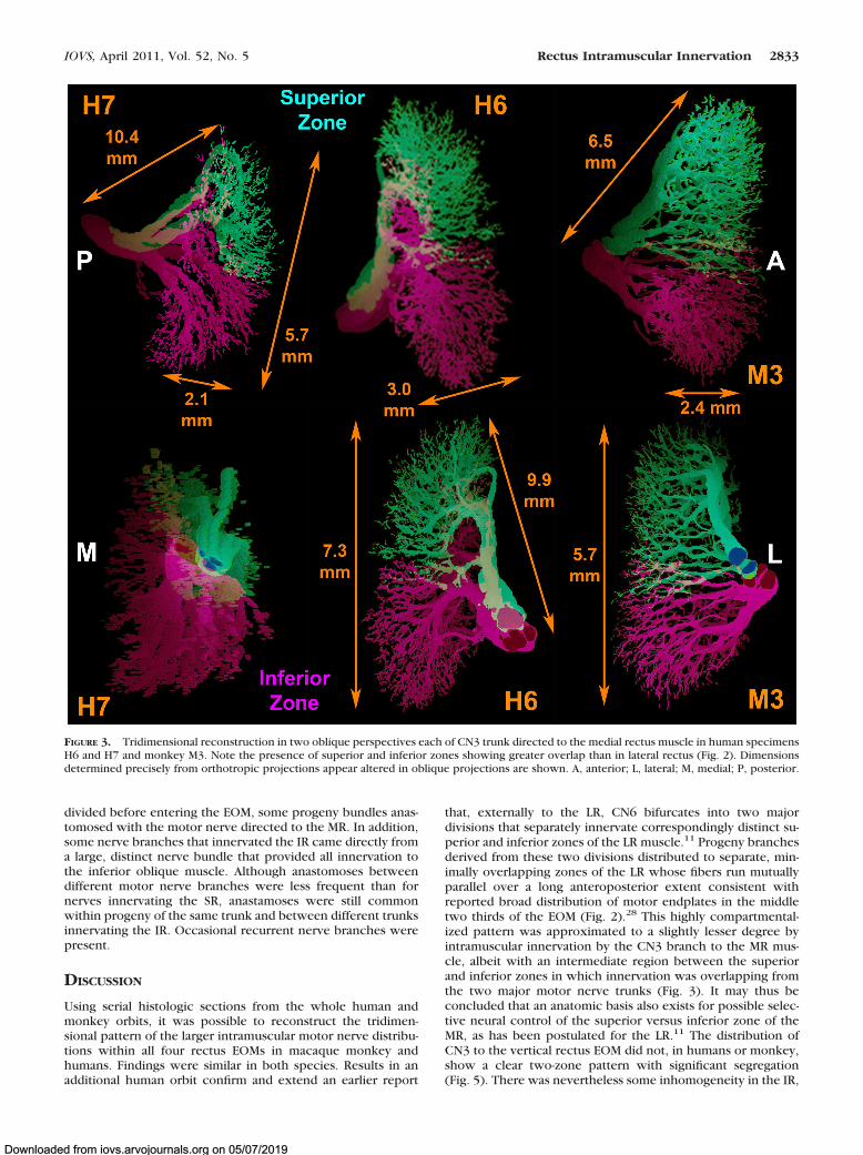
Although the MR did not show the same discrete two-zoneinnervational pattern found in CN6, the intramuscularbranches of CN3 innervating the MR divided externally to theEOM and subsequently distributed within it in an organized,topographic pattern that was maintained along the length ofthe MR. Anastomoses between branches either from the sameor from different main trunks of intramuscular CN3, althoughpresent, were less frequent than in the SR and IR (Fig. 3).Occasional CN3 branches in the MR took a recurrent coursefrom anterior to posterior, mainly in the initial divisions of thenerve inside the EOM.
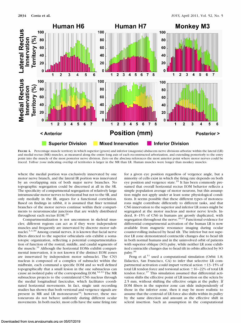
A quantitative comparison of nerve segregation within su-perior and inferior zones of the LR and MR was obtained bycomparing, in transverse sections all along the length of eachEOM where intramuscular nerves could be traced, the percent-age of transverse EOM width occupied by exclusively segre-gated nerve projections, as well as the proportion containingoverlapping projections (Fig. 4). These three percentages donot sum to 100% because traceable nerve fibers were notidentified in all portions of each EOM cross section. As seen inFigure 4 (top), 1.7–4.1% of LR width contained nerve fibersfrom both the superior and inferior divisions of CN6, while thebulk of the LR contained nerve arborizations from either thesuperior or inferior division in both humans and monkey. Bycontrast, Figure 4 (bottom) illustrates that, in both humans andmonkey, 2–41% of MR width contains mixed branches of boththe superior and inferior divisions of the MR branch of CN3;the remaining 20–40% of MR width received segregated inner-vation from either superior or inferior division, but not both.
Superior Rectus
The intramuscular nerve distribution in the SR was thoroughly“mixed,” with a large number of anastomoses betweenbranches from the same trunk and from different trunks withinthe EOM. In human orbit H6, the superior division of CN3 was
IOVS, April 2011, Vol. 52, No. 5 Rectus Intramuscular Innervation 2831
Downloaded from iovs.arvojournals.org on 05/07/2019
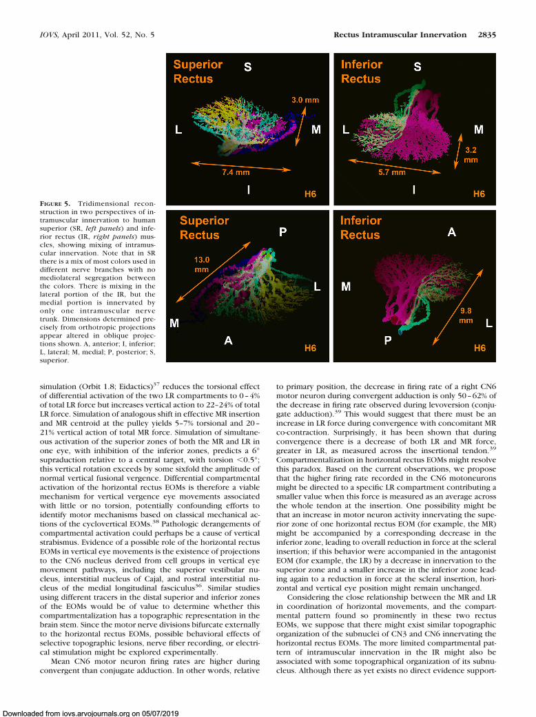
already divided in several branches in the orbital apex in thedeepest serial section examined. Therefore, we followed thesebranches with multiple different color overlay schemes seek-ing any possible pattern of segregation, but no pattern wasfound (Fig. 5). In the SR, many nerve branches showed arecurrent course, turning posteriorly after running anteriorly inthe deep orbit.
Inferior Rectus
Similarly to the SR, motor nerves within the IR were thor-oughly “mixed” in the lateral portion of the EOM; however, themedial portion was exclusively innervated by one the medialdivision of the motor nerve (Fig. 5). Additionally, even after theanteriorly coursing main nerve trunk to the IR had already
FIGURE 1. Photomicrographs fromhuman H6 medial (MR, left panels)and superior (SR, right panels) rec-tus muscles showing the proximaldivisions of the motor nerves deep inthe orbit (upper panels) and theirrespective branches in anterior sec-tions (lower panels). Note the mixedcolors in nerves resulting from anas-tomoses between branches in SR.
FIGURE 2. Tridimensional recon-struction of human CN6 within theLR muscle, in two oblique perspec-tives of its 12 mm total anterior (A) toposterior (P), 6.6 mm inferior to su-perior, and 1.2 mm medial (M) tolateral (L) extent showing segregatedsuperior and inferior zones. Notethat there is minimal overlap be-tween the two zones. Dimensionsdetermined precisely from orthotro-pic projections may appear altered inoblique projections shown.
2832 Costa et al. IOVS, April 2011, Vol. 52, No. 5
Downloaded from iovs.arvojournals.org on 05/07/2019
divided before entering the EOM, some progeny bundles anas-tomosed with the motor nerve directed to the MR. In addition,some nerve branches that innervated the IR came directly froma large, distinct nerve bundle that provided all innervation tothe inferior oblique muscle. Although anastomoses betweendifferent motor nerve branches were less frequent than fornerves innervating the SR, anastamoses were still commonwithin progeny of the same trunk and between different trunksinnervating the IR. Occasional recurrent nerve branches werepresent.
DISCUSSION
Using serial histologic sections from the whole human andmonkey orbits, it was possible to reconstruct the tridimen-sional pattern of the larger intramuscular motor nerve distribu-tions within all four rectus EOMs in macaque monkey andhumans. Findings were similar in both species. Results in anadditional human orbit confirm and extend an earlier report
that, externally to the LR, CN6 bifurcates into two majordivisions that separately innervate correspondingly distinct su-perior and inferior zones of the LR muscle.11 Progeny branchesderived from these two divisions distributed to separate, min-imally overlapping zones of the LR whose fibers run mutuallyparallel over a long anteroposterior extent consistent withreported broad distribution of motor endplates in the middletwo thirds of the EOM (Fig. 2).28 This highly compartmental-ized pattern was approximated to a slightly lesser degree byintramuscular innervation by the CN3 branch to the MR mus-cle, albeit with an intermediate region between the superiorand inferior zones in which innervation was overlapping fromthe two major motor nerve trunks (Fig. 3). It may thus beconcluded that an anatomic basis also exists for possible selec-tive neural control of the superior versus inferior zone of theMR, as has been postulated for the LR.11 The distribution ofCN3 to the vertical rectus EOM did not, in humans or monkey,show a clear two-zone pattern with significant segregation(Fig. 5). There was nevertheless some inhomogeneity in the IR,
FIGURE 3. Tridimensional reconstruction in two oblique perspectives each of CN3 trunk directed to the medial rectus muscle in human specimensH6 and H7 and monkey M3. Note the presence of superior and inferior zones showing greater overlap than in lateral rectus (Fig. 2). Dimensionsdetermined precisely from orthotropic projections appear altered in oblique projections are shown. A, anterior; L, lateral; M, medial; P, posterior.
IOVS, April 2011, Vol. 52, No. 5 Rectus Intramuscular Innervation 2833
Downloaded from iovs.arvojournals.org on 05/07/2019
where the medial portion was exclusively innervated by onemotor nerve branch, and the lateral IR portion was innervatedby an overlapping mix of both major nerve branches. Notopographic segregation could be discerned at all in the SR.The specificity of compartmental segregation of relatively largeintramuscular motor nerves to horizontal but not to the SR, andonly medially in the IR, argues for a functional correlation.Based on findings in rabbit, it is assumed that finer terminalbranches of the motor nerves continue within their compart-ments to neuromuscular junctions that are widely distributedthroughout each rectus EOM.29
Compartmentalization is not uncommon in skeletal mus-cles; different regions can act as if they were independentmuscles and frequently are innervated by discrete motor sub-nuclei.3,4,30 Among cranial nerves, it is known that facial nervefibers directed to the superior orbicularis oris exhibit a soma-totopic organization, reflecting a potential compartmentaliza-tion of function of the rostral, middle, and caudal segments ofthe muscle.31 Although the horizontal EOMs exhibit compart-mental innervation, it is not known if the distinct EOM zonesare innervated by independent motor subnuclei. The CN3nucleus is composed of a complex of subnuclei within themidbrain, each command a specific EOM and so well definedtopographically that a small lesion in the one subnucleus cancause an isolated palsy of the corresponding EOM.32,33 The MRsubnucleus projects to the contralateral CN6 nucleus throughthe medial longitudinal fasciculus, which facilitates coordi-nated horizontal movements. In fact, single unit recordingstudies has shown that both versional and vergence signals arepresent in MR and LR motoneurons34; however, these mo-toneurons do not behave uniformly during different ocularmovements. In both nuclei, most cells have the same firing rate
for a given eye position regardless of vergence angle, but aminority of cells exist in which the firing rate depends on botheye position and vergence state.35 It has been commonly pre-sumed that overall horizontal rectus EOM behavior reflects asimple population average of motor neurons, but this assump-tion might not apply under at least some physiological condi-tions. It seems possible that these different types of motoneu-rons might contribute differently to different tasks, and thatCN6 innervation to the superior and inferior LR zones might besegregated at the motor nucleus and motor nerve levels. In-deed, 8–15% of CN6 in humans are grossly duplicated, withsegregation throughout the nerve.20,21 Functional evidence fordifferential compartmental activation of the human LR is nowavailable from magnetic resonance imaging during ocularcounter-rolling induced by head tilt. The inferior but not supe-rior LR zone demonstrated contractile changes due to head tiltin both normal humans and in the uninvolved orbit of patientswith superior oblique (SO) palsy, while neither LR zone exhib-ited contractile changes due to head tilt in orbits affected by SOpalsy.36
Peng et al.11 used a computational simulation (Orbit 1.8;Eidactics, San Francisco, CA) to infer that selective LR com-partmental activation could impart vertical action �13–15% oftotal LR tendon force and torsional action �16–22% of total LRtendon force.37 This simulation assumed that differential acti-vation shifts the effective point of LR insertion on the sclera by2.5 mm, without shifting the effective origin at the pulley. IfEOM fibers in the superior zone can slide independently ofthose in the inferior zone, then it may be more realistic toassume that the centroid of LR force also shifts within its pulleyby the same direction and amount as the effective shift inscleral insertion. Such an assumption in the computational
FIGURE 4. Percentage muscle territory in which superior (green) and inferior (magenta) abducens nerve divisions arborize within the lateral (LR)and medial rectus (MR) muscles, as measured along the entire long axis of each reconstructed arborization, and extending posteriorly to the entrypoint into the muscle of the most posterior nerve division. Zero on the abscissa references the most anterior point where motor nerves could betraced. Yellow zone indicating overlap of territories is larger in the MR than LR. Human muscles were longer than monkey muscles.
2834 Costa et al. IOVS, April 2011, Vol. 52, No. 5
Downloaded from iovs.arvojournals.org on 05/07/2019
simulation (Orbit 1.8; Eidactics)37 reduces the torsional effectof differential activation of the two LR compartments to 0–4%of total LR force but increases vertical action to 22–24% of totalLR force. Simulation of analogous shift in effective MR insertionand MR centroid at the pulley yields 5–7% torsional and 20–21% vertical action of total MR force. Simulation of simultane-ous activation of the superior zones of both the MR and LR inone eye, with inhibition of the inferior zones, predicts a 6°supraduction relative to a central target, with torsion �0.5°;this vertical rotation exceeds by some sixfold the amplitude ofnormal vertical fusional vergence. Differential compartmentalactivation of the horizontal rectus EOMs is therefore a viablemechanism for vertical vergence eye movements associatedwith little or no torsion, potentially confounding efforts toidentify motor mechanisms based on classical mechanical ac-tions of the cyclovertical EOMs.38 Pathologic derangements ofcompartmental activation could perhaps be a cause of verticalstrabismus. Evidence of a possible role of the horizontal rectusEOMs in vertical eye movements is the existence of projectionsto the CN6 nucleus derived from cell groups in vertical eyemovement pathways, including the superior vestibular nu-cleus, interstitial nucleus of Cajal, and rostral interstitial nu-cleus of the medial longitudinal fasciculus36. Similar studiesusing different tracers in the distal superior and inferior zonesof the EOMs would be of value to determine whether thiscompartmentalization has a topographic representation in thebrain stem. Since the motor nerve divisions bifurcate externallyto the horizontal rectus EOMs, possible behavioral effects ofselective topographic lesions, nerve fiber recording, or electri-cal stimulation might be explored experimentally.
Mean CN6 motor neuron firing rates are higher duringconvergent than conjugate adduction. In other words, relative
to primary position, the decrease in firing rate of a right CN6motor neuron during convergent adduction is only 50–62% ofthe decrease in firing rate observed during levoversion (conju-gate adduction).39 This would suggest that there must be anincrease in LR force during convergence with concomitant MRco-contraction. Surprisingly, it has been shown that duringconvergence there is a decrease of both LR and MR force,greater in LR, as measured across the insertional tendon.39
Compartmentalization in horizontal rectus EOMs might resolvethis paradox. Based on the current observations, we proposethat the higher firing rate recorded in the CN6 motoneuronsmight be directed to a specific LR compartment contributing asmaller value when this force is measured as an average acrossthe whole tendon at the insertion. One possibility might bethat an increase in motor neuron activity innervating the supe-rior zone of one horizontal rectus EOM (for example, the MR)might be accompanied by a corresponding decrease in theinferior zone, leading to overall reduction in force at the scleralinsertion; if this behavior were accompanied in the antagonistEOM (for example, the LR) by a decrease in innervation to thesuperior zone and a smaller increase in the inferior zone lead-ing again to a reduction in force at the scleral insertion, hori-zontal and vertical eye position might remain unchanged.
Considering the close relationship between the MR and LRin coordination of horizontal movements, and the compart-mental pattern found so prominently in these two rectusEOMs, we suppose that there might exist similar topographicorganization of the subnuclei of CN3 and CN6 innervating thehorizontal rectus EOMs. The more limited compartmental pat-tern of intramuscular innervation in the IR might also beassociated with some topographical organization of its subnu-cleus. Although there as yet exists no direct evidence support-
FIGURE 5. Tridimensional recon-struction in two perspectives of in-tramuscular innervation to humansuperior (SR, left panels) and infe-rior rectus (IR, right panels) mus-cles, showing mixing of intramus-cular innervation. Note that in SRthere is a mix of most colors used indifferent nerve branches with nomediolateral segregation betweenthe colors. There is mixing in thelateral portion of the IR, but themedial portion is innervated byonly one intramuscular nervetrunk. Dimensions determined pre-cisely from orthotropic projectionsappear altered in oblique projec-tions shown. A, anterior; I, inferior;L, lateral; M, medial; P, posterior; S,superior.
IOVS, April 2011, Vol. 52, No. 5 Rectus Intramuscular Innervation 2835
Downloaded from iovs.arvojournals.org on 05/07/2019
ing the foregoing suppositions, neuroanatomical and neuro-physiological studies of the selective intramuscular innervationzones are warranted to determine whether this segregation ismaintained within the brain stem.
References
1. English AW, Wolf SL, Segal RL. Compartmentalization of musclesand their motor nuclei: the partitioning hypothesis. Phys. Ther.1993;73:857–867.
2. Holtermann A, Roeleveld K, Mork PJ, et al. Selective activation ofneuromuscular compartments within the human trapezius muscle.J Electromyogr Kinesiol. 2009;29:896–902.
3. Urquhart DM, Hodges PW. Differential activity of regions of trans-versus abdominis during trunk rotation. Eur Spine. 2005;14:393–400.
4. Lucas-Osma AM, Collazos-Castro JE. Compartmentalization in thetriceps brachii motoneuron nucleus and its relation to musclearchitecture. J Comp Neurol. 2009;516:226–239.
5. Goldberg SJ, Wilson KE, Shall MS. Summation of extraocular motorunit tensions in the lateral rectus muscle of the cat. Muscle Nerve.1997;20:1229–1235.
6. Goldberg SJ, Meredith MA, Shall MS. Extraocular motor unit andwhole-muscle responses in the lateral rectus muscle of the squirrelmonkey. J Neurosci. 1998;18:10629–10639.
7. Demer JL, Oh SY, Poukens V. Evidence for active control of rectusextraocular muscle pulleys. Invest Ophthalmol Vis Sci. 2000;41:1280–1290.
8. Demer JL. Pivotal role of orbital connective tissues in binocularalignment and strabismus. The Friedenwald lecture. Invest Oph-thalmol Vis Sci. 2004;45:729–738.
9. Demer JL. Current concepts of mechanical and neural factors inocular motility. Curr Opin Neurol. 2006;19:4–13.
10. Demer JL. Mechanics of the orbita. Dev Ophthalmol. 2007;40:132–157.
11. Peng M, Poukens V, da Silva Costa RM, Yoo L, Tychsen L, DemerJL. Compartmentalized innervation of primate lateral rectus mus-cle. Invest Ophthalmol Vis Sci. 2010;51:4612–4617.
12. Govsa F, Kayalioglu G, Erturk M, Ozgur T. The superior orbitalfissure and its contents. J Surg Radiol Anat. 1999;21:181–185.
13. Spencer RF, Porter JD. Biological organization of the extraocularmuscles. Prog Brain Res. 2006;151:43–80.
14. Gilbert PW. The origin and development of the extrinsic ocularmuscles in the domestic cat. Contrib Embryol. 1957;36:61–78.
15. Neal HV. The history of the eye muscles. J Morphol. 1918;30:433–453.
16. Wahl CM, Noden DM, Baker R. Developmental relations betweensixth nerve motor neurons and their targets in the chick embryo.Dev Dynam. 1994;20:191–202.
17. Evans DJ, Noden DM. Spatial relations between avian craniofacialneural crest and paraxial mesoderm cells. Dev Dynam. 2006;235:1310–1325.
18. Nathan H, Ouaknine G, Kosary IZ. The abducens nerve: anatomicalvariations in its course. J Neurosurg. 1974;42:561–566.
19. Jain KK. Aberrant roots of the abducens nerve. J Neurosurg.1964;21:349–351.
20. Iaconetta G, Tessitore E, Samii M. Duplicated abducent nerve andits course: microanatomical study and surgery-related consider-ations. J Neurosurg. 2001;95:853–858.
21. Ozeren MF, Sam B, Akdemir I, Lalkan A, Tekdemir I, Deda H.Duplication of the abducens nerve at the petroclival region: ananatomic study. Neurosurgery. 2003;52:645–651.
22. Demer JL, Clark RA, Engle EC. Magnetic resonance imaging evi-dence for widespread orbital dysinnervation in congenital fibrosisof extraocular muscles due to mutations in KIF21A. Invest Oph-thalmol Vis Sci. 2005;46:530–539.
23. Okanobu H, Kono R, Miyake K, Ohtsuki H. Splitting of the extra-ocular horizontal rectus muscle in congenital cranial dysinnerva-tion disorders. Am J Ophthalmol. 2009;147:550–556.
24. Demer JL, Clark RA, Lim KH, Engle EC. Magnetic resonance imag-ing evidence for widespread orbital dysinnervation in dominantDuane’s retraction syndrome linked to the DURS2 locus. InvestOphthalmol Vis Sci. 2007;48:194–202.
25. Demer JL, Clark RA, Lim K-H, Engle EC. Magnetic resonanceimaging of innervational and extraocular muscle abnormalities inDuane-radial ray syndrome. Invest Ophthalmol Vis Sci. 2007;48:5505–5511.
26. Okanobu H, Kono R, Ohtsuki H, Miyake K. Magnetic resonanceimaging findings in Duane’s retraction syndrome type III. RinshoGanka (Jpn Clin Ophthalmol). 2008;62:65–69.
27. Sheehan DC, Hrapchak BB. Theory and Practice of Histotechnol-ogy. St. Louis: Mosby; 1973:95–116.
28. Mayr R, Gottschall J, Gruber H, Neuhuber W. Internal structure ofcat extraocular muscle. Anat Embryol. 1975;148:25–34.
29. Harrison AR, Anderson BC, Thompson LV, McLoon LK. Myofiberlength and three-dimensional localization of NMJs in normal andbotulinum toxin treated adult extraocular muscles. Invest Oph-thalmol Vis Sci. 2007;48:3594–3601.
30. Mu L, Sanders I. The human cricothyroid muscle: three musclebellies and their innervation patterns. J Voice. 2007;23:21–28.
31. Marshall CD, Hsu RH, Herring SW. Somatotopic organization ofperioral musculature innervation within the pig facial motor nu-cleus. Brain Behav Evol. 2005;66:22–34.
32. Chou TK, Demer JL. Isolated inferior rectus palsy caused by ametastasis to the oculomotor nucleus. Am J Ophthalmol. 1998;126:737–740.
33. Castro OL, Johnson N, Mamourian AC. Isolated inferior obliqueparesis from brain-stem infarction. Perspective on oculomotorfascicular organization in the ventral midbrain tegmentum. ArchNeurol. 1990;47:235–237.
34. Robinson DA, Keller E. The behavior of eye movement motoneu-rons in the alert monkey. Bibl Ophthalmol. 1972;82:7–16.
35. Mays LE, Porter JD. Neural control of vergence eye movements:activity of abducens and oculomotor neurons. J Neurophysiol.1984;52:743–761.
36. Clark RA, Demer JL. Differential lateral rectus (LR) compartmentalcontraction: A novel mechanism accounts for impaired ocularcounter-rolling (OCR) in superior oblique (SO) palsy. Abstr 37thAnnual Mtg Am Ass’n Ped Ophthalmol Strab. In press.
37. Miller JM, Pavlovski DS, Shaemeva I. Orbit 1.8 Gaze MechanicsSimulation. San Francisco: Eidactics; 1999.
38. Mudgil AV, Walker M, Steffen H, Guyton DL, Zee DS. Motormechanisms of vertical fusion in individuals with superior obliqueparesis. J AAPOS. 2002;6:145–153.
39. Miller JM, Bockisch CJ, Pavlovski DS. Missing lateral rectus forceand absence of medial rectus co-contraction in ocular conver-gence. J Neurophysiol. 2002;87:2421–2433.
2836 Costa et al. IOVS, April 2011, Vol. 52, No. 5
Downloaded from iovs.arvojournals.org on 05/07/2019