Intraductal papillary mucinous tumors of the pancreas · Intraductal papillary mucinous tumors of...
7
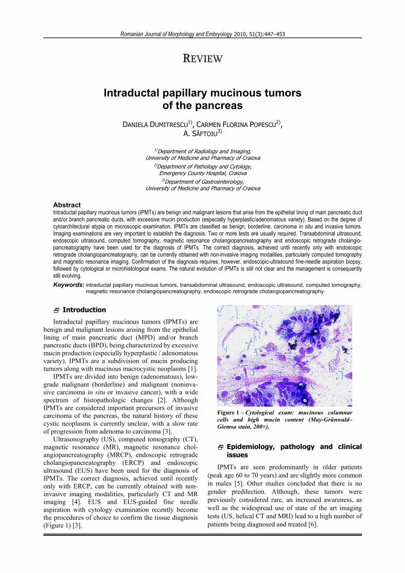
Romanian Journal of Morphology and Embryology 2010, 51(3):447–453 REVIEW Intraductal papillary mucinous tumors of the pancreas DANIELA DUMITRESCU 1) , CARMEN FLORINA POPESCU 2) , A. SĂFTOIU 3) 1) Department of Radiology and Imaging, University of Medicine and Pharmacy of Craiova 2) Department of Pathology and Cytology, Emergency County Hospital, Craiova 3) Department of Gastroenterology, University of Medicine and Pharmacy of Craiova Abstract Intraductal papillary mucinous tumors (IPMTs) are benign and malignant lesions that arise from the epithelial lining of main pancreatic duct and/or branch pancreatic ducts, with excessive mucin production (especially hyperplastic/adenomatous variety). Based on the degree of cytoarchitectural atypia on microscopic examination, IPMTs are classified as benign, borderline, carcinoma in situ and invasive tumors. Imaging examinations are very important to establish the diagnosis. Two or more tests are usually required. Transabdominal ultrasound, endoscopic ultrasound, computed tomography, magnetic resonance cholangiopancreatography and endoscopic retrograde cholangio- pancreatography have been used for the diagnosis of IPMTs. The correct diagnosis, achieved until recently only with endoscopic retrograde cholangiopancreatography, can be currently obtained with non-invasive imaging modalities, particularly computed tomography and magnetic resonance imaging. Confirmation of the diagnosis requires, however, endoscopic-ultrasound fine-needle aspiration biopsy, followed by cytological or microhistological exams. The natural evolution of IPMTs is still not clear and the management is consequently still evolving. Keywords: intraductal papillary mucinous tumors, transabdominal ultrasound, endoscopic ultrasound, computed tomography, magnetic resonance cholangiopancreatography, endoscopic retrograde cholangiopancreatography. Introduction Intraductal papillary mucinous tumors (IPMTs) are benign and malignant lesions arising from the epithelial lining of main pancreatic duct (MPD) and/or branch pancreatic ducts (BPD), being characterized by excessive mucin production (especially hyperplastic / adenomatous variety). IPMTs are a subdivision of mucin producing tumors along with mucinous macrocystic neoplasms [1]. IPMTs are divided into benign (adenomatous), low- grade malignant (borderline) and malignant (noninva- sive carcinoma in situ or invasive cancer), with a wide spectrum of histopathologic changes [2]. Although IPMTs are considered important precursors of invasive carcinoma of the pancreas, the natural history of these cystic neoplasms is currently unclear, with a slow rate of progression from adenoma to carcinoma [3]. Ultrasonography (US), computed tomography (CT), magnetic resonance (MR), magnetic resonance chol- angiopancreatography (MRCP), endoscopic retrograde cholangiopancreatography (ERCP) and endoscopic ultrasound (EUS) have been used for the diagnosis of IPMTs. The correct diagnosis, achieved until recently only with ERCP, can be currently obtained with non- invasive imaging modalities, particularly CT and MR imaging [4]. EUS and EUS-guided fine needle aspiration with cytology examination recently become the procedures of choice to confirm the tissue diagnosis (Figure 1) [3]. Figure 1 – Cytological exam: mucinous columnar cells and high mucin content (May-Grünwald– Giemsa stain, 200×). Epidemiology, pathology and clinical issues IPMTs are seen predominantly in older patients (peak age 60 to 70 years) and are slightly more common in males [5]. Other studies concluded that there is no gender predilection. Although, these tumors were previously considered rare, an increased awareness, as well as the widespread use of state of the art imaging tests (US, helical CT and MRI) lead to a high number of patients being diagnosed and treated [6].
Transcript of Intraductal papillary mucinous tumors of the pancreas · Intraductal papillary mucinous tumors of...
Romanian Journal of Morphology and Embryology 2010, 51(3):447–453
RREEVVIIEEWW
Intraductal papillary mucinous tumors of the pancreas
DANIELA DUMITRESCU1), CARMEN FLORINA POPESCU2), A. SĂFTOIU3)
1)Department of Radiology and Imaging, University of Medicine and Pharmacy of Craiova
2)Department of Pathology and Cytology, Emergency County Hospital, Craiova
3)Department of Gastroenterology, University of Medicine and Pharmacy of Craiova
Abstract Intraductal papillary mucinous tumors (IPMTs) are benign and malignant lesions that arise from the epithelial lining of main pancreatic duct and/or branch pancreatic ducts, with excessive mucin production (especially hyperplastic/adenomatous variety). Based on the degree of cytoarchitectural atypia on microscopic examination, IPMTs are classified as benign, borderline, carcinoma in situ and invasive tumors. Imaging examinations are very important to establish the diagnosis. Two or more tests are usually required. Transabdominal ultrasound, endoscopic ultrasound, computed tomography, magnetic resonance cholangiopancreatography and endoscopic retrograde cholangio-pancreatography have been used for the diagnosis of IPMTs. The correct diagnosis, achieved until recently only with endoscopic retrograde cholangiopancreatography, can be currently obtained with non-invasive imaging modalities, particularly computed tomography and magnetic resonance imaging. Confirmation of the diagnosis requires, however, endoscopic-ultrasound fine-needle aspiration biopsy, followed by cytological or microhistological exams. The natural evolution of IPMTs is still not clear and the management is consequently still evolving. Keywords: intraductal papillary mucinous tumors, transabdominal ultrasound, endoscopic ultrasound, computed tomography,
magnetic resonance cholangiopancreatography, endoscopic retrograde cholangiopancreatography.
Introduction
Intraductal papillary mucinous tumors (IPMTs) are benign and malignant lesions arising from the epithelial lining of main pancreatic duct (MPD) and/or branch pancreatic ducts (BPD), being characterized by excessive mucin production (especially hyperplastic / adenomatous variety). IPMTs are a subdivision of mucin producing tumors along with mucinous macrocystic neoplasms [1].
IPMTs are divided into benign (adenomatous), low-grade malignant (borderline) and malignant (noninva-sive carcinoma in situ or invasive cancer), with a wide spectrum of histopathologic changes [2]. Although IPMTs are considered important precursors of invasive carcinoma of the pancreas, the natural history of these cystic neoplasms is currently unclear, with a slow rate of progression from adenoma to carcinoma [3].
Ultrasonography (US), computed tomography (CT), magnetic resonance (MR), magnetic resonance chol-angiopancreatography (MRCP), endoscopic retrograde cholangiopancreatography (ERCP) and endoscopic ultrasound (EUS) have been used for the diagnosis of IPMTs. The correct diagnosis, achieved until recently only with ERCP, can be currently obtained with non-invasive imaging modalities, particularly CT and MR imaging [4]. EUS and EUS-guided fine needle aspiration with cytology examination recently become the procedures of choice to confirm the tissue diagnosis (Figure 1) [3].
Figure 1 – Cytological exam: mucinous columnar cells and high mucin content (May-Grünwald–Giemsa stain, 200×).
Epidemiology, pathology and clinical issues
IPMTs are seen predominantly in older patients (peak age 60 to 70 years) and are slightly more common in males [5]. Other studies concluded that there is no gender predilection. Although, these tumors were previously considered rare, an increased awareness, as well as the widespread use of state of the art imaging tests (US, helical CT and MRI) lead to a high number of patients being diagnosed and treated [6].
Daniela Dumitrescu et al.
448
Over 95% of epithelial tumors of pancreas are solid ductal adenocarcinomas. The remaining ones are repre-sented by serous cystadenomas, mucin producing forms (mucinous cystic neoplasms and IPMTs), cystic endo-crine tumors, solid pseudopapillary neoplasms and other rare tumors [6]. Pancreatic cystic neoplasms usually represent less than 5 to 10 percent of all pancreatic neo-plasms [2, 3, 6]. Mucin producing tumors should be considered in the differential diagnosis of every cystic lesion in the pancreas, due to their pre-malignant potential [7]. There are two types of mucin producing tumors: mucinous cystic neoplasms (cystadenomas and cystadenocarcinomas) and IPMTs [6]. Mucinous cystic neoplasms do not communicate with the pancreatic ductal system, whereas IPMTs characteristically com-municate with the ductal system [8].
Mucin producing tumors comprise a spectrum of cyst and papillary formation; some cases are almost exclusively cystic and others are predominantly papi-llary [9]. The unique papillary epithelium arises from the ductal epithelium, while mucinous cystic neoplasms are composed of discrete individual cysts lined with mucin-producing cells in a columnar epithelium [6]. As a category, IPMTs have been delineated from mucinous cystic neoplasms, which are characterised by a dense mesenchymal ovarian-like stroma [10]. Based on the degree of cytoarchitectural atypia on microscopic exa-mination, IPMTs are classified as benign (adenoma), borderline, carcinoma in situ and invasive tumors.
Differential diagnosis between various malignant characteristics is difficult [11]. The degree of dysplasia ranges from mild to moderate to severe (carcinoma in situ) and the foci of early malignancy may be evident as intraductal mural nodules. Invasive carcinoma is seen in approximately 30 to 50 percent of the resected cases and determines a poor prognosis, as well as the presence of lymph node metastasis and vascular invasion [12]. The mucinous material distends the duct system produ- cing an apparently multiloculated mass [3], while the surrounding pancreatic parenchyma is atrophied, thus becoming a “pseudocapsule” of the mass. The dilated duct may be found in only a part of the gland or may involve the entire duct system [5]. The most notable imaging characteristics of IPMT relate to the large amount of mucin it produces and the resulting impaction, obstruction and dilatation of the pancreatic ducts.
Two types of IPMT are described: main duct type (MDT-IPMT) and branch duct type (BDT-IPMT) [3]. Branch duct type is usually benign, while main duct type and mixed type tumor may be malignant [13]. A combined type IPMT affects both the MPD and the branch duct. IPMTs are typically located in the head and uncinate process, but distal forms affecting the body and tail of the pancreas are also described [14].
Pathogenesis of these tumors is unclear, but several molecular abnormalities were described, including K-ras mutations, p53 overexpression, Dpc4 expression, MUC2 and MUC5 mucin mRNA overexpression [3, 5]. The frequency of these mutations increases with higher degrees of dysplasia in the neoplasms [15].
Most pancreatic cystic neoplasms are asymptomatic, being discovered during US or CT examinations with
other indications [6]. The patients may be symptomatic, with recurrent acute pancreatitis, chronic abdominal pain, nausea or vomiting and jaundice, indicating a lesion that obstructs the biliary or pancreatic ducts. The clinical symptoms and signs of IPMT are due to the gradual distension of the excretory duct that is induced by the hypersecretion of mucin. The impaired outflow of pancreatic juice may cause pain and produces the laboratory test abnormalities of pancreatitis [16]. Advanced IPMTs with invasive carcinoma have a simi-lar clinical presentation with pancreatic ductal adeno-carcinoma, including intractable pain, weight loss and progressive jaundice. In these stages IPMT share similar clinico-pathological characteristics with pancreatic ductal carcinoma, including sex and age distribution and location within the pancreas [17].
The natural evolution of IPMTs is still not clear and consequently the management is still evolving [3]. It is believed that IPMTs of the pancreas are slow-growing tumors because patients with IPMT often have a long history of chronic pancreatitis-like symptoms [11]. Most of the papers report a mean time of progression for IPMTs ranging from 5.5 to 6.9 years [18], but the evolution may be faster, with major imaging changes. All suspected IPMTs should be resected, due to the malignant potential. Several factors should be taken into consideration when deciding a surgical intervention that carries a risk of significant morbidity and mortality. These factors include the patients’ age and symptoms, the degree of surgical risk, as well as the location and size of the tumor. Some authors consider that the BDT-IPMT represents a subset of patients with distinct features and a lower malignant potential that may not require surgical management.
Imaging assessment
Imaging examinations are very important to esta-blish the diagnosis. Usually two or more tests are requi-red [6]. Imaging can reveal the papilla within the duo-denal lumen, the main pancreatic duct, the cystic lesion, the communication between the main pancreatic duct and the cystic lesion, the papillary projection into the main pancreatic duct or the cystic lesion, and the infil-tration within the pancreatic parenchyma or other organs [4]. Transabdominal ultrasound and computed tomogra-phy scanning show cystic pancreatic lesions and/or dilatation of the main pancreatic duct or side branches [7]. However, these changes are nonspecific, and cannot differentiate IPMTs from severe chronic pancreatitis, pancreatic pseudocysts or other cystic neoplasms. The presence of a communication between the main pancreatic duct and the cystic lesion is also one of the most reliable findings for the diagnosis of IPMT on CT, MRCP, and ERCP, because serous cystadenoma and mucinous cystic tumors do not usually communicate with the main pancreatic duct. Although pseudocysts often communicate with the main pancreatic duct, the size of the communicating duct is small. Visualization of a communicating duct on helical CT, dynamic MR imaging and MRCP is probably more specific for this
Intraductal papillary mucinous tumors of the pancreas
449
entity because the visualized communicating duct is more than 2 mm in diameter [4].
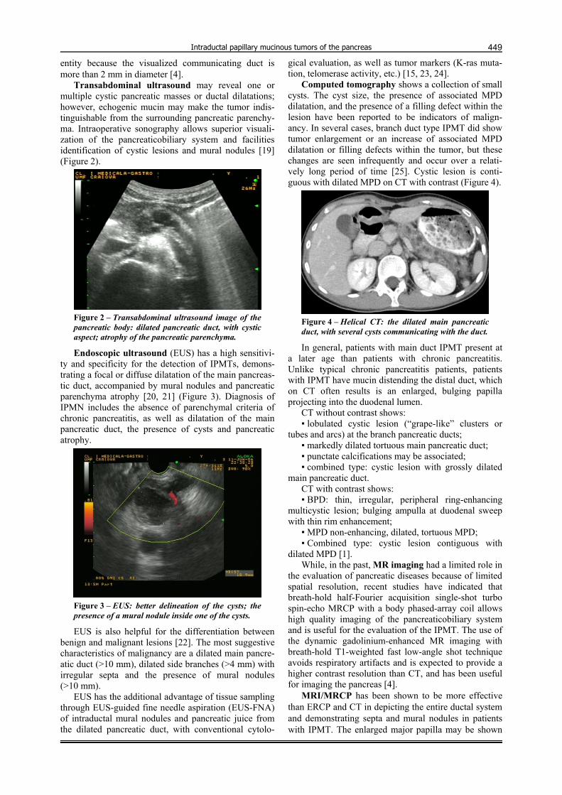
Transabdominal ultrasound may reveal one or multiple cystic pancreatic masses or ductal dilatations; however, echogenic mucin may make the tumor indis-tinguishable from the surrounding pancreatic parenchy-ma. Intraoperative sonography allows superior visuali-zation of the pancreaticobiliary system and facilities identification of cystic lesions and mural nodules [19] (Figure 2).
Figure 2 – Transabdominal ultrasound image of the pancreatic body: dilated pancreatic duct, with cystic aspect; atrophy of the pancreatic parenchyma.
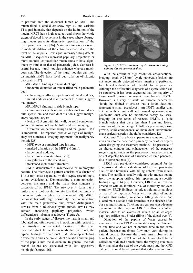
Endoscopic ultrasound (EUS) has a high sensitivi-ty and specificity for the detection of IPMTs, demons-trating a focal or diffuse dilatation of the main pancreas-tic duct, accompanied by mural nodules and pancreatic parenchyma atrophy [20, 21] (Figure 3). Diagnosis of IPMN includes the absence of parenchymal criteria of chronic pancreatitis, as well as dilatation of the main pancreatic duct, the presence of cysts and pancreatic atrophy.
Figure 3 – EUS: better delineation of the cysts; the presence of a mural nodule inside one of the cysts.
EUS is also helpful for the differentiation between benign and malignant lesions [22]. The most suggestive characteristics of malignancy are a dilated main pancre-atic duct (>10 mm), dilated side branches (>4 mm) with irregular septa and the presence of mural nodules (>10 mm).
EUS has the additional advantage of tissue sampling through EUS-guided fine needle aspiration (EUS-FNA) of intraductal mural nodules and pancreatic juice from the dilated pancreatic duct, with conventional cytolo-
gical evaluation, as well as tumor markers (K-ras muta-tion, telomerase activity, etc.) [15, 23, 24].
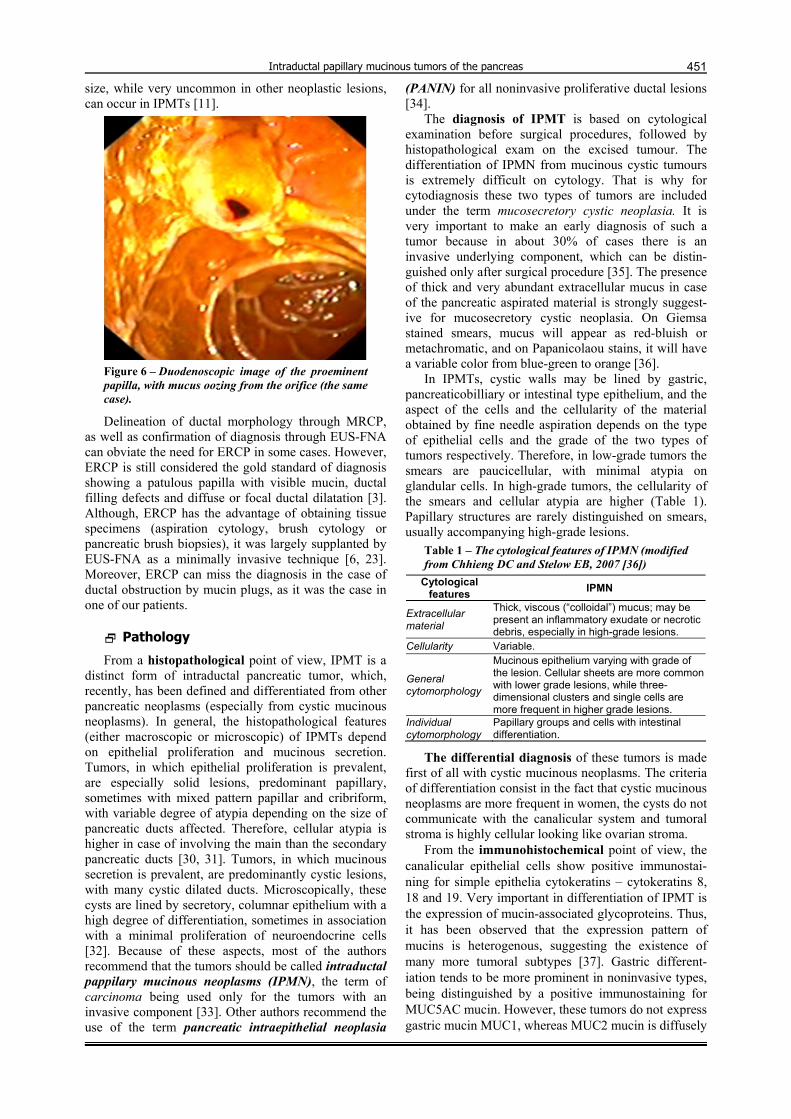
Computed tomography shows a collection of small cysts. The cyst size, the presence of associated MPD dilatation, and the presence of a filling defect within the lesion have been reported to be indicators of malign-ancy. In several cases, branch duct type IPMT did show tumor enlargement or an increase of associated MPD dilatation or filling defects within the tumor, but these changes are seen infrequently and occur over a relati-vely long period of time [25]. Cystic lesion is conti-guous with dilated MPD on CT with contrast (Figure 4).
Figure 4 – Helical CT: the dilated main pancreatic duct, with several cysts communicating with the duct.
In general, patients with main duct IPMT present at a later age than patients with chronic pancreatitis. Unlike typical chronic pancreatitis patients, patients with IPMT have mucin distending the distal duct, which on CT often results is an enlarged, bulging papilla projecting into the duodenal lumen.
CT without contrast shows: ▪ lobulated cystic lesion (“grape-like” clusters or
tubes and arcs) at the branch pancreatic ducts; ▪ markedly dilated tortuous main pancreatic duct; ▪ punctate calcifications may be associated; ▪ combined type: cystic lesion with grossly dilated
main pancreatic duct. CT with contrast shows: ▪ BPD: thin, irregular, peripheral ring-enhancing
multicystic lesion; bulging ampulla at duodenal sweep with thin rim enhancement;
▪ MPD non-enhancing, dilated, tortuous MPD; ▪ Combined type: cystic lesion contiguous with
dilated MPD [1]. While, in the past, MR imaging had a limited role in
the evaluation of pancreatic diseases because of limited spatial resolution, recent studies have indicated that breath-hold half-Fourier acquisition single-shot turbo spin-echo MRCP with a body phased-array coil allows high quality imaging of the pancreaticobiliary system and is useful for the evaluation of the IPMT. The use of the dynamic gadolinium-enhanced MR imaging with breath-hold T1-weighted fast low-angle shot technique avoids respiratory artifacts and is expected to provide a higher contrast resolution than CT, and has been useful for imaging the pancreas [4].
MRI/MRCP has been shown to be more effective than ERCP and CT in depicting the entire ductal system and demonstrating septa and mural nodules in patients with IPMT. The enlarged major papilla may be shown
Daniela Dumitrescu et al.
450
to protrude into the duodenal lumen on MRI. The mucin-filled, dilated ducts show high T2 and variable T1 signal intensity that depends on the hydration of the mucin. MRCP has a high accuracy and shows the whole extent of ductal involvement in the cases where obstruc-ting mucus prevents diagnostic opacification of the main pancreatic duct [26]. Main duct tumors can result in moderate dilation of the entire pancreatic duct to the level of the ampulla. Low signal intensity filling defects on MRCP sequences represent papillary projections or mural nodules; extracellular mucin tends to have signal intensity similar to that of pancreatic juice. Contrast is useful because mural nodules enhance whereas mucin does not. The detection of the mural nodules can help distinguish IPMT from focal duct dilation of chronic pancreatitis [27].
MRI/MRCP findings in main duct type: ▪ moderate dilatation of mucin-filled main pancreatic
duct; ▪ enhancing papillary projections and mural nodules; ▪ mural nodules and duct diameter >15 mm suggest
malignancy. MRI/MRCP findings in side branch type: ▪ communicates with normal caliber duct mural no-
dule, cyst >3cm, and main duct dilation suggest malign-ancy; requires surgery;
▪ lesion <2.5 cm with thin wall, no solid component, and normal main duct can be followed with imaging.
Differentiation between benign and malignant IPMT is important. The reported predictive signs of malign-ancy are numerous. Imaging arguments for malignancy include [28]:
▪ MPD type or combined type lesions, ▪ marked dilatation of the MPD (>10mm), ▪ large mural nodules, ▪ large tumors (greater than 3 cm), ▪ irregularities of the ductal wall, ▪ thickened septum-like structures. IPMTs demonstrate a macrocystic or microcystic
pattern. The microcystic pattern consists of a cluster of 1 to 2 mm cysts separated by thin septa, resembling a serous cystadenoma. Demonstrating a communication between the mass and the main duct suggests a diagnosis of an IPMT. The macrocystic form has a unilocular or multilocular architecture that can mimic a mucinous cystic neoplasm or pseudocyst [29]. MRCP demonstrates with high sensibility the comunication with the main pancreatic duct, which distinguishes IPMTs from a mucinous cystic tumor. IPMTs may contain enhancing papillary projection, which differentiates it from a pseudocyst (Figure 5).
In the early stages of disease, the mass is small and lobulated and often eccentric in position with respect to the visualized or expected location of the main pancreatic duct. If the lesion seeds the main duct, the typical findings of main duct IPMT develop including diffuse main and side branch duct dilatation and bulging of the papilla into the duodenum. In general, the side branch lesions are associated with less aggressive histologic features [26].
Figure 5 – MRCP: multiple cysts communicating with the dilated pancreatic duct.
With the advent of high-resolution cross-sectional imaging, small (<25 mm) cystic pancreatic lesions are not uncommonly detected when imaging is performed for clinical indication not referable to the pancreas. Although the differential diagnosis of a cystic lesion can be extensive, it has been suggested that the majority of these small lesions represent side branch IPMTs. However, a history of acute or chronic pancreatitis should be elicited to ensure that a lesion does not represent a small pseudocyst. An IPMT smaller than 2.5 cm with a thin wall and normal appearing main pancreatic duct can be monitored safely by serial imaging. In one series of resected IPMTs, all side branch lesions that were less than 3 cm and lacked mural nodules were benign. If follow-up imaging shows growth, solid components, or main duct involvement, than surgical resection should be considered [26].
MRI and CT are both superior in evaluation of the invasion into the pancreatic parenchyma or other organs when designing the treatment method. The presence of an altered contour and enhancement of the pancreas suggesting invasion in the pancreatic parenchyma may be not depicted because of associated chronic pancreas-titis in some patients [4].
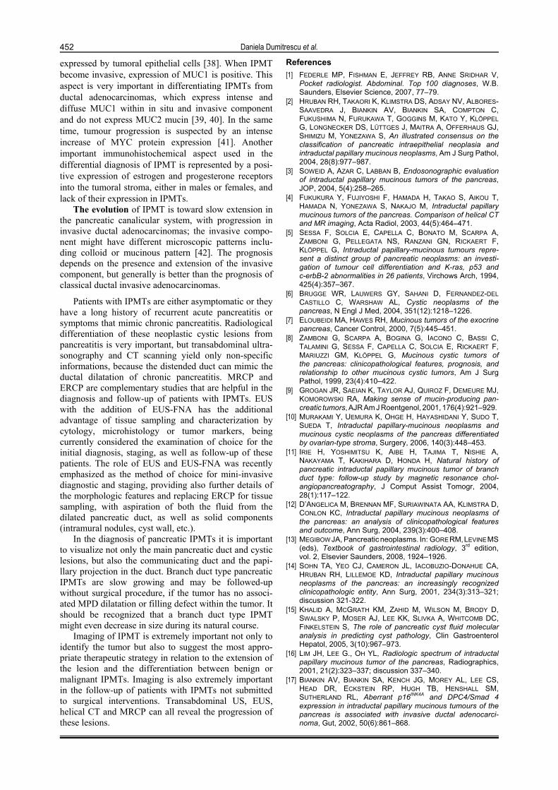
ERCP was previously considered essential for the diagnosis and indicates dilatation of the main pancreatic duct or side branches, with filling defects from mucus plugs. The papilla is usually bulging with mucus oozing from the gapping orifice, this representing a specific finding (Figure 6) [28]. However, ERCP is an invasive procedure with an additional risk of morbidity and even mortality. ERCP findings include a bulging or patulous orifice of the papilla of Vater with secretion of mucin through this orifice, mucinous filling defects, and dilated main duct and side branches in the absence of an obstructing stricture. Thick mucus can prevent adequate evaluation of the ducts on ERCP. Reflux of contrast material due to an excess of mucin or an enlarged papillary orifice may hinder filling of the ductal tree [4].
Dilatation of the papilla of Vater caused by mucinous flow on ERCP examination may be observed at one time and yet not at another time in the same patient, because mucinous flow may vary during its natural course. Because the cystic mass observed in branch duct type IPMT is not the tumor itself but a collection of dilated branch ducts, the varying mucinous flow may alter the size of the cystic mass and the MPD caliber. It should be recognized that a decrease in tumor
Intraductal papillary mucinous tumors of the pancreas
451
size, while very uncommon in other neoplastic lesions, can occur in IPMTs [11].
Figure 6 – Duodenoscopic image of the proeminent papilla, with mucus oozing from the orifice (the same case).
Delineation of ductal morphology through MRCP, as well as confirmation of diagnosis through EUS-FNA can obviate the need for ERCP in some cases. However, ERCP is still considered the gold standard of diagnosis showing a patulous papilla with visible mucin, ductal filling defects and diffuse or focal ductal dilatation [3]. Although, ERCP has the advantage of obtaining tissue specimens (aspiration cytology, brush cytology or pancreatic brush biopsies), it was largely supplanted by EUS-FNA as a minimally invasive technique [6, 23]. Moreover, ERCP can miss the diagnosis in the case of ductal obstruction by mucin plugs, as it was the case in one of our patients.
Pathology
From a histopathological point of view, IPMT is a distinct form of intraductal pancreatic tumor, which, recently, has been defined and differentiated from other pancreatic neoplasms (especially from cystic mucinous neoplasms). In general, the histopathological features (either macroscopic or microscopic) of IPMTs depend on epithelial proliferation and mucinous secretion. Tumors, in which epithelial proliferation is prevalent, are especially solid lesions, predominant papillary, sometimes with mixed pattern papillar and cribriform, with variable degree of atypia depending on the size of pancreatic ducts affected. Therefore, cellular atypia is higher in case of involving the main than the secondary pancreatic ducts [30, 31]. Tumors, in which mucinous secretion is prevalent, are predominantly cystic lesions, with many cystic dilated ducts. Microscopically, these cysts are lined by secretory, columnar epithelium with a high degree of differentiation, sometimes in association with a minimal proliferation of neuroendocrine cells [32]. Because of these aspects, most of the authors recommend that the tumors should be called intraductal pappilary mucinous neoplasms (IPMN), the term of carcinoma being used only for the tumors with an invasive component [33]. Other authors recommend the use of the term pancreatic intraepithelial neoplasia
(PANIN) for all noninvasive proliferative ductal lesions [34].
The diagnosis of IPMT is based on cytological examination before surgical procedures, followed by histopathological exam on the excised tumour. The differentiation of IPMN from mucinous cystic tumours is extremely difficult on cytology. That is why for cytodiagnosis these two types of tumors are included under the term mucosecretory cystic neoplasia. It is very important to make an early diagnosis of such a tumor because in about 30% of cases there is an invasive underlying component, which can be distin-guished only after surgical procedure [35]. The presence of thick and very abundant extracellular mucus in case of the pancreatic aspirated material is strongly suggest-ive for mucosecretory cystic neoplasia. On Giemsa stained smears, mucus will appear as red-bluish or metachromatic, and on Papanicolaou stains, it will have a variable color from blue-green to orange [36].
In IPMTs, cystic walls may be lined by gastric, pancreaticobilliary or intestinal type epithelium, and the aspect of the cells and the cellularity of the material obtained by fine needle aspiration depends on the type of epithelial cells and the grade of the two types of tumors respectively. Therefore, in low-grade tumors the smears are paucicellular, with minimal atypia on glandular cells. In high-grade tumors, the cellularity of the smears and cellular atypia are higher (Table 1). Papillary structures are rarely distinguished on smears, usually accompanying high-grade lesions.
Table 1 – The cytological features of IPMN (modified from Chhieng DC and Stelow EB, 2007 [36])
Cytological features IPMN
Extracellular material
Thick, viscous (“colloidal”) mucus; may be present an inflammatory exudate or necrotic debris, especially in high-grade lesions.
Cellularity Variable.
General cytomorphology
Mucinous epithelium varying with grade of the lesion. Cellular sheets are more common with lower grade lesions, while three-dimensional clusters and single cells are more frequent in higher grade lesions.
Individual cytomorphology
Papillary groups and cells with intestinal differentiation.
The differential diagnosis of these tumors is made first of all with cystic mucinous neoplasms. The criteria of differentiation consist in the fact that cystic mucinous neoplasms are more frequent in women, the cysts do not communicate with the canalicular system and tumoral stroma is highly cellular looking like ovarian stroma.
From the immunohistochemical point of view, the canalicular epithelial cells show positive immunostai-ning for simple epithelia cytokeratins – cytokeratins 8, 18 and 19. Very important in differentiation of IPMT is the expression of mucin-associated glycoproteins. Thus, it has been observed that the expression pattern of mucins is heterogenous, suggesting the existence of many more tumoral subtypes [37]. Gastric different-iation tends to be more prominent in noninvasive types, being distinguished by a positive immunostaining for MUC5AC mucin. However, these tumors do not express gastric mucin MUC1, whereas MUC2 mucin is diffusely
Daniela Dumitrescu et al.
452
expressed by tumoral epithelial cells [38]. When IPMT become invasive, expression of MUC1 is positive. This aspect is very important in differentiating IPMTs from ductal adenocarcinomas, which express intense and diffuse MUC1 within in situ and invasive component and do not express MUC2 mucin [39, 40]. In the same time, tumour progression is suspected by an intense increase of MYC protein expression [41]. Another important immunohistochemical aspect used in the differential diagnosis of IPMT is represented by a posi-tive expression of estrogen and progesterone receptors into the tumoral stroma, either in males or females, and lack of their expression in IPMTs.
The evolution of IPMT is toward slow extension in the pancreatic canalicular system, with progression in invasive ductal adenocarcinomas; the invasive compo-nent might have different microscopic patterns inclu-ding colloid or mucinous pattern [42]. The prognosis depends on the presence and extension of the invasive component, but generally is better than the prognosis of classical ductal invasive adenocarcinomas.
Patients with IPMTs are either asymptomatic or they have a long history of recurrent acute pancreatitis or symptoms that mimic chronic pancreatitis. Radiological differentiation of these neoplastic cystic lesions from pancreatitis is very important, but transabdominal ultra-sonography and CT scanning yield only non-specific informations, because the distended duct can mimic the ductal dilatation of chronic pancreatitis. MRCP and ERCP are complementary studies that are helpful in the diagnosis and follow-up of patients with IPMTs. EUS with the addition of EUS-FNA has the additional advantage of tissue sampling and characterization by cytology, microhistology or tumor markers, being currently considered the examination of choice for the initial diagnosis, staging, as well as follow-up of these patients. The role of EUS and EUS-FNA was recently emphasized as the method of choice for mini-invasive diagnostic and staging, providing also further details of the morphologic features and replacing ERCP for tissue sampling, with aspiration of both the fluid from the dilated pancreatic duct, as well as solid components (intramural nodules, cyst wall, etc.).
In the diagnosis of pancreatic IPMTs it is important to visualize not only the main pancreatic duct and cystic lesions, but also the communicating duct and the papi-llary projection in the duct. Branch duct type pancreatic IPMTs are slow growing and may be followed-up without surgical procedure, if the tumor has no associ-ated MPD dilatation or filling defect within the tumor. It should be recognized that a branch duct type IPMT might even decrease in size during its natural course.
Imaging of IPMT is extremely important not only to identify the tumor but also to suggest the most appro-priate therapeutic strategy in relation to the extension of the lesion and the differentiation between benign or malignant IPMTs. Imaging is also extremely important in the follow-up of patients with IPMTs not submitted to surgical interventions. Transabdominal US, EUS, helical CT and MRCP can all reveal the progression of these lesions.
References [1] FEDERLE MP, FISHMAN E, JEFFREY RB, ANNE SRIDHAR V,
Pocket radiologist. Abdominal. Top 100 diagnoses, W.B. Saunders, Elsevier Science, 2007, 77–79.
[2] HRUBAN RH, TAKAORI K, KLIMSTRA DS, ADSAY NV, ALBORES-SAAVEDRA J, BIANKIN AV, BIANKIN SA, COMPTON C, FUKUSHIMA N, FURUKAWA T, GOGGINS M, KATO Y, KLÖPPEL G, LONGNECKER DS, LÜTTGES J, MAITRA A, OFFERHAUS GJ, SHIMIZU M, YONEZAWA S, An illustrated consensus on the classification of pancreatic intraepithelial neoplasia and intraductal papillary mucinous neoplasms, Am J Surg Pathol, 2004, 28(8):977–987.
[3] SOWEID A, AZAR C, LABBAN B, Endosonographic evaluation of intraductal papillary mucinous tumors of the pancreas, JOP, 2004, 5(4):258–265.
[4] FUKUKURA Y, FUJIYOSHI F, HAMADA H, TAKAO S, AIKOU T, HAMADA N, YONEZAWA S, NAKAJO M, Intraductal papillary mucinous tumors of the pancreas. Comparison of helical CT and MR imaging, Acta Radiol, 2003, 44(5):464–471.
[5] SESSA F, SOLCIA E, CAPELLA C, BONATO M, SCARPA A, ZAMBONI G, PELLEGATA NS, RANZANI GN, RICKAERT F, KLÖPPEL G, Intraductal papillary-mucinous tumours repre-sent a distinct group of pancreatic neoplasms: an investi-gation of tumour cell differentiation and K-ras, p53 and c-erbB-2 abnormalities in 26 patients, Virchows Arch, 1994, 425(4):357–367.
[6] BRUGGE WR, LAUWERS GY, SAHANI D, FERNANDEZ-DEL CASTILLO C, WARSHAW AL, Cystic neoplasms of the pancreas, N Engl J Med, 2004, 351(12):1218–1226.
[7] ELOUBEIDI MA, HAWES RH, Mucinous tumors of the exocrine pancreas, Cancer Control, 2000, 7(5):445–451.
[8] ZAMBONI G, SCARPA A, BOGINA G, IACONO C, BASSI C, TALAMINI G, SESSA F, CAPELLA C, SOLCIA E, RICKAERT F, MARIUZZI GM, KLÖPPEL G, Mucinous cystic tumors of the pancreas: clinicopathological features, prognosis, and relationship to other mucinous cystic tumors, Am J Surg Pathol, 1999, 23(4):410–422.
[9] GROGAN JR, SAEIAN K, TAYLOR AJ, QUIROZ F, DEMEURE MJ, KOMOROWSKI RA, Making sense of mucin-producing pan-creatic tumors, AJR Am J Roentgenol, 2001, 176(4):921–929.
[10] MURAKAMI Y, UEMURA K, OHGE H, HAYASHIDANI Y, SUDO T, SUEDA T, Intraductal papillary-mucinous neoplasms and mucinous cystic neoplasms of the pancreas differentiated by ovarian-type stroma, Surgery, 2006, 140(3):448–453.
[11] IRIE H, YOSHIMITSU K, AIBE H, TAJIMA T, NISHIE A, NAKAYAMA T, KAKIHARA D, HONDA H, Natural history of pancreatic intraductal papillary mucinous tumor of branch duct type: follow-up study by magnetic resonance chol-angiopancreatography, J Comput Assist Tomogr, 2004, 28(1):117–122.
[12] D’ANGELICA M, BRENNAN MF, SURIAWINATA AA, KLIMSTRA D, CONLON KC, Intraductal papillary mucinous neoplasms of the pancreas: an analysis of clinicopathological features and outcome, Ann Surg, 2004, 239(3):400–408.
[13] MEGIBOW JA, Pancreatic neoplasms. In: GORE RM, LEVINE MS
(eds), Textbook of gastrointestinal radiology, 3rd edition, vol. 2, Elsevier Saunders, 2008, 1924–1926.
[14] SOHN TA, YEO CJ, CAMERON JL, IACOBUZIO-DONAHUE CA, HRUBAN RH, LILLEMOE KD, Intraductal papillary mucinous neoplasms of the pancreas: an increasingly recognized clinicopathologic entity, Ann Surg, 2001, 234(3):313–321; discussion 321-322.
[15] KHALID A, MCGRATH KM, ZAHID M, WILSON M, BRODY D, SWALSKY P, MOSER AJ, LEE KK, SLIVKA A, WHITCOMB DC, FINKELSTEIN S, The role of pancreatic cyst fluid molecular analysis in predicting cyst pathology, Clin Gastroenterol Hepatol, 2005, 3(10):967–973.
[16] LIM JH, LEE G., OH YL, Radiologic spectrum of intraductal papillary mucinous tumor of the pancreas, Radiographics, 2001, 21(2):323–337; discussion 337–340.
[17] BIANKIN AV, BIANKIN SA, KENCH JG, MOREY AL, LEE CS, HEAD DR, ECKSTEIN RP, HUGH TB, HENSHALL SM, SUTHERLAND RL, Aberrant p16INK4A and DPC4/Smad 4 expression in intraductal papillary mucinous tumours of the pancreas is associated with invasive ductal adenocarci-noma, Gut, 2002, 50(6):861–868.
Intraductal papillary mucinous tumors of the pancreas
453[18] ABU-HILAL M, SALVIA R, CASARIL A, PEARCE NW, BASSI C,
CAPELLI P, NICOLI N, Obstructive chronic pancreatitis and/or intraductal papillary mucinous neoplasms (IPMNs): a 21-year long case report, JOP, 2006, 7(2):218–221.
[19] SILAS AM, MORRIN MM, RAPTOPOULOS V, KEOGAN M, Intraductal papillary mucinous tumors of the pancreas, AJR Am J Roentgenol, 2001, 176(1):179–185.
[20] SUGIYAMA M, ATOMI Y, SAITO M, Intraductal papillary tumors of the pancreas: evaluation with endoscopic ultrasonogra-phy, Gastrointest Endosc, 1998, 48(2):164–171.
[21] AITHAL GP, CHEN RY, CUNNINGHAM JT, DURKALSKI V, KIM EY, PATEL RS, WALLACE MB, HAWES RH, HOFFMAN BJ, Accuracy of EUS for detection of intraductal papillary mucinous tumor of the pancreas, Gastrointest Endosc, 2002, 56(5):701–707.
[22] KUBO H, CHIJIIWA Y, AKAHOSHI K, HAMADA S, HARADA N, SUMII T, TAKASHIMA M, NAWATA H, Intraductal papillary-mucinous tumors of the pancreas: differential diagnosis between benign and malignant tumors by endoscopic ultra-sonography, Am J Gastroenterol, 2001, 96(5):1429–1434.
[23] STELOW EB, STANLEY MW, BARDALES RH, MALLERY S, LAI R, LINZIE BM, PAMBUCCIAN SE, Intraductal papillary-mucinous neoplasm of the pancreas. The findings and limitations of cytologic samples obtained by endoscopic ultrasound-guided fine-needle aspiration, Am J Clin Pathol, 2003, 120(3):398–404.
[24] INOUE H, TSUCHIDA A, KAWASAKI Y, FUJIMOTO Y, YAMASAKI S, KAJIYAMA G, Preoperative diagnosis of intraductal papillary-mucinous tumors of the pancreas with attention to telome-rase activity, Cancer, 2001, 91(1):35–41.
[25] KWON RS, SCHEIMAN JM, New advances in pancreatic imaging, Curr Opin Gastroenterol, 2006, 22(5):512–519.
[26] KOITO K, NAMIENO T, ICHIMURA T, YAMA N, HAREYAMA M, MORITA K, NISHI M, Mucin-producing pancreatic tumors: comparison of MR cholangiopancreatography with endos-copic retrograde cholangiopancreatography, Radiology, 1998, 208(1):231–237.
[27] SIEGELMAN ES, Body MRI, Elsevier Sauders, 2005, 89–128. [28] RAIJMAN I, KORTAN P, WALDEN D, KANDEL G, MARCON NE,
HABER GB, Mucinous ductal ectasia: cholangiopancrea-tographic and endoscopic findings, Endoscopy, 1994, 26(3):303–307.
[29] LEE JKT, SAGEL SS, STANLEY RJ, HEIKEN JP, Computed body tomography with MRI correlation, vol. 2, 4th edition, Lippincott Williams & Wilkins, 2006, 1007–1100.
[30] NISHIHARA K, FUKUDA T, TSUNEYOSHI M, KOMINAMI T, MAEDA S, SAKU M, Intraductal papillary neoplasm of the pancreas, Cancer, 1993, 72(3):689–696.
[31] TERRIS B, PONSOT P, PAYE F, HAMMEL P, SAUVANET A, MOLAS G, BERNADES P, BELGHITI J, RUSZNIEWSKI P, FLÉJOU JF, Intraductal papillary mucinous tumors of the pancreas confined to the secondary ducts show less aggressive pathologic features as compared with those involving the main pancreatic duct, Am J Surg Pathol, 2000, 24(10):1372–1377.
[32] TERADA T, OHTA T, KITAMURA Y, ASHIDA K, MATSUNAGA Y, KATO M, Endocrine cells in intraductal papillary-mucinous neoplasms of the pancreas. A histochemical and immuno-histochemical study, Virchows Arch, 1997, 431(1):31–36.
[33] ROSAI J, Pancreas and ampullary region. In: ROSAI J, Rosai and Ackerman’s surgical pathology, vol. 1, Elsevier, 2004, 1061–1114.
[34] HRUBAN RH, ADSAY NV, ALBORES-SAAVEDRA J, COMPTON C, GARRETT ES, GOODMAN SN, KERN SE, KLIMSTRA DS, KLÖPPEL G, LONGNECKER DS, LÜTTGES J, OFFERHAUS GJ, Pancreatic intraepithelial neoplasia: a new nomenclature and classification system for pancreatic duct lesions, Am J Surg Pathol, 2001, 25(5):579–586.
[35] TANAKA M, CHARI S, ADSAY V, FERNANDEZ-DEL CASTILLO C, FALCONI M, SHIMIZU M, YAMAGUCHI K, YAMAO K, MATSUNO S; INTERNATIONAL ASSOCIATION OF PANCREATOLOGY, Interna-tional consensus guidelines for management of intraductal papillary mucinous neoplasms and mucinous cystic neo-plasms of the pancreas, Pancreatology, 2006, 6(1–2):17–32.
[36] CHHIENG DC, STELOW EB, Cystic mucus-producing neoplasia: intraductal papillary mucinous neoplasms and mucinous cystic neoplasms. In: CHHIENG DC, STELOW EB, Pancreatic cytopathology, Springer, 2007, 124–144.
[37] LÜTTGES J, ZAMBONI G, LONGNECKER D, KLÖPPEL G, The immunohistochemical mucin expression pattern distin-guishes different types of intraductal papillary mucinous neoplasms of the pancreas and determines their relation-ship to mucinous noncystic carcinoma and ductal adeno-carcinoma, Am J Surg Pathol, 2001, 25(7):942–948.
[38] LÜTTGES J, FEYERABEND B, BUCHELT T, PACENA M, KLÖPPEL G, The mucin profile of noninvasive and invasive mucinous cystic neoplasms of the pancreas, Am J Surg Pathol, 2002, 26(4):466–471.
[39] ADSAY NV, MERATI K, ANDEA A, SARKAR F, HRUBAN RH, WILENTZ RE, GOGGINS M, IOCOBUZIO-DONAHUE C, LONGNECKER DS, KLIMSTRA DS, The dichotomy in the pre-invasive neoplasia to invasive carcinoma sequence in the pancreas: differential expression of MUC1 and MUC2 supports the existence of two separate pathways of carcinogenesis, Mod Pathol, 2002, 15(10):1087–1095.
[40] ZHANG H, MAITRA A, TABACKA P, WILENTZ RE, HRUBAN RH, ADSAY NV, Differential MUC1, MUC2 and MUC5AC expression in colorectal, ampullary and pancreatobiliary carcinomas: potential biologic and diagnostic implications, Mod Pathol, 2003, 16:138a.
[41] SWARTZ MJ, BATRA SK, VARSHNEY GC, HOLLINGSWORTH MA, YEO CJ, CAMERON JL, WILENTZ RE, HRUBAN RH, ARGANI P, MUC4 expression increases progressively in pancre- atic intraepithelial neoplasia, Am J Clin Pathol, 2002, 117(5):791–796.
[42] BRAT DJ, LILLEMOE KD, YEO CJ, WARFIELD PB, HRUBAN RH, Progression of pancreatic intraductal neoplasias to infiltra-ting adenocarcinoma of the pancreas, Am J Surg Pathol, 1998, 22(2):163–169.
Corresponding author Daniela Dumitrescu, Assistant Professor, MD, PhD, Department of Radiology and Medical Imagery, University of Medicine and Pharmacy of Craiova, 1 Tabaci Street, 200642 Craiova, Romania; Phone/Fax +40251–502 263, e-mail: [email protected] Received: November 1st, 2009
Accepted: July 25th, 2010