Intracranial Extra-Axial Primary Tumors - SNO · Part IV Intracranial Extra-Axial Primary Tumors...
33
Part IV Intracranial Extra-Axial Primary Tumors 3601_e11_p267-299 2/15/02 4:38 PM Page 267
Transcript of Intracranial Extra-Axial Primary Tumors - SNO · Part IV Intracranial Extra-Axial Primary Tumors...

Part IV
Intracranial Extra-Axial Primary Tumors
3601_e11_p267-299 2/15/02 4:38 PM Page 267

3601_e11_p267-299 2/15/02 4:38 PM Page 268
269
11
Meningiomas
MICHAEL W. MCDERMOTT, ALFREDO QUINONES-HINOSA,GREGORY N. FULLER, AND CHARLES B. WILSON
In 1614, the Swiss physician Felix Plater provided thefirst documented account of a meningioma (Netsky andLapresle, 1956). In the eighteenth century, AntoineLouis, from France, presented a series of patients with“fungueuses de la dure-mere” (Louis, 1771) or “fun-gating mass of the dura matter.” In the United States,W.W. Keen successfully resected a meningioma in 1887(Bingham and Keen, 1986). Years later, Harvey Cush-ing coined the term “meningioma” to describe any pri-mary discrete mass attached to the meninges (Cush-ing, 1922; Cushing and Eisenhardt, 1938).
The classic publication of Cushing and Eisenhardt’s32 chapter book in 1938 did much to bring togetherthe classic neurology with which these tumors can pres-ent. Even today this book is an essential review for allthose who deal on a regular basis with patients har-boring these tumors. Cushing thought that menin-giomas were a formidable problem and that the “prog-nosis hinges on the surgeon’s wide experience with theproblem in all its many aspects than is true of almostany other operation that can be named.”
A recent update on the surgical management ofmeningiomas by Al-Mefty (1991) indicates that manyof the challenges presented by these tumors cannotbe overcome even with modern-day technology. Yetonly in the last 15 years has the true long-term clin-ical problem of meningiomas come to be appreci-ated, largely because of the availability of magneticresonance imaging (MRI). This imaging modality al-lows us to see with great detail small residual or re-current tumors on the convexity and at the skull base.
Meningiomas can be found throughout the centralnervous system, from the optic nerve sheath to the
spinal cord but rarely, however, below the thoracicspine region. Their biologic behavior is almost alwaysone of continuous slow growth, with metastases oc-curring only with the malignant form of the disease.These tumors affect women more commonly thanmen and are mostly found in the fifth and sixthdecades of life (Burger et al., 1991; Martuza et al.,1985; Russell and Rubinstein, 1989). The expressionof steroid hormone receptors on meningiomas hasbeen used to try to explain this predilection for thefemale sex and has been the center of numerous stud-ies (Carroll et al., 1997; Halper et al., 1989; Schwartzet al., 1984; Smith and Cahill, 1994).
Recently, new discoveries about the molecular bi-ology of brain tumors have changed the way in whichwe view them; for example, techniques for their di-agnosis have evolved and now include everything fromminimally invasive image-directed biopsy to extensivecraniofacial exposures for total removal of the tumorand dural attachments, aided by cytologic, chromo-somal, genetic, and molecular studies. The insightsprovided by these sophisticated analytical laboratorytools provide the basis for the rapid translation of newdiscoveries from the bench to the bedside. This chap-ter reviews the current understanding of the basic bi-ology of meningiomas and the current standards ofclinical management.
INCIDENCE
Meningiomas account for 20% of all primary in-tracranial neoplasms and 25% of all intraspinal tu-
3601_e11_p267-299 2/15/02 4:38 PM Page 269
mors (Burger and Vogel, 1991; Carroll et al., 1993,1995a,b, 1997, 1999). In the original surgical seriesof Cushing (1938), meningiomas accounted for13.4% of more than 2000 brain tumor patients hehad treated. The incidence of meningiomas in thegeneral population varies between 2 and 15 per100,000 people, the incidence increasing with age(Young, 1981; Kurland et al., 1982). The 1997 re-port of the Central Brain Tumor Registry of the UnitedStates shows that for the period 1990 to 1994, menin-giomas accounted for 24% (4989) of primary braintumors, with an incidence ranging from 0.08 (ages 0to 19 years) to 13.72 (ages 75 to 84 years) per100,000 population.
Two recent studies from the Kumamoto Prefec-ture in Japan (Kuratsu and Ushio, 1996a; Kuratsu etal., 2000) (population 1.85 million) have provideda longitudinal follow up from 5 to 7 years regard-ing the incidence of meningiomas. The first report(Kuratsu and Ushio, 1996a) found that the age-adjusted incidence of meningiomas in males and fe-males was 1.56 and 3.95 per 100,000, respectively.Overall, meningiomas were the most common pri-mary brain tumor in this population group, with anage-adjusted incidence of 2.76 per 100,000, withthe highest incidence of 13.02 per 100,000 amongwomen aged 70 to 79 years. A subsequent studyfrom the same group, which included two moreyears of data, showed that meningiomas accountedfor 32% of all primary tumors and that 39% of thesetumors were asymptomatic (Kuratsu et al., 2000).Meningiomas tended to be asymptomatic most oftenin females and in particular those over the age of70 years.
Other epidemiologic observations also note that in-tracranial and intraspinal meningiomas afflict womentwofold and nearly ninefold as often as men, respec-tively (Earle and Richany, 1969; McDermott, 1996;Schmidek, 1991). Studies performed during the pasttwo decades have demonstrated that meningiomas ex-press estrogen (infrequently), progesterone, and an-drogen hormone receptors, leading to the hypothe-sis that steroid sex hormones may contribute to theirgrowth, which could explain the higher incidence ofmeningiomas in women (Black et al., 1996; Black,1993, 1997).
Race may also be important, as a higher percent-age of all brain tumors are meningiomas in Africancountries. In one study of brain tumors in Los Ange-les County, the male and female meningioma tumor
rates were highest in the black population (Preston-Martin, 1989).
In children, meningiomas are uncommon, com-prising only 1% to 4% of all brain tumors and withno female preponderance. In the recent review byErdincler and colleagues (1998), 62% of pediatricpatients presented between the ages of 10 to 15 years,and 41% had associated neurofibromatosis.
LOCATION
Meningiomas can occur anywhere along the course ofthe intracranial arachnoid and dura, but tend to clus-ter along sites where arachnoid granulations returncerebrospinal fluid to the venous system, that is, theconvexity and basal venous sinuses. Larger clinical se-ries with more than 500 patients document that thethree most common locations for intracranial menin-giomas are all supratentorial: parasagittal, convexity,and sphenoid wing. In the surgical series of Cushing(1922), 8% of meningiomas were in the posteriorfossa, and they occurred along the tentorium, in thecerebellopontine angle, and at the foramen magnum,in decreasing order of frequency. A modern series thatincluded patients treated surgically as well as those sim-ply observed revealed convexity, falcine, sphenoidwing, and tentorium as the four most common loca-tions. In the first two decades of life, Germano (1994)found that 67% of meningiomas were supratentorialand 14.4% were infratentorial. Intraventricular menin-giomas are most commonly within the lateral ventricle(80%) near the atrium and most often on the left sideof the brain. Three-fourths of the remaining 20% of in-traventricular meningiomas occur in the third ventri-cle, and the rest occur in the fourth ventricle.
ETIOLOGY
Exogenous and Endogenous FactorsAssociated with Meningiomas
Meningiomas are thought to mainly arise from capcells of the arachnoid layer around the major sinusesand large cerebral veins where arachnoid granula-tions are most prominent (Kida et al., 1988; O’Rahillyand Muller, 1986). Cushing pointed out that menin-giomas are almost always attached to the dura eventhough they do not arise from it. Exogenous and en-
270 INTRACRANIAL EXTRA-AXIAL PRIMARY TUMORS
3601_e11_p267-299 2/15/02 4:38 PM Page 270

dogenous factors acting alone or in combination arethought to account for the tumorigenesis of menin-giomas. Trauma, viral infection, and prior brain ir-radiation are some of the exogenous factors impli-cated in the development of meningiomas (Harrisonet al., 1991; McDermott, 1996; Musa et al., 1995; Ronet al., 1988a,b). Presently, studies of radiation ther-apy for tinea capitis in childhood present the strongestand most convincing epidemiologic evidence linkingionizing radiation to the occurrence of meningiomas(Ron et al., 1988a,b). There are numerous other eti-ologic risk factors reported in the literature, includ-ing head trauma, cigarette smoking, nitrite con-sumption, and even elevated cholesterol levels(Longstreth et al., 1993).
Endogenous factors associated with meningiomasinclude progestins (McDermott et al., 1996), estro-gen (Carroll et al., 1999), prolactin (Black, 1996),glucocorticoids (Carroll et al., 1993), dopamine, so-matostatin (Black, 1993) and growth factors, in-cluding platelet-derived growth factor (PDGF) (Blacket al., 1994) and epidermal growth factor (EGF) (Car-roll et al., 1997).
The Role of Hormones and Growth Factors
The finding that meningiomas occur twice as often infemales than in males raises the question of the pos-sible role of estrogen and progestins in their growth(Black, 1997). Donnell and colleagues (1979)showed in the 1970s that estrogen binding sites werepreserved in the cytosol of meningiomas. More re-cently, with highly sensitive detection methods, at leasttwo isotypes of estrogen receptors have been foundin meningiomas (Carroll et al., 1999). Many groupshave investigated the potential role of estrogen, pro-gesterone, and androgen on the growth of menin-giomas (Carroll et al., 1993, 1999; Halper et al.,1989; Hsu et al., 1997; Martuza and Eldridge, 1988;Martuza et al., 1981; Martuza et al., 1981, 1985;Maxwell et al., 1990, 1993, 1998; Schwartz et al.,1984; Tilzer et al., 1982). The function of estrogenreceptors in meningiomas, however, is still unclear(Table 11–1).
There appears to be a consensus in the literaturethat most meningiomas are progesterone receptor
Meningiomas 271
Table 11–1. Tumor Suppressor Genes, Proto-oncogenes, and Receptors Identified inMeningiomas
MerlinTumor Suppressor Genes p53 (rarely involved)
Proto-oncogenes c-myc
N-myc
c-sys
IGF-I
IGF-II
Receptors Androgens
Dopamine
Endothelin A
Epidermal growth factor
Fibroblast growth factor receptor-1
Glucocorticoids
Interferon-�
Interleukin 6
Neurotensin
Platelet-derived growth factor-�
Progesterone
Somatostatin
Transforming growth factor-�1
3601_e11_p267-299 2/15/02 4:38 PM Page 271

positive and some are estrogen receptor positive(Blankenstein et al., 1995; Bouillot et al., 1994; Car-roll et al., 1999). In one study 33 meningiomas wereexamined for the presence of the progesterone re-ceptor by Northern blot analysis, and 11 of these wereanalyzed by immunohistochemistry (Carroll et al.,1993). In this analysis the progesterone receptormRNA was expressed in 81% of tumors and the pro-gesterone receptor was found in the nucleus in 40%of meningiomas (Carroll et al., 1993). By transfect-ing a construct with a progesterone-responsive ele-ment and a reporter sequence into primary culturesof meningiomas, it was later shown that endogenouslyexpressed progesterone receptors were activated inthese tumors (Black, 1997; Carroll et al., 1993). Theprecise function of progesterone receptors in vivo is,however, unknown.
The antiprogesterone agent medroxyprogesteroneacetate (MPA) has been used in an attempt to exploitthe known positive progesterone receptor status ofmost meningiomas. MPA failed to decrease tumor sizein four of five patients treated for 17 to 29 weeks(Markwalder et al., 1987; Olson et al., 1987). Theantiprogesterone agent mifepristone (RU-486) hasalso been used with only modestly better results.Grunberg and colleagues (1991) studied the effectsof long-term oral RU-486 therapy in 14 patients withunresectable meningiomas. Five patients showedsome signs of reduced tumor measurement on com-puterized tomography (CT) scan, MRI, or improvedvisual field examination. Three patients experiencedimproved extraocular muscle function or relief fromheadache. Lamberts and colleagues (1992) examinedmifepristone treatment of meningiomas. In this study,6 of 10 patients were said to be responsive to thetherapy, and 3 of them experienced tumor shrinkage.Although these studies suggest that long-term therapywith mifepristone may be effective in cases of unre-sectable benign meningiomas, the extremely smallsample size and nonblinded methodology make thesignificance of the results difficult to determine.
In the mid-1980s, Weisman and colleagues dem-onstrated the enhancing effect of EGF on DNA syn-thesis and cell growth in primary cultures of men-ingioma cells (Weisman et al., 1986, 1987).Subsequently, they characterized the EGF receptor(EGFR) in meningiomas and suggested the involve-ment of this growth factor in the proliferation and/ordifferentiation of meningioma cells (Weisman et al.,1986, 1987). More recent studies demonstrate that
the EGFR is expressed in human meningioma speci-mens and that the EGFRs are activated in some menin-giomas (Carroll et al., 1997).
Several studies suggest that the growth of menin-giomas may also be mediated by PDGF (Black et al.,1994, 1996; Shamah et al., 1997; Todo et al., 1996;Zhang et al., 1996). The PDGF ligand is found in twosubunits, A and B, and there are two isoforms of thereceptors, � and �. The role of PDGF has been eval-uated in 20 meningiomas, and it has been observedthat PDGF-A, PDGF-B, and PDGF-�-R are expressedin meningiomas (Black et al., 1994). This analysissuggests that the �-receptor is functionally involvedin meningioma growth. When activated, c-fos levelswere increased, and an increase in meningioma celldivision was observed in response to addition ofPDGF-BB. These findings support the hypothesis thatPDGF acts as a growth factor in meningiomas (Blacket al., 1994, 1996).
Meningioma and Pregnancy
Some have noted that the growth of meningiomas isaccelerated during the luteal phase of the menstrualcycle and during pregnancy (Narayansingh et al.,1992; Roelvink et al., 1987; Saitoh et al., 1989; Wanet al., 1990). A literature review of 62 cases of preg-nancy-related symptomatic meningiomas by Roelvinkand colleagues (1991), however, failed to support acause and effect relationship between pregnancy andmeningiomas. They indicated that that if a causal re-lationship between pregnancy and meningiomas ex-isted, the number of cases reported in the literatureup to 1991 should have been greater than 62 with ahigher incidence in the reproductive years. In reality,however, the incidence of meningiomas is highest dur-ing the fifth, sixth, and seventh decades of a woman’slife (McCarthy et al., 1998), when progestins andestradiol are low, if not absent. Interestingly, it hasbeen noted that while progestins and estradiol are nolonger being produced by the female reproductive sys-tem of older women, peripheral tissues such as adi-pose cells are still producing progesterone.
Relationships with Breast Cancer
A positive correlation between meningiomas andbreast cancer has been reported (Rachlin et al., 1991;Rubinstein et al., 1989; Schoenberg et al., 1975,1977). This finding, however, has since been called
272 INTRACRANIAL EXTRA-AXIAL PRIMARY TUMORS
3601_e11_p267-299 2/15/02 4:38 PM Page 272
into question. In a study of 238 patients with menin-giomas, after a person-year method from age- andsex-matched cancer incidence, Jacobs and colleagues(1992) found that the number of breast cancers inthe meningioma group was not significantly higherthan predicted for age and sex.
In direct support of the correlation betweenmeningiomas and breast cancer, Sulman and col-leagues (1998) found that 34% had a loss of het-erozygosity in the short arm of chromosome 1p32 re-gion. Loss of heterozygosity in the same region, 1p32,has been reported for breast carcinoma, melanoma,and other cancers (Sulman et al., 1998). Other stud-ies, however, have failed to find a genetic link be-tween meningiomas and breast cancer. For instance,the putative tumor suppressor genes BRCA1 andBRCA2, associated with familial and sporadic formsof breast and ovarian cancer, do not appear to sharea common pathogenic pathway with meningiomas(Kirsch et al., 1997). Lauge and colleagues (1999)searched for germline mutations in the PTEN gene,known to be affected in Cowden’s disease, a geneticcondition associated with increased risk of breastcancers, but found no evidence for germline PTENmutations in families with breast cancer and menin-giomas. The issue of the responsiveness of menin-giomas to hormones, in analogy to breast cancer, isstill unresolved.
ANGIOGENESIS
Meningiomas, while most often benign, can grow to alarge size before being detected, can encircle criticalstructures, and can even be accompanied by peritu-moral edema (Bitzer et al., 1997; Bradac et al., 1986;New et al., 1980). Any of these occurrences can causedistortion of local structures and, if left untreated, cancause life-threatening mass effect. Meningiomas canalso cause diffuse elevation of intracranial pressure,with a subsequent decrease in cerebral blood flow,leading to a slowing of metabolism in nearby cells (Al-Mefty, 1991; Al-Mefty et al., 1994; Black, 1993). Asmall subset of meningiomas exhibits very aggressivebehavior and can invade and destroy adjacent brain;these rare tumors are malignant.
Several groups have studied the role of peritumoralvasogenic edema in meningiomas and its complica-tions (Bitzer et al., 1997, 1998; Black, 1993; Bradacet al., 1986; Kalkanis et al., 1996; Kondziolka et al.,
1998; Park et al., 2000; Provias et al., 1997; Tsai etal., 1999; Vuorinen et al., 1996). The source of suchedema is not well known but is thought to result fromincreased microvascular permeability and extravasa-tion of proteinaceous and plasma fluid into the adja-cent peritumoral space (Bradac et al., 1986; Kalka-nis et al., 1996). Several studies have confirmed theimportance of vascular endothelial growth factor(VEGF) production, a 40 to 46 kDa protein that is10,000 to 50,000 times more potent than histaminein increasing vascular permeability (Guha, 1998), intumorigenesis, neovascularization, and edema pro-duction of some tumors (Bielenberg et al., 1999; Caoet al., 1996; Folkman and Shing, 1992; Goto et al.,1993; Kenyon et al., 1996; Miller et al., 1994; Rosen-thal et al., 1994; Shima et al., 1995), including menin-giomas (Bitzer et al., 1998; Kalkanis et al., 1996; Parket al., 2000; Provias et al., 1997; Tsai et al., 1999).
To demonstrate the strong link between VEGFmRNA expression and peritumoral edema in menin-giomas, 31 meningioma specimens were subjected toNorthern blot analysis, hybridization with a comple-mentary DNA VEGF probe, and laser densitometry todetermine the relative levels of VEGF mRNA expres-sion (Kalkanis et al., 1996). Magnetic resonanceimaging was used in a double-blind fashion to cor-relate the neuropathologic tissue samples with thepresence of preoperative peritumoral edema. Of 31patients studied, 14 exhibited no edema and 17 ex-hibited some level of peritumoral fluid accumulation.The results demonstrated that meningiomas with per-itumoral edema exhibited 3.4 times the level of VEGFmRNA as those without edema. These data indicatethat VEGF expression is an important factor in the eti-ology of edema around meningiomas (Kalkanis et al.,1996).
Regulation of the expression of VEGF by menin-giomas has been studied by means of an enzyme-linked immunosorbent assay of CH-157MN menin-gioma cell supernatants (Tsai et al., 1999). Tsai andcolleagues (1999) demonstrated that epidermal andbasic fibroblast growth factors similarly induce VEGFsecretion by CH-157MN meningioma cells to 160%above baseline constitutive secretion. The sex hor-mones estradiol, progesterone, and testosterone didnot stimulate or inhibit VEGF secretion in CH-157MNmeningioma cells. Dexamethasone in this study wasshown to decrease VEGF secretion to 32% of base-line constitutive secretion, thus providing a potentialexplanation for the effect of corticosteroids in allevi-
Meningiomas 273
3601_e11_p267-299 2/15/02 4:38 PM Page 273

ating peritumoral brain edema in meningiomas. Theresults from this study suggest that growth factors andcorticosteroids, but not sex hormones, may regulateVEGF secretion (Tsai et al., 1999).
Recent evidence suggests that both mitogenic sig-nals and angiogenic signals (via induction of VEGF)share a common link by activation of the ras signal-ing pathway (Feldkamp et al., 1999a,b; Guha, 1998).Consequently, inhibition of ras activity may lead tocontrol of both tumor cell and tumor angiogenicgrowth. The relevance of the ras pathway in menin-gioma proliferation was recently studied by Shu andcolleagues (1999) using nine primary meningiomacell cultures infected with the recombinant adenovi-rus Ad-rasN17, encoding the dominant negative rasprotein or control adenovirus Ad-pAC. Ras-N17 is aras mutant protein that inhibits function of all en-dogenous cellular ras proteins. The results demon-strated that infection of meningioma cells with Ad-rasN17 increased the expression levels of the ras-N17mutant protein and inhibited phosphorylation of themitogen-activated protein kinases. Suppression of rasproteins inhibited proliferation of all exponentiallygrowing and growth-arrested meningioma cells stim-ulated with serum, suggesting that proliferation ofprimary meningioma cells depends on the presenceof functional ras proteins. Inhibiting the ras pathwaymay be of great value in preventing growthfactor–stimulated meningioma proliferation (Shu etal., 1999).
APOPTOSIS
Studies with 20 different low passage meningioma cellcultures and the addition of hydroxyurea resulted ina decrease in cell proliferation and arrested cellgrowth in the S phase in vitro. Characteristic signs ofapoptosis, including DNA fragmentation, detected byin situ DNA strand break labeling, and discreteoligonucleosomal fragments (DNA ladder) were ob-served (Schrell et al., 1997). In vivo studies showedthat when tissues from five different meningiomaswere transplanted into mice followed by treatmentwith hydroxyurea, in situ DNA fragmentation was ob-served in all hydroxyurea-treated meningioma trans-plants, providing evidence that hydroxyurea can causeapoptosis in tumor cells (Schrell et al., 1997).
Recent reports indicate that preoperative em-bolization of intracranial meningiomas, used for se-
lected patients to reduce tumor vascularity and bloodloss during surgery, may produce ischemic changesconsistent with apoptosis (Matyja et al., 1999; Nakasuet al., 1997, 1998). Alterations of p53, bcl-2, and baxexpression, genes involved in an apoptotic deathpathway, have been observed in embolized menin-giomas (Nakasu et al., 1998). p53 and its down-stream effector p21 accumulated mainly in peri-necrotic areas, where apoptosis was also observed(Nakasu et al., 1997, 1998). bcl-2 was often ex-pressed in the areas distant from necrosis, whereasbax was immunostained more intensely in the per-inecrotic areas (Nakasu et al., 1998). Matyja and col-leagues (1999), in addition to finding similar resultsin four biopsy specimens of embolized meningiomaswith p53 and bcl-2, also found that the anti-CD-68immunostained cells were distributed around orwithin the necrotic foci. These results are consistentwith the hypothesis that cell injury by preoperative tu-mor embolization correlates with the expression ofapoptosis-related proteins (Matyja et al., 1999).
Apoptotic cell death in some meningiomas andschwannomas has been associated with the use ofgamma knife radiosurgery. Indeed, radiation-inducedapoptosis is thought to contribute to the low-dose ef-fects of gamma knife radiosurgery (Tsuzuki et al.,1996).
CYTOGENETICS AND MOLECULARGENETICS
As early as the 1960s, monosomy of chromosome 22was reported in meningiomas (Zang and Singer,1967), a feature found in 40% to 80% of these tu-mors (Collins et al., 1990; Dumanski et al., 1990).There also seems to be an association with partial ortotal loss of chromosomes 14 (Katsuyama et al.,1986) and 17 (Maltby et al., 1988) and with the Ychromosome (Maltby et al., 1988). Abnormalities ofchromosomes 1, 3, 6, 7, 8, 10, 12, 18, and X havealso been reported in meningiomas (Arnoldus et al.,1992; Smith and Cahill, 1994; Vagner-Capodano etal., 1993).
In the mid-1980s, several groups described par-tial losses such as the terminal deletion of chromo-some 22 (Dumanski et al., 1987, 1990; Seizinger etal., 1987a–d). With restriction fragment-length poly-morphisms (RFLP), a tumor suppressor gene in-volved in meningiomas has been narrowed to a locus
274 INTRACRANIAL EXTRA-AXIAL PRIMARY TUMORS
3601_e11_p267-299 2/15/02 4:38 PM Page 274

on the long arm of chromosome 22 (Dumanski et al.,1987, 1990; Lekanne Deprez et al., 1991; Rouleau etal., 1987; Seizinger et al., 1987a–d) between the myo-globin locus and the 22q12.3-qter (Dumanski et al.,1990; Leon et al., 1994; Peyrard et al., 1999).
TUMOR SUPPRESSOR GENES PROTO-ONCOGENES
The concept of tumor suppressor genes originated inthe late 1960s when Harris and colleagues (1969)demonstrated that normal cells contain genes that cansuppress neoplastic growth via inhibition of cell divi-sion and cell proliferation. In meningiomas, the p53tumor suppressor gene has been studied for pointmutations, but such mutations were rarely found(Mashiyama et al., 1991; Ohgaki et al., 1993). In con-trast, the putative tumor suppressor gene mapping tochromosome 22 has more often been implicated inthe tumorigenesis of meningiomas (Arnoldus et al.,1992; Black, 1993; Collins et al., 1990; Dumanski etal., 1987; Gutmann et al., 1997; Kazumoto et al.,1990; Merel et al., 1995a,b; Parry et al., 1996;Rouleau et al., 1987, 1989; Ruttledge et al., 1994;Seizinger et al., 1987a,b; Sulman et al., 1998; Tro-fatter et al., 1993; Twist et al., 1994).
The NF1 gene resides on chromosome 17. Itsproduct, neurofibromin, is a large protein of 2818amino acids. The protein acts as a negative regulatorin the ras signal transduction pathway and might alsoact downstream of ras (Sundaram et al., 1997;Zwarthoff, 1996). A 360 amino acid region of neu-rofibromin shows significant homology to the catalyticdomain of the mammalian p21 ras-specific 120 kDaGTPase-activating protein. In the cell types that areaffected in NF1 patients, the absence of neurofibrominleads to increased proliferation, resulting in benign,and in some cases malignant, tumors. A study by Sun-daram and colleagues (1997) analyzed the expres-sion and functional status of neurofibromin in estab-lished human leptomeningeal LTAg2B cells, in 17sporadic meningiomas, and in a meningioma from apatient affected by NF2. The expression of neurofi-bromin was determined via immunoblotting and im-munoprecipitation with antineurofibromin antibod-ies, whereas its functional status was determinedthrough its ability to stimulate the intrinsic GTPase ac-tivity of p21 ras. This study showed for the first timethat neurofibromin is expressed at high levels in lep-
tomeningeal cells and in sporadic meningiomas, anddiminished GAP activity of neurofibromin was foundin approximately 28% of the tumors analyzed. Theseresults suggested also for the first time that decreasedlevels of neurofibromin in these tumors might con-tribute to their tumorigenesis (Sundaram et al.,1997).
Another tumor suppression gene, known as NF2,merlin, or schwannomin, is found to be mutated inmost sporadic meningiomas, and it has been recentlyimplicated in their tumorigenesis (Gutmann et al.,1997; Kimura et al., 1998; Lee et al., 1997). Schwan-nomin is a member of the band 4.1 superfamily ofproteins that have been shown to play important rolesin linking cell membrane proteins with the cy-toskeleton, a site of activation of tumor suppressorgenes in humans (Rouleau et al., 1993). Lee and col-leagues (1997) used immunoblotting and immuno-precipitation experiments to determine the size andsubcellular distribution of normal schwannomin inrabbit and human brain tissue, and established hu-man leptomeningeal LTAg2B cells. Subsequently, theyused similar techniques to determine the expressionlevel of schwannomin in 14 human sporadic menin-giomas. Their results showed that schwannomin is aprotein of approximately 66 kDa predominantly ex-pressed in the Triton X-100–insoluble fraction of thebrain and LTAg2B cells. The expression of schwan-nomin was severely reduced in almost 60% of pri-mary sporadic meningiomas, which raises the issuethat this may be an important factor in the tumorige-nesis of meningiomas. The development ofmeningotheliomatous meningiomas is probablylinked to alterations in other oncogenes or tumorsuppressor genes as results showed that all six tu-mors with normal schwannomin expression were ofthis type (Lee et al., 1997).
Proto-oncogenes, or genes that stimulate cell divi-sion, can become oncogenes when mutated. Onco-genes encode proteins that stimulate proliferation andsometimes mediate biologic activities that contributeto invasion. In a study of 19 meningiomas, Kazumotoand colleagues (1990) found that there was at leasta fivefold expression of the sis oncogene and of thec-myc oncogene in a great percentage of these tu-mors. The n-myc gene has also been found to be in-volved in meningiomas (Detta et al., 1993; McDon-ald and Dohrmann, 1988; Tanaka et al., 1989). IGF-Iand IGF-II have been found in a large number ofmeningiomas and may play an important role in tu-
Meningiomas 275
3601_e11_p267-299 2/15/02 4:38 PM Page 275

mor growth and cell division of meningiomas (Anto-niades et al., 1992; Friend et al., 1999; Glick et al.,1997; Nordqvist et al., 1997).
PATHOLOGY
In the classic monograph by Cushing and Eisenhardt(1938), meningiomas were divided into 9 major mor-phologic types; 7 of these categories were further di-vided into a total of 20 subtypes. Subsequent recog-nition of the lack of associated prognostic significancefor most of these subtypes led to a widely adoptedsimplified scheme of three commonly encountered“classic” patterns: meningothelial, fibrous (fibro-blastic), and transitional (mixed) (Fig. 11–1). In ad-dition to these three basic patterns, the newly revisedWorld Health Organization (WHO) classification(Kleihues and Cavenee, 2000) includes six additionalmorphologically distinct variants of low-grade menin-gioma (WHO grade I): psammomatous, angiomatous,microcystic, secretory, lymphoplasmacyte rich, andmetaplastic. There is no prognostic difference be-tween these morphologic variants and those that ex-hibit a “classic” pattern; the formal codification andmorphologic description of these entities is providedin acknowledgment of the importance of recognizingthem as meningiomas despite their unusual pheno-typic appearance.
Four specific meningeal tumor morphologies rec-ognized by the WHO classification are associated witha greater likelihood of recurrence and/or aggressivebehavior and thus warrant brief comment: the clearcell, chordoid, rhabdoid, and papillary variants. Clearcell and chordoid meningiomas are classified as WHOgrade II tumors, and rhabdoid and papillary menin-giomas are classified as WHO grade III. Papillarymeningiomas were originally described as an ag-gressive variant characterized by a high rate of re-currence and distant metastases (Ludwin et al.,1975). This description was subsequently confirmedby other investigators (Pasquier et al., 1986) and iscurrently recognized in the WHO classification (Fig.11–2). Any discussion of aggressive meningeal tu-mors includes the issue of meningeal hemangioperi-cytoma. The meningeal hemangiopericytoma is amorphologically distinct neoplasm (Fig. 11–3) thathistorically has been variously considered as eitheran aggressive variant of meningioma (Russell and Ru-binstein, 1989) or as the meningeal equivalent of sys-
temically occurring hemangiopericytomas (Burger etal., 1991). The current WHO schema classifiesmeningeal hemangiopericytoma as a malignant mes-enchymal neoplasm of nonmeningotheliomatous ori-gin. It is agreed by all investigators that these tumorsexhibit a generally more aggressive behavior than“typical” meningiomas and therefore must be distin-guished from them.
Any of the various subtypes of meningiomas dis-cussed above may exhibit morphologic features asso-
276 INTRACRANIAL EXTRA-AXIAL PRIMARY TUMORS
Figure 11–1. Meningioma. The three “classic” histologicpatterns of meningioma are meningothelial (A), fibrous (B),and transitional (C).
3601_e11_p267-299 2/15/02 4:38 PM Page 276

ciated with aggressive clinical behavior and warrant up-grading to either atypical (WHO grade II) or anaplas-tic (WHO grade III) meningioma. Morphologic crite-ria for atypical meningioma include either increasedmitotic activity (defined as greater than or equal to 4mitoses per 10 high power fields), brain invasion (in-vasion of the dura and cranial bone does not by itselfconstitute an indication of atypicality), or at least threeof the following five features: dense cellularity, smallcell formation, prominent nucleoli, patternless orsheet-like growth, and foci of tumor necrosis. Criteriafor anaplastic meningioma (WHO grade III; WHO ter-minology prefers the term anaplastic over malignant)include either a very high mitotic rate (defined as 20or more mitotic figures per 10 high power fields) ormorphologic features far in excess of those seen inatypical meningiomas, which would include appear-ances similar to sarcoma, carcinoma, or melanoma.Frank brain invasion may be seen in low-grade, atypi-cal, or anaplastic meningiomas. When present in anordinary low-grade meningioma, this feature was pre-viously used as a criterion that warranted a diagnosisof anaplastic (or malignant) meningioma; however, re-cent studies have shown that the clinical behavior ofotherwise ordinary meningiomas that show brain in-vasion is much closer to that of atypical meningiomasthan to truly anaplastic (malignant) meningiomas(Perry et al., 1999), and current WHO criteria rec-ommend classification as such. Much research effortis currently directed toward the evaluation of prolifer-
ation markers, such as MIB-1 (a monoclonal antibodythat recognizes the Ki-67 antigen in routinely processedformalin-fixed, paraffin-embedded tissue), as poten-tially powerful prognostic indicators of aggressive be-havior.
CLINICAL PRESENTATION BY LOCATION
In general, meningiomas present with the usual triadof symptoms of brain tumors in an adult patient,namely, headache, seizures, progressive focal neuro-logic deficit, and/or change in personality. Given thatmany of these tumors grow slowly, the onset of symp-toms can be insidious. Those symptoms caused bydelayed effects of chronic increased intracranial pres-sure, such as deteriorating vision from papilledema,or double vision from sixth nerve palsies, are un-common today.
Supratentorial Meningiomas
Olfactory groove meningiomas arise in the midlineadjacent to the crista galli and the planum sphe-noidale. As they enlarge, they compress the inferiorfrontal lobe, elevating it, and posteriorly displace the
Meningiomas 277
Figure 11–2. Papillary meningioma. This unusual variant ischaracterized by prominent perivascular arrangement ofmeningothelial cells, often with distinctive perivascular anu-clear zones that resemble the perivascular pseudorosettes ofependymoma. Separation of adjacent angiocentric phalangesgives rise to the pseudopapillae that typify the lesion.
Figure 11–3. Meningeal hemangiopericytoma. Distinctivefeatures of this aggressive tumor include the densely packed,disorganized cellularity and characteristic angular, branchingvasculature (“staghorn” pattern). Meningeal hemangiopericy-tomas also differ from meningiomas immunohistochemically(lack of epithelial membrane antigen expression), ultrastruc-turally (presence of basement membrane-like material andlack of abundant intercellular desmosomal attachments), andin incidence between the sexes (equal frequency for heman-giopericytomas compared with a definite female predilectionfor meningiomas).
3601_e11_p267-299 2/15/02 4:38 PM Page 277

optic apparatus. Typically these patients have slow on-set of change in mental status, depressed mood, andimpaired insight, judgment, and motivation. It is rarefor patients to complain of a loss of the sense of smell.Family members usually notice a change in person-ality first; late in the course of their disease, patientsmay complain of headaches and reduced vision. Therare syndrome described by Foster and Kennedy ofanosmia, unilateral optic atrophy, and contralateralpapilledema was originally attributed to tumors in thislocation.
Immediately behind the olfactory grove is anotherlocation where meningiomas develop, the tubercu-lum sellae. This is the bone on the anterior aspect ofthe sella turcica, and most patients with tumors inthis location have progressive visual loss, which isusually asymmetric. Changes in personality and men-tal status are uncommon because the patients cometo medical attention with their visual disturbance be-fore their tumors become large. They occur morecommonly in women than in men and typically pres-ent in the fifth and sixth decades of life. Visual fieldfindings may resemble those of pituitary adenomawith a bitemporal field defect.
The sphenoid wing is one of the most common lo-cations for an intracranial, supratentorial meningioma.These tumors typically arise anywhere along the courseof the sphenoid wing, from the anterior clinoid medi-ally to the pterion laterally. Symptomatology is relatedto location along the sphenoid wing. Those patientswith medial clinoid meningiomas usually have slowprogressive unilateral visual loss, headaches, andseizures. Middle-third sphenoid wing meningiomaspresent with headaches, seizures, and mild proptosis,without prominent visual disturbance. Lateral sphenoidmeningiomas present with headaches, seizures, andswelling in the region of the temporal fossa. A type ofmeningioma in this location that occurs much morecommonly in women is the hyperostosing “en plaque”meningioma. The typical chronology is “a middle-agedwomen who for long periods has slowly had increas-ing unilateral exophthalmos with ultimate impairmentof vision and with palpable swelling in the temporal re-gion,” as described by Cushing and Eisenhardt (1938).Edema of the inferior lid may be prominent in thesepatients. An impairment of lateral gaze may also bepresent due to involvement of lateral periorbital and/orlateral rectus muscle.
Cavernous sinus meningiomas are a difficult clin-ical problem. Typically, patients with these tumors
present with double vision, facial numbness, and,later in their course, headache and reduced visualacuity. Seizures are uncommon and usually occuronly if there is a large exophytic middle fossa com-ponent to the tumor. These symptoms make senseanatomically given the presence of the third, fourth,fifth, and sixth cranial nerves within the cavernous si-nus compartment.
Parasagittal meningiomas fill the angle betweenthe convexity dura and the midline falx. The classicclinical presentation is a patient with a headache andfocal motor or sensory seizures, beginning with symp-toms in the lower extremity progressing up into thebody, arm, and face. Frequently these patients willhave unilateral upper motor neuron signs or corticalsensory disturbance. Papilledema is relatively un-common except in the largest tumors.
Meningiomas that arise truly from the falx are cov-ered on their superior aspects by cortical tissue ofthe frontal, parietal, or occipital lobes. Clinical signsor symptoms depend partly on where along the falxthese tumors grow. Anatomically these tumors are de-fined as occurring in the anterior, middle, or poste-rior third of the falx. Anterior-third falx meningiomasmay present with headache, seizure, and mental sta-tus change. Middle-third falx meningiomas presentwith headache, seizures, and focal motor sensorydeficit. Posterior-third falx meningiomas present withheadache and visual loss, or irritative visual phe-nomenon with visual hallucinations. It is rare for alarge bilateral dumbbell-shaped falx tumor to pres-ent with a spastic paraparesis.
Convexity meningiomas arise in the dura over thefrontal, temporal, parietal, or occipital lobes. Fre-quently these tumors are asymptomatic and are dis-covered on imaging carried out for another reason.However, when the tumors are large, they presentwith headache, seizures, and focal neurologic deficitdepending on whether the tumor is on the right orleft side and over speech, motor, sensory, or occipi-tal cortex. These tumors are the most favorable forsurgical treatment.
Patients with intraventricular meningiomas haveprotean manifestations. In the series of Cushing andEisenhardt (1938), tumors within the atrium of thelateral ventricle typically presented with headache,nausea, vomiting, seizures, and speech disturbance.Patients with large tumors may also have a hemi-anopsia and unilateral sensorimotor deficit. Third-ventricular meningiomas present with nonspecific
278 INTRACRANIAL EXTRA-AXIAL PRIMARY TUMORS
3601_e11_p267-299 2/15/02 4:38 PM Page 278
symptoms, which are predominantly from increasedintracranial pressure.
Posterior Fossa Meningiomas
Tentorial meningiomas present most often with head-ache, extremity or gait ataxia, nausea, and vomiting.Seizures and mental status changes are uncommonbecause of their posterior fossa location. Commonclinical findings include evidence of cranial nerve III,IV, V, and VI deficits and appendicular cerebellumdisturbance. The more medial and anterior the loca-tion of the tentorial meningioma, the more likely thepatients are to have extraocular muscle disturbance.Tentorial meningiomas that have a significant com-ponent extending up into the inferior temporal or oc-cipital lobes may cause seizure or visual disturbance.A peculiar and surgically unfavorable type of menin-gioma involving the tentorium is the falcotentorialmeningioma, which involves the junction of the falxcerebri and tentorium, usually including the straightsinus. These patients typically present with symptomsof increased intracranial pressure without focal neu-rologic deficit.
Petroclival meningiomas usually have an insidiousonset of headache, gait disturbance, double vision,reduced hearing, and vertigo. Common clinical signsinclude cranial neuropathy, particularly involvingnerves IV, V, VI, and VIII, along with papilledema,ataxia, and cerebellar dysmetria. For meningiomasthat arise along the posterior petrous bone within theCP angle, Sami and Ammarati (1988) distinguishedsymptoms of those tumors arising anterior to the in-ternal auditory canal (IAC) from those arising poste-rior to it. Anteriorly placed lesions usually presentwith facial pain, facial numbness, and reduced hear-ing. With tumors posterior to the IAC, cerebellar signsare predominant and gait disturbance is common. Itis not infrequent for a tumor in this location to beconfused with a vestibular schwannoma. Menin-giomas of the cerebellar convexity usually presentwith symptoms and signs of increased intracranialpressure, hydrocephalus, or cerebellar dysfunctionipsilateral to the tumor. Papilledema, appendicular,and truncal ataxia are common.
Foramen magnum meningiomas are difficult todiagnose clinically and, before MR imaging, were fre-quently confused with demyelinating disorders. Mostpatients have a history of neck and suboccipital painthat is worse with flexion and Valsalva maneuvers.
Motor and sensory deficits usually develop first in thearm and then in the legs and may involve all extrem-ities to the point of spastic quadraparesis. The so-called crural palsy, with atrophy in the intrinsic mus-cles of the hand, trapezius, and shoulder, can beparticularly confusing for the clinician. Secondarydamage from increased venous pressure in the cer-vical spinal cord may be the cause for some of thesesymptoms and signs. In a clinical series from theMayo Clinic, 94% of patients complained of upper ex-tremity dysesthesia; 75% of suboccipital or neck pain;49% of upper extremity weakness; 47% of gait dis-turbance; and 42% of clumsiness of their hands.Problems with lower cranial nerve function may leadto complaints of dysarthria and dysphasia. Differen-tial diagnosis in these patients can also include sy-ringomyelia and intramedullary cervical spinal cordtumors.
Meningiomas occurring near the torcular ofHerophilus, or confluence of the superior sagittal,straight, transverse, and occipital sinuses, presentwith symptoms of increased intracranial pressure.The patients of Cushing and Eisenhardt (1938) com-plained of visual impairment related to papilledema,headaches, and neck pain. The authors noted thatlower quadrants of the visual field appeared to be af-fected before the upper quadrant of the visual field,and recovery of these fields postoperatively pro-ceeded in a reverse direction. The last location formeningiomas of the posterior fossa is the fourth ven-tricle. Despite the abundance of critical nuclei im-mediately below the pial surface of the floor of theventricle, these tumors usually present only withsymptoms of obstructive hydrocephalus and in-creased pressure.
IMAGING STUDIES
Plain X-Rays
For the most part the use of plain radiographs for thediagnosis of intracranial meningiomas is of historicinterest only. In the past, evidence of intracranial cal-cification, hyperostosis of the sphenoid wing or cal-varium, displacement of the pineal gland or enlargedvascular channels were used as indirect supportiveevidence of the presence of a long-standing, vasculartumor attached to the dura and/or invading the bone.Computed tomography followed by MR imaging have
Meningiomas 279
3601_e11_p267-299 2/15/02 4:38 PM Page 279
become standards of imaging for the diagnosis andfollow up of intracranial meningiomas. Sometimes,when only MR imaging has been done and the patienthas a history of prior craniotomy or craniectomy,plain X-rays are helpful in delineating the extent ofbone removal, which may assist with preoperativeplanning and intraoperative cranial pin fixation.
Computed Axial Tomography
Computed tomography is inferior to MRI for delin-eating soft tissue details of both the meningioma un-der investigation and the surrounding brain. It is su-perior to MRI for defining the extent of bonyhyperostosis, a key point in the consideration of ba-sal and convexity meningiomas with associated hy-perostosis. Pathologic studies have demonstrated thathyperostotic bone contains meningiothelial cells, andfailure to remove this involved bone may lead to laterrecurrence of a soft tissue tumor or progression pri-marily within bone. Thin-cut CT imaging may now beused with image-guided surgical systems primarily forthe purpose of helping the surgeon define the bonymargins of tumor intraoperatively. In these circum-stances, image fusion technologies allow the surgeonto use MRI data to determine the soft tissue marginsof the tumor adjacent to brain (e.g., determining thedistance to the medial or deep aspect of the tumor)and then switch to CT-based images for the resectionof tumor-involved bone.
On non-contrast CT imaging, meningiomas appearisodense to slightly hyperdense. Calcification withinthe tumor can be punctate or confluent and is easilyseen. Optic nerve sheath meningiomas may show thetraditional “tram track” linear calcification outliningthe orbital course of the optic nerve. When hyperos-tosis of the calvarium is extensive the bone can havea mottled to radiating starburst appearance. Menin-giomas along the sphenoid wing produce dense scle-rotic hyperostosis of the greater wing and pterionproducing associated exophthalmos. These tumorsare characteristically seen in women. With the ad-ministration of iodinated contrast, meningiomas usu-ally enhance homogenously and intensely. The “duraltail” so often referred to with MRI is not well visual-ized with CT as it is thinly opposed to the underlyingbone.
Mantle et al. (1999) reported on the use of quan-tifying peritumoral brain edema on CT scanning forpredicting the probability for meningioma recur-
rence. Between 1980 and 1998, 135 patients wereevaluated. Edema grade was linearly related to edemavolume by digitizing the CT scans. The completenessof resection was the most powerful predictor of re-currence, followed by edema grade and brain inva-sion. The chance of recurrence within 10 years aftercomplete resection was exponentially related to themaximum linear dimension of surrounding peritu-moral edema. The chance of recurrence increased byapproximately 10% for each centimeter grade in-crease in the amount of edema. The authors alsofound that the histopathologic documentation of braininvasion increased by 20% per centimeter of edemathickness on the actual CT scan. Thus, valuable in-formation can be derived by this simple imagingstudy, which may influence treatment and outcome.Furthermore, Kuratsu and colleagues (2000) in eval-uating the clinical features of asymptomatic menin-giomas found that patients with evidence of tumoralcalcification on CT scans had a significantly lowerlikelihood of tumor growth after longer than a 1 yearfollow up (p � 0.008).
One disadvantage of CT is that multiplanar imag-ing is limited. Direct coronal imaging without muchartifact is limited to the anterior and middle cranialfossa. Direct sagittal images cannot be achieved. Onthe other hand, bony foramina within the skull baseare clearly seen, and three-dimensional reconstruc-tions available from image-guided system worksta-tions give bony spatial resolution and detail superiorto that obtained from MRI.
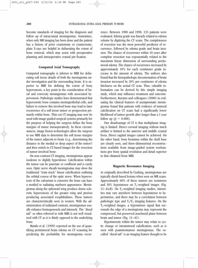
Magnetic Resonance Imaging
As originally described by Cushing, meningiomas aretypically dural-based lesions when seen on MR scans.Approximately 60% of these tumors are isointenseand 30% hypointense on T1-weighted images (Fig.11–4A,B). On T2-weighted imaging studies, intensi-ties may vary anywhere between hypointense to hy-perintense, and there may be a correlation betweenpathologic type and T1/T2 imaging features. On theT2-weighted images, a hypointense signal that sur-rounds the edge of a meningioma may represent thecompressed, but preserved arachnoid plane betweenbrain and tumor (Fig. 11–4C).
Hypointensity within the tumor may relate to cys-tic change or intratumoral calcification, such as isseen with psammomatous meningiomas. The so-called “dural tail” is an imaging feature thought to be
280 INTRACRANIAL EXTRA-AXIAL PRIMARY TUMORS
3601_e11_p267-299 2/15/02 4:38 PM Page 280
characteristic of meningiomas. This tail typically ex-tends several millimeters from the edge of the tumorand is thought to represent hypervascularity ratherthan tumor infiltration. Magnetic resonance imagingis the preferred modality for deriving a volumetric
study for the use of image-guided surgical systems,and these systems help with planning skin flaps, bonyopenings, and extent of removal as surgery proceeds.
Paleologos et al. (2000) evaluated the use of im-age-guided surgical systems in 100 patients treated
Meningiomas 281
Figure 11–4. (A) Axial T1-weighted MRI scan of right frontoparietal parasagittal meningioma with homogenous enhancementtypical of benign meningioma. (B) Coronal T1-weighted MRI showing meningioma filling the angle between falx and convexity dura,characteristic of parasagittal meningioma. Signal flow void near midline is consistent with patent superior sagittal sinus. Note rel-ative lack of mass effect given large size of tumor, suggesting slow tumor growth over a long interval of time. (C) Axial T2, secondecho image showing no edema in surrounding brain and thin rim of hyperintensity between tumor edge and brain, consistent withpreserved arachnoid plane.
3601_e11_p267-299 2/15/02 4:38 PM Page 281

surgically for meningiomas. This group was comparedwith 170 patients operated on without the use of in-traoperative navigational systems. Although the studywas not randomized, surgical times were shorter forthe image-guided group, and blood loss and mean hos-pital days spent were less for the image-guided group.Importantly, surgical complications, either permanentneurologic deficit or those complications requiring anadditional surgical procedure, were significantly less inthe image-guided group. There are technical consid-erations with the use of image-guided systems for skull-based neoplasms, such as the type of imaging, imag-ing sequence, and convenience issues referable to thetype of image guidance system, either light-emittingdiode or passive mechanical arm, to be used. At ourinstitution, The University of California, San Francisco,image-guided surgical systems are the standard for op-erations on large convexity or skull-based menin-giomas, and we prefer an older passive arm system be-cause it avoids problems with line of sight when theoperating microscope is in position.
Magnetic resonance imaging has been reported tobe of value in predicting the relationship between per-itumoral edema as evidenced by hyperintensity on T2-weighted images and the cleavage plane between tu-mor and surrounding brain. In the study by Ildan etal. (1989) increasing degrees of peritumoral edemaon T2 images correlated significantly with worseningof surgical cleavage plane between tumor and sur-rounding brain. On angiographic studies, those tu-mors with pial arterial supply were much more likelyto demonstrate significant peritumoral edema. Theauthors proposed that MRI findings might predict thedifficulty of microsurgical dissection for the surgeonbefore operation.
In another study of cystic meningioma, Zee et al.(1995) evaluated the MRI features of 15 cysticmeningiomas classifying them into three differenttypes. Type I cystic meningiomas were those withcysts wholly within tumor. Type II were those withcysts at the periphery, but still within margins of thetumor, and type III were cysts peripheral to the tu-mor in adjacent brain. Enhancement of cyst wallswas seen in type II cystic meningiomas, but not intype III. The authors suggested that this pattern ofenhancement required surgical excision of the en-hancing wall. They also found that the pathology inthese cystic meningiomas tended to be more ag-gressive.
Functional MRI techniques are also useful in pre-dicting the location of visual, motor, sensory, andspeech language cortex relative to meningiomas. Pre-suming that the meningiomas do not invade the ad-jacent brain, information about the adjacent cortexmay nonetheless be important for predicting tempo-rary neurologic disability that may result after removalof large meningiomas.
Cerebral Angiography
Conventional cerebral angiography is still useful inthe management of patients with meningiomas despitethe advent of MR angiography and MR venography.Whereas the blood supply for the variety of tumor lo-cations is predictable based on anatomy, this infor-mation can still assist the surgeon with planning asurgical approach, and embolization of very hypo-vascular tumors may assist with surgical removal. An-giographic information about tumor blood supply andthe displacement of major arteries and their positionsrelative to the margins of the tumor are important.On the venous phase of these studies, the positionsof draining cortical veins are critical as well becausethese must be preserved. The later phase of angiog-raphy, which gives information about venous anat-omy, is still the gold standard for determining whetheror not a major venous sinus is still patent (Fig. 11–5).
Bendszus et al. (2000) recently tried to evaluatethe benefit of preoperative embolization of menin-giomas. Their perspective was drawn from a non-randomized, noncontrolled study of 60 consecutivepatients in two different neurosurgical centers whowere operated on and followed up. In Center A, noembolization was performed. In Center B, all patientsunderwent embolization. Mean tumor sizes and meanblood losses with surgery did not differ betweengroups, but, in the subgroup of patients who hadsubtotal devascularization in more than 90% of thetumor, blood loss was significantly less than in pa-tients who were not embolized (p � 0.05). Therewere no differences in surgeons’ observations re-garding hemostasis, tumor consistency or intratu-moral necrosis. There was one new permanent neu-rologic deficit related to embolization (3%).
At our institution, preoperative embolization is re-served for the largest meningiomas even if the exter-nal carotid supply can be accessed easily during theopening (Fig. 11–6). A variety of embolic agents may
282 INTRACRANIAL EXTRA-AXIAL PRIMARY TUMORS
3601_e11_p267-299 2/15/02 4:38 PM Page 282

283
Figure 11–5. Sagittal, venous phase of cerebral angiogram in same tumor as in Figure 11–1. Study confirms patency of superiorsagittal sinus and displacement of draining veins.
Figure 11–6. (A) Lateral external carotid injection of middle meningeal artery supplying tumor. (B) Preoperative T1-weighted MRIwith contrast (for image-guided system) showing dramatic effect of embolization on central blood supply to tumor. Persistent medialenhancement is due to supply from falcine artery originating off the anterior ethmoid arteries, not suitable for embolization.
3601_e11_p267-299 2/15/02 4:38 PM Page 283

get into the smallest blood vessels supplying tumors.We have used a combination of gelfoam powder,polyvinyl alcohol foam, and platinum coils. It hasbeen our practice to embolize these large menin-giomas the day before operation without waiting forfurther thrombosis to occur. In the principal author’sexperience there have been no problems with this ap-proach in more than 100 surgical cases. If em-bolization is performed, a surgeon must rememberthat compromise of the blood supply to the scalp mayhave occurred and consider this when planning skinincisions.
OPTIONS FOR TREATMENT
Observation
Not every patient with an intracranial meningioma re-quires surgical intervention, and many factors, patientand tumor related, are involved in the decision to rec-ommend surgery. One of the first questions to askwhen a tumor is found on imaging studies is whetherthe imaged tumor is responsible for the patient’ssymptoms and signs. If not, and the imaging featuresare consistent with a benign tumor (homogeneousenhancement, smooth rounded margins, no associ-ated brain edema, no satellite lesions), a period ofobservation is recommended. More and more asymp-tomatic meningiomas are discovered on imagingstudies done for some other reason.
Olivero et al. (1995) followed 57 patients withsymptomatic meningiomas over an average of 32months (range 6 months to 15 years). None of thepatients became symptomatic from their enlarging tu-mor during follow up, and tumor growth was ob-served in only 10 of 45 patients (22%) imaged. Theaverage growth rate in these patients was a 0.24 cmincrease in maximum diameter per year. In anotherstudy, Kuratsu et al. (2000) studied 109 patients. Ofthese, 63 (57.7%) had imaging follow ups of morethan 1 year. Thirty-one percent of this subgroupshowed tumor growth over an average follow-up pe-riod of 27.8 months (range, 12 to 87 months). Whilethe average age in the two groups of patients, withand without tumor growth, was similar (67.5 versus66.0 years), tumors that did not grow were morelikely to be calcified on CT or hypointense on T2-weighted MRI, consistent with intratumoral calcifica-tion. Clearly, then, not all tumors grow under obser-
vation, and it is our policy to recommend 6 monthinterval follow-up imaging with MRI for 2 years andthen once a year if tumors are stable and the patientremains asymptomatic.
Surgery
A discussion of the specific surgical approach foreach tumor location is beyond the scope of this text,but some general comments can be made. First, forsymptomatic meningiomas, surgery is the mainstay ofdiagnosis and is the first step in treatment. With sur-gical removal there is no delay in “tumor response”as with other forms of therapy. Symptoms related toincreased intracranial pressure or those related to lo-cal compression of brain can be improved quickly.Chozick et al. (1996) found that 39.9% of 158 pa-tients with meningiomas had preoperative seizuresand 88.9% of these patients had complete control ofseizures postoperatively following tumor removal.
The surgeon must consider the indications for,risks associated with, reasonable goals of, and pro-jected outcomes for expected or unexpected patholo-gies associated with surgery. These issues must be re-viewed frankly with patients but without frighteningthem. Patient factors such as age, life expectancy, neu-rologic condition, and associated medical conditionsshould be taken into account. A determination of theresectability of the tumor should also be made. Insome skull-base locations, such as cavernous sinus,complete removal of all microscopic tumor tissue isvery difficult, if not impossible. Larson et al. (1995),in their experience with 36 patients who had cav-ernous sinus meningiomas, documented microscopicinvasion into cranial nerves by tumor. Sen and Hague(1997) examined six patients with benign menin-giomas of the cavernous sinus at autopsy. There wasa tendency for infiltration of the carotid artery, the pi-tuitary gland, and the connective tissue between fas-cicles of nerves. The trigeminal nerve and ganglionwere particularly prone to invasion. These pathologicstudies, as well as clinical experience, have prompteda conservative surgical approach with these tumor lo-cations, treating residual or recurrent disease withradiotherapy.
The degree of surgical removal is also related tothe risk of recurrence. This was outlined in the sem-inal paper of Donald Simpson (1957) (Table 11–2).Tumor-infiltrated bone left behind increases the riskof recurrence as does simple coagulation rather than
284 INTRACRANIAL EXTRA-AXIAL PRIMARY TUMORS
3601_e11_p267-299 2/15/02 4:38 PM Page 284

excision of the tumor’s dural attachment (Fig. 11–7).Surgical decision-making during the operation withrespect to the degree of tumor removal attempted andthe length of the operation may also affect outcome.Condra et al. (1997) classified “total excision” (TE)as a Simpson grade I, II, or III excision and foundthat of the 174 of 229 (76%) patients with this de-gree of excision, local control rates were 93%, 80%,and 76% at 5, 10, and 15 years, respectively. In con-trast, the “subtotal excision” (SE; Simpson grade IV)results for equivalent time periods were 53%, 40%,and 30%, with cause-specific survival results mirror-ing local control for the TE and SE groups. Condraet al. (1997) thought that SE alone was inadequatetherapy. In contrast, Jung et al. (2000), in reportingtheir results for the removal of 38 petroclival menin-giomas, found that the median progression-free sur-vival (PFS) was 66 months after SE and that the growthrate was slow (0.37 cm/year). The mean tumor dou-bling time was 8 years. These authors thought thatsubtotal resection, with or without radiation, was anoption for patients with petroclival meningiomas.Similarly, Couldwell et al. (1996) reported their ex-perience with 109 patients with petroclival menin-giomas, many of who had subtotal resection of theposterior cavernous sinus component. In 69%, grosstotal tumor resection was achieved with a recurrencerate of only 13% over a 6.1 year mean follow up. Inthe 20 patients with known subtotal resection of thecavernous sinus component, 12 (60%) demonstratedradiographic progression and went on to furthertreatment.
Whether complications result from surgery formeningiomas depends on a number of factors, withpatient age and tumor location as major considera-tions. In the series of Kuratsu et al. (2000), asymp-tomatic meningiomas that underwent surgical re-
moval had a perioperative morbidity of 23.3% forthose over the age of 70 years and 3.5% for thoseyounger. The neurologic, medical, and surgical mor-bidity rates in the entire group were 6.9%, 3.4%, and2.3%, respectively. For supratentorial meningiomas,a parietal location is a risk factor for the developmentof postoperative seizures. Infratentorial and skull-base meningiomas present challenges for cranialnerve preservation. In the series of Couldwell et al.(1996), of 109 petroclival meningiomas, permanentnew cranial nerve deficits developed in 33% and themortality rate was 3.7%. Modern neurosurgical se-ries also reveal that neurologic morbidity after oper-ation is site dependent.
At open operation, meningiomas are attached tothe dura, and displacing the adjacent arachnoid andbrain maintaining the “arachnoid plane” assists thesurgeon with dissection. Two common principles em-ployed during meningioma surgery are (1) to debulkthe tumor centrally, first folding the thinned-out wallsback into the cavity created rather than retractingbrain to define the tumor margin; and (2) to respectthe arachnoid membranes, which help separate tu-mor from the adjacent brain, blood vessels, and cra-nial nerves. A variety of instruments are now usedroutinely to assist with the surgical removal of thesetumors, including the operating microscope, neuro-physiologic monitoring, image-guided surgical navi-gation systems, ultrasonic aspirators, and, more re-cently, intraoperative MRI. For many of the complexskull-base tumors, especially those in the middle andposterior fossa, a team approach is taken, combin-ing the skills of neurosurgeons and neuro-otologists.For very long operations this method allows co-sur-geons to share the workload, with a rest between op-erative sessions of 2 to 4 hours helping to maintaintheir concentration and stamina. Without going into
Meningiomas 285
Table 11–2. Simpson Grade by Extent of Tumor, Dura, and Bone/Venous Sinus Excision
Bone/Sinus
Tumor Removal Dural Attachment ExcisedGrade Complete Partial Biopsy Excised Coagulated
I X X X
II X X X
III X
IV X
V X
3601_e11_p267-299 2/15/02 4:38 PM Page 285

exhaustive detail, a few specific comments can bemade about the most common tumor locations.
Supratentorial
Common locations in the supratentorial compartmentinclude convexity, falx/parasagittal, sphenoid wing, andparasellar. Convexity meningiomas, when small, are
straightforward. Image-guided surgical systems can beused to map out the location of the tumor and then toplan a margin of normal dural excision. Al-Mefty(1991) has coined the term “grade zero” excision fortumors in these locations to include a cuff of 2 cm ofnormal dura around the tumor base. When the tumorsare very large, the most medial surface may have a poorbrain–tumor interface even with benign pathology.
286 INTRACRANIAL EXTRA-AXIAL PRIMARY TUMORS
Figure 11–7. Postoperative axial (A,B) and coronal (C)T1-weighted MRIs taken postoperatively confirm gross tu-mor resection of the same tumor as in Figures 11–4 to11–6. Inferior two-thirds of falx giving rise to a portionof the tumor base was also excised. Intraoperative andimaging findings consistent with Simpson grade II removalas the lateral wall of the superior sagittal sinus was co-agulated, not excised.
3601_e11_p267-299 2/15/02 4:38 PM Page 286
For falx/parasagittal meningiomas, preoperativeassessments of the venous sinuses and parasagittaldraining veins with MR venography or angiographywill help the surgeon decide whether total excision ispossible and which is the best route for avoiding im-portant veins. Use of surgical navigational systems hasbecome almost routine for the approach to these tu-mors. When the superior sagittal sinus is occludedon the preoperative angiogram, the point along thesinus where flow is present or not can be determinedwith a small intraoperative Doppler probe.
For sphenoid wing and parasellar tumors, the in-creased use of skull-base approaches limits theamount of brain retraction needed, reducing earlyand late complications. Orbitozygomatic osteotomies,combined with removal of the roof, lateral walls ofthe orbit, and the pterion, can be performed beforethe dura is opened for tumor removal. Similarly, forlarge olfactory groove, planum sphenoidale, and tu-berculum meningiomas, a bifrontal, extended frontal(bilateral supraorbital osteotomy) craniotomy pro-vides excellent exposure with the least brain retrac-tion. Microdissection of the olfactory nerves back tothe optic nerves can be done to preserve smell.
Infratentorial
Medial anterior tentorial tumors with extension intoMeckel’s cave or the posterior cavernous sinus andpetroclival tumors are best approached with a retro-labyrinthine petrosal craniotomy. An incision in thedura along the temporal floor, crossing the superiorpetrosal sinus and then down in front of the sigmoidsinus, is combined with incision of the tentorium toits medial free edge. Care is taken to avoid injury tothe vein of Labbe entering the tentorium and to thefourth nerve at its free edge. This provides the short-est route to the center of the tumor without signifi-cant cerebellar retraction and, when combined withphysiologic monitoring, hearing is not inadvertentlyaffected.
For foramen magnum meningiomas, the far lateraltranscondylar approach provides a corridor to the tu-mor without spinal cord or brain retraction. Aftersuboccipital craniotomy and C-1 hemilaminectomy,the posterior one-third of the occipital condyle isdrilled off and the dura is opened in a curvilinearfashion just medial to the entry of the vertebral arteryinto the posterior fossa dura. With intraoperativemonitoring of cranial nerves IX to XII, internal de-
bulking of the tumor allows displacement of the cap-sule away from the nerves, brain stem, and upperspinal cord. Arnautovic et al. (2000) reported grosstotal resection in 12 of 18 patients using this approach.
RADIATION THERAPY
External Beam Irradiation
It seems somewhat paradoxical that a form of treat-ment implicated in the development of meningiomaswould be recommended for benign residual or re-current disease. Radiation therapy for residual be-nign meningiomas is still somewhat controversial,although there is good evidence that subtotal exci-sion plus radiotherapy produces local control andoverall survival that is superior to subtotal removalalone. For surgeons the problem of arachnoid scar-ring created by radiotherapy makes reoperation forrecurrence much more difficult. This concern needsto be balanced against the risk of earlier recurrence.Modern series of external irradiation (XRT) usingthree-dimensional treatment planning limits theamount of dose delivered to surrounding normalbrain compared with bilateral opposed fields. Theadvent of intensity-modulated radiation therapy(IMRT) provides for even greater dose conformity,although clinical experience with this technology isin its early stages.
Radiation affects tumor cells (reproductive andapoptotic cell death) and tumor vasculature by bothdirect and indirect means. The indirect form of DNAdamage that results from the ionization of water andproduction of free radical species accounts for ap-proximately 80% of the observed clinical effect. Thiseffect is observed following a latent interval charac-terized by slowly proliferating tumors, which takelonger to shrink after radiation than quickly prolif-erating tissues (e.g., lymphoma). Several studiessince 1990 document the effectiveness of this treat-ment (Table 11–3). McCarthy et al. (1998), in anevaluation of factors associated with meningioma pa-tient survival from the National Cancer Data Base,found that radiation was a significant factor associ-ated with improved survival for both benign (N �8891; p � 0.0001) and malignant (N � 771; p �0.001) meningiomas. No details about treatment weregiven.
Meningiomas 287
3601_e11_p267-299 2/15/02 4:38 PM Page 287

At the University of California, San Francisco, the5 and 10 year PFSs for residual benign meningiomastreated with XRT were 89% and 77%, respectively(Goldsmith et al., 1994). Frontal and olfactory loca-tions had slightly higher recurrence rates, and therisk of recurrence increased 2.2-fold for every 100cm2 increase in tumor size. A dose–response effecton tumor control was observed for benign and ma-lignant tumors: For benign tumors doses �52 Gy andfor malignant tumors doses �53 Gy were associatedwith significantly improved local control.
Condra et al. (1997) analyzed the experience with262 patients at the University of Florida, dividing theminto treatment groups of total excision (TE), subtotalexcision (SE), and subtotal excision plus radiother-apy (SE � RT). The median follow up for the entiregroup was 8.2 years, and in this time no radiation-induced malignancy was reported as a complicationof treatment. Of the 25 patients with SE alone who re-curred, salvage therapy of any type was less success-ful in regaining long-term tumor control. Local con-trol (LC) and cause-specific survival (CSS) at 15 yearswere significantly reduced after SE alone (30%LC/51% CSS) compared with TE (76% LC/88% CSS)or SE + RT (87% LC/86% CSS) (p � 0.0001 LC; p �0.0003 CSS). Multivariate analysis confirmed the
prognostic importance of treatment selection, with SEalone inferior to others (p � 0.0001). Atypicalpathologic features and Karnofsky performance scorewere also predictive of CSS.
Radiosurgery has been used most often for small,well-defined meningiomas, residual or recurrent, thatare commonly seen in skull-base locations. Nutting etal. (1999) published their results with fractionatedXRT, proposing these as a baseline for the evaluationof new treatment strategies such as radiosurgery andskull-base surgery. There were 82 patients with his-tologically confirmed benign meningiomas, with amedian follow up of 9 years included in the study.The 5 and 10 year rates of freedom from progressionwere 92% and 83%, respectively. Sphenoid ridge tu-mor locations had a higher recurrence rate thanparasellar locations (31% versus 10%). There wereno cases of secondary tumor development, and onlyone patient had radiation retinopathy.
Complications of XRT with current delivery meth-ods are few. Toxicity is usually described in terms ofthe time interval from treatment as acute (hours todays), early delayed (weeks to months), and late de-layed (months to years). Goldsmith et al. (1994) pro-vided a detailed account of complications in their series. Of 140 patients, 5 (3.6%) had permanent
288 INTRACRANIAL EXTRA-AXIAL PRIMARY TUMORS
Table 11–3. Results of External Irradiation for Meningiomas Since 1990
No.Author Pathology Patients Dose (Gy) Control Rate
Glaholm et al. (1990) Benign 177 50–55 (range) 84% 5 yr74% 10 yr
Miralbell et al. (1992) Primary 17 54 (median) 88% 8 yrRecurrent 16 54 (median) 78% 8 yr
Goldsmith et al. (1994) Benign 117 54 (median) 89% 5 yr77% 10 yr
Malignant 23 54 (median) 48% 5 yr
Maire et al. (1995) Mixed* 91 50.9 (mean) 91% 5 yr†
72% 10 yr
Milosevic et al. (1996) Atypical 17 50 (40–60) 51%†
Malignant 42 27%†
Condra et al. (1997) Benign 21 53.3 (median) 87% 15 yr86% 15 yr†
Maguire et al. (1999) Mixed* 28 53.1 (median) 81% 8 yr
Nutting et al. (1999) Benign 82 55–60 (range) 92% 5 yr83% 10 yr
*Majority of cases benign; see reference for details.†Cause-specific survival.
3601_e11_p267-299 2/15/02 4:38 PM Page 288

(late-delayed) complications of treatment. Three pa-tients had sudden blindness 20 to 22 months after thecompletion of radiation therapy, and 2 patients de-veloped cerebral necrosis at 13 and 30 months aftertreatment. In the series of Nutting et al. (1999) ofcavernous sinus meningiomas, 61 of 82 patients wereavailable for long-term follow up. Six patients had vi-sual impairment (9.8%), five due to cataracts and onedue to retinopathy. Three patients developed hypopi-tuitarism (4.9%), and four had impairment of short-term memory (6.5%).
Radiosurgery
The technique of radiosurgery delivers a high dose ofradiation to a defined intracranial target using stereo-tactic methods in a single treatment session. Typically,a stereotactic frame is applied to the patient’s skullunder local anesthesia. Relocatable frames usingstraps and dental bite blocks are used for fraction-ated stereotactic radiotherapy. Radiosurgical treat-ments can be delivered with a specially adapted lin-ear accelerator or gamma knife unit. Each relies ona steep dose gradient outside the edge of the targetto limit normal tissue effects. Published results arenow maturing, and there appear to be good data thatthis treatment is an effective form of therapy for small,well-defined tumors that are more than 4 mm fromthe optic nerve or chiasm (Table 11–4).
For meningiomas located outside the cranial base,complete excision of the tumor and dural attachmentsis the goal. For parasagittal meningiomas, achievingthis goal is problematic due to involvement of the su-perior sagittal sinus and draining veins. Kondziolkaand colleagues (1998) in a multicenter study of ra-diosurgery for benign parasagittal meningiomas col-lected 203 cases with a median follow up of 3.5 years.The mean tumor volume was 10 cc. The 5 year over-all tumor control rate was 67% � 8.77% for the en-tire series. Considering just the “in-field” control ratefor the targeted lesion, it was 85% � 6.2% at 5 years.Patients who had radiosurgery as the primary modeof therapy had a better control rate than those whohad undergone prior resection (93% versus 60%;p � 0.08). In multivariate analysis, predictors of tu-mor progression were pre-existing neurologic deficitand tumor volume greater than 7.5 cc. A marginaldose of 15 Gy or greater, or the maximum dose, didnot improve tumor control. The 3 and 5 year actu-arial rate of symptomatic edema was 16% � 3.8%.This is similar to the 14.8% rate of symptomaticedema reported by Singh et al. (2000). Outcomeswere also dependent on neurologic status at the timeof treatment. For those with deficit before radio-surgery 65% were improved or stable compared witha rate of 83% for those without deficit before treat-ment. A follow-up article on perspectives of 99 pa-tients who underwent radiosurgery for meningiomas
Meningiomas 289
Table 11–4. Radiosurgery Results for Meningiomas Based on Selected Series Published Since 1990
No. Min./Max. DoseAuthor Pathology Patients (Gy) Control Rate
Engenhart et al. (1990) Benign 17 29 (mean max.), LN 76% at 3.3 yr
Kondziolka and Lundsford (1992) Benign 50 16.9 (mean marginal), GK 96% at 2 yr
Duma et al. (1993) Benign 34 16 (median marginal), GK 100% at 2.2 yr
Valentino et al. (1993) Benign 72 37 (median max.), LN 94% at 2.5–8 yr
Chang et al. (1998) Benign 55 18.3 (median marginal), LN 98% at 2 yr
Kondziolka et al. (1998) Benign 185 15 (median marginal), GK 85% at 5 yr
Hakim et al. (1998) Benign 106 15 (median marginal), LN 89% at 5 yr
Subach et al. (1998) Benign 62 15 (mean marginal), GK 87% at 8 yr
Shafron et al. (1999) Benign 38 12.7 (mean marginal), LN 100% at 2 yr
Iwai et al. (1999) Benign 24 10.6 (median marginal), GK 100% at 1.5 yr
Morita et al. (1999) Benign 88 16 (median marginal), GK 95% at 5 yr
Roche et al. (2000) Benign 92 15 (median marginal), GK 93% at 5 yr
Ojemann et al. (2000) Malignant 22 15.5 (median marginal), GK 48% at 2 yr
LN, linear accelerator; GK, Gamma knife
3601_e11_p267-299 2/15/02 4:38 PM Page 289

revealed that 5 to 10 years after treatment 96% ofthose surveyed believed that radiosurgery provided asatisfactory outcome and 93% had required no fur-ther treatment (Kondziolka et al., 1999).
Hakim et al. (1998) reported similar results in127 patients with 155 meningiomas treated using alinear accelerator, 52.9% of which were categorizedas being located in the skull base. Their median tu-mor volume was 4.1 cc, and median follow up was31 months. Of the tumors studied, 106 were benign,26 atypical, and 18 malignant. The 1, 2, 3, 4, and5 year tumor control rates for benign meningiomasoutside versus within the cranial base were100/100%, 92.0/93.8%, 92.0/88.9%, 92.0/88.9%,and 92.0/88.9%, respectively. The median time toprogression for atypical and malignant menin-giomas was 24.4 and 13.9 months, respectively.When death from intercurrent disease was excluded,the 2 and 4 year survivals for benign, atypical, andmalignant meningiomas following radiosurgerywere 94.8/91%, 83.3/83.3%, and 64.6/21.5%, re-spectively. Six patients (4.7%) developed permanentcomplications, including two deaths, one from cere-bral infarction and a second from hypothalamic dysfunction.
For many skull-base meningiomas with extensioninto the cavernous sinus complete surgical removalwould seem to be the exception rather than the rule.Radiosurgery for residual or progressive disease iseffective and with acceptable risks. Conformal treat-ment plans are a must to keep complication rates low.Chang et al. (1998) reported preliminary results withradiosurgery for 55 patients with skull-base menin-giomas. The 2 year actuarial control rate was 98%,with 29% of tumors decreasing in size and 69% re-maining stable. Twenty-seven percent of patients hadimprovement in neurologic status, and 22% (12) de-veloped new cranial nerve deficits 6 to 12 months af-ter treatment (transient in 10, permanent in 2).Roche et al. (2000) reported the largest series of 80cavernous sinus meningiomas treated with radio-surgery with a minimum 1 year follow up. The meanpatient age was 49 years, and mean tumor volumewas 5.8 cc (range 0.9 to 18.6 cc). The mean maxi-mum prescription dose was 28 Gy (range 12 to 50Gy) delivered with a median planning isodose line of50% (range 30% to 70%). With a median follow upof 30.5 months (range 12 to 79 months) the actuar-ial 5 year PFS was 92.8%. No new oculomotor deficitwas observed.
Morita et al. (1999) reported that in their seriesof 88 skull-base meningiomas treated with radio-surgery, the risk of trigeminal neuropathy was asso-ciated with doses of 19 Gy or more, and the optic ap-paratus appeared to tolerate doses greater than 10Gy. If lower doses to cranial nerves are associatedwith reduced side effects, the question is what effectlowering the dose will have on tumor control rates.Iwai et al. (1999) achieved similar control rates toother series over a shorter term of follow up (me-dian 17.1 months) with a much lower marginal me-dian prescription dose of 10.6 Gy. Only one patientin their series (1/24) developed worsening of a pre-existing cranial nerve deficit, and no patient had anynew deficit.
Brachytherapy
Interstitial brachytherapy is an irradiation techniquethat is not commonly used for newly diagnosed or re-current meningiomas. Permanent 125I-sources can beimplanted either at open craniotomy or stereotacti-cally. These low-activity implants have a half-life of 60days and produce low-energy photons (27 to 35 keV)with a half value in tissue of 20 mm. Continuous low-dose irradiation has many theoretical biologic ad-vantages for tumor control and protects the sur-rounding normal brain tissue. One additional methodof protecting the brain stem from high doses is toplace a small amount of sterile gold foil between theimplant site and surrounding neural tissue.
Gutin et al. (1987) reported one of the first seriesof interstitial brachytherapy for recurrent skull-basetumors, six of which were meningiomas: three benignand three malignant. Five to 36 sources were im-planted at open operation, delivering 80 to 150 Gy tothe periphery of the tumors over the lifetime of thesources. Two patients recurred outside the implantedvolume, surviving only 8 and 9 months, respectively.The remaining four patients were stable at follow upfrom 2� to 54� months. At last review, 13 patientshad been treated in a similar manner, with 8 stableat a median follow up of 10.5 months (range 5months to 6.5 years). Kumar et al. (1993) treated 15patients with primary and recurrent skull-basemeningiomas using stereotactic implantation of 125I-sources with a median follow up of 29 months. Theyreported an impressive 73% complete radiographicresponse rate and stabilization of tumors in the re-maining patients (100% control). No patients were
290 INTRACRANIAL EXTRA-AXIAL PRIMARY TUMORS
3601_e11_p267-299 2/15/02 4:38 PM Page 290
reported to have developed early or late delayed ra-diation toxicity.
Vuorinen et al. (1996) published the largest ex-perience with interstitial brachytherapy for 25parasellar-clival meningiomas and 19 globoid menin-giomas in elderly patients. The dose to the margin ofthe tumors ranged from 100 to 150 Gy, with sourcesimplanted using stereotactic methods. In 2 of the 44cases, sources were found on postoperative imagingstudies to lie on the surface of the tumor. In theparasellar-clival group, studied for a median of 19months, 16% were moderately smaller, 52% slightlysmaller, and 20% were stable in size. Of the 17 pa-tients with III, V, VI cranial neuropathy before treat-ment, 36% showed improvement. One patient suf-fered a third nerve injury with the implant procedure,and facial numbness developed or increased in 47%of patients. There was no serious bleeding and noprocedural mortality. Even this small amount of datamay indicate a role for brachytherapy for recurrentmeningiomas undergoing reoperation where the re-sidual tumor, or involved cranial base dura, can beimplanted at the same sitting.
CHEMOTHERAPY
Cytotoxic Chemotherapy
The treatment of recurrent benign or malignantmeningiomas with standard alkylating agents, whileused in a number of cancer centers since the 1980s,did not appear in the medical literature until the mid-1990s. Wilson (1994) reported an experience withcyclophosphamide, adriamycin, and vincristine formalignant meningiomas recurrent after surgery andradiation therapy. In 11 patients, 73% had progres-sion at 1 year and 100% at 2 years. Chamberlain(1996) reported prospective results giving the sameagents to 14 patients. Patients who had gross total tu-mor resection received three cycles of chemotherapy,and those with subtotal removal received six cyclesof treatment. Four patients required dose reductionsfor neutropenia, and for three the planned course oftreatment could not be completed. There were 3 par-tial responses, and 11 patients had stable disease. Themedian time to tumor progression was 4.6 years, andthe median survival was 5.3 years.
Other reports documenting the use of intra-arte-rial or intravenous use of drugs such as cisplatin,
doxorubicin, and/or dacarbazine have not produceddramatic results. Schrell et al. (1997) reported onfour patients with 15% to 74% reductions in tumorvolumes who received 1000 to 1500 mg per day ofhydroxyurea over a period of 5 to 24 months. De-spite this report, there are no other published datadocumenting a similar experience, and so thesearch for a well-tolerated, effective form of therapyis ongoing.
Biologic Therapy
The largest series of immunomodulatory therapyfor recurrent benign and malignant meningiomasinvolves the use of interferon-�2B. This leukocyte-produced cytokine may have two mechanisms of ac-tion: one tumor antiproliferative effect and a sec-ond antiangiogenic effect. Kaba et al. (1997)reported on the use of this agent by six patients ata dosage of 4 mU/m2 per day, 5 days per week. Pa-tients were treated for 4 to more than 14 months,and side effects were limited. Five of six patientsshowed radiographic response, four had stabiliza-tion of disease, and one had slight tumor reduc-tion. One of the responding patients had progres-sion of his tumor on two occasions after stoppingtreatment. Further investigations with this agent andother biologic response modifiers seem to be indi-cated.
Hormone Receptor Antagonists
As previously mentioned, because meningiomas aremore common in women than in men, and some tu-mors become symptomatic during pregnancy, a rolefor female sex hormones has been suggested in theirdevelopment. Laboratory work has shown that estro-gen receptors are present at reduced levels comparedwith progesterone receptors. Clinical trials of theantiestrogen agent tamoxifen have not been encour-aging. Markwalder et al. (1987) studied six patientswith recurrent inoperable meningiomas treated withtamoxifen over a 6 to 12 month period. One patientappeared to show an initial tumor response; two hadstabilization of tumor; and three others had diseaseprogression at follow up.
The Southwest Oncology Group (Goodwin et al.,1993) studied 21 patients, and, after a median fol-low up of 15.1 months, 32% of patients had no tu-mor growth and 53% had disease progression on
Meningiomas 291
3601_e11_p267-299 2/15/02 4:38 PM Page 291

imaging studies. In subsequent studies with the an-tiprogestational agent medroxyprogesterone acetate(MPA), the drug could reduce the level of tumorprogesterone receptor compared with historic con-trols, but it did not reduce tumor size in four of fivewomen who took the drug once a week for 17 to29 weeks. As noted at the beginning of this chapter,another antiprogestational drug, RU-486, mifepris-tone, was given to 14 patients with recurrent andunresectable meningiomas, and 4 had a minor de-crease in tumor size on imaging studies. Anotherpatient had an improved visual field examination.Lamberts et al. (1992), using the same dosage of200 mg per day, noted that of 12 patients 3 had tran-sient tumor regression and 1 had a sustained re-gression (70% volume reduction). A recent case re-port by Oura et al. (2000) also showed a volumereduction (73%) in a presumed meningioma after2 years of treatment with MPA. Clearly, there maybe some role for the treatment of benign recurrentmeningiomas with antiprogesterone agents, and it ishoped that responses can be correlated with pro-gesterone receptor status derived from surgicalspecimens.
MALIGNANT MENINGIOMA
Malignant meningiomas generally account for lessthan 10% of all meningiomas and deserve separatemention as they are one of the most difficult primarybrain tumors to control. Survivals from a number ofclinical series range from 2 to 9 years (Table 11–5).In the recent series of Palma et al. (1997), 8.6% ofintracranial meningiomas were atypical or malignant,and in the personal series of Wilson (1994) the in-cidence was 12%. Unlike benign meningiomas, theredoes not appear to be the same predominance of fe-male sex, and, in fact, the ratio may be reversed. You-nis et al. (1995) found that 67% of malignant menin-giomas occurred in males.
The most common presenting symptoms are head-ache, seizures, personality change, and painless sub-cutaneous scalp lumps. The duration of symptoms isshorter than with benign meningiomas. Spread of tu-mor outside the nervous system has been documentedin as many as 24% of patients before death, and com-mon sites for metastases are lung, liver, and bone.On imaging studies, these tumors reflect their ag-gressive nature with irregular borders, surrounding
292 INTRACRANIAL EXTRA-AXIAL PRIMARY TUMORS
Table 11–5. Malignant and Anaplastic Meningioma Recurrence Rates and Survivals in Selected Series Since 1990
No. RecurrenceAuthor Pathology Patients Rate Survival
Maier et al. (1992) Anaplastic 14 73% na
Mahmood et al. (1993) Atypical 20 50% at 5 yr 5.95 yrAnaplastic 5 33% at 5 yr 8.75 yr
Goldsmith et al. (1994) Malignant 23 52% at 5 yr 58% at 3.3 yr
Wilson (1994) Malignant 24 na 2.0 yrs. (median)
Younis et al. (1995) Atypical 6 67% at 2 yr 50% at 5 yrMalignant 12 44% at 2 yr 62% at 5 yr
Milosevic et al. (1996) Atypical 17 66% 28% at 5 yrMalignant 42 (all patients) (all patients)
Palma et al. (1997) Atypical 42 52% at 5 yr 95% at 5 yrMalignant 29 84% at 5 yrs. 64.3% at 5 yr
Hakim et al. (1998) Atypical 26 24.4 mo. PFS 83.3% at 4 yrMalignant 18 13.9 mo. PFS 21.5% at 4 yr
Hug et al. (2000) Atypical 15 62% at 5 yr 93%Malignant 16 48% at 5 yrs. 38% (mean 59 mo.)
Ojemann et al. (2000) Malignant 22 68% at 2 yr 75% at 2 yr74% at 5 yr 40% at 5 yr
na, Not available; PFS, progression-free survival.
3601_e11_p267-299 2/15/02 4:38 PM Page 292
edema, and nonhomogeneous contrast enhancement.“Mushrooming” is also a feature of these tumors,where the main body of the dural-based tumor pushesoff nodules of tumor into the dura adjacent to it. Inone series where both CT and MRI had been used indiagnostic imaging, 50% of tumors showed indistinctmargins, 41% “mushrooming,” 33% had soft tissueinvolvement, and none exhibited intratumoral calci-fication.
The mainstay of therapy for these tumors is surgi-cal removal. As expected, time to recurrence andoverall survival are shorter in those with partial re-sections versus complete resection. Palma et al.(1997) found that for atypical meningiomas, thosewith a Simpson grade I excision did significantly bet-ter than those with Simpson grade II or III (p �0.0071). Tumor location in the convexity was stronglyrelated to grade I surgical excision (r � 0.75). Formalignant meningiomas, even though a grade I exci-sion was not significantly associated with improvedsurvival versus grade II resection, patients with tu-mors of the convexity did significantly better thanthose with basal/parasagittal malignant tumors. Inmultivariate analysis, convexity location proved to bea positive factor for survival.
Radiotherapy has been shown in most series tolengthen time to recurrence and survival. Goldsmithet al. (1994) found a dose–response relationship forXRT with a 5 year PFS of 63% for those receiving adose of �53 Gy versus 17% for those whose dosewas �53 Gy. Milosevic et al. (1996) also found a sig-nificant relationship between cause-specific survivaland dose above or below 50 Gy (p � 0.0005). Boost-ing the tumor site with additional photon or protonradiation may improve local control. At the Harvardproton therapy unit, significantly improved local con-trol was achieved for both atypical and malignantmeningiomas with the addition of a proton boost fortotal doses �60 Gy. Actuarial 5 and 8 year survivalrates for malignant meningioma were significantly im-proved with proton over photon therapy and doses�60 Gy.
Radiosurgery has been used to treat malignantmeningioma immediately following XRT as a boost,but more often for recurrent disease. Hakim et al.(1998) treated 26 atypical and 18 malignant menin-giomas with radiosurgery in an overall series of 127patients of whom 51% were being treated for recur-rence. The median time to progression (TTP) foratypical and malignant meningioma was 24.4 and
13.9 months, respectively. Five year disease-free sur-vivals were 83.3% and 21.5% for atypical menin-giomas and malignant meningiomas. Ojemann et al.(2000) recently reported the results of radiosurgeryfor malignant meningioma in 22 patients, 3 of whomwere treated in a boost setting. The median pre-scription dose was 15.5 Gy (range 12 to 18 Gy), andthe median target volume was 7.35 cc (range 0.59 to35.0 cc). Nineteen patients were treated for recur-rence and had 37 lesions treated in 30 sessions. Forthe 31 malignant meningioma tumors treated, the 2and 5 year PFS rates were 48% and 34%, respectively.On multivariate analysis of factors affecting time toprogression, age �50 years (p � 0.0003) and tu-mor volume �8 cc (p � 0.05) were significant. Anumber of patient and treatment factors were also an-alyzed for effect on overall survival, but no significantrelationships were found. Five of the 22 patientstreated (23%) developed radiation necrosis as a com-plication of treatment with a median time to onset of77 weeks after treatment (range 15 to 120 weeks).Given its effectiveness for recurrent malignant menin-gioma, the role and timing of radiosurgery in the man-agement of malignant meningioma requires furtherevaluation.
CONCLUSION
Despite their benign histology in the majority of cases,meningiomas located around the venous sinuses andbase of the skull can be difficult to control and attimes impossible to eradicate. Whereas selected se-ries reporting the results of specific therapies showfavorable outcomes, population-based data have sug-gested otherwise. The availability of cytogenetic andmolecular techniques for the identification of exoge-nous and endogenous factors associated with menin-giomas will improve our understanding of the basicmechanisms involved in tumorigenesis and providefor new methods of treatment. At present, it is notclear what roles hormones, growth factors, and onco-genes, as well as tumor suppressor genes, play in thedevelopment of meningiomas. These tumors remaina clinical problem to be best solved by the combinedapproaches of basic scientists and those clinicianswho treat patients suffering from these tumors. Cush-ing and Eisenhardt (1938) were correct more than60 years ago when they said that “the ultimate prog-nosis hinges more on the surgeon’s wide experience
Meningiomas 293
3601_e11_p267-299 2/15/02 4:38 PM Page 293
with the problem in all its many aspects than is true of almost any other operation that can benamed.”
REFERENCES
Al-Mefty O.1991. Meningiomas. New York: Raven Press, p 630.Al-Mefty O, Origitano T. 1994. Meningiomas. In: Regachary S,
Wilkins R (eds), Principles of Neurosurgery. London: WolfePublishing, pp 28.21–28.12
Antoniades HN, Galanopoulos T, Neville-Golden J, Maxwell M.1992. Expression of insulin-like growth factors I and II andtheir receptor mRNAs in primary human astrocytomas andmeningiomas; in vivo studies using in situ hybridization andimmunocytochemistry. Int J Cancer 50:215–222.
Arnautovic K I, Al-Mefty O, et al. 2000. Ventral foramen mag-num meninigiomas. J Neurosurg 92:71–80.
Arnoldus EP, Wolters LB, Voormolen JH, et al. 1992. Inter-phase cytogenetics: a new tool for the study of geneticchanges in brain tumors. J Neurosurg 76:997–1003.
Bendszus M, Rao G, Burger R, et al. 2000. Is there a benefitof preoperative meningioma embolization? Neurosurgery47:1306–1312.
Bielenberg DR, Bucana CD, Sanchez R, Mulliken JB, FolkmanJ, Fidler IJ. 1999. Progressive growth of infantile cutaneoushemangiomas is directly correlated with hyperplasia andangiogenesis of adjacent epidermis and inversely correlatedwith expression of the endogenous angiogenesis inhibitor,IFN-beta. Int J Oncol 14:401–408.
Bingham WF. 1986. W. W. Keen and the dawn of Americanneurosurgery. J Neurosurg 64:705–712.
Bitzer M, Opitz H, Popp J, et al. 1998. Angiogenesis and brainoedema in intracranial meningiomas: influence of vascularendothelial growth factor. Acta Neurochir 140:333–340.
Bitzer M, Wockel L, Luft AR, et al. 1997. The importance ofpial blood supply to the development of peritumoral brainedema in meningiomas. J Neurosurg 87:368–373.
Black P, Carroll R, Zhang J. 1996. The molecular biology ofhormone and growth factor receptors in meningiomas. ActaNeurochir Suppl (Wien) 65:50–53.
Black PM. 1997. Hormones, radiosurgery and virtual reality:new aspects of meningioma management. Can J Neurol Sci24:302–306.
Black PM. 1993. Meningiomas. Neurosurgery 32:643–657.Black PM, Carroll R, Glowacka D, Riley K, Dashner K. 1994.
Platelet-derived growth factor expression and stimulationin human meningiomas. J Neurosurg 81:388–393.
Blankenstein MA, Koehorst SG, van der Kallen CJ, et al. 1995.Oestrogen receptor independent expression of progestin re-ceptors in human meningioma—a review. J SteroidBiochem Mol Biol 53:361–365.
Bouillot P, Pellissier JF, Devictor B, et al. 1994. Quantitativeimaging of estrogen and progesterone receptors, estrogen-regulated protein, and growth fraction: immunocytochem-ical assays in 52 meningiomas. Correlation with clinical andmorphological data. J Neurosurg 81:765–773.
Bradac GB, Ferszt R, Bender A, Schorner W. 1986. Peritumoraledema in meningiomas. A radiological and histologicalstudy. Neuroradiology 28:304–312.
Burger PC, Scheithauer F, Vogel FS. 1991. Surgical Pathologyof the Nervous System and its Coverings, 3rd ed. New York:Churchill Livingstone, p 737.
Cao Y, Linden P, Shima D, Browne F, Folkman J. 1996. In vivoangiogenic activity and hypoxia induction of heterodimersof placenta growth factor/vascular endothelial growth fac-tor. J Clin Invest 98:2507–2511.
Carroll RS, Black PM, Zhang J, et al. 1997. Expression and ac-tivation of epidermal growth factor receptors in menin-giomas. J Neurosurg 87:315–323.
Carroll RS, Glowacka D, Dashner K, Black PM. 1993. Proges-terone receptor expression in meningiomas. Cancer Res53:1312–1316.
Carroll RS, Zhang J, Black PM. 1999. Expression of estrogenreceptors alpha and beta in human meningiomas. J Neu-rooncol 42:109–116.
Carroll RS, Zhang J, Dashner K, Black PM. 1995a. Progester-one and glucocorticoid receptor activation in meningiomas.Neurosurgery 37:92–97.
Carroll RS, Zhang J, Dashner K, Sar M, Wilson EM, Black PM.1995b. Androgen receptor expression in meningiomas. JNeurosurg 82:453–460.
Chamberlain MC. 1996. Adjuvant combined modality therapyfor malignant meningiomas. J Neurosurg 84:733–736.
Chang SD, Adler JR, Martin DP. 1998. LINAC radiosurgery forcavernous sinus meningiomas. Stereotact Funct Neurosurg71:43–50.
Chozick BS, Reinert SE, Greenblatt SH, et al. 1996. Incidenceof seizures after surgery for supratentorial meningiomas: amodern analysis. J Neurosurg 84:382–386.
Collins VP, Nordenskjold M, Dumanski JP. 1990. The molec-ular genetics of meningiomas. Brain Pathol 1:19–24.
Condra KS, Buatti JM, Mendenhall WM, Friedman WA, MarcusRB, Rhoton AL, et al. 1997. Benign meningiomas: primarytreatment selection affects survival. Int J Radiat Oncol BiolPhys 39:427–436.
Couldwell WT, Fukushima T, Giannotta SL, Weiss MH. 1996.Petroclival meningiomas: surgical experience in 109 cases.J Neurosurg 84:20–28.
Cushing H. 1922. The meningiomas (dural endotheliomas):their souce, and favoured seats of origin. Brain 45:282–316.
Cushing H, Eisenhardt L. 1938. Meningiomas: Their Classifi-cation, Regional Behavior, Life History and Surgical End Re-sults. Springfield, IL: Thomas, p 785.
Detta A, Kenny BG, Smith C, Logan A, Hitchcock E. 1993. Cor-relation of proto-oncogene expression and proliferationand meningiomas. Neurosurgery 33:1065–1074.
Donnell MS, Meyer GA, Donegan WL. 1979. Estrogen-recep-tor protein in intracranial meningiomas. J Neurosurg50:499–502.
Duma CM, Lunsford LD, Kondziolka D, et al. 1993. Stereotac-tic radiosurgery of cavernous sinus meningiomas as an ad-dition or alternative to microsurgery. Neurosurgery 32:699–705.
Dumanski JP, Carlbom E, Collins VP, Nordenskjold M. 1987.
294 INTRACRANIAL EXTRA-AXIAL PRIMARY TUMORS
3601_e11_p267-299 2/15/02 4:38 PM Page 294
Deletion mapping of a locus on human chromosome 22 in-volved in the oncogenesis of meningioma. Proc Natl AcadSci USA 84:9275–9279.
Dumanski JP, Rouleau GA, Nordenskjold M, Collins VP. 1990.Molecular genetic analysis of chromosome 22 in 81 casesof meningioma. Cancer Res 50:5863–5867.
Earle KM, Richany SF. 1969. Meningiomas. A study of the his-tology, incidence, and biologic behavior of 243 cases fromthe Frazier-Grant collection of brain tumors. Med Ann DistColumbia 38:353–356 passim.
Engenhart R, Kimmig BN, Hover KH, et al. 1990. Stereotacticsingle high dose radiation therapy of benign intracranialmeningiomas. Int J Radiat Oncol Biol Phys 19:1021–1026.
Erdincler P, Lena G, Sarioglu AC, Kuday C, Choux M. 1998. In-tracranial meningiomas in children: review of 29 cases.Surg Neurol 49:136–140.
Feldkamp MM, Lau N, Guha A. 1999a. Growth inhibition of as-trocytoma cells by farnesyl transferase inhibitors is medi-ated by a combination of antiproliferative, proapoptotic andanti-angiogenic effects. Oncogene 18:7514–7526.
Feldkamp MM, Lau N, Rak J, Kerbel RS, Guha A. 1999b. Nor-moxic and hypoxic regulation of vascular endothelialgrowth factor (VEGF) by astrocytoma cells is mediated byRas. Int J Cancer 81:118–124.
Folkman J, Shing Y. 1992. Control of angiogenesis by heparinand other sulfated polysaccharides. Adv Exp Med Biol313:355–364.
Friend KE, Radinsky R, McCutcheon IE. 1999. Growth hormonereceptor expression and function in meningiomas: effect of aspecific receptor antagonist. J Neurosurg 91:93–99.
Germano IM, Edwards MSB, Davis RL, Schiffer D. 1994. In-tracranial meningiomas of the first 2 decades of life. J Neu-rosurg 80:447–453.
Glaholm J, Bloom HJG, Crow JH. 1990. The role of radio-therapy in the management of intracranial meningiomas:The Royal Marsden Hospital experience with 186 patients.Int J Radiat Oncol Biol Phys 18:755–761.
Glick RP, Lichtor T, Unterman TG. 1997. Insulin-like growthfactors in central nervous system tumors. J Neurooncol35:315–325.
Goldsmith BJ, Wara WM, Wilson CB, Larson DA. 1994. Post-operative irradiation for subtotally resected menin-giomas—a retrospective analysis of 140 patients treatedfrom 1967 to 1990 [published erratum appears in J Neu-rosurg 1994 Apr; 80(4):777]. J Neurosurg 80:195–201.
Goodwin JW, Crowley J, Eyre HJ, Stafford B, Jaeckle KA,Townsend JJ. 1993. A phase II evaluation of tamoxifen inunresectable or refractory meningiomas: a Southwest On-cology Group study. J Neurooncol 15:75–77.
Goto F, Goto K, Weindel K, Folkman J. 1993. Synergistic ef-fects of vascular endothelial growth factor and basic fibro-blast growth factor on the proliferation and cord formationof bovine capillary endothelial cells within collagen gels.Lab Invest 69:508–517.
Grunberg SM, Weiss MH, Spitz IM, et al. 1991. Treatment ofunresectable meningiomas with the antiprogesterone agentmifepristone. J Neurosurg 74:861–866.
Guha A. 1998. Ras activation in astrocytomas and neurofibro-mas. Can J Neurol Sci 25:267–281.
Gutin PH, Leibel SA, Hosobuchi Y, et al. 1987. Brachytherapyof recurrent tumors of the skull base and spine with io-dine-125 sources. Neurosurgery 20:938–945.
Gutmann DH, Giordano MJ, Fishback AS, Guha A. 1997. Lossof merlin expression in sporadic meningiomas, ependy-momas and schwannomas. Neurology 49:267–270.
Hakim RH, E. Alexander E 3d, Loeffler JS, et al. 1998. Resultsof linear accelerator-based radiosurgery for intracranialmeningiomas. Neurosurgery 42:446–454.
Halper J, Colvard DS, Scheithauer BW, et al. 1989. Estrogenand progesterone receptors in meningiomas: comparisonof nuclear binding, dextran-coated charcoal, and im-munoperoxidase staining assays. Neurosurgery 25:546–552; discussion, 552–543.
Harris H, Miller OJ, Klein G, Worst P, Tachibana T. 1969. Sup-pression of malignancy by cell fusion. Nature 223:363–368.
Harrison MJ, Wolfe DE, Lau TS, Mitnick RJ, Sachdev VP. 1991.Radiation-induced meningiomas: experience at the MountSinai Hospital and review of the literature. J Neurosurg75:564–574.
Hsu DW, Efird JT, Hedley-Whyte ET. 1997. Progesterone andestrogen receptors in meningiomas: prognostic considera-tions. J Neurosurg 86:113–120.
Hug EB, Devries A, Thornton AF, et al. 2000. Management ofatypical and mailgnant meningiomas: role of high-dose, 3D-conformal radiation therapy. J Neurooncol 48:151–160.
Ildan F, Tuna A, Gocer AI, et al. 1999. Correlation of the re-lationships of brain-tumor interfaces, magnetic resonanceimaging, and angiographic findings to predict cleavage ofmeningiomas. J Neurosurg 91:384–390.
Iwai Y, Yamanaka K, Yasui T, et al. 1999. Gamma knife sur-gery for skull base meningiomas—the effectiveness of low-dose treatment. Surg Neurol 52:40–45.
Jacobs DH, Holmes FF, McFarlane MJ. 1992. Meningiomas arenot significantly associated with breast cancer. Arch Neu-rol 49:753–756.
Jung HW, Yoo H, Paek SH, Choi KS. 2000. Long-term outcomeand growth rate of subtotally resected petroclival menin-giomas—experience with 38 cases. Neurosurgery 46:567–575.
Kaba SE, DeMonte F, Bruner JM, et al. 1997. The treatment ofrecurrent unresectable and malignant meningiomas with in-terferon alpha-2B. Neurosurgery 40:271–275.
Kalkanis SN, Carroll RS, Zhang J, Zamani AA, Black PM. 1996.Correlation of vascular endothelial growth factor messen-ger RNA expression with peritumoral vasogenic cerebraledema in meningiomas. J Neurosurg 85:1095–1101.
Katsuyama J, Papenhausen PR, Herz F, Gazivoda P, Hirano A,Koss LG. 1986. Chromosome abnormalities in menin-giomas. Cancer Genet Cytogenet 22:63–68.
Kazumoto K, Tamura M, Hoshino H, Yuasa Y. 1990. Enhancedexpression of the sis and c-myc oncogenes in human menin-giomas. J Neurosurg 72:786–791.
Kenyon BM, Voest EE, Chen CC, Flynn E, Folkman J, D’AmatoRJ. 1996. A model of angiogenesis in the mouse cornea.Invest Ophthalmol Vis Sci 37:1625–1632.
Kida S, Yamashima T, Kubota T, Ito H, Yamamoto S. 1988. Alight and electron microscopic and immunohistochemicalstudy of human arachnoid villi. J Neurosurg 69:429–435.
Meningiomas 295
3601_e11_p267-299 2/15/02 4:38 PM Page 295

Kimura Y, Koga H, Araki N, et al.1998. The involvement of cal-pain-dependent proteolysis of the tumor suppressor NF2(merlin) in schwannomas and meningiomas. Nat Med4:915–922.
Kirsch M, Zhu JJ, Black PM. 1997. Analysis of the BRCA1 andBRCA2 genes in sporadic meningiomas. Genes Chromo-somes Cancer 20:53–59.
Kleihues P, Cavenee WK. 2000. Pathology and Genetics of Tu-mours of the Nervous System. Lyon: IARC Press.
Kondziolka D, Flickinger JC, Perez B. 1998. Judicious resec-tion and/or radiosurgery for parasagittal meningiomas: out-comes from a multicenter review. Gamma Knife Menin-gioma Study Group. Neurosurgery 43:405–413; discussion,413–404.
Kondziolka D, Levy EI, Niranjan A, Flickinger JC, Lunsford LD.1999. Long-term outcomes after meningioma radiosurgery:physician and patient perspectives. J Neurosurg 91:44–50.
Kondziolka D, Lunsford LD. 1992. Radiosurgery of menin-giomas. Neurosurg Clin North Am 3:219–230.
Kumar PP, Patil AA, Leibrock LJ, et al. 1993. Continuous lowdose rate brachytherapy with high activity iodine-125 seedsin the management of meningiomas. Int J Radiat Oncol BiolPhys 25:325–328.
Kuratsu J, Ushio Y. 1996a. Epidemiological study of primaryintracranial tumors: a regional survey in Kumamoto Pre-fecture in the southern part of Japan. J Neurosurg 84:946–950.
Kuratsu J, Ushio Y. 1996b. Epidemiological study of primaryintracranial tumors in childhood. A population-based sur-vey in Kumamoto Prefecture, Japan. Pediatr Neurosurg25:240–247.
Kuratsu J, Kochi M, Ushio Y. 2000. Incidence and clinical fea-tures of asymptomatic meningiomas. J Neurosurg 92:766–770.
Kurland LT, Schoenberg BS, Annegers JF, Okazaki H, MolgaardCA. 1982. The incidence of primary intracranial neoplasmsin Rochester, Minnesota, 1935–1977. Ann NY Acad Sci381:6–16.
Lamberts SW, Tanghe HL, Avezaat CJ, et al. 1992. Mifepristone(RU 486) treatment of meningiomas. J Neurol NeurosurgPsychiatry 55:486–490.
Larson JJ, van Loveren HR, Balko MG, Tew JM. 1995. Evidenceof meningioma infiltration into cranial nerves: clinical im-plications for cavernous sinus meningiomas. J Neurosurg83:596–599.
Lauge A, Lefebvre C, Laurent-Puig P, et al. 1999. No evidencefor germline PTEN mutations in families with breast andbrain tumours. Int J Cancer 84:216–219.
Lee JH, Sundaram V, Stein DJ, Kinney SE, Stacey DW, GolubicM. 1997. Reduced expression of schwannomin/merlin inhuman sporadic meningiomas. Neurosurgery 40:578–587.
Lekanne Deprez RH, Groen NA, van Biezen NA, et al. 1991. At(4;22) in a meningioma points to the localization of a putative tumor-suppressor gene. Am J Hum Genet 48:783–790.
Leon SP, Zhu J, Black PM. 1994. Genetic aberrations in hu-man brain tumors. Neurosurgery 34:708–722.
Longstreth WT Jr, Dennis LK, McGuire VM, Drangsholt MT,Koepsell TD. 1993. Epidemiology of intracranial menin-gioma. Cancer 72:639–648.
Louis A. 1771. Memoire sur les Tumeurs Fongueuses de laDure-Mere. In: Memoires de L’Academie Royale deChirurgie, vol 5. Paris: P.F. Didot le Jeune, pp 1–59.
Ludwin SK, Rubinstein LJ, Russell DS. 1975. Papillary menin-gioma: a malignant variant of meningioma. Cancer36:1363–1373.
Maguire PD, Clough R, Friedman AH, Halperin EC. 1999. Frac-tionated external-beam radiation therapy for meningiomasof the cavernous sinus. Int J Radiat Oncol Biol Phys44:75–79.
Mahmood A, Caccamo DV, Tomecek FJ, Malik GM. 1993. Atyp-ical and malignant meningiomas: a clinicopathological re-view. Neurosurgery 33:955–963.
Maier H, Ofner D, Hittmair A, Kitz K, Budka H. 1992. Classic,atypical, and anaplastic meningioma: three histopathologi-cal subtypes of clinical relevance. J Neurosurg 77:616–623.
Maire JP, Caudry M, Guerin J, et al. 1995. Fractionated radi-ation therapy in the treatment of intracranial meningiomas:local control, functional efficacy, and tolerance in 91 pa-tients. Int J Radiat Oncol Biol Phys 33:315–321.
Maltby EL, Ironside JW, Battersby RD. 1988. Cytogenetic stud-ies in 50 meningiomas. Cancer Genet Cytogenet 31:199–210.
Mantle RE, Lach B, Delgado MR, Baeesa S, Belanger G. 1999.Predicting the probability of meningioma recurrence basedon the quantity of peritumoral brain edema on computer-ized tomography scanning. J Neurosurg 91:375–383.
Markwalder TM, Waelti E, Konig MP. 1987. Endocrine ma-nipulation of meningiomas with medroxyprogesterone ac-etate. Effect of MPA on receptor status of meningioma cy-tosols. Surg Neurol 28:3–9.
Martuza RL, Eldridge R. 1988. Neurofibromatosis 2 (bilateralacoustic neurofibromatosis). N Engl J Med 318:684–688.
Martuza RL, MacLaughlin DT, Ojemann RG. 1981. Specificestradiol binding in schwannomas, meningiomas, and neu-rofibromas. Neurosurgery 9:665–671.
Martuza RL, Miller DC, MacLaughlin DT. 1985. Estrogen andprogestin binding by cytosolic and nuclear fractions of hu-man meningiomas. J Neurosurg 62:750–756.
Mashiyama S, Murakami Y, Yoshimoto T, Sekiya T, Hayashi K.1991. Detection of p53 gene mutations in human brain tu-mors by single-strand conformation polymorphism analy-sis of polymerase chain reaction products. Oncogene6:1313–1318.
Matyja E, Taraszewska A, Marszalek P. 1999. Necrosis andapoptosis of tumor cells in embolized meningiomas:histopathology and P53, BCL-2, CD-68 immunohistochem-istry. Folia Neuropathol 37:93–98.
Maxwell M, Galanopoulos T, Hedley-Whyte ET, Black PM, An-toniades HN. 1990. Human meningiomas co-expressplatelet-derived growth factor (PDGF) and PDGF-receptorgenes and their protein products. Int J Cancer 46:16–21.
Maxwell M, Galanopoulos T, Neville-Golden J, AntoniadesHN. 1993. Expression of androgen and progesterone re-ceptors in primary human meningiomas J Neurosurg78:456–462.
Maxwell M, Shih SD, Galanopoulos T, Hedley-Whyte ET, Cos-grove GR. 1998. Familial meningioma: analysis of expres-sion of neurofibromatosis 2 protein Merlin. Report of twocases. J Neurosurg 88:562–569.
296 INTRACRANIAL EXTRA-AXIAL PRIMARY TUMORS
3601_e11_p267-299 2/15/02 4:38 PM Page 296

McCarthy BJ, Davis FG, Freels S, et al. 1998. Factors associ-ated with survival in patients with meningioma. J Neurosurg88:831–839.
McDermott M, Wilson C. 1996. Meningiomas. In: Youmans JR(ed), Neurological Surgery: A Comprehensive ReferenceGuide to the Diagnosis and Management of NeurosurgicalProblems, 4th ed. Philadelphia: Saunders, pp 2782–2825.
McDermott MW. 1996. Current treatment of meningiomas.Curr Opin Neurol 9:409–413.
McDonald JD, Dohrmann GJ. 1988. Molecular biology of braintumors. Neurosurgery 23:537–544.
Merel P, Haong-Xuan K, Sanson M, et al. 1995a. Predominantoccurrence of somatic mutations of the NF2 gene in menin-giomas and schwannomas. Genes Chromosomes Cancer13:211–216.
Merel P, Khe HX, Sanson M, et al. 1995b. Screening for germ-line mutations in the NF2 gene. Genes Chromosomes Can-cer 12:117–127.
Miller JW, Adamis AP, Shima DT, et al. 1994. Vascular endo-thelial growth factor/vascular permeability factor is tempo-rally and spatially correlated with ocular angiogenesis in aprimate model. Am J Pathol 145:574–584.
Milosevic MF, Frost PJ, Laperriere NJ, Wong CS, Simpson WJ.1996. Radiotherapy for atypical or malignant intracranialmeningioma. Int J Radiat Oncol Biol Phys 34:817–822.
Miralbell R, Linggood RM, de la Monte S, Vonvery K, Mun-zenrider JE, Mirimanoff RO. 1992. The role of radiother-apy in the treatment of subtotally resected benign menin-giomas. J Neurooncol 13:157–164.
Morita A, Coffey RJ, Foote RL, Schiff D, Gorman D. 1999. Riskof injury to cranial nerves after gamma knife radiosurgeryfor skull base meningiomas: experience in 88 patients. JNeurosurg 90:42–49.
Musa BS, Pople IK, Cummins BH. 1995. Intracranial menin-giomas following irradiation—a growing problem? Br JNeurosurg 9:629–637.
Nakasu S, Nakajima M, Nakazawa T, Nakasu Y, Handa J. 1998.Alteration of bcl-2 and bax expression in embolized menin-giomas. Brain Tumor Pathol 15:13–17.
Nakasu S, Nakajima M, Nakazawa T, Nakasu Y, Handa J. 1997.p53 accumulation and apoptosis in embolized menin-giomas. Acta Neuropathol (Berl) 93:599–605.
Narayansingh GV, Ramsewak S, Cross JN, Adam RU, KissoonW. 1992. Worsening neurological status in late pregnancy:consider meningioma. Postgrad Med J 68:486.
Netsky M, Lapresle J. 1956. The first account of a meningioma.Bull Hist Med 30:465–468.
New PF, Aronow S, Hesselink JR. 1980. National Cancer Insti-tute study: evaluation of computed tomography in the di-agnosis of intracranial neoplasms. IV. Meningiomas. Radi-ology 136:665–675.
Nordqvist AC, Peyrard M, Pettersson H, et al. 1997. A high ra-tio of insulin-like growth factor II/insulin-like growth fac-tor binding protein 2 messenger RNA as a marker foranaplasia in meningiomas. Cancer Res 57:2611–2614,1997.
Nutting C, Brada M, Brazil L, et al. 1999. Radiotherapy in thetreatment of benign meningioma of the skull base. J Neu-rosurg 90:823–827.
Ohgaki H, Eibl RH, Schwab M, et al. 1993. Mutations of the
p53 tumor suppressor gene in neoplasms of the humannervous system. Mol Carcinog 8:74–80.
Ojemann SG, Sneed PK, Larson DA, et al. 2000. Radiosurgeryfor malignant meningioma: results in 22 patients. J Neuro-surg 93:62–67.
Olivero WC, Lister JR, Elwood PW. 1995. The natural historyand growth rate of asymptomatic meningiomas: a review of60 patients. J Neurosurg 83:222–224.
Olson JJ, Beck DW, Schlechte JA, Loh PM. 1987. Effect of theantiprogesterone RU-38486 on meningioma implanted intonude mice. J Neurosurg 66:584–587.
O’Rahilly R, Muller F. 1986. The meninges in human devel-opment. J Neuropathol Exp Neurol 45:588–608.
Oura S, Sakurai T, Yoshimura G, et al. 2000. Regression of apresumed meningioma with the antiestrogen agent mepi-tiostane. Case report. J Neurosurg 93:132–135.
Paleologos TS, Wadley JP, Kitchen ND, Thomas DG. 2000. Clin-ical utility and cost-effectiveness of interactive image-guidedcraniotomy: clinical comparison between conventional andimage-guided meningioma surgery Neurosurgery 47:40–48.
Palma L, Celli P, Franco C, Cervoni L, Cantore G. 1997. Long-term prognosis for atypical and malignant meningiomas: astudy of 71 surgical cases. J Neurosurg 86:793–800.
Park K, Kim JH, Nam DH, et al. 2000. Vascular endothelialgrowth factor expression under ischemic stress in humanmeningiomas. Neurosci Lett 283:45–48.
Parry DM, MacCollin MM, Kaiser-Kupfer MI, et al. 1996. Germ-line mutations in the neurofibromatosis 2 gene: correla-tions with disease severity and retinal abnormalities. Am JHum Genet 59:529–539.
Pasquier B, Gasnier F, Pasquier D, Keddari E, Morens A, Coud-erc P. 1986. Papillary meningioma. Clinicopathologic studyof seven cases and review of the literature. Cancer58:299–305.
Perry A, Scheithauer BW, Stafford SL, Lohse CM, Wollan PC.1999. “Malignancy” in meningiomas. A clinicopathologicstudy of 116 patients, with grading implications. Cancer85:2046–2056.
Peyrard M, Seroussi E, Sandberg-Nordqvist AC, et al. 1999.The human LARGE gene from 22q12.3–q13.1 is a new, dis-tinct member of the glycosyltransferase gene family. ProcNatl Acad Sci USA 96:598–603.
Provias J, Claffey K, delAguila L, Lau N, Feldkamp M, Guha A.1997. Meningiomas: role of vascular endothelial growthfactor/vascular permeability factor in angiogenesis and per-itumoral edema. Neurosurgery 40:1016–1026.
Preston-Martin S. 1989. Descriptive epidemiology of primarytumors of the brain, cranial nerves and cranial meningesin Los Angeles County. Neuroepidemiology 8:283–295.
Rachlin J, Rosenblum M. 1991. Etiology and biology of menin-giomas. In: Al-Mefty O (ed), Meningiomas. New York: RavenPress, pp 27–36.
Roche PH, Regis J, Dufour H, et al. 2000. Gamma knife ra-diosurgery in the management of cavernous sinus menin-giomas. J Neurosurg 93:68–73.
Roelvink NC, Kamphorst W, August H, van Alphen M, Rao BR.1987a. Literature statistics do not support a growth stimu-lating role for female sex steroid hormones in haeman-giomas and meningiomas. J Neurooncol 11:243–253.
Meningiomas 297
3601_e11_p267-299 2/15/02 4:38 PM Page 297
Roelvink NC, Kamphorst W, van Alphen HA, Rao BR. 1987b.Pregnancy-related primary brain and spinal tumors. ArchNeurol 44:209–215.
Ron E, Modan B, Boice JD Jr. 1988a. Mortality after radio-therapy for ringworm of the scalp. Am J Epidemiol127:713–725.
Ron E, Modan B, Boice JD Jr,, et al. 1988b. Tumors of thebrain and nervous system after radiotherapy in childhood.N Engl J Med 319:1033–1039.
Rosenthal RA, Moses MA, Shintani Y, Megyesi JF, Langer R,Folkman J. 1994. Purification and characterization of twocollagenase inhibitors from mouse sarcoma 180 condi-tioned medium. J Cell Biochem 56:97–105.
Rouleau GA, Haines JL, Bazanowski A, et al. 1989. A geneticlinkage map of the long arm of human chromosome 22.Genomics 4:1–6.
Rouleau GA, Merel P, Lutchman M, et al. 1993. Alteration ina new gene encoding a putative membrane-organizing pro-tein causes neuro-fibromatosis type 2. Nature 363:515–521.
Rouleau GA, Wertelecki W, Haines JL, et al. 1987. Genetic link-age of bilateral acoustic neurofibromatosis to a DNA markeron chromosome 22. Nature 329:246–248.
Rubinstein AB, Schein M, Reichenthal E. 1989. The associa-tion of carcinoma of the breast with meningioma. Surg Gy-necol Obstet 169:334–336.
Russell DS, Rubinstein LJ. 1989. Pathology of Tumours of theNervous System, 5th ed. Baltimore: Williams & Wilkins, p1012.
Ruttledge MH, Sarrazin J, Rangaratnam S, et al. 1994. Evidencefor the complete inactivation of the NF2 gene in the ma-jority of sporadic meningiomas. Nat Genet 6:180–184.
Saitoh Y, Oku Y, Izumoto S, Go J. 1989. Rapid growth of ameningioma during pregnancy: relationship with estrogenand progesterone receptors—case report. Neurol Med Chir(Tokyo) 29:440–443.
Samii M, Ammirati M. 1988. The combined supra-infratento-rial pre-sigmoid sinus avenue to the petro-clival region. Sur-gical technique and clinical applications. Acta Neurochir(Wien) 95:6–12.
Schmidek HH. 1991. Meningiomas and their Surgical Man-agement. Philadelphia: Saunders, p 557.
Schoenberg BS. 1977. Multiple primary neoplasms and thenervous system. Cancer 40:1961–1967.
Schoenberg BS, Christine BW, Whisnant JP. 1975. Nervous sys-tem neoplasms and primary malignancies of other sites. Theunique association between meningiomas and breast can-cer. Neurology 25:705–712.
Schrell UM, Rittig MG, Anders M, et al. 1997. Hydroxyurea fortreatment of unresectable and recurrent meningiomas. I.Inhibition of primary human meningioma cells in cultureand in meningioma transplants by induction of the apop-totic pathway. J Neurosurg 86:845–852.
Schwartz MR, Randolph RL, Cech DA, Rose JE, Panko WB.1984. Steroid hormone binding macromolecules in menin-giomas. Failure to meet criteria of specific receptors. Can-cer 53:922–927.
Seizinger BR, de la Monte S, Atkins L, Gusella JF, Martuza RL.1987a. Molecular genetic approach to human meningioma:
loss of genes on chromosome 22. Proc Natl Acad Sci USA84:5419–5423.
Seizinger BR, Martuza RL, Rouleau G, Breakefield XO, GusellaJF. 1987b. Models for inherited susceptibility to cancer inthe nervous system: a molecular-genetic approach to neu-rofibromatosis. Dev Neurosci 9:144–153.
Seizinger BR, Rouleau G, Lane AH, et al. 1987c. DNA linkageanalysis in von Recklinghausen neurofibromatosis. J MedGenet 24:529–530
Seizinger BR, Rouleau G, Ozelius LJ, et al. 1987d. Commonpathogenetic mechanism for three tumor types in bilateralacoustic neurofibromatosis. Science 236:317–319.
Sen C, Hague K. 1997. Meningiomas involving the cavernoussinus: histological factors affecting the degree of resection.J Neurosurg 87:535–543.
Shafron DH, Friedman WA, Buatti JM, Bova FJ, MendenhallWM. 1999. Linac radiosurgery for benign meningiomas. IntJ Radiat Oncol Biol Phys 43:321–327.
Shamah SM, Alberta JA, Giannobile WV, et al. 1997. Detectionof activated platelet-derived growth factor receptors in hu-man meningioma. Cancer Res 57:4141–4147.
Shima DT, Adamis AP, Ferrara N, et al. 1995. Hypoxic induc-tion of endothelial cell growth factors in retinal cells: iden-tification and characterization of vascular endothelialgrowth factor (VEGF) as the mitogen. Mol Med 1:182–193.
Shu J, Lee JH, Harwalkar JA, Oh-Siskovic S, Stacey DW, Golu-bic M. 1999. Adenovirus-mediated gene transfer of domi-nant negative Ha-Ras inhibits proliferation of primarymeningioma cells. Neurosurgery 44:579–587; discussion,587–578.
Simpson D. 1957. The recurrence of intracranial meningiomasafter surgical treatment. J Neurol Neurosurg Psychiatry20:22–39.
Singh VP, Kansai S, Vaishya S, Julka PK, Mehta VS. 2000. Earlycomplications following gamma knife radiosurgery for in-tracranial meningiomas. J Neurosurg 93(suppl 3):57–61.
Smith DA, Cahill DW. 1994. The biology of meningiomas. Neu-rosurg Clin North Am 5:201–215.
Subach BR, Lunsford LD, Kondziolka D, Maltz AH, FlickingerJC. 1998. Management of petroclival meningiomas bystereotactic radiosurgery. Neurosurgery 42:437–445.
Sulman EP, Dumanski JP, White PS, et al. 1998. Identificationof a consistent region of allelic loss on 1p32 in menin-giomas: correlation with increased morbidity. Cancer Res58:3226–3230.
Sundaram V, Lee JH, Harwalkar JA, et al. 1997. Reduced ex-pression of neurofibromin in human meningiomas. Br JCancer 76:747–756.
Tanaka K, Sato C, Maeda Y, et al. 1989. Establishment of a hu-man malignant meningioma cell line with amplified c-myconcogene. Cancer 64:2243–2249.
Tilzer LL, Plapp FV, Evans JP, Stone D, Alward K. 1982. Ste-roid receptor proteins in human meningiomas. Cancer49:633–636.
Todo T, Adams EF, Fahlbusch R, Dingermann T, Werner H.1996. Autocrine growth stimulation of human meningiomacells by platelet-derived growth factor. J Neurosurg84:852–858; discussion, 858–859.
Trofatter JA, MacCollin MM, Rutter JL, et al. 1993. A novel
298 INTRACRANIAL EXTRA-AXIAL PRIMARY TUMORS
3601_e11_p267-299 2/15/02 4:38 PM Page 298

moesin-, ezrin-, radixin-like gene is a candidate for the neu-rofibromatosis 2 tumor suppressor [published erratum ap-pears in Cell 1993 Nov 19;75(4):826]. Cell 72:791–800.
Tsai JC, Hsiao YY, Teng LJ, et al. 1999. Regulation of vascularendothelial growth factor secretion in human meningiomacells. J Formos Med Assoc 98:111–117.
Tsuzuki T, Tsunoda S, Sakaki T, et al. 1996. Tumor cell pro-liferation and apoptosis associated with the Gamma Knifeeffect. Stereotact Funct Neurosurg 66:39–48.
Twist EC, Ruttledge MH, Rousseau M, et al. 1994. The neu-rofibromatosis type 2 gene is inactivated in schwannomas.Hum Mol Genet 3:147–151.
Vagner-Capodano AM, Grisoli F, Gambarelli D, Sedan R, Pel-let W, De Victor B. 1993. Correlation between cytogeneticand histopathological findings in 75 human meningiomas.Neurosurgery 32:892–900; discussion, 900.
Valentino V, Schinaia G, Raimondi AJ. 1993. The results of ra-diosurgical management of 72 middle fossa meningiomas.Acta Neurochir (Wien) 122:60–70.
Vuorinen V, Heikkonen J, Brander A, et al. 1996. Interstitialradiotherapy of 25 parasellar/clival meningiomas and 19 meningiomas in the elderly. Analysis of short-term tol-erance and responses. Acta Neurochir (Wien)138:495–508.
Wan WL, Geller JL, Feldon SE, Sadun AA. 1990. Visual losscaused by rapidly progressive intracranial meningiomasduring pregnancy. Ophthalmology 97:18–21.
Weisman AS, Raguet SS, Kelly PA. 1987. Characterization of theepidermal growth factor receptor in human meningioma.Cancer Res 47:2172–2176.
Weisman AS, Villemure JG, Kelly PA. 1986. Regulation of DNAsynthesis and growth of cells derived from primary humanmeningiomas. Cancer Res 46:2545–2550.
Wilson CB. 1994. Meningiomas: genetics, malignancy, and therole of radiation in induction and treatment. The RichardC. Schneider Lecture. J Neurosurg 81:666–675.
Young JL Jr, Percy CL, Asire AJ. 1981. Surveillance, epidemi-ology, and end results: incidence and mortality data,1973–1977. Prepared by Demographic Analysis Section,Division of Cancer Cause and Prevention, National CancerInstitute.
Younis GA, Sawaya R, DeMonte F, Hess KR, Albrecht S, BrunerJM. 1995. Aggressive meningeal tumors: review of a series.J Neurosurg 82:17–27.
Zang KD, Singer H. 1967. Chromosomal consitution of menin-giomas. Nature 216:84–85.
Zee CS, Chen T, Hinton DR, Tan M, Segall HD, Apuzzo ML.1995. Magnetic resonance imaging of cystic meningiomasand its surgical implications. Neurosurgery 36:482–488.
Zhang ZJ, Muhr C, Wang JL. 1996. Interferon-alpha inhibitsthe DNA synthesis induced by PDGF and EGF in culturedmeningioma cells. Anticancer Res 16:717–723.
Zwarthoff EC. 1996. Neurofibromatosis and associated tumoursuppressor genes. Pathol Res Pract 192:647–657.
Meningiomas 299
3601_e11_p267-299 2/15/02 4:38 PM Page 299